Cardioverter defibrillator therapy in the primary and ... · Cardioverter defibrillator therapy in...
18
174 E. Zitron, D. Thomas, H. A. Katus, R. Becker Introduction Since the first cardioverter defibrillator was implanted in 1980 in a cardiac arrest survivor, overwhelming technological progress has en- abled a continuous expansion in indications and clinical use of ICD systems. Starting with thoracotomy systems with epicardial elec- trodes exclusively used in survivors of cardiac arrest, the vast majority of modern transve- nous ICDs are implanted in patients with heart failure at risk for future cardiac arrest (1, 2). Reflecting this development, implantation rates have grown steadily: An estimated num- ber of 70,000 ICDs have been implanted in Europe in the year 2009 (3). This review aims to provide a brief overview of our current understanding of ICD use in the primary and secondary pre- vention of sudden cardiac death. Following the chronological development of ICD thera- py, secondary prevention indications are re- viewed first, succeeded by current primary prevention indications. Then, determinants for the choice of device type are reviewed briefly with a focus on cardiac resynchroniza- tion therapy systems (CRT-D). Finally, major limitations of ICD therapy are discussed and current trends are outlined. ICD therapy for secondary prevention of sudden cardiac death Initially, the ICD was developed to prevent sudden cardiac death from recurrent arrhyth- mia in high-risk patients who had survived one or more resuscitations because of ven- tricular tachycardia or ventricular fibrillation (1, 2). This group of indications in patients having already experienced a life-threatening event of documented or presumed ventricu- lar tachyarrhythmia was later classified as “secondary prevention” (fig. 1) (1, 2). Current recommendations regarding ICD Applied Cardiopulmonary Pathophysiology 16: 174-191, 2012 Cardioverter defibrillator therapy in the primary and secondary prevention of sudden cardiac death Edgar Zitron, Dierk Thomas, Hugo A. Katus, Ruediger Becker Department of Cardiology, Medical University Hospital, Heidelberg, Germany Abstract Implantable cardioverter defibrillators (ICDs) are the mainstay of modern device-based thera- py for ventricular arrhythmias. Originally developed for patients resuscitated from cardiac ar- rest, the vast majority of today’s ICDs are implanted prophylactically in patients with heart fail- ure at increased risk for ventricular arrhythmias. The objective of this review is to provide a con- cise overview of current ICD indications and device selection criteria. Furthermore, remaining limitations of ICD therapy are discussed and current trends are outlined. Key words: implantable cardioverter defibrillator, primary prevention, secondary prevention, sudden cardiac death, heart failure, cardiac resynchronization therapy
Transcript of Cardioverter defibrillator therapy in the primary and ... · Cardioverter defibrillator therapy in...

174 E. Zitron, D. Thomas, H. A. Katus, R. Becker
Introduction
Since the first cardioverter defibrillator wasimplanted in 1980 in a cardiac arrest survivor,overwhelming technological progress has en-abled a continuous expansion in indicationsand clinical use of ICD systems. Starting withthoracotomy systems with epicardial elec-trodes exclusively used in survivors of cardiacarrest, the vast majority of modern transve-nous ICDs are implanted in patients withheart failure at risk for future cardiac arrest (1,2). Reflecting this development, implantationrates have grown steadily: An estimated num-ber of 70,000 ICDs have been implanted inEurope in the year 2009 (3).
This review aims to provide a briefoverview of our current understanding ofICD use in the primary and secondary pre-vention of sudden cardiac death. Followingthe chronological development of ICD thera-py, secondary prevention indications are re-viewed first, succeeded by current primary
prevention indications. Then, determinantsfor the choice of device type are reviewedbriefly with a focus on cardiac resynchroniza-tion therapy systems (CRT-D). Finally, majorlimitations of ICD therapy are discussed andcurrent trends are outlined.
ICD therapy for secondary prevention of sudden cardiacdeath
Initially, the ICD was developed to preventsudden cardiac death from recurrent arrhyth-mia in high-risk patients who had survivedone or more resuscitations because of ven-tricular tachycardia or ventricular fibrillation(1, 2). This group of indications in patientshaving already experienced a life-threateningevent of documented or presumed ventricu-lar tachyarrhythmia was later classified as“secondary prevention” (fig. 1) (1, 2).
Current recommendations regarding ICD
Applied Cardiopulmonary Pathophysiology 16: 174-191, 2012
Cardioverter defibrillator therapy in the primary and secondary prevention of sudden cardiac death
Edgar Zitron, Dierk Thomas, Hugo A. Katus, Ruediger Becker
Department of Cardiology, Medical University Hospital, Heidelberg, Germany
Abstract
Implantable cardioverter defibrillators (ICDs) are the mainstay of modern device-based thera-py for ventricular arrhythmias. Originally developed for patients resuscitated from cardiac ar-rest, the vast majority of today’s ICDs are implanted prophylactically in patients with heart fail-ure at increased risk for ventricular arrhythmias. The objective of this review is to provide a con-cise overview of current ICD indications and device selection criteria. Furthermore, remaininglimitations of ICD therapy are discussed and current trends are outlined.
Key words: implantable cardioverter defibrillator, primary prevention, secondary prevention,sudden cardiac death, heart failure, cardiac resynchronization therapy

175Cardioverter defibrillator therapy in the primary and secondary prevention of ...
indications in secondary prevention aremainly based on a group of prospective ran-domized trials that were conducted in the1990s (overview in table 1) (1, 4-7). Thelargest and most important of those trials wasAVID (4). It included approximately 1000 pa-tients who had either survived resuscitationfrom ventricular fibrillation or who had expe-rienced a symptomatic ventricular tachycar-dia. Notably, the index ventricular tachycar-dia had to be associated with either syncopeor at least with hemodynamic instability andstructural heart disease with a reduced leftventricular ejection fraction (LVEF ≤ 40%).Patients were prospectively randomized toreceive either ICD implantation or amio-darone therapy. After a mean follow up ofmerely 1.5 years, the trial was stopped pre-maturely because of a significant mortalitybenefit in the ICD group. Two other large tri-als with similar design – CIDS and CASH – al-so found a reduction of overall mortality and
arrhythmic mortality in association with ICDtherapy (5, 7). However, in CIDS and CASHthose effects did not reach statistical signifi-cance, possibly as a consequence of smallersample sizes, use of early invasive thoracoto-my ICD systems and less well-tailored inclu-sion criteria (5, 7). Eventually, a meta-analysisof pooled data from AVID, CIDS and CASHdemonstrated an overall mortality benefitwith a risk reduction of 28% in death fromany cause and 50% in arrhythmic death (6).
The majority of patients included in thoselarge secondary prevention trials had coro-nary heart disease with reduced ejectionfraction (4-7). However, observational andregistry data support the use of ICDs also insecondary prevention patients with othertypes of structural heart disease such as dilat-ed non-ischemic cardiomyopathy and hyper-trophic cardiomyopathy (1).
Figure 1: ICD Holter recording showing an episode of fast ventricular tachycardia (cycle length 230-240 ms) that was effectively terminated by an ICD shock.Abbreviations: AP – atrial pacing, F – signal detected in the ventricular fibrillation zone, HV – ICDshock (high voltage therapy), VP – ventricular pacing

176 E. Zitron, D. Thomas, H. A. Katus, R. Becker
Hence, present guidelines recommendICD implantation in patients who have sur-vived a prior cardiac arrest or sustained ven-tricular tachycardia regardless of the type ofunderlying structural heart disease (1, 2). No-tably, however, possible transient reversiblecauses for cardiac arrest such as acute myo -cardial infarction and acute myocarditisshould be evaluated thoroughly (1, 2). Fur-thermore, idiopathic ventricular tachycardiain the absence of structural heart diseaseshould primarily be treated with catheter ab-lation (2).
ICD therapy for primary prevention of sudden cardiacdeath
The term “primary prevention of sudden car-diac death” refers to ICD implantation in in-dividuals who are at risk for, but have not yet
experienced an episode of sustained ventric-ular tachycardia, ventricular fibrillation or re-suscitated cardiac arrest (1, 2). A consider-able number of large trials have evaluated theuse of ICDs in those patients after risk strati-fication depending on the type and severityof structural heart disease (overview in table2) (8-12).
Ischemic heart disease. ICD therapy inchronic ischemic heart disease has particular-ly been influenced by MADIT II, DINAMITand IRIS (9, 11, 12). The MADIT II trial en-rolled more than 1200 patients with priormyo cardial infarction and a LVEF ≤ 30% (11).Notably, the median time between myocar-dial infarction and study enrolment was 6years, indicating that most patients were inthe chronic post-infarction phase. Patientswere prospectively randomized to receiveoptimal medical therapy either with or with-out ICD implantation. After less than 2 years
Table 1: Brief overview of major clinical trials that compared ICD therapy and medical therapy for thesecondary prevention of sudden cardiac death. Patients with a reversible cause of VF/VT such asacute myocardial infarction or electrolyte imbalance were excluded in all trials.
Trial Patients Follow Up Main Inclusion Criteria Main Findings
AVID 1016 1.5 years
(early termination)
(i) resuscitation from VF
(ii) VT with syncope
(iii) VT with haemodynamic
instability and LVEF ≤
40%
ICD therapy reduced all-
cause death in comparison
to amiodarone therapy (rela-
tive risk reduction 33%).
CIDS 659 3 years (i) resuscitation from VT/VF
(ii) VT with syncope
(iii) VT with haemodynamic
instability and LVEF
≤ 35%
(iv) unexplained syncope
and VT inducible by
PVS
ICD therapy was associated
with a non-significant re-
duction of both all-cause
death and arrhythmic death
in comparison to amiodaro-
ne therapy (relative risk re-
duction 20% and 33%, re-
spectively).
CASH 288 3 years resuscitation from VT/VF ICD therapy was associated
with a non-significant re-
duction of all-cause death in
comparison to therapy with
either amiodarone or meto-
prolol (relative risk reduction
23%)
Abbreviations: LVEF – left ventricular ejection fraction, PVS – programmed ventricular stimulation, VF –
ventricular fibrillation, VT – ventricular tachycardia

177Cardioverter defibrillator therapy in the primary and secondary prevention of ...
of follow up, the trial was stopped due to asignificant reduction of overall mortality inthe ICD group. By contrast, the DINAMIT tri-al focused on the direct post-infarction phaseand enrolled patients 6-40 days after anacute myocardial infarction with a LVEF≤ 35% who also had electrocardiographicrisk markers on Holter monitoring (9). Sur-prisingly, in this high-risk group of patientsICD therapy did not confer an overall survivalbenefit (9). Instead, ICD implantation was as-sociated with a reduction of arrhythmic
death that was offset by an increase of non-arrhythmic death (9, 13). This observationhas been attributed to a conversion of thecause of death in high-risk individuals withpatients dying from the progression of is-chemic heart disease instead of dying fromventricular arrhythmia (9, 13). This notionwas affirmed by the IRIS trial that had a simi-lar objective and also focused on patients inthe early phase (5-31 days) after an acute my-ocardial infarction with risk markers (12). Asin DINAMIT, ICD therapy did not reduce
Table 2: Brief overview of landmark clinical trials that compared ICD therapy and optimal medical the-rapy (or amiodarone (SCD-HeFT)) for the primary prevention of sudden cardiac death.
Trial Patients Follow Up Main Inclusion Criteria Main Findings
MADIT II 1232 1.7 years
(early termination)
Prior myocardial
infarction and
LVEF ≤ 30%
Prophylactic ICD implan-
tation significantly redu-
ced overall mortality in
comparison to OMT (rela-
tive risk reduction 31%).
SCD-HeFT 2521 3.8 years Congestive heart failure
NYHA II/III and LVEF ≤
35%
Prophylactic ICD implan-
tation significantly redu-
ced overall mortality in
comparison to amiodaro-
ne or placebo (relative
risk reduction 23%).
DEFINITE 458 2.4 years Non-ischemic DCM and
LVEF ≤ 36% and PVB or
NSVT
In comparison to OMT,
prophylactic ICD implan-
tation significantly redu-
ced the risk of sudden
death and was associa-
ted with an nonsignificant
reduction of overall mor-
tality (p=0.08, relative risk
reduction 35%).
DINAMIT 674 2.5 years Myocardial infarction
within the preceding 6-40
days, LVEF ≤ 35% and
impaired cardiac autono-
mic function on Holter
Prophylactic ICD therapy
did not reduce overall
mortality in those high-
risk patients in compari-
son to OMT.
IRIS 898 3.1 years Myocardial infarction
within the preceding 5-31
days, LVEF ≤ 40% and
initial HR>90 bpm or
NSVT on Holter
As in DINAMIT, prophy-
lactic ICD therapy did not
reduce overall mortality in
those high-risk patients in
comparison to OMT.
Abbreviations: DCM – dilated cardiomyopathy, HR – heart rate, LVEF – left ventricular ejection fraction,
NSVT – non-sustained ventricular tachycardia, NYHA – New York Heart Association, OMT – optimal medi-
cal therapy, PVB – premature ventricular beats

178 E. Zitron, D. Thomas, H. A. Katus, R. Becker
overall mortality in those patients and a re-duction in arrhythmic death was offset by anincrease in non-arrhythmic death (12).
Current guidelines recommend ICD im-plantation in patients with left ventricular dys-function due to prior myocardial infarctionand a LVEF ≤ 30-35% (1, 2). However, anICD should not be implanted within the first40 days after an acute myocardial infarction,resulting from the negative results of the DI-NAMIT and IRIS trials (1, 2, 9, 12). Further-more, in individuals with an LVEF ≤ 40% andnon-sustained VT, inducible sustained ven-tricular tachycardia on programmed ventricu-lar stimulation may identify high-risk patientswho benefit from an ICD (1, 2, 14, 15).
Non-ischemic dilated cardiomyopathy.For chronic heart failure of non-ischemic ori-gin, existing clinical trial data are more het-erogeneous. DEFINITE was the largest re-spective trial, enrolling nearly 500 individualswith non-ischemic dilated cardiomyopathyand LVEF ≤ 36% (10). In those patients, ICDtherapy reduced both the risk of sudden car-diac death and of all-cause death. However,due to a low event rate the latter effect close-ly missed statistical significance (10). TheSCD-HeFT trial had a principally different de-sign: It included patients with symptomaticheart failure (NYHA classes II and III) andLVEF ≤ 35% irrespective of the type of un-derlying structural heart disease (8). 48% ofpatients had non-ischemic dilated cardiomyo -pathy and 52% had ischemic heart disease.In the SCD-HeFT cohort, ICD therapy was as-sociated with a significant reduction of totalmortality. Interestingly, although the eventrate was lower in non-ischemic heart failurethan in ischemic heart failure, the treatmenteffect did not vary according to the etiologyof heart failure (8).
Hence, present guidelines recommendICD implantation in patients with non-is-chemic dilated cardiomyopathy with a LVEF≤ 35% and symptomatic heart failure NYHAII or III (1, 2). In asymptomatic patients (i.e.NYHA I), the recommendation for ICD ther-apy is more restrictive because of a lack of
data and lower event rates than in NYHA II-III (1, 2).
Hypertrophic cardiomyopathy and pri-mary electrical heart disease. In less com-mon cardiomyopathies such as hypertrophiccardiomyopathy and in primary electricalheart diseases such as Long QT or Brugadasyndrome, much less data are available tosupport the use of ICDs, and the lack of ran-domized clinical trials limits the value of cur-rent recommendations for defibrillator im-plantation in this disease entities. There isgeneral consent, however, that in cases ofsurvived cardiac arrest in the setting of hyper-trophic cardiomyopathy or ion channel dis-ease, an ICD implantation is indicated as sec-ondary prevention (1, 2). However, risk strat-ification for ICD implantation as primary pre-vention in those individuals can be very com-plex and relies on disease-specific schemesthat are based mainly on observational stud-ies and registries (brief summary shown intable 3) (1, 16, 17).
Choice of device type in ICDtherapy
Standard single-chamber ICD systems arebased on a single RV lead for sensing/pacingand cardioversion/defibrillation; they are ca-pable of ventricular bradycardia support, an-titachycardia pacing, cardioversion and defib-rillation. Dual-chamber ICDs have an addi-tional RA lead, thus enabling AV sequential(physiological) pacing and holding the poten-tial to improve the differentiation betweensupraventricular and ventricular tachyarrhyth-mia based on dual-chamber detection algo-rithms. Triple-chamber systems (RA, RV andLV leads) are designed to provide cardiac re-synchronization therapy in addition to the ca-pabilities of dual-chamber systems (CRT-de-fibrillators). In clinical practice the choice ofICD type is influenced by many variables,and recent evidence suggests that the selec-tion of device hardware affects importantoutcomes in ICD patients (1).

179Cardioverter defibrillator therapy in the primary and secondary prevention of ...
Table 3: Brief overview of major risk factors and principles of risk stratification with respect to ICD im-plantation in genetic cardiomyopathies and primary electrical heart diseases. For details, please re-fer to the listed references.
Major Risk Factors for
SCD
Principle Recommendations References
Hypertrophic cardio-
myopathy
Prior cardiac arrest, sponta-
neous sustained VT
Family history of SCD, un-
explained syncope, NSVT,
abnormal blood pressure re-
sponse to exercise, massive
LV hypertrophy
ICD implantation is recommended
in patients with prior VF or sustai-
ned VT. Prophylactic ICD implanta-
tion should be considered in high-
risk patients.
Patients with end-stage hypertro-
phic cardiomyopathy may have an
ICD indication due to heart failure
(LVEF≤35%, NYHA II/III).
(1, 16)
Arrhythmogenic right
ventricular cardio-
myopathy
Prior cardiac arrest, sponta-
neous sustained VT
Extensive disease, one or
more affected family mem-
bers with SCD, unexplained
syncope
ICD implantation is recommended
in patients with prior VF or sustai-
ned VT. Prophylactic ICD implanta-
tion should be considered in high-
risk patients.
(1)
Long QT syndrome Prior cardiac arrest, sponta-
neous sustained VT
Unexplained syncope, QT
duration, genotype, sex
ICD implantation is recommended
in patients with prior VF or sustai-
ned VT despite adequate medical
therapy. Prophylactic ICD implanta-
tion should be considered in high-
risk patients.
(1, 17)
Brugada syndrome Prior cardiac arrest, sponta-
neous sustained VT
Unexplained syncope
Role of EP testing contro-
versial
ICD implantation is recommended
in patients with prior VF or sustai-
ned VT or unexplained syncope.
Prophylactic ICD implantation
should be considered in high-risk
patients.
(1)
Catecholaminergic
polymorphic ventri-
cular tachycardia
Prior cardiac arrest, sponta-
neous sustained VT
Unexplained syncope
ICD implantation is recommended
in patients with prior VF or sustai-
ned VT or unexplained syncope de-
spite beta blocker therapy.
(1)
Short QT syndrome Prior cardiac arrest ICD implantation is recommended
in patients with prior cardiac arrest.
Due to the small number of pa-
tients, no evidence-based recom-
mendation with respect to the treat-
ment of asymptomatic patients can
be made.
(1)
Idiopathic ventricular
fibrillation
Prior cardiac arrest ICD implantation is recommended
in patients with prior cardiac arrest.
(1)
Abbreviations: EP – electrophysiological (study), LVEF – left ventricular ejection fraction, NSVT – non-sus-
tained ventricular tachycardia, NYHA – New York Heart Association, SCD – sudden cardiac death, VF –
ventricular fibrillation, VT – ventricular tachycardia

180 E. Zitron, D. Thomas, H. A. Katus, R. Becker
Dual- versus single-chamber ICD. Dual-chamber ICD systems may provide specificadvantages in two patient groups: (i) patientswith symptomatic sick-sinus-syndrome (incl.bradycardia-tachycardia-syndrome) and (ii)patients with known slow ventricular tachy-cardia or supraventricular tachycardia notamenable to catheter ablation.
In patients with dual-chamber ICDs, how-ever, unnecessary RV pacing needs to beavoided. This is based on data demonstratingworse outcome with dual-chamber (70 bpm)vs. single chamber backup (40 bpm) pacing,if RV ventricular pacing rate in the dual-cham-ber mode is high (18, 19). Novel algorithmsdesigned to minimize RV pacing in dual-chamber systems should be used to avoid ad-verse effects of RV pacing (20). These algo-rithms are appropriate for use in patients withsick-sinus-syndrome and patients with grade IAV block and well tolerated grade II AV block(e.g. asymptomatic mobitz I block).
The issue of dual-chamber versus single-chamber tachycardia discrimination has beenaddressed by a number of studies, but withconflicting results. Although theoreticallydual-chamber discrimination should be supe-rior in specificity for VT detection, clinical da-ta do not unanimously demonstrate signifi-cant reductions in inappropriate ICD thera-pies (21-25). The performance of tachycardiadiscrimination algorithms does not only de-pend on the quality of algorithms per se, butalso on the way how VT/VF detection is pro-grammed in individual patients (e.g. choiceof tachycardia detection rate/number of de-tection intervals, choice of specific combina-tions of detection criteria and VT therapies).This implies that optimization of clinical re-sults with these algorithms largely correlateswith the quality of patient-tailored program-ming, which clearly is operator-dependent toa certain degree. A few clinical studies withdual-chamber defibrillators have addressedthe question whether dual-chamber ICDs im-prove detection and therapy of slow VTs. Ba-sically, the detection of slow VTs can be opti-mized with dual-chamber ICDs (25). Howev-er, it must be mentioned that the problem of
slow VTs can not necessarily be solved sim-ply by using dual-chamber ICDs with sophis-ticated discrimination algorithms, but that VTablation plays a major role in this subset ofpatients. Furthermore, one should rememberthat initiation of antiarrhythmic medication(e.g. with amiodarone) may prolong the cy-cle length of VT (i.e. reduce the rate in bpm),so that ensuring VT detection while avoidinginappropriate therapies may be very chal-lenging, even in modern dual-chamber de-vices. Therefore, symptomatic sustained slowVT despite appropriate drug treatment (espe-cially if unresponsive to antitachycardia pac-ing) remains an indication for catheter abla-tion.
CRT- vs. single-/dual-chamber defibrilla-tor. Cardiac resynchronization therapy (CRT)requires the placement of a left ventricularlead, targeting a posterolateral or posteriorcardiac vein through a transvenous access;rarely, epicardial lead implantation is requiredfor anatomical reasons. Thus, AV-sequentialbiventricular pacing can be accomplished, in-tended to reduce electrical and mechanicaldyssynchrony at multiple levels (atrioventricu-lar, interventricular, intraventricular and intra-mural). Marked dyssynchrony is typicallyfound in patients with wide QRS complex,particularly in those with left bundle branchblock. Early CRT defibrillator trials have fo-cused on patients with moderate to severeheart failure (NYHA III-IV), left ventricular EF≤ 35% and QRS ≥ 120–130 ms (MIRACLEICD, COMPANION) (26, 27). The MIRACLEICD trial (n=369) demonstrated that CRT-Dcompared to single-chamber ICD program-ming improved QoL, NYHA functional classand peak oxygen consumption. The muchlarger COMPANION study (n=1520) showedthat both CRT pacemakers and CRT defibril-lators reduced the combined endpoint ofdeath or hospitalisation by 20% comparedwith optimal pharmacologic therapy (27).Subsequently, CRT-D trials were extended topatients with mild to moderate heart failure(NYHA I-III, left ventricular EF ≤ 30-40% andQRS ≥ 120–130 ms (REVERSE, MADIT-CRT

181Cardioverter defibrillator therapy in the primary and secondary prevention of ...
and RAFT) (28-31). REVERSE was the first tri-al in mildly symptomatic patients (NYHA I/II)with LVEF ≤ 40% and QRS ≥ 120 ms,demonstrating in the European cohort thatCRT-pacing reduced the risk of clinical wors-ening measured with a composite index inpatients with NYHA I/II heart failure. This wasinterpreted in a way that CRT prevents theprogression of heart failure in patients withasymptomatic and mildly symptomatic heartfailure. Two large randomized trials (MADIT-CRT and RAFT) have recently demonstratedin cohorts with mild to moderate heart failure(NYHA I-II/II-III), left ventricular EF ≤ 30%and QRS ≥ 130/≥ 120 ms that CRT defibril-lators reduce the combined endpoint ofdeath or hospitalization as compared to ICDtherapy alone. In RAFT, the patients with aCRT-D system also benefitted with respect toall-cause mortality. The most relevant CRTdefibrillator trials are summarized in table 4.Unfortunately, not all patients benefit fromCRT defibrillators as far as alleviation of heartfailure symptoms is concerned; the percent-age of non-responders in major clinical trialswas around 30%. Subgroup analyses fromthe largest CRT defibrillator studies (COM-PANION, MADIT-CRT and RAFT) consistent-ly revealed that patients with left bundlebranch block and QRS width ≥ 150 ms ben-efit most from CRT defibrillators. Because wehave learned that there are patients withwide QRS but minimal mechanical dyssyn-chrony and vice versa, the predictive value ofvarious echocardiographic dyssynchrony pa-rameters to identify responders to CRT is ofmajor interest. However, two recent prospec-tive studies on this issue (PROSPECT andRethinQ) were disappointing (32, 33). Cur-rently the focus of research is on novelechocardiographic techniques (such as“speckle tracking”) as well as on CT- andMRI-based measurements of dyssynchrony,the latter modalities also providing data onscar localization and coronary venous anato-my (34).
Current ESC guidelines have defined aclass I indication for CRT-pacemakers/CRT-defibrillators in patients with NYHA III/IV
heart failure, LVEF ≤ 35%, QRS ≥ 120 msand sinus rhythm (35). In less symptomaticpatients (NYHA I/II), a class I indication wasonly assigned to candidates with QRS ≥ 150ms (35)
Limitations and complicationsof ICD therapy
“Electrical storm”. The term electrical stormrefers to repetitive appropriate ICD therapiesdelivered in a short period of time. In the ab-sence of a formally approved definition, usu-ally at least three appropriate VT/VF detec-tions within 24 h, either associated withshocks or antitachycardia pacing (or untreat-ed sustained VT documented in a devicemonitor zone), would be classified as “electri-cal storm” (36). Up to 25% of ICD patientshave been demonstrated to experience suchan event within 3 years (36). The reasons un-derlying the development of electrical stormare truly manifold: Progression of heart fail-ure/underlying cardiac disease, acute coro-nary syndrome, electrolyte imbalance (e.g.hypocalemia due to diarrhea), psychologicalstress, changes in antiarrhythmic medica-tion/proarrhythmia, infective diseases andmany other medical conditions have beenshown to be associated with episodes of“electrical storm”. It is to be rememberedthat such events are associated with in-creased mortality (36), and every effortneeds be made to minimize the risk for thepatient. In the management of electricalstorm, appropriate sedation (e.g. i.v. midazo-lam) and administration of antiarrhythmicdrugs (i.v. amiodarone, i.v. betablocker if he-modynamically tolerated) are basic measureswhich usually need to be taken as early aspossible. Correction of electrolyte imbalance(target serum potassium level 4.5-5.0 mmol/l;additional magnesium supplementation in in-dividual cases, e.g. with torsade-de-pointestachycardia) and modifications of ICD pro-gramming (e.g. optimization of antitachycar-dia pacing; overdrive pacing; etc.) usuallyrepresent the next steps. Once the situation

182 E. Zitron, D. Thomas, H. A. Katus, R. Becker
is stabilized, the components of further diag-nostic workup are to be selected on an indi-vidual basis. If a reversible/correctable causeof electrical storm can be excluded, long-term amiodarone treatment is usually imple-mented (combined with betablocker therapywhenever possible). Sotalol may be tried inpatients with contraindications against amio-darone, although a randomized trial demon-strated only a strong tendency towards shock
reduction with sotalol versus betablockertreatment within one year (37). Ablation ofventricular tachycardia (usually guided by 3Dmapping systems) is a valuable alternative,particularly for monomorphic VT in postin-farction patients. The long-term success ratesof VT ablation in patients with electricalstorm have been shown to largely depend onthe primary result, the best outcome beingachieved if no VT at all remains inducible af-
Table 4: Brief overview of major CRT defibrillator trials. In all trials shown here except for RAFT, pa-tients with atrial fibrillation were excluded. Furthermore, it should be noted that in the European co-hort of the REVERSE trial, several important patient baseline characteristics differed from the remain-der of the REVERSE trial population.
Trial Patients Follow Up Main Inclusion
Criteria
Main Findings
MIRACLE-
ICD
369 6 months NYHA III/IV, LVEF
≤ 35%, QRS ≥ 130
ms
CRT-D improved QoL and NY-
HA class in comparison to ICD
alone.
COMPANION 1520 1.3 years NYHA III/IV, LVEF
≤ 35%, QRS ≥ 120
ms
CRT-P / CRT-D reduced the
combined endpoint of all-cau-
se death or hospitalisation.
REVERSE 610 1 year NYHA I/II, LVEF ≤
40%, QRS ≥ 120
ms
CRT-pacing “on” non-signifi-
cantly reduced the risk of clini-
cal worsening measured with
a composite index (p=0.10) in
comparison to CRT-pacing
“off” (all study patients implan-
ted with either CRT-D (83%) or
CRT-P (17%)).
REVERSE
European
Cohort
262 2 years NYHA I/II, LVEF ≤
40%, QRS ≥ 120
ms
CRT-pacing “on” significantly
reduced the risk of clinical
worsening measured with a
composite index (p=0.01) in
comparison to CRT-pacing
“off” (all study patients implan-
ted with either CRT-D (68%) or
CRT-P (32%)).
MADIT-CRT 1820 2.4 years NYHA I/II, LVEF ≤
30%, QRS ≥ 130
ms
CRT-D reduced the combined
endpoint of all-cause death or
hospitalisation in comparison
to ICD alone.
RAFT 1798 3.3 years NYHA II/III, LVEF
≤ 30%, QRS ≥ 120
ms or paced QRS
≥ 200 ms
CRT-D reduced the combined
endpoint of all-cause death or
hospitalisation in comparison
to ICD alone.
Abbreviations: 6MWT – six minute walk test, LVEF – left ventricular ejection fraction, CRT-D – CRT with de-
fibrillator function, CRT-P – CRT with pacemaker function, NYHA – New York Heart Association, OMT – op-
timal medical therapy, pVO2 – peak oxygen consumption, QoL – quality of life, QRS – QRS width

183Cardioverter defibrillator therapy in the primary and secondary prevention of ...
ter ablation (38). International guidelines rec-ommend i.v. amiodarone (or procainamide)followed by VT ablation in patients with fre-quently recurring or incessant monomorphicVT (2).
Inappropriate ICD shocks. ICD shocksdelivered for reasons other than ventriculartachycardia or ventricular fibrillation are de-fined as inappropriate shocks. In the MADITII trial inappropriate shocks occurred in11.5% of patients within two years of followup, with about one third of total shockepisodes (31.5%) being inappropriate (39).Probably explained by the longer follow up inSCD-HeFT (3.8 years), the percentage of pa-tients with inappropriate shocks was evengreater (17.4%) in this primary prevention tri-al (40). A similar percentage of inappropriateshocks (13% within a mean follow up of 3.4years) was found in a Dutch single-centre ob-servational study in 1,544 ICD recipients im-planted between 1996 and 2006, suggestingthat randomized studies and routine ICD useare comparable in this respect (41). Inappro-priate shocks have a significant impact onprognosis, with doubled all-cause mortalityrates compared to patients free of shockshown in both MADIT II and SCD-HeFT. Thereason why inappropriate shocks have thisnegative impact on prognosis are not fullyunderstood: It has been speculated that thedevelopment of atrial fibrillation in patientswith heart failure plays a role, because this ar-rhythmia is associated with both adverseprognosis and inappropriate shocks. Further-more, negative inotropic effects of the shockitself may increase mortality, particularly ifpatients receive multiple shocks due to over-sensing or ongoing supraventricular tachycar-dia. Rarely, inappropriate shocks can provokeventricular tachycardia of fibrillation, i.e. ex-ert proarrhythmic effects.
The most common reason for inappropri-ate shocks is atrial fibrillation with rapid ven-tricular response, followed by (regular)supraventricular tachycardia and oversens-ing. The latter most commonly results fromlead defects, but can also reflect (external)
electromagnetic interference with the deviceor T wave oversensing (fig. 2). In a large ICDcohort, a history of atrial fibrillation, age be-low 70 years, no statin use and interim appro-priate shocks were independent predictors ofinappropriate shocks (41).
Minimizing the risk for inappropriateshocks remains a challenging goal, even withmodern ICD systems. Basic considerations in-clude tailoring of heart failure medication(with an appropriate dose of beta-blocker,etc.) and an appropriate choice of VT detec-tion rate (no lower than necessary). ActiveVT zones (including shocks) below 170-180bpm (particularly in single-chamber devices)should only be programmed if sustained“slow VT” is known (42). Standard tachycar-dia discrimination algorithms – such as stabil-ity, morphology and sudden onset criteria insingle-chamber devices, and manufacturer-specific dual-chamber discrimination algo-rithms in dual-/triple-chamber devices –should routinely be programmed “on”, be-cause the risk of inappropriate shocks usual-ly is by far greater than the risk of VT under-detection. A concise overview published byJosephson and coworkers covers major as-pects of shock reduction in ICD patients, ad-dressing both appropriate and inappropriatetherapies (43).
Necessity of testing defibrillation effec-tiveness. In order to ensure effective defibril-lation, ICDs used to be tested routinely dur-ing implantation by repetitive induction ofventricular fibrillation and determination ofan estimated minimal effective energy levelfor defibrillation commonly termed “defibril-lation threshold” (DFT). Implanters aimed toreach a low DFT in order to acquire a safetymargin between the DFT and the maximumoutput of the ICD. The value of this approachwas self-evident in the early era of ICDs withsignificant rates of defibrillation failure andhigh rates of appropriate shocks (44). Later,this practice was modified in the way thatsuccessful termination of VF at an energy lev-el 10 J below the maximal output of the de-vice was considered appropriate to ensure

184 E. Zitron, D. Thomas, H. A. Katus, R. Becker
defibrillation effectiveness (e.g. terminationof 2 induced VF episodes with 25 J each in a35 J device). However, in modern high-out-put ICDs with left pectoral implantation theprimary shock success rates have been esti-mated to be as high as 95% for submaximaland 99% for maximal shocks in the setting ofinduced ventricular fibrillation (44). Addition-ally, the incidence of shocks has declined, be-cause most ICDs are implanted for primaryprevention indications today and antitachy-cardia pacing is used as a first-line therapy forthe termination of ventricular tachycardia.Hence, it has been questioned whether rou-tine implant testing still is mandatory, and sur-vey data indicate that many implanting cen-tres have stopped this practice (44). This is-sue is still unresolved but novel data can beexpected from current randomized trials (e.g.the SIMPLE trial) that prospectively test riskand benefit of routine DFT testing.
ICD system infections. Infections of ICDsystems remain a significant problem in clini-cal practice. Infection rates may actually have
risen in recent years with the use of increas-ingly complex ICD systems and the extensionof ICD indications to older patients withmore co-morbidities (45). As a matter of fact,ICD system infections are associated withconsiderable morbidity and mortality. Be-cause antibiotic treatment alone is generallyinsufficient to manage this condition, com-plete ICD explantation (including all implant-ed leads) is mandatory to cure these patients.
Defibrillation lead failure. Defibrillationlead failure can occur as lead fracture, insula-tion defect, lead perforation, loss of captureand sensing defects. As may be expected, ithas been shown that lead failure rates pro-gressively increase with time after implanta-tion (46). Reported rates of lead failure varywidely. For example, two large analyses havefound cumulative rates of 2.5% after 5 yearsas opposed to 15% after 5 years and 40% af-ter 8 years, respectively (46, 47). Long-termstability and safety of ICD leads remains animportant goal of technological develop-ment.
Figure 2: ICD Holter recording of an episode of T wave oversensing leading to an inappropriate ICDshock. In the near-field signal (“V”), large T wave amplitudes are detected and interpreted as ventri-cular fibrillation by the ICD.Abbreviations: FF – far-field signal, V – near-field (bipolar) signal, Vs – signal detected, VF – signaldetected in the ventricular fibrillation zone

185Cardioverter defibrillator therapy in the primary and secondary prevention of ...
Current trends in ICD technology
Remote patient monitoring. A growing num-ber of ICD platforms is supplemented by tele-monitoring options, and a recent EHRA con-sensus paper has underlined the potential ofthis new technology (3). Remote monitoringof ICD patients may be realized throughtransmission of automatically measured datafrom the ICD to a service center with the useof a telephone station at the patient’s home.It has been shown that remote monitoringholds the potential to detect technical prob-lems such as lead failure and T wave over-sensing earlier if compared with the conven-tional practice of scheduled in-house visits atregular intervals; thus, the risk of inappropri-ate shocks can be reduced (3). Furthermore,several ICD systems can detect and recordadditional physiological parameters such asatrial fibrillation burden, intrathoracic volumestatus and patient activity status, which mayallow for timely and individualized therapyadjustments. It is to be expected that remotemonitoring of ICDs will become increasinglycommon in the near future, e.g. as part oftelecardiology networks in ambulatory heartfailure patients.
Shock reduction. It is well-documentedthat ICD shocks are a major cause of re-duced quality of life in ICD recipients due topsychological stress and anxiety (48, 49).Hence, there is an ongoing effort to reducethe incidence of shocks. Major progress wasmade by establishing the effectiveness of an-titachycardia pacing for fast ventricular tachy-cardia (188–250 bpm), which is associatedwith a marked reduction of shocked arrhyth-mias (48, 49). Further advances may be ex-pected from optimization of detection algo-rithms to prevent inappropriate shocks (i.e.SVT discrimination, lead noise discrimina-tion, etc.) and respective clinical evaluation(50). In patients with stable VT, prophylacticVT ablation before ICD implantation hasbeen demonstrated to prolong survival freefrom VT or VF (51). Other aspects of shock
reduction are discussed above (see “inappro-priate ICD shocks” and “electrical storm”).
“Wearable cardioverter-defibrillator”.The “wearable cardioverter-defibrillator”(WCD) is an external device that is able toautomatically detect and terminate ventricu-lar tachycardia and ventricular fibrillation(52). It uses a set of monitoring and defibril-lation electrodes and a defibrillation unit thatis worn on a belt. Registry data show that theWCD is a feasible option for patients at tem-porary high risk for VT/VF, as bridge to deci-sion for ICD implantation or as bridge to re-implantation after a device infection (53).Hence, use of the WCD is likely to further in-crease in the near future, e.g. in high-risk pa-tients with myocarditis or in the early phaseafter the initial diagnosis of a cardiomyopathy(52).
Subcutaneous ICD systems. In 2010,Bardy et al. presented a comprehensive clini-cal evaluation of a new ICD system withouttransvenous leads, denoted “entirely subcuta-neous ICD” (54). The system consists of apulse generator that is implanted subcuta-neously on the left lateral thorax and a subcu-taneous electrode that is placed parasternallyon the left side. It could be demonstratedthat this system reliably detected and effec-tively defibrillated ventricular fibrillation(both induced and spontaneous episodes)with 65J shocks (54). However, defibrillationthresholds can be higher than 65J in individ-ual cases. Basically, an “entirely subcuta-neous ICD” is a valuable alternative for pa-tients with limited central venous access. Itmay also offer some further advantages,mainly the absence of complications such asendocarditis, pericardial effusion, thrombosisand venous occlusion. However, clinical trialsdirectly comparing subcutaneous with trans-venous ICDs need to be performed, in orderto clarify whether this concept may be devel-oped towards a first line therapy.
MRI conditional systems. Magnetic reso-nance imaging (MRI) has rapidly become the
186 E. Zitron, D. Thomas, H. A. Katus, R. Becker
imaging modality of choice in many diagnos-tic areas. This includes a wide variety of dis-eases particularly in the fields of neurology,orthopedics and gastroenterology. But also inmodern cardiology, MRI scans play a majorrole, both in the characterization of car-diomyopathies and the diagnosis of regionalmyocardial ischemia. To date, ICD recipientsare excluded from MRI use except in cases ofurgent need (“firm relative contraindication”)due to potential electromagnetic interferencewith consequent device failure or damage tothe myocardium. MRI conditional pacemakersystems, however, have already been devel-oped and are in clinical use; the latest gener-ation has been approved for both extratho-racic and thoracic MRI scans (55). It is to beexpected that MRI conditional pacemakerswill become the standard of care, and the de-velopment of MRI conditional ICD systemshas now entered the phase of early clinicaltrials.
Conclusions
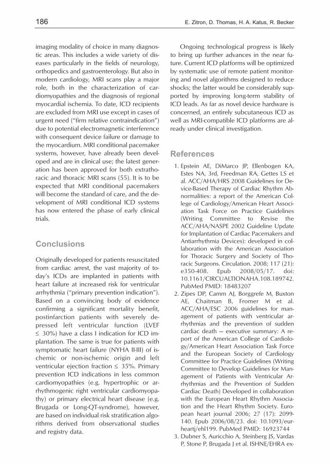
Originally developed for patients resuscitatedfrom cardiac arrest, the vast majority of to-day’s ICDs are implanted in patients withheart failure at increased risk for ventriculararrhythmia (“primary prevention indication”).Based on a convincing body of evidenceconfirming a significant mortality benefit,postinfarction patients with severely de-pressed left ventricular function (LVEF≤ 30%) have a class I indication for ICD im-plantation. The same is true for patients withsymptomatic heart failure (NYHA II-III) of is-chemic or non-ischemic origin and leftventricular ejection fraction ≤ 35%. Primaryprevention ICD indications in less commoncardiomyopathies (e.g. hypertrophic or ar-rhythmogenic right ventricular cardiomyopa-thy) or primary electrical heart disease (e.g.Brugada or Long-QT-syndrome), however,are based on individual risk stratification algo-rithms derived from observational studiesand registry data.
Ongoing technological progress is likelyto bring up further advances in the near fu-ture. Current ICD platforms will be optimizedby systematic use of remote patient monitor-ing and novel algorithms designed to reduceshocks; the latter would be considerably sup-ported by improving long-term stability ofICD leads. As far as novel device hardware isconcerned, an entirely subcutaneous ICD aswell as MRI-compatible ICD platforms are al-ready under clinical investigation.
References
1. Epstein AE, DiMarco JP, Ellenbogen KA,Estes NA, 3rd, Freedman RA, Gettes LS etal. ACC/AHA/HRS 2008 Guidelines for De-vice-Based Therapy of Cardiac Rhythm Ab-normalities: a report of the American Col-lege of Cardiology/American Heart Associ-ation Task Force on Practice Guidelines(Writing Committee to Revise theACC/AHA/NASPE 2002 Guideline Updatefor Implantation of Cardiac Pacemakers andAntiarrhythmia Devices): developed in col-laboration with the American Associationfor Thoracic Surgery and Society of Tho-racic Surgeons. Circulation. 2008; 117 (21):e350-408. Epub 2008/05/17. doi:10.1161/CIRCUALTIONAHA.108.189742.PubMed PMID: 18483207
2. Zipes DP, Camm AJ, Borggrefe M, BuxtonAE, Chaitman B, Fromer M et al.ACC/AHA/ESC 2006 guidelines for man-agement of patients with ventricular ar-rhythmias and the prevention of suddencardiac death — executive summary: A re-port of the American College of Cardiolo-gy/American Heart Association Task Forceand the European Society of CardiologyCommittee for Practice Guidelines (WritingCommittee to Develop Guidelines for Man-agement of Patients with Ventricular Ar-rhythmias and the Prevention of SuddenCardiac Death) Developed in collaborationwith the European Heart Rhythm Associa-tion and the Heart Rhythm Society. Euro-pean heart journal 2006; 27 (17): 2099-140. Epub 2006/08/23. doi: 10.1093/eur-heartj/ehl199. PubMed PMID: 16923744
3. Dubner S, Auricchio A, Steinberg JS, VardasP, Stone P, Brugada J et al. ISHNE/EHRA ex-

187Cardioverter defibrillator therapy in the primary and secondary prevention of ...
pert consensus on remote monitoring ofcardiovascular implantable electronic de-vices (CIEDs). Europace: European pacing,arrhythmias, and cardiac electrophysiology:journal of the working groups on cardiacpacing, arrhythmias, and cardiac cellularelectrophysiology of the European Societyof Cardiology 2012; 14 (2): 278-93. Epub2012/01/11. doi: 10.1093/europace/eur303. PubMed PMID: 22232544
4. A comparison of antiarrhythmic-drug thera-py with implantable defibrillators in patientsresuscitated from near-fatal ventricular ar-rhythmias. The Antiarrhythmics versus Im-plantable Defibrillators (AVID) Investigators.The New England journal of medicine1997; 337 (22): 1576-83. Epub1997/12/31. doi: 10.1056/NEJM199711273372202. PubMed PMID: 9411221
5. Connolly SJ, Gent M, Roberts RS, Dorian P,Roy D, Sheldon RS et al. Canadian im-plantable defibrillator study (CIDS): a ran-domized trial of the implantable cardiovert-er defibrillator against amiodarone. Circula-tion 2000; 101 (11): 1297-302. Epub2000/03/22. PubMed PMID: 10725290
6. Connolly SJ, Hallstrom AP, Cappato R,Schron EB, Kuck KH, Zipes DP et al. Meta-analysis of the implantable cardioverter de-fibrillator secondary prevention trials. AVID,CASH and CIDS studies. Antiarrhythmics vsImplantable Defibrillator study. Cardiac Ar-rest Study Hamburg. Canadian ImplantableDefibrillator Study. European heart journal2000; 21 (24): 2071-8. Epub 2000/12/05.doi: 10.1053/euhj.2000.2476. PubMedPMID: 11102258
7. Kuck KH, Cappato R, Siebels J, Ruppel R.Randomized comparison of antiarrhythmicdrug therapy with implantable defibrillatorsin patients resuscitated from cardiac arrest:the Cardiac Arrest Study Hamburg (CASH).Circulation 2000; 102 (7): 748-54. Epub2000/08/16. PubMed PMID: 10942742
8. Bardy GH, Lee KL, Mark DB, Poole JE, Pack-er DL, Boineau R et al. Amiodarone or animplantable cardioverter-defibrillator forcongestive heart failure. The New Englandjournal of medicine 2005; 352 (3): 225-37.Epub 2005/01/22. doi: 10.1056/NEJ-Moa043399. PubMed PMID: 15659722
9. Hohnloser SH, Kuck KH, Dorian P, RobertsRS, Hampton JR, Hatala R et al. Prophylac-tic use of an implantable cardioverter-defib-
rillator after acute myocardial infarction.The New England journal of medicine2004; 351 (24): 2481-8. Epub 2004/12/14.doi: 10.1056/NEJMoa041489. PubMedPMID: 15590950
10. Kadish A, Dyer A, Daubert JP, Quigg R,Estes NA, Anderson KP et al. Prophylacticdefibrillator implantation in patients withnonischemic dilated cardiomyopathy. TheNew England journal of medicine 2004;350 (21): 2151-8. Epub 2004/05/21. doi:10.1056/NEJMoa033088. PubMed PMID:15152060
11. Moss AJ, Zareba W, Hall WJ, Klein H,Wilber DJ, Cannom DS et al. Prophylacticimplantation of a defibrillator in patientswith myocardial infarction and reducedejection fraction. The New England journalof medicine 2002; 346 (12): 877-83. Epub2002/03/22. doi: 10.1056/NEJ-Moa013474. PubMed PMID: 11907286
12. Steinbeck G, Andresen D, Seidl K, Brach-mann J, Hoffmann E, Wojciechowski D etal. Defibrillator implantation early after my-ocardial infarction. The New England jour-nal of medicine 2009; 361 (15): 1427-36.Epub 2009/10/09. doi: 10.1056/NEJ-Moa0901889. PubMed PMID: 19812399
13. Dorian P, Hohnloser SH, Thorpe KE,Roberts RS, Kuck KH, Gent M et al. Mecha-nisms underlying the lack of effect of im-plantable cardioverter-defibrillator therapyon mortality in high-risk patients with recentmyocardial infarction: insights from the De-fibrillation in Acute Myocardial InfarctionTrial (DINAMIT). Circulation 2010; 122(25): 2645-52. Epub 2010/12/08. doi:10.1161/CIRCULATIONAHA.109.924225.PubMed PMID: 21135366
14. Buxton AE, Lee KL, Fisher JD, JosephsonME, Prystowsky EN, Hafley G. A random-ized study of the prevention of suddendeath in patients with coronary artery dis-ease. Multicenter Unsustained TachycardiaTrial Investigators. The New England journalof medicine 1999; 341 (25): 1882-90. Epub1999/12/22. doi: 10.1056/NE-JM199912163412503. PubMed PMID:10601507
15. Buxton AE, Lee KL, DiCarlo L, Gold MR,Greer GS, Prystowsky EN et al. Electrophys-iologic testing to identify patients with coro-nary artery disease who are at risk for sud-den death. Multicenter Unsustained Tachy-

188 E. Zitron, D. Thomas, H. A. Katus, R. Becker
cardia Trial Investigators. The New Englandjournal of medicine 2000; 342 (26): 1937-45. Epub 2000/06/30. doi: 10.1056/NE-JM200006293422602. PubMed PMID:10874061
16. Maron BJ, McKenna WJ, Danielson GK,Kappenberger LJ, Kuhn HJ, Seidman CE etal. American College of Cardiology/Euro-pean Society of Cardiology clinical expertconsensus document on hypertrophic car-diomyopathy. A report of the American Col-lege of Cardiology Foundation Task Forceon Clinical Expert Consensus Documentsand the European Society of CardiologyCommittee for Practice Guidelines. Journalof the American College of Cardiology2003; 42 (9): 1687-713. Epub 2003/11/11.PubMed PMID: 14607462
17. Schwartz PJ, Spazzolini C, Priori SG, CrottiL, Vicentini A, Landolina M et al. Who arethe long-QT syndrome patients who receivean implantable cardioverter-defibrillator andwhat happens to them?: data from the Euro-pean Long-QT Syndrome Implantable Car-dioverter-Defibrillator (LQTS ICD) Registry.Circulation 2010; 122 (13): 1272-82. Epub2010/09/15. doi: 10.1161/CIRCULATION-AHA.110.950147. PubMed PMID:20837891
18. Wilkoff BL, Cook JR, Epstein AE, Greene HL,Hallstrom AP, Hsia H et al. Dual-chamberpacing or ventricular backup pacing in pa-tients with an implantable defibrillator: theDual Chamber and VVI Implantable Defib-rillator (DAVID) Trial. JAMA : the journal ofthe American Medical Association 2002;288 (24): 3115-23. Epub 2003/01/03.PubMed PMID: 12495391
19. Wilkoff BL, Kudenchuk PJ, Buxton AE, Shar-ma A, Cook JR, Bhandari AK et al. TheDAVID (Dual Chamber and VVI Im-plantable Defibrillator) II trial. Journal of theAmerican College of Cardiology 2009; 53(10): 872-80. Epub 2009/03/07. doi:10.1016/j.jacc.2008.10.057. PubMedPMID: 19264245
20. Sweeney MO. Minimizing right ventricularpacing: a new paradigm for cardiac pacingin sinus node dysfunction. American heartjournal 2007; 153 (4 Suppl): 34-43. Epub2007/03/31. doi: 10.1016/j.ahj.2007.01.019. PubMed PMID: 17394901
21. Kuhlkamp V, Dornberger V, Mewis C,Suchalla R, Bosch RF, Seipel L. Clinical expe-
rience with the new detection algorithmsfor atrial fibrillation of a defibrillator withdual chamber sensing and pacing. Journalof cardiovascular electrophysiology 1999;10 (7): 905-15. Epub 1999/07/21. PubMedPMID: 10413370
22. Theuns DA, Klootwijk AP, Goedhart DM,Jordaens LJ. Prevention of inappropriatetherapy in implantable cardioverter-defibril-lators: results of a prospective, randomizedstudy of tachyarrhythmia detection algo-rithms. Journal of the American College ofCardiology 2004; 44 (12): 2362-7. Epub2004/12/21. doi: 10.1016/j.jacc.2004.09.039. PubMed PMID: 15607399
23. Deisenhofer I, Kolb C, Ndrepepa G,Schreieck J, Karch M, Schmieder S et al. Docurrent dual chamber cardioverter defibrilla-tors have advantages over conventional sin-gle chamber cardioverter defibrillators in re-ducing inappropriate therapies? A random-ized, prospective study. Journal of cardio-vascular electrophysiology 2001; 12 (2):134-42. Epub 2001/03/10. PubMed PMID:11232608
24. Friedman PA, McClelland RL, Bamlet WR,Acosta H, Kessler D, Munger TM et al. Dual-chamber versus single-chamber detectionenhancements for implantable defibrillatorrhythm diagnosis: the detect supraventricu-lar tachycardia study. Circulation 2006; 113(25): 2871-9. Epub 2006/06/14. doi:10.1161/CIRCULATIONAHA.105.594531.PubMed PMID: 16769912
25. Sadoul N, Mletzko R, Anselme F, Bowes R,Schols W, Kouakam C et al. Incidence andclinical relevance of slow ventricular tachy-cardia in implantable cardioverter-defibrilla-tor recipients: an international multicenterprospective study. Circulation 2005; 112(7): 946-53. Epub 2005/08/17. doi:10.1161/CIRCULATIONAHA.105.533513.PubMed PMID: 16103252
26. Young JB, Abraham WT, Smith AL, Leon AR,Lieberman R, Wilkoff B et al. Combined car-diac resynchronization and implantable car-dioversion defibrillation in advanced chron-ic heart failure: the MIRACLE ICD Trial. JA-MA: the journal of the American MedicalAssociation 2003; 289 (20): 2685-94. Epub2003/05/29. doi: 10.1001/jama.289.20.2685. PubMed PMID: 12771115
27. Bristow MR, Saxon LA, Boehmer J, KruegerS, Kass DA, De Marco T et al. Cardiac-resyn-

189Cardioverter defibrillator therapy in the primary and secondary prevention of ...
chronization therapy with or without an im-plantable defibrillator in advanced chronicheart failure. The New England journal ofmedicine 2004; 350 (21): 2140-50. Epub2004/05/21. doi: 10.1056/NEJMoa032423. PubMed PMID: 15152059
28. Linde C, Abraham WT, Gold MR, St JohnSutton M, Ghio S, Daubert C. Randomizedtrial of cardiac resynchronization in mildlysymptomatic heart failure patients and inasymptomatic patients with left ventriculardysfunction and previous heart failuresymptoms. Journal of the American Collegeof Cardiology 2008; 52 (23): 1834-43. Epub2008/11/29. doi: 10.1016/j.jacc.2008.08.027. PubMed PMID: 19038680
29. Daubert C, Gold MR, Abraham WT, Ghio S,Hassager C, Goode G et al. Prevention ofdisease progression by cardiac resynchro-nization therapy in patients with asympto-matic or mildly symptomatic left ventriculardysfunction: insights from the European co-hort of the REVERSE (Resynchronization Re-verses Remodeling in Systolic Left Ventricu-lar Dysfunction) trial. Journal of the Ameri-can College of Cardiology 2009; 54 (20):1837-46. Epub 2009/10/06. doi:10.1016/j.jacc.2009.08.011. PubMedPMID: 19800193
30. Moss AJ, Hall WJ, Cannom DS, Klein H,Brown MW, Daubert JP et al. Cardiac-resyn-chronization therapy for the prevention ofheart-failure events. The New England jour-nal of medicine 2009; 361 (14): 1329-38.Epub 2009/09/03. doi: 10.1056/NEJ-Moa0906431. PubMed PMID: 19723701
31. Tang AS, Wells GA, Talajic M, Arnold MO,Sheldon R, Connolly S et al. Cardiac-resyn-chronization therapy for mild-to-moderateheart failure. The New England journal ofmedicine 2010; 363 (25): 2385-95. Epub2010/11/16. doi: 10.1056/NEJ-Moa1009540. PubMed PMID: 21073365
32. Chung ES, Leon AR, Tavazzi L, Sun JP, Ni-hoyannopoulos P, Merlino J et al. Results ofthe Predictors of Response to CRT(PROSPECT) trial. Circulation 2008; 117(20): 2608-16. Epub 2008/05/07. doi:10.1161/CIRCULATIONAHA.107.743120.PubMed PMID: 18458170
33. Beshai JF, Grimm RA, Nagueh SF, Baker JH,2nd, Beau SL, Greenberg SM et al. Cardiac-resynchronization therapy in heart failurewith narrow QRS complexes. The New Eng-
land journal of medicine 2007; 357 (24):2461-71. Epub 2007/11/08. doi:10.1056/NEJMoa0706695. PubMed PMID:17986493
34. Singh JP, Gras D. Biventricular pacing: cur-rent trends and future strategies. Europeanheart journal 2012; 33 (3): 305-13. Epub2011/09/29. doi: 10.1093/eurheartj/ehr366. PubMed PMID: 21951629
35. Dickstein K, Vardas PE, Auricchio A,Daubert JC, Linde C, McMurray J et al.2010 Focused Update of ESC Guidelineson device therapy in heart failure: an up-date of the 2008 ESC Guidelines for the di-agnosis and treatment of acute and chronicheart failure and the 2007 ESC guidelinesfor cardiac and resynchronization therapy.Developed with the special contribution ofthe Heart Failure Association and the Euro-pean Heart Rhythm Association. Europeanheart journal 2010; 31 (21): 2677-87. Epub2010/08/31. doi: 10.1093/eurheartj/ehq337. PubMed PMID: 20801924
36. Israel CW, Barold SS. Electrical storm in pa-tients with an implanted defibrillator: a mat-ter of definition. Annals of noninvasive elec-trocardiology: the official journal of the In-ternational Society for Holter and Noninva-sive Electrocardiology, Inc. 2007; 12 (4):375-82. Epub 2007/11/01. doi: 10.1111/j.1542-474X.2007.00187.x. PubMed PMID:17970963
37. Connolly SJ, Dorian P, Roberts RS, Gent M,Bailin S, Fain ES et al. Comparison of beta-blockers, amiodarone plus beta-blockers, orsotalol for prevention of shocks from im-plantable cardioverter defibrillators: the OP-TIC Study: a randomized trial. JAMA: thejournal of the American Medical Associa-tion 2006; 295 (2): 165-71. Epub2006/01/13. doi: 10.1001/jama.295.2.165.PubMed PMID: 16403928
38. Carbucicchio C, Santamaria M, Trevisi N,Maccabelli G, Giraldi F, Fassini G et al.Catheter ablation for the treatment of elec-trical storm in patients with implantable car-dioverter-defibrillators: short- and long-termoutcomes in a prospective single-centerstudy. Circulation 2008; 117 (4): 462-9.Epub 2008/01/04. doi: 10.1161/CIRCULA-TIONAHA.106.686534. PubMed PMID:18172038
39. Daubert JP, Zareba W, Cannom DS, McNittS, Rosero SZ, Wang P et al. Inappropriate

190 E. Zitron, D. Thomas, H. A. Katus, R. Becker
implantable cardioverter-defibrillator shocksin MADIT II: frequency, mechanisms, pre-dictors, and survival impact. Journal of theAmerican College of Cardiology 2008; 51(14): 1357-65. Epub 2008/04/05. doi:10.1016/j.jacc.2007.09.073. PubMedPMID: 18387436
40. Poole JE, Johnson GW, Hellkamp AS, An-derson J, Callans DJ, Raitt MH et al. Prog-nostic importance of defibrillator shocks inpatients with heart failure. The New Eng-land journal of medicine 2008; 359 (10):1009-17. Epub 2008/09/05. doi:10.1056/NEJMoa071098. PubMed PMID:18768944; PubMed Central PMCID:PMC2922510
41. van Rees JB, Borleffs CJ, de Bie MK, StijnenT, van Erven L, Bax JJ et al. Inappropriate im-plantable cardioverter-defibrillator shocks:incidence, predictors, and impact on mor-tality. Journal of the American College ofCardiology 2011; 57 (5): 556-62. Epub2011/01/29. doi: 10.1016/j.jacc.2010.06.059. PubMed PMID: 21272746
42. Wilkoff BL, Williamson BD, Stern RS, MooreSL, Lu F, Lee SW et al. Strategic program-ming of detection and therapy parametersin implantable cardioverter-defibrillators re-duces shocks in primary prevention pa-tients: results from the PREPARE (PrimaryPrevention Parameters Evaluation) study.Journal of the American College of Cardiol-ogy 2008; 52 (7): 541-50. Epub2008/08/09. doi: 10.1016/j.jacc.2008.05.011. PubMed PMID: 18687248
43. Germano JJ, Reynolds M, Essebag V,Josephson ME. Frequency and causes of im-plantable cardioverter-defibrillator thera-pies: is device therapy proarrhythmic? TheAmerican journal of cardiology 2006; 97(8): 1255-61. Epub 2006/04/18. doi:10.1016/j.amjcard.2005.11.048. PubMedPMID: 16616037
44. Swerdlow CD, Russo AM, Degroot PJ. Thedilemma of ICD implant testing. Pacing andclinical electrophysiology: PACE 2007; 30(5): 675-700. Epub 2007/04/28. doi:10.1111/j .1540-8159.2007.00730.x.PubMed PMID: 17461879
45. Baddour LM, Epstein AE, Erickson CC,Knight BP, Levison ME, Lockhart PB et al.Update on cardiovascular implantable elec-tronic device infections and their manage-ment: a scientific statement from the Amer-
ican Heart Association. Circulation 2010;121 (3): 458-77. Epub 2010/01/06. doi:10.1161/CIRCULATIONAHA.109.192665.PubMed PMID: 20048212
46. Kleemann T, Becker T, Doenges K, Vater M,Senges J, Schneider S et al. Annual rate oftransvenous defibrillation lead defects in im-plantable cardioverter-defibrillators over aperiod of >10 years. Circulation 2007; 115(19): 2474-80. Epub 2007/05/02. doi:10.1161/CIRCULATIONAHA.106.663807.PubMed PMID: 17470696
47. Eckstein J, Koller MT, Zabel M, Kalusche D,Schaer BA, Osswald S et al. Necessity forsurgical revision of defibrillator leads im-planted long-term: causes and manage-ment. Circulation 2008; 117 (21): 2727-33.Epub 2008/05/21. doi: 10.1161/CIRCULA-TIONAHA.107.740670. PubMed PMID:18490526
48. Wathen MS, DeGroot PJ, Sweeney MO,Stark AJ, Otterness MF, Adkisson WO et al.Prospective randomized multicenter trial ofempirical antitachycardia pacing versusshocks for spontaneous rapid ventriculartachycardia in patients with implantable car-dioverter-defibrillators: Pacing Fast Ventricu-lar Tachycardia Reduces Shock Therapies(PainFREE Rx II) trial results. Circulation2004; 110 (17): 2591-6. Epub 2004/10/20.doi: 10.1161/01.CIR.0000145610.64014.E4. PubMed PMID: 15492306
49. Wathen MS, Sweeney MO, DeGroot PJ,Stark AJ, Koehler JL, Chisner MB et al.Shock reduction using antitachycardia pac-ing for spontaneous rapid ventricular tachy-cardia in patients with coronary artery dis-ease. Circulation 2001; 104 (7): 796-801.Epub 2001/08/15. PubMed PMID:11502705
50. Auricchio A, Meijer A, Kurita T, Schloss E,Brinkman K, Claessens-van Ooijen M et al.Safety, efficacy, and performance of newdiscrimination algorithms to reduce inap-propriate and unnecessary shocks: thePainFree SST clinical study design. Eu-ropace: European pacing, arrhythmias, andcardiac electrophysiology: journal of theworking groups on cardiac pacing, arrhyth-mias, and cardiac cellular electrophysiologyof the European Society of Cardiology2011; 13 (10): 1484-93. Epub 2011/06/15.doi: 10.1093/europace/eur133. PubMedPMID: 21669960

191Cardioverter defibrillator therapy in the primary and secondary prevention of ...
51. Kuck KH, Schaumann A, Eckardt L, WillemsS, Ventura R, Delacretaz E et al. Catheterablation of stable ventricular tachycardiabefore defibrillator implantation in patientswith coronary heart disease (VTACH): amulticentre randomised controlled trial.Lancet 2010; 375 (9708): 31-40. Epub2010/01/30. doi: 10.1016/S0140-6736(09)61755-4. PubMed PMID:20109864
52. Klein HU, Meltendorf U, Reek S, Smid J,Kuss S, Cygankiewicz I et al. Bridging a tem-porary high risk of sudden arrhythmicdeath. Experience with the wearable car-dioverter defibrillator (WCD). Pacing andclinical electrophysiology: PACE 2010; 33(3): 353-67. Epub 2009/11/06. doi:10.1111/j .1540-8159.2009.02590.x.PubMed PMID: 19889186
53. Chung MK, Szymkiewicz SJ, Shao M, ZishiriE, Niebauer MJ, Lindsay BD et al. Aggregatenational experience with the wearable car-dioverter-defibrillator: event rates, compli-ance, and survival. Journal of the AmericanCollege of Cardiology 2010; 56 (3): 194-203. Epub 2010/07/14. doi: 10.1016/j.jacc.2010.04.016. PubMed PMID:20620738; PubMed Central PMCID:PMC2962668
54. Bardy GH, Smith WM, Hood MA, CrozierIG, Melton IC, Jordaens L et al. An entirelysubcutaneous implantable cardioverter-de-fibrillator. The New England journal of med-icine 2010; 363 (1): 36-44. Epub2010/05/14. doi: 10.1056/NEJMoa0909545. PubMed PMID: 20463331
55. Jung W, Zvereva V, Hajredini B, Jackle S.Safe magnetic resonance image scanning ofthe pacemaker patient: current technolo-gies and future directions. Europace: Euro-pean pacing, arrhythmias, and cardiac elec-trophysiology: journal of the workinggroups on cardiac pacing, arrhythmias, andcardiac cellular electrophysiology of the Eu-ropean Society of Cardiology 2012. Epub2012/01/13. doi: 10.1093/europace/eur391. PubMed PMID: 22237585
Correspondence address
Prof. Ruediger Becker, M.D.Department of CardiologyMedical University Hospital HeidelbergIm Neuenheimer Feld 41069120 [email protected]