Cardiovascular System2
56
Cardiovascular System Lecture 2 Properties of Cardiac Muscles
-
Upload
mbbs-ims-msu -
Category
Education
-
view
262 -
download
0
Transcript of Cardiovascular System2

Cardiovascular System
Lecture 2Properties of Cardiac Muscles

Properties of the cardiac muscle:
I. Excitability
II. Conductivity
III. Contractility
IV. Rhythmicity

Properties of the cardiac Muscle
I. Excitability (Irritability)
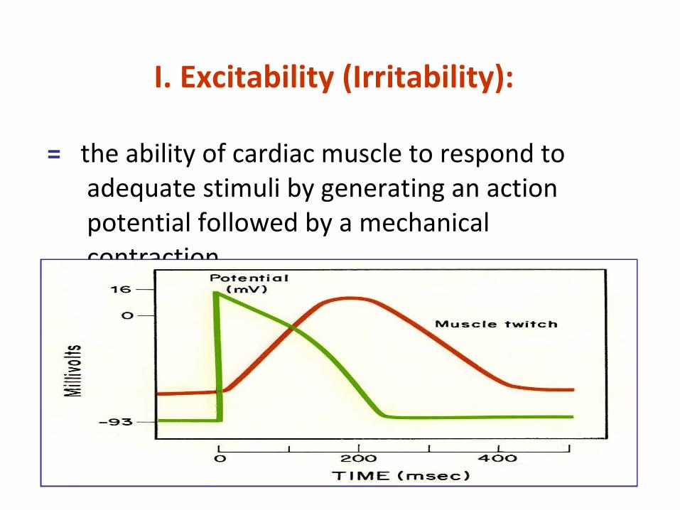
I. Excitability (Irritability):
= the ability of cardiac muscle to respond to adequate stimuli by generating an action potential followed by a mechanical contraction.

■ The mechanical response consists of contraction (systole) & relaxation (diastole).
■ Cardiac muscle begins to contract few milliseconds after the AP begins, & continues to contract until few milliseconds after the AP ends.
■ Duration of contraction:
≈ 0.2 sec in arial muscle, &
≈ 0.3 sec in ventricular muscle.
Relation between the action potential & the mechanical response

■ Diastole begins at the end of the plateau.
■ 2nd rapid repolarization is completed at about the middle of diastole.
Relation between the action potential & the mechanical response (continued)

Action potential of different types of cardiac muscle

Action potential of ventricular muscle
■ Ventricular ms has a RMP of –90 mV. (≈ –85 to –95mV).
■ The trans-membranous AP overshoots to a potential of (≈ +20mV).

AP of ventricular muscle (continued)
Phase 0 = Rapid depolarization.
Phase 1 = Rapid repolarization/ 1st rapid repolarization.
Phase 2 = A plateau.Phase 3 = Slow repolarization/
2nd rapid repolarization.
Phase 4 = Complete repolarization.
■ Trans-membranous AP of ventricular ms is characterized by presence of 5 phases.

Phase 0 = Rapid depolarization. ■ opening of fast Na+ channels → ↑ Na + influx.Phase 1 = Rapid repolarization/ 1st rapid repolarization. ■ closing of Na+ channels, ↓ K+ permeability, with Cl - influx.Phase 2 = A plateau. ■ opening of slow Ca2+ channels (slow Ca2+ Na+ channels) → ↑
Ca 2+ influx.Phase 3 = Slow repolarization/ 2nd rapid repolarization. ■ closing of slow Ca2+ channels, with ↑ K+ permeability → ↑ K +
efflux.Phase 4 = Complete repolarization. ■ active Na+ K+ pump → 2K + in/ 3Na + out.
AP of ventricular muscle (continued)
0
1
2
3
4
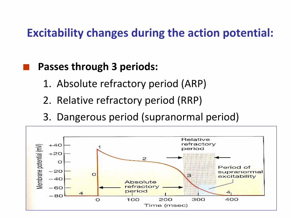
Excitability changes during the action potential:
■ Passes through 3 periods:
1. Absolute refractory period (ARP)
2. Relative refractory period (RRP)
3. Dangerous period (supranormal period)

Refractory Periods

1. Absolute refractory period (ARP):
■ The excitability of cardiac ms is completely lost during this period, i.e. doesn’t respond to 2nd stimulus.
■ V. longV. long.
■ Occupies the whole period of systole.
■ Corresponds to the period of depolarization (phase 0), & the first 2 phases of repolarization.
■ Ht can’t be tetanized (continuous contraction), as its ARP occupies the whole contraction phase.

■ The excitability of cardiac ms is partially recovered during this period, i.e. stronger stimuli than normal are required to excite the muscle.
■ Occupies the time of diastole.
■ Corresponds to the 3rd phase of repolarization.
■ Can be affected by the HR, temp., bacterial toxins, vagal stimulation, sympathetic stimulation & drugs.
2. Relative Refractory Period (RRP):

■ The excitability of cardiac ms is supranormal just at the end of the AP, i.e. weaker stimuli than normal can excite the ms.
■ ? result in ventricular fibrillation.
3. Dangerous Period (Supranormal):

1. Cardiac innervation.
2. Effect of ions concentration in ECF.
3. Physical factors.
4. Blood flow.
5. Chemical factors (drugs).
Factors affecting myocardial excitability:

1. Cardiac Innervation:
■ Sympathetic NS → ↑ excitability.
■ Parasympathetic NS (vagus) → ↓ excitability.
2. Effect of ions concentration in ECF:
■ ↑ Ca2+ → ↑ excitability.
■ ↑ K+ → ↓ excitability.
3. Physical factors: ■ ↑ temperature → ↑ excitability.
■ ↓ temperature → ↓ excitability.
Factors affecting myocardial excitability (continued)

4. Blood flow:
■ Insufficient bl flow to cardiac ms ↓ excitability &
myocardial metabolism for 3 reasons:
(1) lack of O2,
(2) excess accumulation of CO2, &
(3) lack of sufficient food nutrients.
5. Chemical factors (drugs): ■ Digitalis → ↑ excitability.
Factors affecting myocardial excitability (continued)

Properties of the cardiac Muscle
II. Conductivity

II. Conductivity:
= the ability of cardiac muscle fibers to conduct the cardiac impulses that are initiated in the SA-node (the pacemaker of the heart).

■ The impulse is conducted:
1st ⇒ Atrial spread ■ from SA-node → conductive tissue → ventricles.
2nd ⇒ Ventricular spread ■ from apex of the heart → base, via Purkinje fibers to the endocardial surface of ventricles.
The direction of the impulse:

The direction of the impulse (continued)

Conduction of Impulse
■ APs from SA node spread quickly at rate of 0.8 - 1.0 m/sec.
■ Time delay occurs as impulses pass through AV node. – Slow conduction of 0.03 – 0.05 m/sec.
■ Impulse conduction ↑ as spread to Purkinje fibers at a velocity of 5.0 m/sec.– Ventricular contraction begins 0.1–0.2 sec. after
contraction of the atria.

Impulse Conduction through the Heart

The conduction velocities of the impulse:
SA-node 0.05 m/sec. AV-node 0.01 m/sec. … (slowest) Bundle of His 1.00 m/sec. Purkinje fibers 4.00 m/sec. …. (fastest) Atrial & Ventricular muscles 0.3 to 0.4 m/sec.

The conduction velocities (continued)
■ The slowest conduction velocity in AV-node:
■ because it has few no. of intercalated discs. ■ Importance: to allow sufficient time for ventricles to be filled with blood before they contract.
■ The fastest Conduction velocity in Purkinje fibers:
■ Importance: to allow the 2 ventricles to contract at the same time simultaneously.

1. Cardiac innervation.
2. Effect of ions concentration in ECF.
3. Physical factors.
4. Blood flow.
5. Chemical factors (drugs).
Factors affecting myocardial conductivity:

1. Cardiac Innervation:
■ Sympathetic NS → ↑ conductivity.
■ Parasympathetic NS (vagus) → ↓ conductivity.
2. Effect of ions concentration in ECF:
■ ↑ Ca2+ → ↑ conductivity.
■ ↑ K+ → ↓ conductivity.
3. Physical factors: ■ ↑ temperature → ↑ conductivity.
■ ↓ temperature → ↓ conductivity.
Factors affecting myocardial conductivity (continued)

4. Blood flow:
■ Insufficient blood flow to cardiac ms ↓ conductivity &
myocardial metabolism for 3 reasons: (1) lack of O2,
(2) excess accumulation of CO2, &
(3) lack of sufficient food nutrients.
5. Chemical factors (drugs):
■ Digitalis → ↑ conductivity.
Factors affecting myocardial conductivity (continued)

Properties of the cardiac Muscle
III. Contractility

III. Contractility:
= the ability of the cardiac muscle to convert chemical energy into mechanical work.
Contractility (continued)
♥ Myocardial fibers have ‘Functional syncytium.
♥ Strength of myocardial contraction determines the heart pumping power.
♥ Mechanism of contraction depends on the contractile filaments, which contain the protein molecules (actin & myosin).

1. Cardiac innervation.
2. Oxygen supply.
3. Calcium & potassium ions concentration in ECF.
4. Physical factors.
5. Hormonal & chemical factors (drugs).
6. Mechanical factors.
Factors affecting myocardial contractility: (Inotropic effectors)

1. Cardiac Innervation: ■ Sympathetic NS → ↑ force of contraction.
■ Parasympathetic NS (vagus) → ↓ atrial force of contraction with no significant effect on ventricular muscle.
Factors affecting myocardial contractility (continued)

2. Oxygen supply: ■ Hypoxia → ↓ contractility.
3. Calcium & potassium ions concentration in ECF:
■ ↑ Ca2+ → ↑ contractility. ■ ↑ K+ → ↓ contractility.
4. Physical factors: ■ Warming → ↑ contractility. ■ Cooling → ↓ contractility.
Factors affecting myocardial contractility (continued)

5. Hormonal & chemical factors (drugs):
■ +ve inotropics: (Adrenaline, noradrenaline, alkalosis, digitalis, Ca2+, caffeine…)
■ -ve inotropics: (Acetylcholine, acidosis, ether, chloroform, some bacterial toxins (e.g. diphtheria toxins).
Factors affecting myocardial contractility (continued)

6. Mechanical factors:
a. Cardiac muscle obeys ‘all or none law’:
i.e. minimal or threshold stimuli lead to maximal
cardiac contraction, because cardiac muscle behaves as a syncytium.
Factors affecting myocardial contractility (continued)

b. Cardiac muscle can’t be stimulated while it is contracted, because its excitability during contraction is zero due to long ARP, so it can’t be tetanized.
c. Cardiac muscle can perform both isometric & isotonic types of contractions.
Factors affecting myocardial contractility (continued)

d. Starling’s law of the heart:
■ “Length-tension relationship” ‘Within limits, the greater the initial length of the fiber, the stronger will be the force of its contraction; However, overstretching the fiber as in heart failure its power of contractility decreases’
i.e. within limits, the power of contraction is directly proportional to the initial length of the ms.
■ Cardiac ms accommodates itself (up to certain limit) to the changes in venous return.
Factors affecting myocardial contractility (continued)

e. Cardiac muscle shows staircase phenomenon (gradation), if providing all other conditions kept constant.
i.e. if an isolated heart is stimulated by successive
equal & effective stimuli, the 1st few contractions
show a gradual ↑ in the magnitude of contraction.
Factors affecting myocardial contractility (continued)

Properties of the cardiac Muscle
IV. Rhythmicity (Automaticity)

IV. Rhythmicity (automaticity):
= the ability of cardiac muscle to contract in a regular
constant manner without nerve supply.
♥ It’s myogenic in origin (i.e. not neurogenic).
♥ Its initiated by the ‘pacemaker’ of the ht, the
SA- node.

The pacemaker of the heart:
= the SA- node.
♥ Contains the P- cells, which are probably the actual pacemaker cells.
♥ Has the fastest rhythm (rate of discharge) of all parts of the heart, 90 impulses/min. its fibers have an unstable RMP.
♥ Has spontaneous (w/out stimulation) depolarization, up to firing level.

Pacemaker potential:
♥ Its RMP is (≈ -60 mV).
♥ Pacemaker tissue is characterized by unstable
membrane potential.
-6
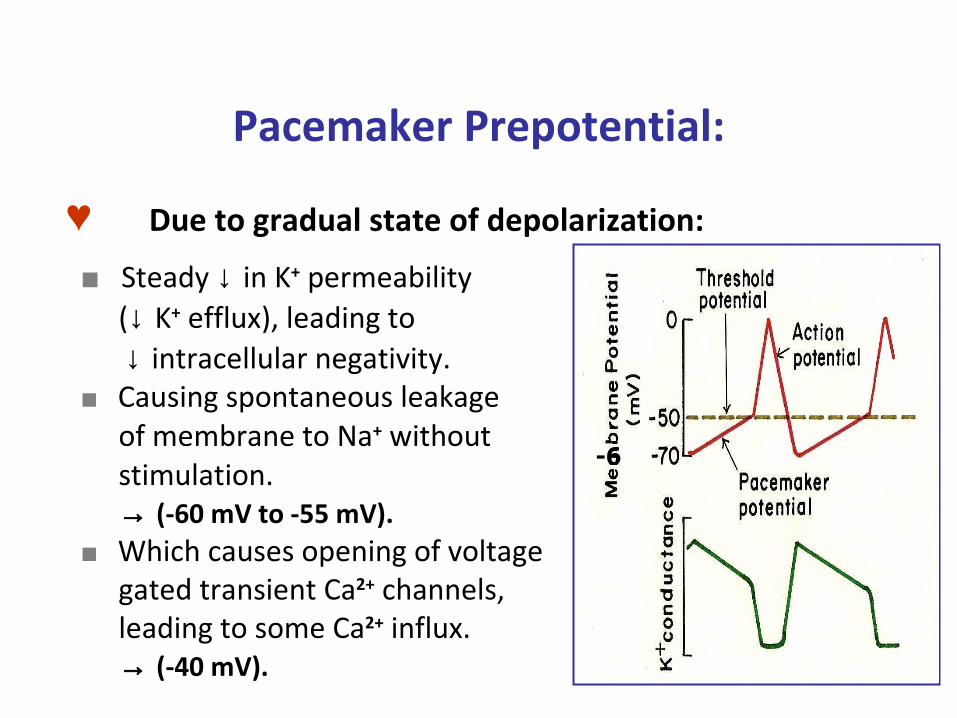
Pacemaker Prepotential:
♥ Due to gradual state of depolarization:
■ Steady ↓ in K+ permeability (↓ K+ efflux), leading to ↓ intracellular negativity. ■ Causing spontaneous leakage of membrane to Na+ without stimulation. → (-60 mV to -55 mV). ■ Which causes opening of voltage gated transient Ca2+ channels, leading to some Ca2+ influx. → (-40 mV).
-6
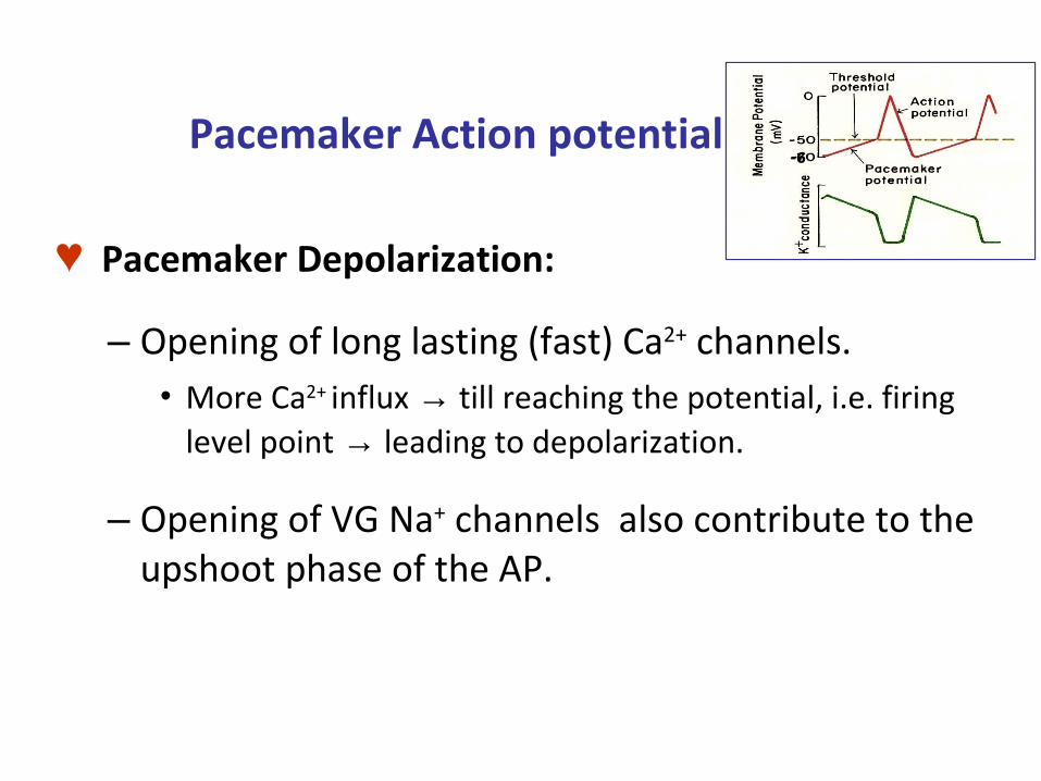
Pacemaker Action potential (AP)
♥ Pacemaker Depolarization:
– Opening of long lasting (fast) Ca2+ channels.• More Ca2+ influx → till reaching the potential, i.e. firing
level point → leading to depolarization.
– Opening of VG Na+ channels also contribute to the upshoot phase of the AP.
-6

Pacemaker Action potential (AP) (continued)
♥ Pacemaker Repolarization:– Opening of VG K+ channels.
• K+ diffuses outward (efflux), … (so +vity will go out of cell).
♥ Pacemaker Hyperpolarization: ■ excessive K+ efflux,
(This will lead to hardship of K+ efflux in 2nd depolarization).
• Ectopic pacemaker:– Pacemaker other than SA node:
• If APs from SA node are prevented from reaching these areas, these cells will generate pacemaker potentials.
-6

-6↓ K+ out
↑ Ca2+ inT Ca2+ L Ca2+
L Ca2+ ↑ K+ out
↑↑↑ K+ out
Na+ in
↑↑↑ Ca2+
in

1. Cardiac innervation.
2. Effect of ions concentration in ECF.
3. Physical factors.
4. Chemical factors (drugs).
Factors affecting myocardial rhythmicity (chronotropic effectors):
a. Sympathetic stimuli:
→ Tachycardia, by ↑ spontaneous depolarization of SA- node.
How? ■ ↓ SA- node membrane permeability to K+ → less K+ efflux. ■ ↑ membrane permeability to Ca2+ → more Ca2+ influx.
■ As a result, the slope of depolarization ↑, causing ↑ rate of SA- node firing & ↑ HR.
Factors affecting myocardial rhythmicity:1. Cardiac Innervation:

b. Parasympathetic stimuli (vagus):
→ Bradycardia, by ↓ spontaneous depolarization of
SA- node. How? ■ ↑ SA- node membrane permeability to K+ → more K+ efflux. ■ ↓ membrane permeability to Ca2+ → less Ca2+ influx.
■ As a result, the prepotential slope ↓, causing ↓ rate of SA- node firing & ↓ HR.
Factors affecting myocardial rhythmicity:1. Cardiac Innervation (continued)


a. K+ ions: ■ If ↓ in ECF → ↑ rhythmicity.
■ If ↑ in ECF → ↓ rhythmicity. (? stop heart in diastole)
b. Na+ ions: ■ If ↑ in ECF → increases rhythmicity,
but can’t maintain it.
Factors affecting myocardial rhythmicity:2. Effect of ion concentrations in ECF:

a. Warming: → ↑ rhythmicity.
b. Cooling: → ↓ rhythmicity.
c. Exercise: → ↑ HR as a result of ↑ sympathetic n.
stimulation & ↓ vagal inhibition to
SA- node.
d. Endurance-trained athletes: Resting bradycardia
due to high vagal activity.
Factors affecting myocardial rhythmicity:3. Physical factors:

a. Thyroid hormones & catecholamines:
→ ↑ rhythmicity.
b. Ach:
→ ↓ rhythmicity.
c. Hypoxia:
→ ↓ rhythmicity.
Factors affecting myocardial rhythmicity:4. Chemical factors (drugs):

Remember:
■ Intrinsic rhythmicity of denervated SA- node is ≈ 90
impulses/min, while that of AV- node is ≈ 60
impulses/min.
■ However, vagal tone controls SA- node to become 70
impulses/min, & AV- node to 40 impulses/min.
■ If SA- node activity is depressed by a disease, AV-
node takes over & becomes the pacemaker instead,
leading to bradycardia.