CARDIOVASCULAR SYSTEM THE HEART Chapter 18. Overview of Cardiovascular System.
description

CardiovascularCardiovascularSystemSystem
Circulation and Gas ExchangeCirculation and Gas Exchange

CirculationCirculation
• Exchange of materials must take place across a wet membrane
• Exchange of materials must take place across a wet membrane

Phylum Cnidaria: gastrovascular cavity
Simple animals have a gastrovascular cavity (digestion and circulation)

CirculationCirculation
• Complex organisms are multi-layered & have Complex organisms are multi-layered & have cells that are isolated and need transport cells that are isolated and need transport systemssystems
• Special organs just for transport (circulation); Special organs just for transport (circulation); heart, vesselsheart, vessels
Circulatory System Overview:Circulatory System Overview:
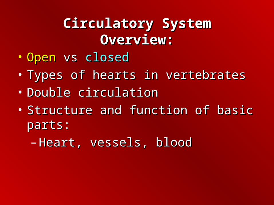
• OpenOpen vs vs closedclosed
• Types of hearts in vertebratesTypes of hearts in vertebrates
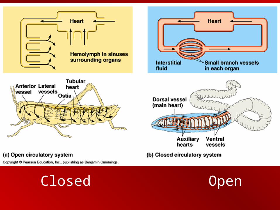
• Double circulationDouble circulation
• Structure and function of basic parts:Structure and function of basic parts:
– Heart, vessels, bloodHeart, vessels, blood

Open CirculatoryOpen Circulatory Systemystem
• No closed vascular tubes; ‘Blood’ (hemolymphhemolymph) circulates freely in sinuses sinuses (spaces around organs)
• Hydrostatic pressureHydrostatic pressure returns the hemolymph to the heart
– Ex. Arthropods, clams; limited in size

Closed Circulatory Closed Circulatory SystemSystem
• Closed vessels; Closed vessels; veinsveins• Blood travels to an Blood travels to an exchange exchange
surfacesurface (pulmonary), then to body (pulmonary), then to body cells (systemic)cells (systemic)
• Blood remains in vesselsBlood remains in vessels; ;
– Much more efficient Much more efficient – Ex. Earthworms, vertebratesEx. Earthworms, vertebrates
Closed Open

Vertebrate PhylogenyVertebrate Phylogeny
Adaptations (Evolution) of the Cardiovascular System

Structural AdaptationsStructural Adaptations
• Heart has chambersHeart has chambers
– AtriaAtria - - Superior chambers - receive bloodSuperior chambers - receive blood
– VentriclesVentricles - - Inferior chambers; pump Inferior chambers; pump blood away from the heartblood away from the heart

Vertebrate HeartsVertebrate Hearts
• Number of chambers is different, demonstrate Number of chambers is different, demonstrate evolutionary adaptationevolutionary adaptation
– 2 chambers = 2 chambers = 11 atriumatrium, , 11 ventricleventricle
– 3 chambers = 3 chambers = 22 atriaatria, , 11 ventricleventricle
– 4 chambers = 4 chambers = 22 atriaatria, , 22 ventriclesventricles

Blood passes through 2 capillary beds; pulmonarypulmonary, (gill) systemicsystemic
–Reduces blood
pressure
–Oxygen-rich blood
slower to circulate

Three ChamberedThree Chambered HeartHeart
• Double circulationDouble circulation:: blood blood travels separately to lungs travels separately to lungs and systemand system
• Oxygenated blood mixes Oxygenated blood mixes with deoxygenated bloodwith deoxygenated blood
• Amphibians, reptilesAmphibians, reptiles
Double circulationDouble circulation; pulmonary and systemic are separated


Mammalian HeartMammalian Heart
4-chambered, double circulation


Pulmonary circuitPulmonary circuit
Systemic circuitSystemic circuit
Systemic circuitSystemic circuit
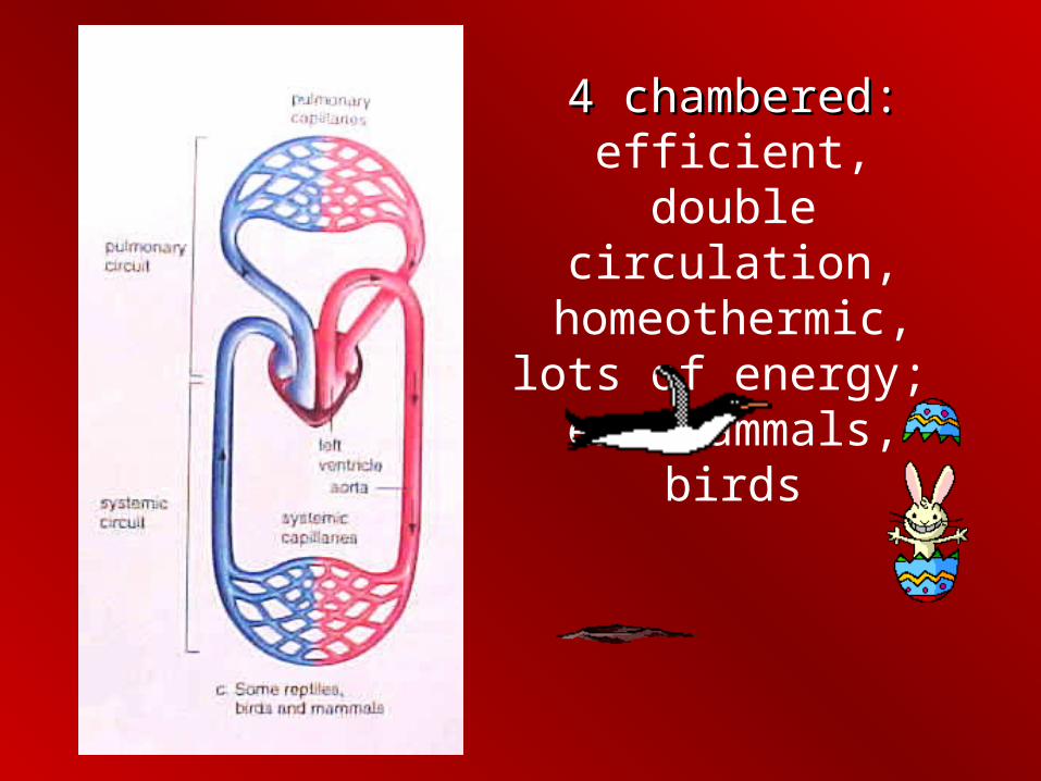
4 chambered:4 chambered: efficient, double circulation,
homeothermic, lots of energy; ex. Mammals,
birds

• Three basic parts:
– Heart
– Blood vessels
– Blood
• Three basic parts:
– Heart
– Blood vessels
– Blood
Structure and Function of the Structure and Function of the Circulatory SystemCirculatory System

• Cardiac muscle;Cardiac muscle; Smooth (rhythmical, persistent)
+ striated (multinucleated, strength)
• Muscle tissue can change shape, in response to electrical or chemical stimulation
• Cardiac muscle;Cardiac muscle; Smooth (rhythmical, persistent)
+ striated (multinucleated, strength)
• Muscle tissue can change shape, in response to electrical or chemical stimulation
HeartHeart
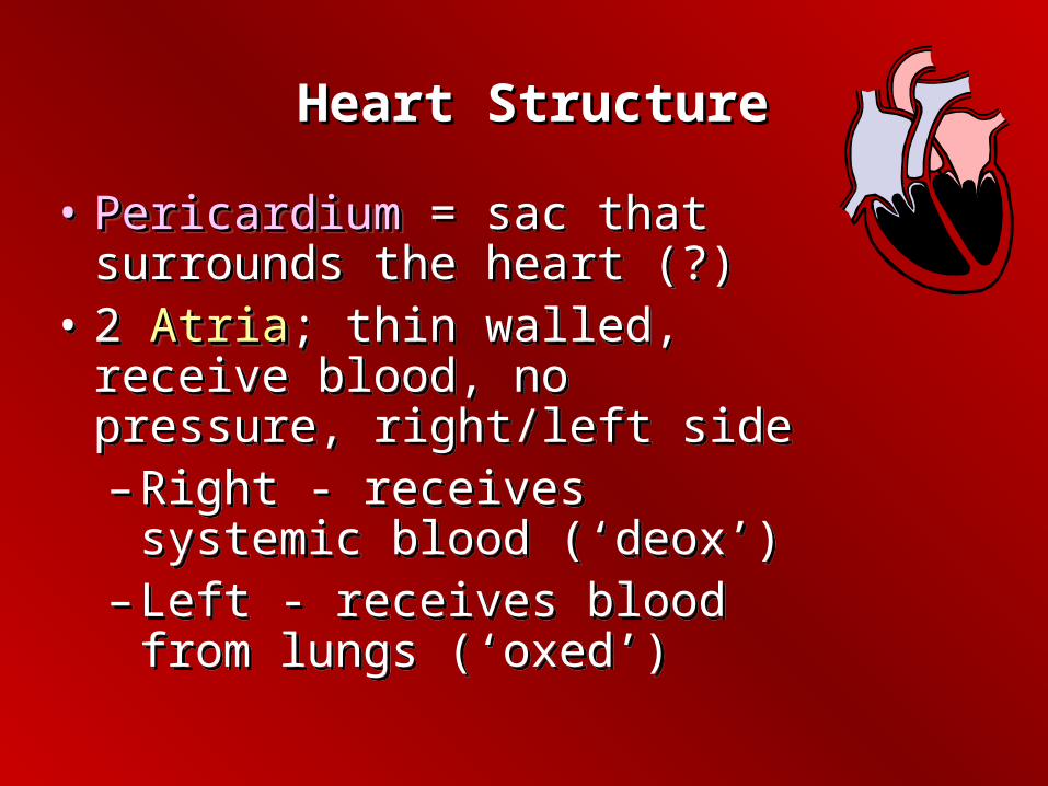
Heart StructureHeart Structure
• PericardiumPericardium = sac that surrounds the heart (?)
• 2 AtriaAtria; thin walled, receive blood, no pressure, right/left side– Right - receives systemic blood
(‘deox’)– Left - receives blood from lungs
(‘oxed’)
• PericardiumPericardium = sac that surrounds the heart (?)
• 2 AtriaAtria; thin walled, receive blood, no pressure, right/left side– Right - receives systemic blood
(‘deox’)– Left - receives blood from lungs
(‘oxed’)

Heart StructureHeart Structure
• 2 VentriclesVentricles; thicker walls, pump blood to body/lungs,
– Right -Right - pumps blood to lungs (pulmonary)
– Left - pumps blood to body (systemic); heaviest muscle
• 2 VentriclesVentricles; thicker walls, pump blood to body/lungs,
– Right -Right - pumps blood to lungs (pulmonary)
– Left - pumps blood to body (systemic); heaviest muscle
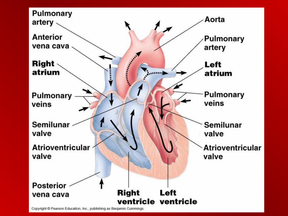
Blood Flow Through the HeartBlood Flow Through the Heart

1. O2 poor systemic blood
4. O2 rich blood to body 2. O2 poor blood to lungs
3. O2 rich blood from lungs

Cardiac CycleCardiac Cycle
• Heart cycle: sequence of events during heartbeat
• SystoleSystole• DiastoleDiastole
• Heart cycle: sequence of events during heartbeat
• SystoleSystole• DiastoleDiastole
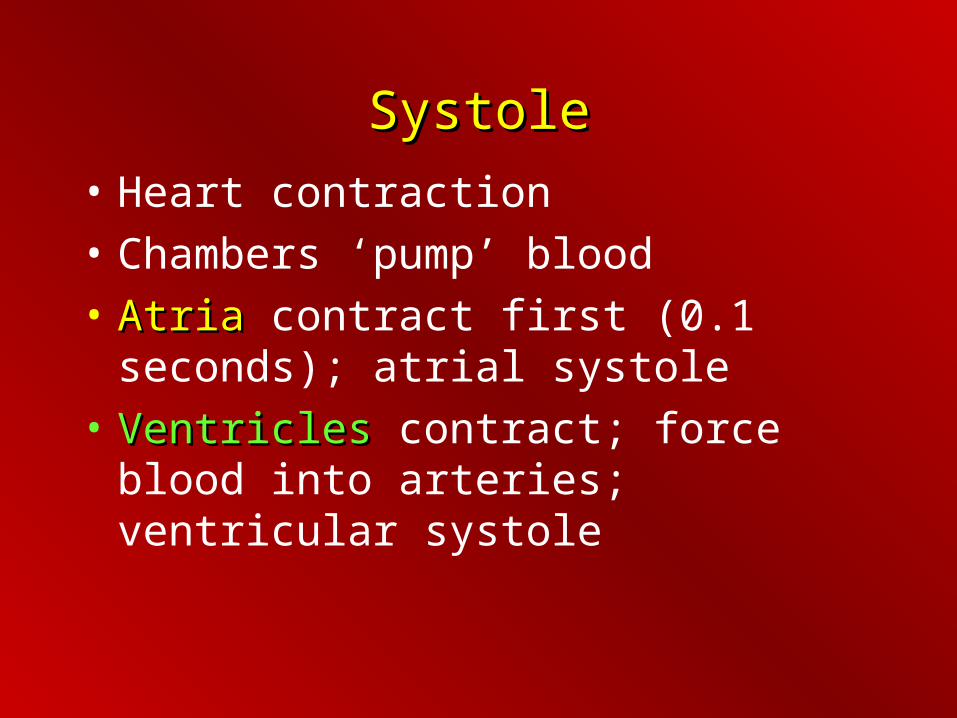
SystoleSystole
• Heart contraction
• Chambers ‘pump’ blood
• AtriaAtria contract first (0.1 seconds); atrial systole
• VentriclesVentricles contract; force blood into arteries; ventricular systole

DiastoleDiastole
• Relaxation phase
– Ventricles refill with blood
– Valves prevent ‘backflow’
Heart CycleHeart Cycle
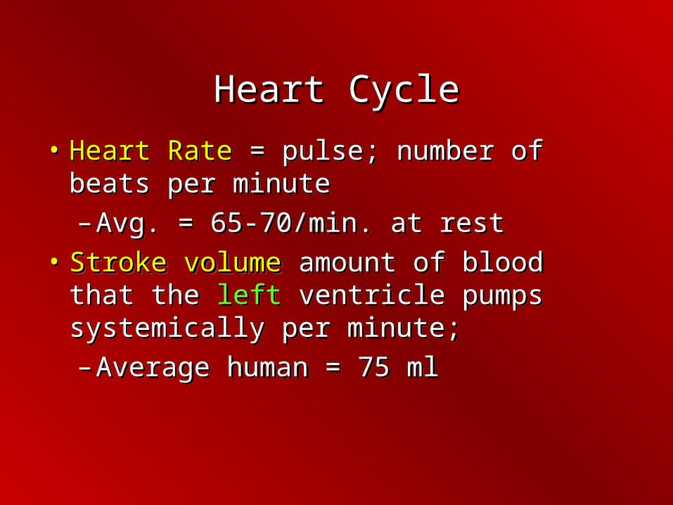
• Heart Rate = pulse; number of beats per minute
– Avg. = 65-70/min. at rest
• Stroke volumeStroke volume amount of blood that the left ventricle pumps systemically per minute;
– Average human = 75 ml
• Heart Rate = pulse; number of beats per minute
– Avg. = 65-70/min. at rest
• Stroke volumeStroke volume amount of blood that the left ventricle pumps systemically per minute;
– Average human = 75 ml
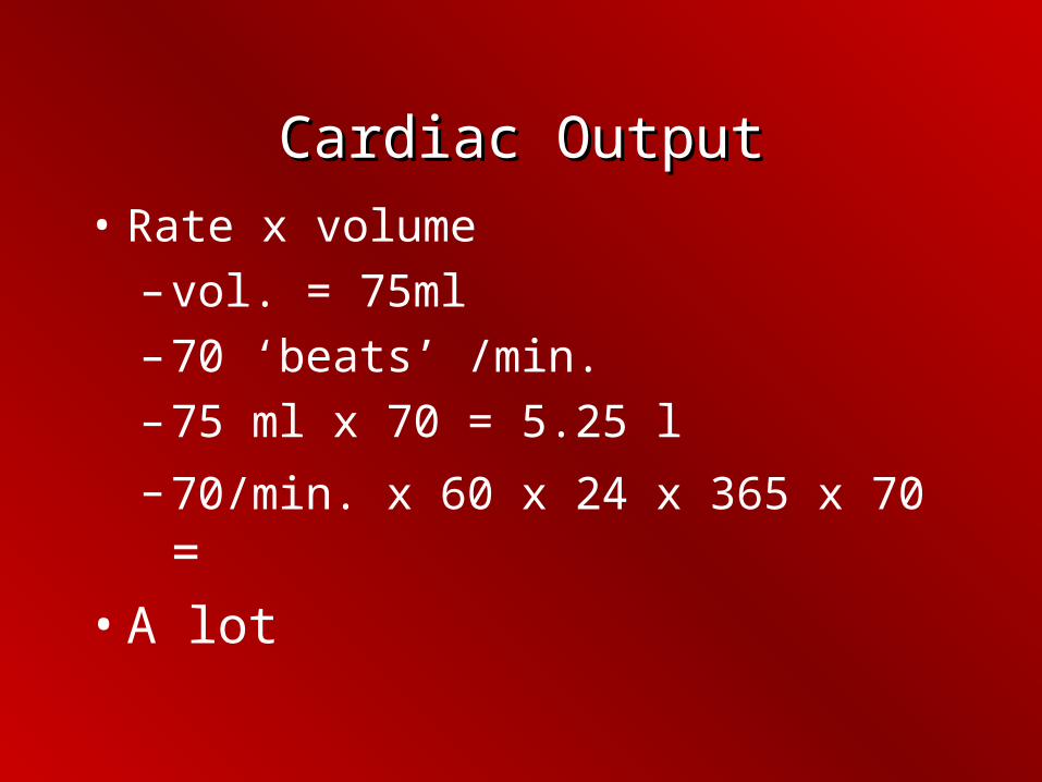
Cardiac OutputCardiac Output
• Rate x volume
– vol. = 75ml
– 70 ‘beats’ /min.
– 75 ml x 70 = 5.25 l
– 70/min. x 60 x 24 x 365 x 70 =
• A lot

Heart CycleHeart Cycle
Inverse relationship between size and heart rate;
Elephants = 25
Shrews = 1560
Inverse relationship between size and heart rate;
Elephants = 25
Shrews = 1560
251560

How Do We Keep Blood From How Do We Keep Blood From Going ‘Backwards’?Going ‘Backwards’?
One-Way Valves

StructureStructure
• Four valves: prevent ‘back flow’
• 2 AtrioventricularAtrioventricular between atria and ventricles
• 2 SemilunarSemilunar; between ventricles and arteries, aorta and pulmonary
• Four valves: prevent ‘back flow’
• 2 AtrioventricularAtrioventricular between atria and ventricles
• 2 SemilunarSemilunar; between ventricles and arteries, aorta and pulmonary
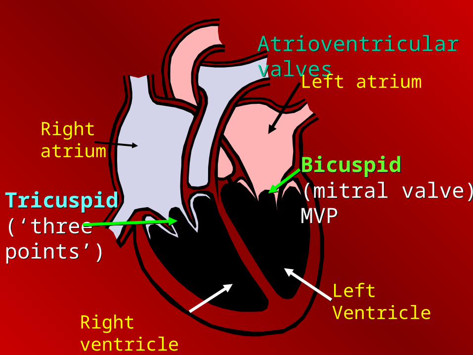
Tricuspid (‘three points’)Tricuspid (‘three points’)
Bicuspid (mitral valve) MVPBicuspid (mitral valve) MVP
Atrioventricular valvesAtrioventricular valves
Right atrium
Left atrium
Left Ventricle
Right ventricle

2 Semilunar; between ventricles and arteries2 Semilunar; between ventricles and arteries
Aortic valveAortic valvePulmonary valvePulmonary valve
RV
LV

Heart CycleHeart Cycle
• Heart sounds: valves opening/closing; “heart beat”
• Stethoscope
• “LubbLubb” = lower pitch, atrioventricular valves closing (bicuspid/tricuspid); ventricles contracting; just before systole
• “DuppDupp” = semilunar valves closing; ventricles relax; just before diastole
• Heart sounds: valves opening/closing; “heart beat”
• Stethoscope
• “LubbLubb” = lower pitch, atrioventricular valves closing (bicuspid/tricuspid); ventricles contracting; just before systole
• “DuppDupp” = semilunar valves closing; ventricles relax; just before diastole

Heart CycleHeart Cycle
• Heart Murmur: defect in valve causing backflow. Serious, corrected with surgery
• Heart Murmur: defect in valve causing backflow. Serious, corrected with surgery
NormalMurmurs

Control of the Heart CycleControl of the Heart Cycle
Control of Heart CycleControl of Heart Cycle
• Intercalated disks = special areas between cells; extraordinary cell-to-cell communication; folds in between like tongue-in-groove
– (Why is this important?) – structure/function
• Intercalated disks = special areas between cells; extraordinary cell-to-cell communication; folds in between like tongue-in-groove
– (Why is this important?) – structure/function


Control of Heart CycleControl of Heart Cycle
• Cardiac muscle is myogenic (self-excitable)
– Contracts without nervous input
• Tempo is controlled by nodes (knots of nervous tissue + cardiac muscle)
• Two ‘nodes’ stimulate muscle contraction
– Sinoatrial Node (SA)
– Atrioventricular node (AV)
• Cardiac muscle is myogenic (self-excitable)
– Contracts without nervous input
• Tempo is controlled by nodes (knots of nervous tissue + cardiac muscle)
• Two ‘nodes’ stimulate muscle contraction
– Sinoatrial Node (SA)
– Atrioventricular node (AV)

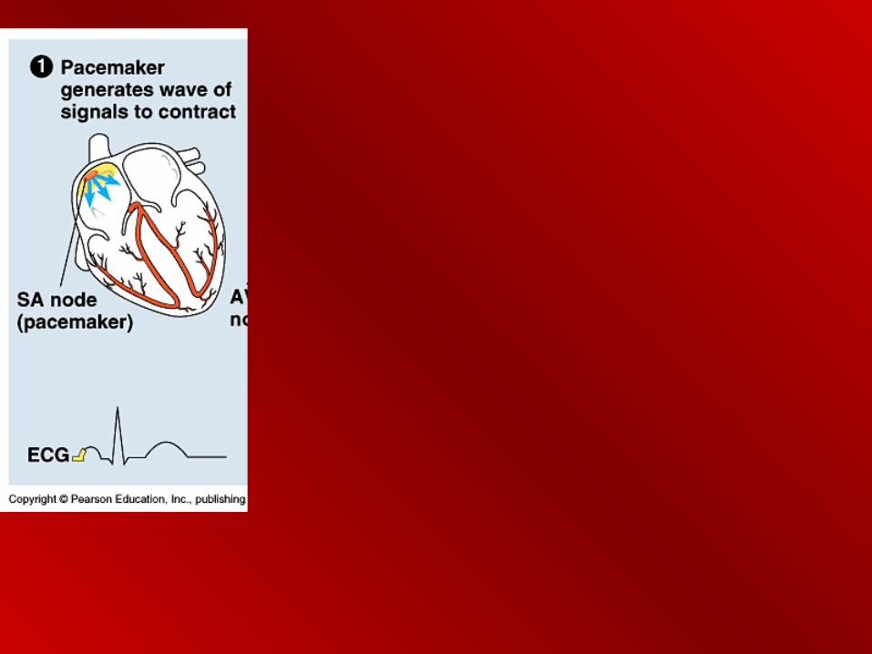
• Sinoatrial node (SA) -Sinoatrial node (SA) - tempo of contraction “Pacemaker”
• Starts a wave of contraction; causes both atria to contract together
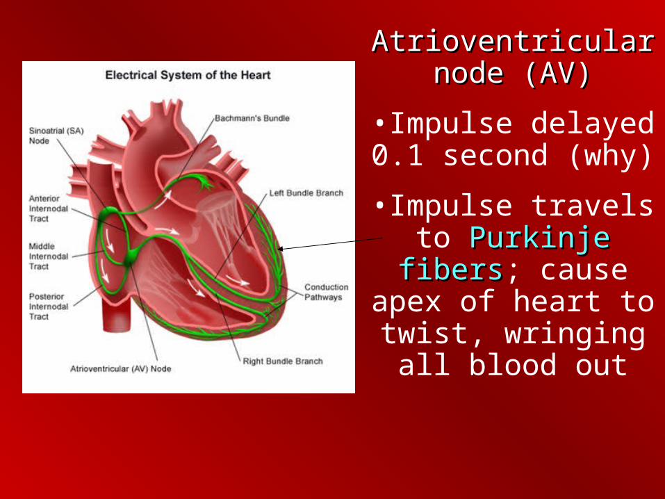
Atrioventricular node Atrioventricular node (AV)(AV)
•Impulse delayed 0.1 second (why)
•Impulse travels to Purkinje fibersPurkinje fibers; cause apex of heart to twist, wringing all blood out
ElectrocardiogramElectrocardiogram
• Detects tiny electrical changes; action potentials
• Depolarization/repolarization detected by electrodes on surface of skin
• Detects tiny electrical changes; action potentials
• Depolarization/repolarization detected by electrodes on surface of skin


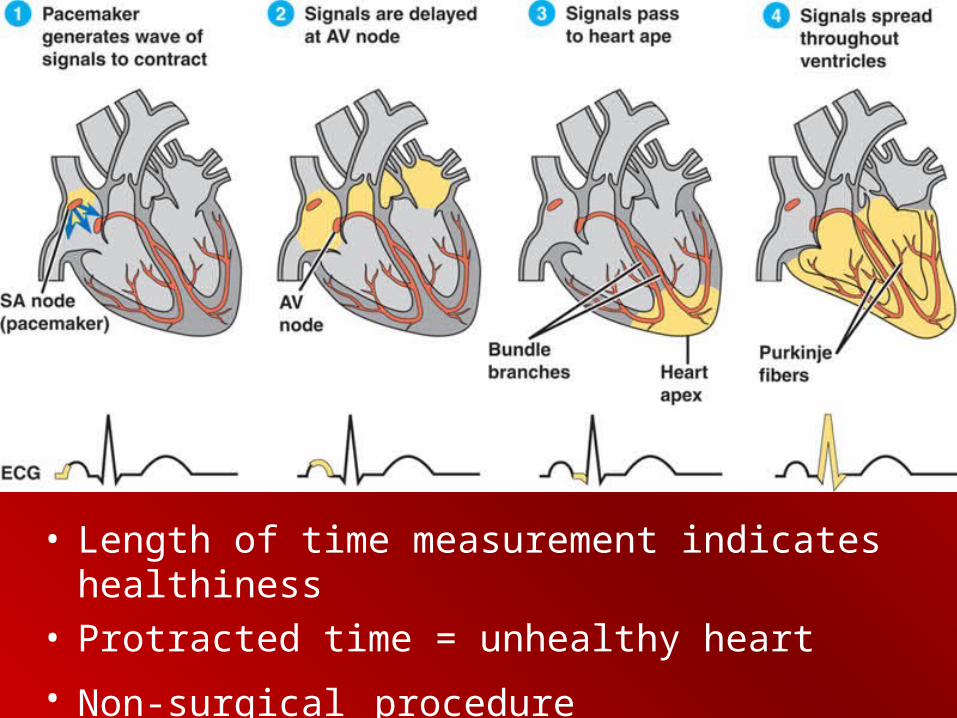
• Length of time measurement indicates healthiness• Protracted time = unhealthy heart
• Non-surgical procedure

Regulation of Heart CycleRegulation of Heart Cycle
• Controlled by SA node• Influenced by:
– Autonomic nervous system– Hormones – Temperature – Exercise
• Balance controlled by medulla
• Controlled by SA node• Influenced by:
– Autonomic nervous system– Hormones – Temperature – Exercise
• Balance controlled by medulla

Regulation of Heart CycleRegulation of Heart Cycle
• Autonomic – ‘automatic’
– Causes release of norepinephrine (hormone)
• Increases heart rate (emotions; fear, ‘love’)
• Impulses come from cerebrum (memory)
• Other causes for heart rate change:
– Pressure
– Ions –potassium, Ca.
• Autonomic – ‘automatic’
– Causes release of norepinephrine (hormone)
• Increases heart rate (emotions; fear, ‘love’)
• Impulses come from cerebrum (memory)
• Other causes for heart rate change:
– Pressure
– Ions –potassium, Ca.

Blood PressureBlood Pressure
• SystoleSystole = contraction of chambers; blood ‘pumped’
• DiastoleDiastole = relaxing of chambers, ventricles, atria fill

Blood PressureBlood Pressure
• Measured by a sphygmomanometer/stethoscopesphygmomanometer/stethoscope
• First number is systolesystole
• Second is diastolediastole
• 120/70 = ‘good’, normal


Blood PressureBlood Pressure
• The hydrostatic force that blood exerts against a vessel wall
• Greater in arteries
• Greatest during systole (contraction)
• Peripheral resistancePeripheral resistance = impedance from arterioles; blood enters arteries faster than it can get out
• Always pressure (even during diastole)

Blood PressureBlood Pressure• What would cause blood pressure?
• Cardiac output and peripheral resistance
• Stress causes neural and hormonal responses which trigger smooth muscle to contract, increases peripheral resistance

Blood PressureBlood Pressure• Pressure is near zero in veins
– Blood is returning to the heart – Movement of muscle
– Valves in the veins– Breathing increases volume in the thoracic cavity
causing vena cavae to dilate


Structural Differences in VesselsStructural Differences in Vessels
Arteries, veins, capillaries

Vessels:Vessels: 2 Types2 Types
• ArteriesArteries; carry blood away from the heart
• Branch out into arteriolesarterioles
• Branch into capillaries (diffusion/osmosis)
• ArteriesArteries; carry blood away from the heart
• Branch out into arteriolesarterioles
• Branch into capillaries (diffusion/osmosis)

ArteriesArteries ArteriesArteries
• 3 layers (tunics): connective epithelium, smooth muscle and endothelium: thick walled; deep in body
• Arteriole = smallest arteries with 3 tunics
• 3 layers (tunics): connective epithelium, smooth muscle and endothelium: thick walled; deep in body
• Arteriole = smallest arteries with 3 tunics
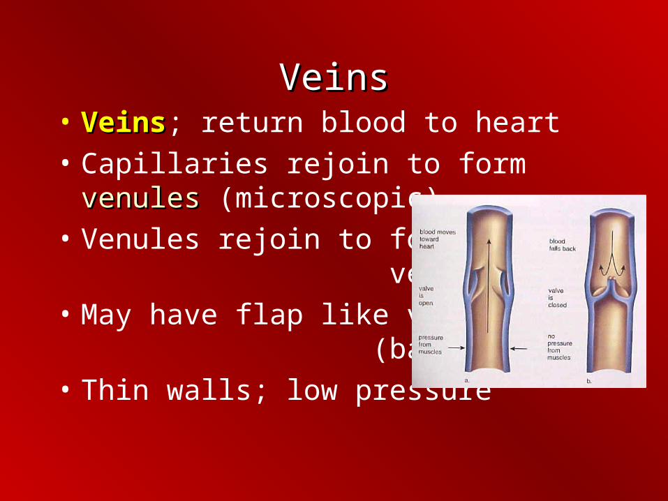
VeinsVeins• VeinsVeins; return blood to heart
• Capillaries rejoin to form venulesvenules (microscopic)
• Venules rejoin to form veins
• May have flap like valves (back flow)
• Thin walls; low pressure


Blood VesselsBlood Vessels
• CapillaryCapillary: endothelial tissue, thin, branched
• Diffusion of O2 to individual cells
• CapillaryCapillary: endothelial tissue, thin, branched
• Diffusion of O2 to individual cells


Water that escapes from capillary diffuses back into lymph

Capillary ExchangeCapillary Exchange
• Passive transport occurs by:
– DiffusionDiffusion
– OsmosisOsmosis
– Hydrostatic pressureHydrostatic pressure; blood pressure
– Gap junctionsGap junctions == pores between cells
• Passive transport occurs by:
– DiffusionDiffusion
– OsmosisOsmosis
– Hydrostatic pressureHydrostatic pressure; blood pressure
– Gap junctionsGap junctions == pores between cells

Capillary ExchangeCapillary Exchange
• Capillary wall is ‘leaky’
• Materials may cross in vesicles
– Active transport (endocytosis, exocytosis)
• Capillary wall is ‘leaky’
• Materials may cross in vesicles
– Active transport (endocytosis, exocytosis)

Capillary ExchangeCapillary Exchange
• Gases move by diffusion
• Fluids move by osmosisosmosis or vesicles
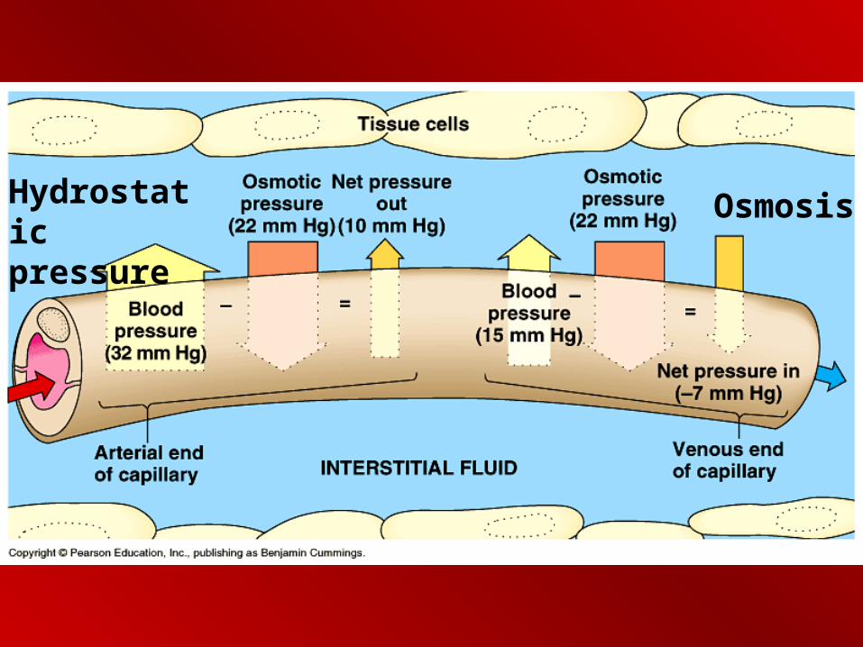
• Direction of fluid flow depends upon the amount of pressure
• Pressure = hydrostatic + osmotic
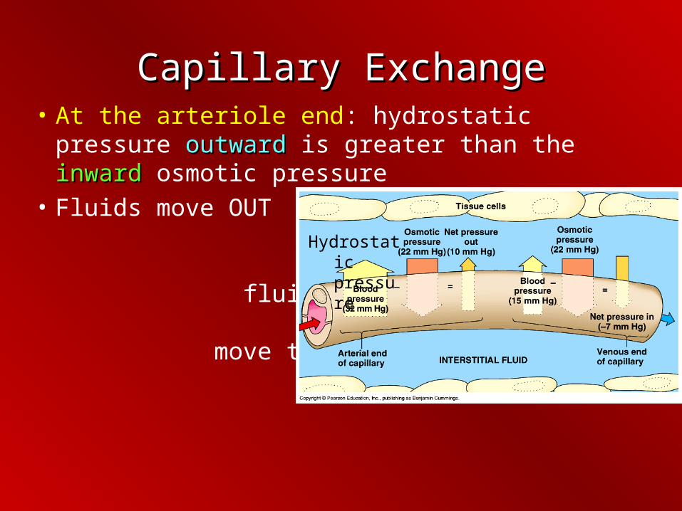
Capillary ExchangeCapillary Exchange• At the arteriole end: hydrostatic pressure
outwardoutward is greater than the inwardinward osmotic pressure
• Fluids move OUT into the interstial fluid (materials move through in dissolved from)
Hydrostatic pressure

Capillary ExchangeCapillary Exchange• At the venule end: outward hydrostatic pressure
is less than the inward osmotic pressure
• Fluids move back into the capillary
OsmosisHydrostatic pressure
Osmosis Hydrostatic pressure

Lymph SystemLymph System
•Return Fluids to the Body
•Immunity

LymphLymph
• 85% of fluids lost in capillary bed exchange is recovered at the venous end of the bed
• Other 15% is recovered by LymphLymph
• Lymphatic fluidLymphatic fluid = similar to interstial fluid (water, proteins, antigens)
LymphLymph
• Movement of skeletal muscle aids circulation of lymph (stay active)
• Trauma or histamines may cause an increase in permeability
• Lymphatic system cannot keep up
• Surrounding tissues swell
• Movement of skeletal muscle aids circulation of lymph (stay active)
• Trauma or histamines may cause an increase in permeability
• Lymphatic system cannot keep up
• Surrounding tissues swell

LymphLymph• Lymph fluid travels through nodes
• ‘checked’ for antigens (invaders)
• Nodes may become swollen
• Also carries fats from digestion

Heart ProblemsHeart Problems
• Rheumatic heart diseaseRheumatic heart disease = streptococcal infection; inflamed endocardium; valves damaged” MVP
• Aneurysm = swelling in blood vessel
• Embolism = moving clot
• Rheumatic heart diseaseRheumatic heart disease = streptococcal infection; inflamed endocardium; valves damaged” MVP
• Aneurysm = swelling in blood vessel
• Embolism = moving clot

Heart ProblemsHeart Problems
• Coronary thrombosisCoronary thrombosis = clot in coronary artery; causes infarction (area of damaged tissue)

Heart ProblemsHeart Problems
• ArrhythmiaArrhythmia = heart out of rhythm:
– Tachycardia = 100 +
– Brachycardia = less than 60
– Flutter = 250+
– Fibrillation = heart muscles not contracting together; defribillator stops heart
• ArrhythmiaArrhythmia = heart out of rhythm:
– Tachycardia = 100 +
– Brachycardia = less than 60
– Flutter = 250+
– Fibrillation = heart muscles not contracting together; defribillator stops heart

Heart ProblemsHeart Problems
• Coronary heart disease = reduced flow to coronary arteries; ½ of all deaths in U.S.
• Stress, hypertension (atherosclerosis)
• Enlarges left ventricle (overwork)
• Coronary heart disease = reduced flow to coronary arteries; ½ of all deaths in U.S.
• Stress, hypertension (atherosclerosis)
• Enlarges left ventricle (overwork)

Heart ProblemsHeart Problems• AtherosclerosisAtherosclerosis = hardening of arterial wall due to
build up of plaque (cholesterol); LDL = ‘bad’; HDL = ‘good’
• ArteriosclerosisArteriosclerosis = type of athersclerosis; addition of Ca deposits
Arteriosclerosis


Normal Arteriosclerosis
Plaque

Heart ProblemsHeart Problems
• Myocardial infarction (‘heart attack’); angina pectoris = pain in chest, left arm, shoulder
• Constriction of chest (angina); crushing, bursting (may fade/return)
• Pain in back, jaw, left arm• Shortness of breath• Nausea, sweat, dizzy, pallor
• Myocardial infarction (‘heart attack’); angina pectoris = pain in chest, left arm, shoulder
• Constriction of chest (angina); crushing, bursting (may fade/return)
• Pain in back, jaw, left arm• Shortness of breath• Nausea, sweat, dizzy, pallor

Heart ProblemsHeart Problems
• Scar tissue replaces cardiac muscle
• Rest, diet (animal fat) reduce stress
• Exercise; dilation of skeletal muscles allows increased flow through systemic, increased O2 flow; brain works better…
• Scar tissue replaces cardiac muscle
• Rest, diet (animal fat) reduce stress
• Exercise; dilation of skeletal muscles allows increased flow through systemic, increased O2 flow; brain works better…

Heart Problems: TreatmentHeart Problems: Treatment
• Digitalis = slows, strengthens heart contraction
• Nitroglycerin = dilates vessels
• Anticoagulants = prevent blood clotting
• Digitalis = slows, strengthens heart contraction
• Nitroglycerin = dilates vessels
• Anticoagulants = prevent blood clotting

Heart ProblemsHeart Problems
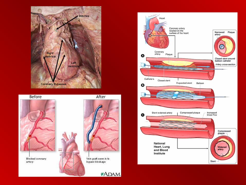
• By-pass surgery; remove vessels (leg) and splice in around coronary vessel that is blocked (‘triple/quadruple by-pass’)
• Stent = ‘balloon’ in artery to swell and break up thrombosis/clot
• By-pass surgery; remove vessels (leg) and splice in around coronary vessel that is blocked (‘triple/quadruple by-pass’)
• Stent = ‘balloon’ in artery to swell and break up thrombosis/clot

Heart ProblemsHeart Problems
• Congenital = from birth; septal defect, cyanosis (‘blue baby’)
• Aging = heart failure: by 70, 30% decrease in heart efficiency; earlier if infections, toxins, anemia, hyperthyroidism, infarction, stress
• Congenital = from birth; septal defect, cyanosis (‘blue baby’)
• Aging = heart failure: by 70, 30% decrease in heart efficiency; earlier if infections, toxins, anemia, hyperthyroidism, infarction, stress

Heart ProblemsHeart Problems• Stroke = loss of blood supply; necrosis
(tissue death); Infarction
• Caused by:
– Thrombosis (stationary clot)
– Hemorrhage (blood leak)
– Arteriosclerosis
• Stroke = loss of blood supply; necrosis (tissue death); Infarction
• Caused by:
– Thrombosis (stationary clot)
– Hemorrhage (blood leak)
– Arteriosclerosis


BloodBlood
• Connective tissue; 2 parts:
– Plasma – fluid – 55%
– Formed elementsFormed elements = solids; 45%
• Makes up 8% of body mass
• Average = 4 to 6 liters of whole blood
• Connective tissue; 2 parts:
– Plasma – fluid – 55%
– Formed elementsFormed elements = solids; 45%
• Makes up 8% of body mass
• Average = 4 to 6 liters of whole blood


FunctionsFunctions
• TransportTransport - - O2, nutrients, enzymes, etc. remove CO2, wastes
• EndothermyEndothermy - (homeostasis)
• BalanceBalance - - fluid, electrolyte, pH (homeostasis)
• ProtectionProtection - - from diseases, infection
• TransportTransport - - O2, nutrients, enzymes, etc. remove CO2, wastes
• EndothermyEndothermy - (homeostasis)
• BalanceBalance - - fluid, electrolyte, pH (homeostasis)
• ProtectionProtection - - from diseases, infection


PlasmaPlasma
• WaterWater = 90% = 90%
• Dissolved gases (CODissolved gases (CO22, O, O2 2 NN22))
• Inorganic saltsInorganic salts (electrolytes; salts) (electrolytes; salts)
• ProteinsProteins
• OtherOther – urea, sugars, aa, hormones

PlasmaPlasma
• ProteinsProteins = buffer blood, osmosis, viscosity (thickness)
– AlbuminsAlbumins = osmotic pressure
– GlobulinsGlobulins = immune (antibodies)
– FibrinogenFibrinogen = clotting agent
• SerumSerum = plasma with no clotting factors; Why serum?
• ProteinsProteins = buffer blood, osmosis, viscosity (thickness)
– AlbuminsAlbumins = osmotic pressure
– GlobulinsGlobulins = immune (antibodies)
– FibrinogenFibrinogen = clotting agent
• SerumSerum = plasma with no clotting factors; Why serum?
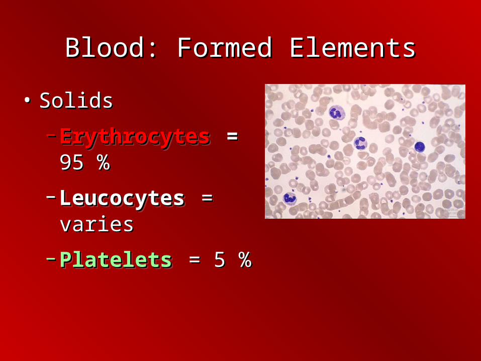
Blood: Formed ElementsBlood: Formed Elements
• Solids
– ErythrocytesErythrocytes = 95 %
– LeucocytesLeucocytes = varies
– PlateletsPlatelets = 5 %
• Solids
– ErythrocytesErythrocytes = 95 %
– LeucocytesLeucocytes = varies
– PlateletsPlatelets = 5 %
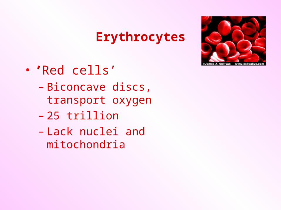
Erythrocytes
• ‘‘Red cells’– Biconcave discs, transport oxygen– 25 trillion– Lack nuclei and mitochondria

Hemoglobin
• Iron-containing protein pigment;
– 250 million molecules per RBC; 1/3 of mass
– Reversibly binds with oxygen
– Oxyhemoglobin -Bright red
• Four heme groups; each contains an iron atom with an affinity for oxygen
Sickle cell Sickle cell

RBC ProductionRBC Production
• Hematopoeisis (‘blood make’)• Red marrow of long bones
– Femur, humerus; skull, ribs, pelvis, sternum, vertebrae
• 2.5 million/sec.
• Nucleus lost during development
• Hematopoeisis (‘blood make’)• Red marrow of long bones
– Femur, humerus; skull, ribs, pelvis, sternum, vertebrae
• 2.5 million/sec.
• Nucleus lost during development

RBC ProductionRBC Production
• RBC production stimulated by ErythropoeitinErythropoeitin (hormone from kidney)
• Negative feedbackNegative feedback mechanismmechanism
– Low oxygen = release of erythropoeitin
• Exercise, altitude (low partial pressure)
• RBC production stimulated by ErythropoeitinErythropoeitin (hormone from kidney)
• Negative feedbackNegative feedback mechanismmechanism
– Low oxygen = release of erythropoeitin
• Exercise, altitude (low partial pressure)
Blood: PhysiologyBlood: Physiology
• After hemoglobin releases oxygen, it has a greater affinity for carbon dioxide
– Carbaminohemoglobin
– Reversible reaction
• Hemoglobin has a greater affinity for carbon arbon monoxidemonoxide than oxygen or carbon dioxide
• After hemoglobin releases oxygen, it has a greater affinity for carbon dioxide
– Carbaminohemoglobin
– Reversible reaction
• Hemoglobin has a greater affinity for carbon arbon monoxidemonoxide than oxygen or carbon dioxide

LossLoss PreventionPrevention
• Vasoconstriction• Platelet plug• Clotting (coagulation)• Clotting factors inactive• Become active when:
– Connective tissue becomes exposed– Chemicals released from injured tissue
• Positive feedback
• Vasoconstriction• Platelet plug• Clotting (coagulation)• Clotting factors inactive• Become active when:
– Connective tissue becomes exposed– Chemicals released from injured tissue
• Positive feedback

ClottingClotting• Prothrombin (globulin protein) converted (by
Ca) into thrombin• Thrombin converts fibrinogen into fibrin
(sticky, thread like)• Fibrin forms a ‘mesh’ net• Platelets and RBC’s clog up = clot • Fragments of cells with membranes• Stick to collagen fibers (connective tissue),
and each other to form platelet ‘plug’
• Prothrombin (globulin protein) converted (by Ca) into thrombin
• Thrombin converts fibrinogen into fibrin (sticky, thread like)
• Fibrin forms a ‘mesh’ net• Platelets and RBC’s clog up = clot • Fragments of cells with membranes• Stick to collagen fibers (connective tissue),
and each other to form platelet ‘plug’


Blood AgglutinationBlood Agglutination
An Example of Immune Response, An Example of Immune Response, Codominance, and Multiple AllelesCodominance, and Multiple Alleles

Blood AgglutinationBlood Agglutination
• Clumping
• Agglutinogens (antigens);
– Glycoproteins on surface of RBC’s
– ‘Flags’
• Agglutinins (antibodies) in plasma
• Clumping
• Agglutinogens (antigens);
– Glycoproteins on surface of RBC’s
– ‘Flags’
• Agglutinins (antibodies) in plasma

Blood AgglutinationBlood Agglutination
• Antibodies ‘attack’ antigens if they don’t match• Ex.
– Anti A clumps B antigens
– Anti B clumps A antigensA antigens
• A person with “A” blood cannot receive “B” blood (?)
• A person with “B” blood cannot receive “A” blood (?)
• Antibodies ‘attack’ antigens if they don’t match• Ex.
– Anti A clumps B antigens
– Anti B clumps A antigensA antigens
• A person with “A” blood cannot receive “B” blood (?)
• A person with “B” blood cannot receive “A” blood (?)

‘A’ antigensA’ antigens
A
‘‘B’ AntibodiesB’ Antibodies

‘‘B’ antigens B’ antigens
B
‘‘A’ AntibodiesA’ Antibodies

‘‘B’ antigens B’ antigens
B
‘‘B’ AntibodiesB’ AntibodiesB antibodies attach B antibodies attach
to B antigens; causes to B antigens; causes blood to agglutinateblood to agglutinate
Person with Person with ‘A’ blood: ‘A’ blood: given ‘B’ given ‘B’ blood blood transfusiontransfusion

‘‘A’ antigens A’ antigens
A
‘A’ Antibodies
Person with ‘B’ blood: given ‘A’ blood transfusion
‘A’ antigens are attacked by ‘A’
antibodies

A, B antigens
AB
No antibodies
Person with AB blood:

What about “O”???

No antigens No antigens
O
A and A and BB antibodies antibodies
Person with O blood:


Blood TypingBlood Typing
• Type “AB” = ‘universal recipient’; has both antigens so neither antibody is present
• Type “O” = ‘universal donor’; has no antigens so nobody’s antibodies are ‘awakened’
• Multi-allelic (more than 2 possible alleles can be inherited; A, B, or O (ABO blood groups)
• Codominant = both A and B are expressed if present in the genes

Genetic Blood ProblemsGenetic Blood Problems
• Sickle-cellSickle-cell – globin molecule misshapen, recessive genetic; advantage for carriers (less malaria)
• HemophiliaHemophilia – lack of clotting factors; sex-linked recessive
Other Blood ProblemsOther Blood Problems
• AnemiaAnemia = deficiency of erythrocytes or hemoglobin in the blood– Lack of energy; tired, listless, pale– Damage to marrow; inability to produce
RBC’s; drugs – Pernicious anemia – lack of vitamin B12
(enables mitosis of RBC); lack of intrinsic factor, absorb B12
• LeukemiaLeukemia – cancer of leukocytes; immature (unable to function); overproduction of WBC prevents normal production of RBC’s/platelets; anemia, bleeding
• MalariaMalaria – protozoa carried by mosquito (vector)
• SepticemiaSepticemia – blood poisoning; surgery, decrease in blood pressure
• AIDSAIDS – HIV; T-cell lymphocytes destroyed
• HepatitisHepatitis – virus affecting liver; virus carried by blood after infection

Blood Problems: InfectionsBlood Problems: Infections
• MononucleosisMononucleosis – lymphocytes are altered by virus, immune system attacks, swelling in lymph nodes
White Blood CellsWhite Blood Cells
• LeukocytesLeukocytes = = ‘white cell’; no hemoglobin‘white cell’; no hemoglobin
– Function in the immune systemFunction in the immune system– Amoeboid movement through tissuesAmoeboid movement through tissues
• Spend most time there (fighting)Spend most time there (fighting)
• LeukocytesLeukocytes = = ‘white cell’; no hemoglobin‘white cell’; no hemoglobin
– Function in the immune systemFunction in the immune system– Amoeboid movement through tissuesAmoeboid movement through tissues
• Spend most time there (fighting)Spend most time there (fighting)

White Blood CellsWhite Blood Cells
• Arise from stem cells in Arise from stem cells in bone marrowbone marrow
• Mature in spleen, thymus, Mature in spleen, thymus, lymph nodes, tonsils, lymph nodes, tonsils, adenoidsadenoids
• Normally 5-10,000 per mmNormally 5-10,000 per mm33

5 Types of Leucocytes5 Types of Leucocytes
• Monocytes – leaves blood becomes macrophage (eats microbes, dead cells)
• Neutrophils - most common; ‘pus’, eat antigen/antibody complexes
• Basophils – least common; release histamine
• Eosinophils – reduce inflammation, eat parasites
• Lymphocytes – produce antibodies
• Monocytes – leaves blood becomes macrophage (eats microbes, dead cells)
• Neutrophils - most common; ‘pus’, eat antigen/antibody complexes
• Basophils – least common; release histamine
• Eosinophils – reduce inflammation, eat parasites
• Lymphocytes – produce antibodies
Gas ExchangeGas Exchange

Gas ExchangeGas Exchange
• Gas exchange = exchange of oxygen and carbon dioxide between the animal and the environment
• CO2 + H2O H2CO3 H++ HCO3-

Gas ExchangeGas Exchange• Environment supplies oxygen and removes
(recycles) carbon dioxide
• Respiratory medium is air for terrestrials
• Aquatics is water

Gas ExchangeGas Exchange
• General knowledge:
• Air = 21% O2 78% N2
• Water = 6-8 ppm O2; mg/liter;
– Called Dissolved Oxygen
– D.O. dependent upon:
• Temperature of water
• Solute concentrations in the water• Movement

Slow moving, warm water = low D.O.

Gas ExchangeGas Exchange
• Respiratory surfaceRespiratory surface = where gas exchange takes place with environment– Must be moist– Diffusion
• Thin, moist, epithelial tissue; highly vascularized
• Single cell layer separates gases from blood

Respiratory OrgansRespiratory Organs
• 4 types of respiratory surfaces:
– SkinSkin
– GillsGills
– TracheaeTracheae
– LungsLungs

GillsGills
• Evaginations of the body surface
• Skin is finely branched to form a feathery surface with large surface area
• Often covered

GillsGills• Have to be very efficient
– Water has less oxygen than air• Ventilation = increase flow of the
respiratory medium over the respiratory surface - brings fresh supply of O2 and removes CO2
• Water is dense; fish have to spend a lot of ATP to ventilate water
• Surface is always moist
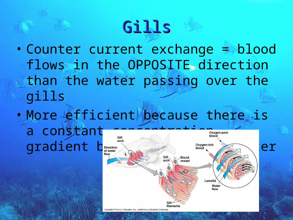
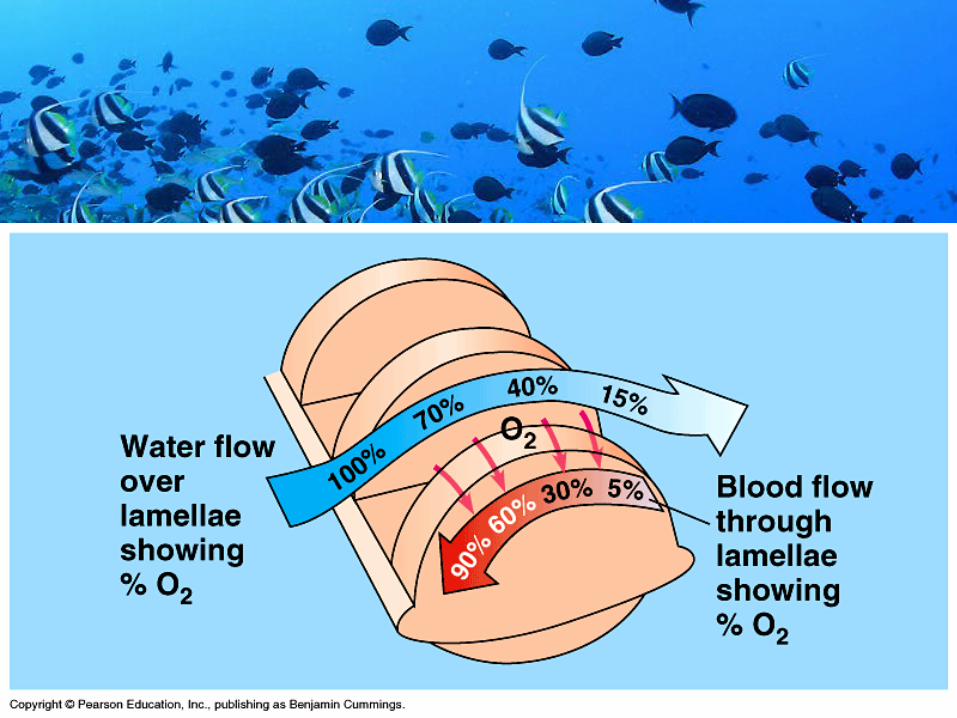
GillsGills• Counter current exchange = blood flows in the
OPPOSITE direction than the water passing over the gills
• More efficient because there is a constant concentration gradient between blood and water

TracheaeTracheae• Insects
• Air has higher O2 content
– Gases diffuse faster
– Surfaces do not have to be ventilated as thoroughly
• Dessicated (dry out)

Tracheal SystemTracheal System
• Trachea = tiny air tubes that branch over the entire body
• Spiracles= pores in the exoskeleton of animals for gas exchange
• Air enters via spiracles and diffuses into the trachea into smaller branches which extend to every cell
– Open circulatory system

Tracheal SystemTracheal System
• Some ‘breathe’, ventilate (muscle contractions)
• Others use diffusion
Tracheal systems; may have ‘pouches’ near major organs

LungsLungs
• InInvaginations of body surface
• Thoracic cavity
• Highly subdivided; many branches
– Surface area
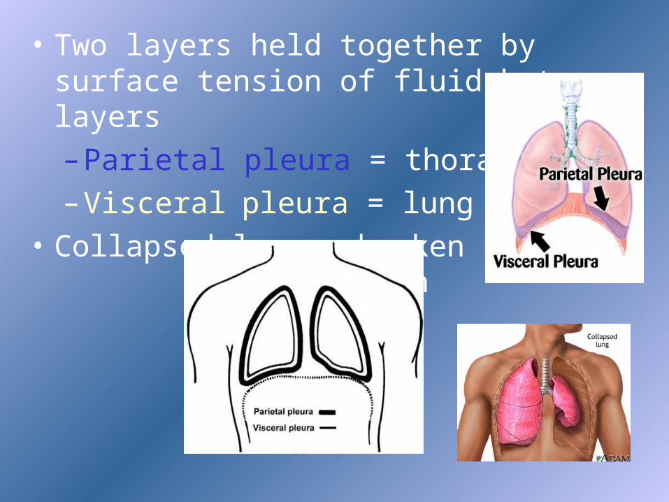
• Two layers held together by surface tension of fluid between layers
– Parietal pleura = thoracic wall
– Visceral pleura = lung surface
• Collapsed lung - broken surface tension

LungsLungs
• Air enters nostrils; filtered by hairs, warmed, humidified
• Pharynx, larynxPharynx, larynx (voice box with vocal cords)
• Cartilage lined tracheatrachea
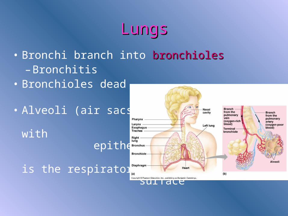
• Forks into 2 bronchibronchi
LungsLungs
• Bronchi branch into bronchiolesbronchioles– Bronchitis
• Bronchioles dead end into alveolialveoli
• Alveoli (air sacs) are lined with epithelium which is the respiratory surface

Skin - CutaneousSkin - Cutaneous• Amphibians, frogs, salamanders
• Small, flat
• Lots of surface area

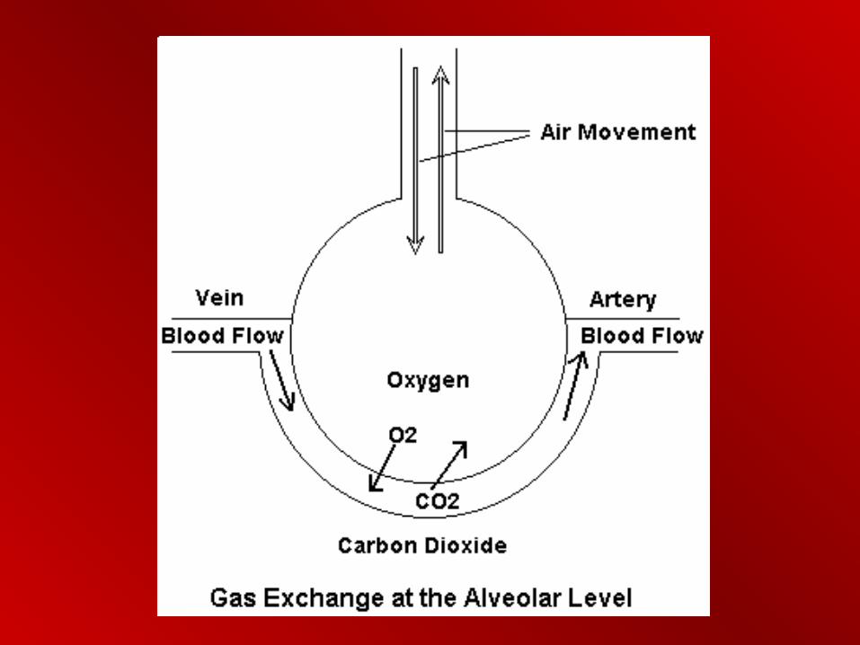
Lungs Lungs • Oxygen dissolves in the moist film covering the
epithelium
• Diffuses into the capillaries surrounding each alveolus
• Carbon dioxide goes opposite

Lungs Lungs
• Vertebrates ventilate by BREATHING
• Inhalation, exhalation
• Positive = frogs
• Negative = mammals

Frog VentilationFrog Ventilation
• Enlarge mouth by lowering the floor of the mouth
• Close mouth and nostrils
• Push floor up
• Air forced into trachea
• Primitive lungs; mostly skin
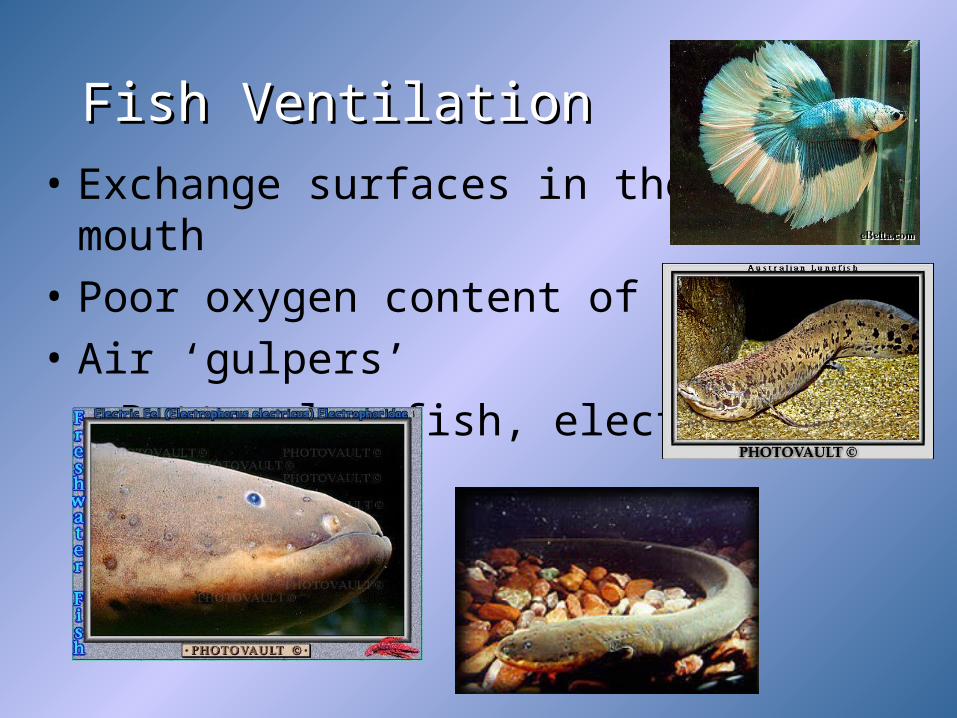
Fish VentilationFish Ventilation
• Exchange surfaces in their mouth
• Poor oxygen content of water
• Air ‘gulpers’
– Betta, lungfish, electric eel

Negative VentilationNegative Ventilation

Ventilating Lungs: MammalsVentilating Lungs: Mammals
• P = 1/V; inverse relationship
• Increase volume, decrease pressure
• Increase size of thoracic cavity decreases the pressure (less than the 760 mm atmospheric pressure)


Ventilating Lungs: MammalsVentilating Lungs: Mammals
• Greater pressure on the outside causes air to ‘push in’ to the lungs (less pressure)
• Diaphragm moves down, ribs expand outward (increased volume)

Mammal BreathingMammal Breathing
• Parietal pleura attached to the ribs
• Visceral pleura attached to the parietal pleura (surface tension from the fluid between)
• As ribs expand, the lungs expand
• Collapsed lung

Mammal BreathingMammal Breathing• Tidal volume = amount of air an organism
inhales and exhales w/ each breath; 500 ml in humans
• Residual volume = amount of air left in the lungs after exhalation
• Vital capacity = maximum volume when forced (running); 4-5000 ml in college males

Breathing: BirdsBreathing: Birds
• Lungs; P. 892
• 8-9 air sacs in abdomen, neck, wings
• Reduces density (lighter)
• Heat sink for heat produced by flying (radiator)


BirdsBirds• Parabronchi = small channels in the lungs
where gas exchange takes place– More efficient; air moves in only one
direction; no dead ends – Air is constantly pumped through– Maximizes complete air exchange (no
residual volume)– Very efficient; altitude

Control Centers in the Brain Control Centers in the Brain
Regulate Rate and Depth of Breathing
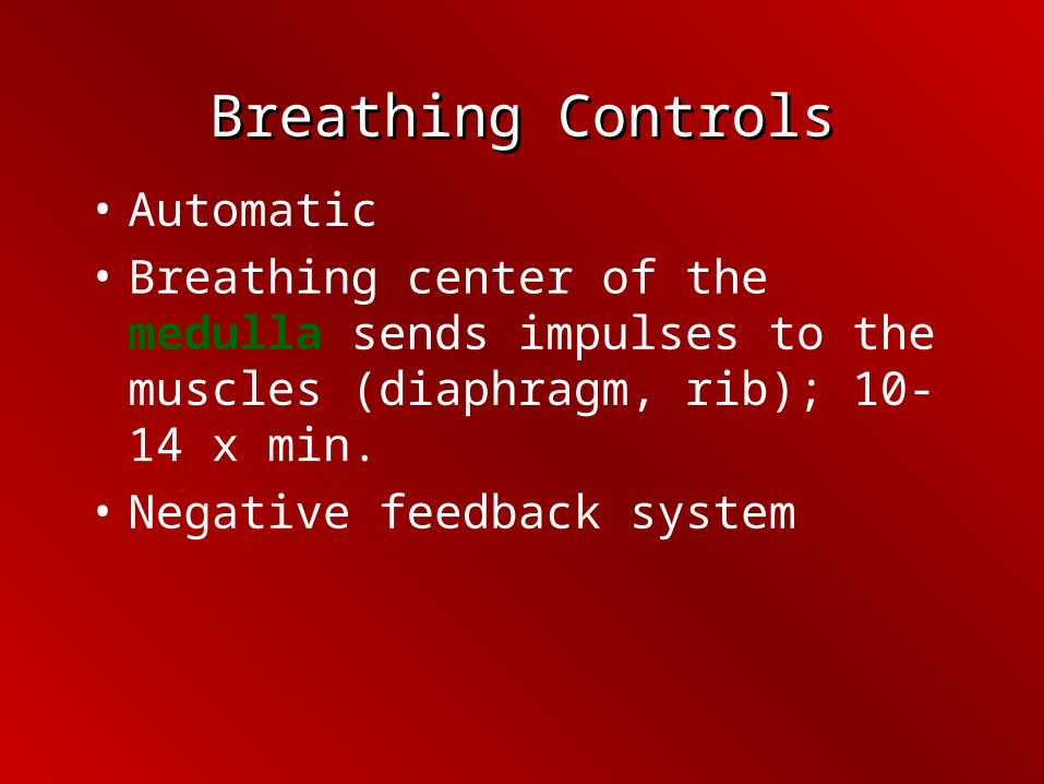
Breathing ControlsBreathing Controls
• Automatic
• Breathing center of the medulla sends impulses to the muscles (diaphragm, rib); 10-14 x min.
• Negative feedback system

Negative Feedback Negative Feedback
• Stretch the lungs; pressure sensors send messages back to medulla inhibiting inspiration
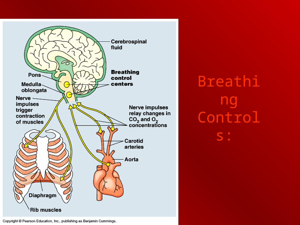
Breathing Controls:

Breathing ControlsBreathing Controls
• Breathing center monitors blood pH; if CO2 levels increase, pH drops (acidic, acidosis)
• CO2 + H2O H2CO3
• Stimulates increase in tempo

Gas ExchangeGas Exchange
• Gas enters or leaves depends upon:

Partial PressurePartial Pressure
• O2 = 21 % of atmosphere
• CO2 = 0.03 %
• Partial pressure = proportion of pressure contributed by a gas in a mixture of gases
• Air = gas mixture
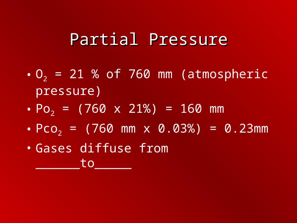
Partial PressurePartial Pressure
• O2 = 21 % of 760 mm (atmospheric pressure)
• Po2 = (760 x 21%) = 160 mm
• Pco2 = (760 mm x 0.03%) = 0.23mm
• Gases diffuse from ______to_____
Partial Pressure: p. 894Partial Pressure: p. 894
• In the alveoli, the PO2 is high and PCO2 is low
• In systemic cells, PO2 is low and PCO2 is high
• O2 diffuses into systemic from the blood; CO2 is opposite
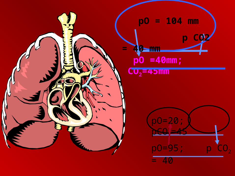
pO =40mm; CO2=45mm
pO=95; p CO2 = 40
pO=20; pCO2=45
pO = 104 mm
p CO2 = 40 mm


Respiratory PigmentsRespiratory Pigments
• Oxygen is carried by pigments because O2 does not diffuse easily into water
• Arthropods (insects, crabs) have Hemocyanin;
• Copper not iron

Respiratory PigmentsRespiratory Pigments
• Dissolved in plasma
• Open circulatory
• Hemoglobin; vertebrates

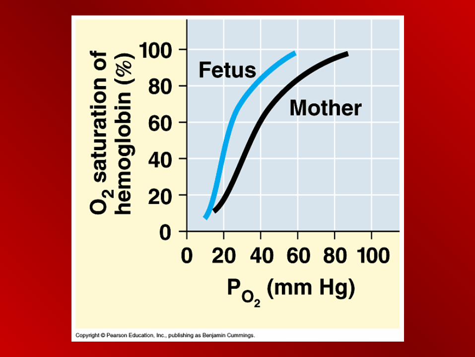
Dissociation; p. 895Dissociation; p. 895
• Release of oxygen by hemoglobin is dependent upon:
– Partial pressure
– pH
DissociationDissociation
• At rest, PO2 in tissues is low;
• Hemoglobin releases only a portion of its O2 (28%)
• If the partial pressure goes down (exercising) then hemoglobin releases more O2

Dissociation curve

Bohr ShiftBohr Shift
• pH causes a change in hemoglobin affinity for oxygen
• During exercise, increased CO2 is dissolved in the plasma
• CO2 becomes carbonic acid (decreases pH)

Bohr ShiftBohr Shift• Conformation of hemoglobin is sensitive to
pH change; loses affinity for O2
• BOHR SHIFT• Active muscle causes Bohr Shift, hemoglobin
releases more O2


COCO22
• Carbon dioxide carried by blood in 3 forms:
– Bicarbonate ions in blood (70%)
– Bound to amino groups (23%)
– Dissolved in plasma (7%)

COCO22
• CO2 diffuses into erythrocytes
• Carbonic anhydrase converts CO2 into bicarbonate
• CO2 + H2O H2CO3 H+ + HCO3-
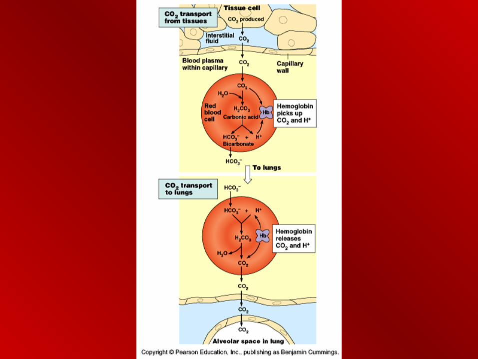

Blood ChemistryBlood Chemistry
• CO2 + H2O H2CO3 H+ + HCO3-
(bicarbonate)
• Carbonic acid lowers pH; H+ is tied up by hemoglobin molecule to prevent drastic lowering of pH

Special Adaptations Special Adaptations
• Seals, whales make lonnnggg underwater dives; deep
• Myoglobin = oxygen storing pigment in muscles
• More O2 in blood
• Twice the blood per kg as humans


Special AdaptationsSpecial Adaptations
• Very large spleen
– Contracts - releasing additional blood
• Diving reflex slows heart rate
– Oxygen consumption slows
• Blood routed to brain, eyes, glands, placenta
• Muscles shift to fermentation