CARDIOVASCULAR SYSTEM Replacing hearts - nova. Cardiovascular System Functions The cardiovascular...
30
CARDIOVASCULAR SYSTEM Replacing hearts - nova
-
Upload
maximillian-cummings -
Category
Documents
-
view
229 -
download
3
Transcript of CARDIOVASCULAR SYSTEM Replacing hearts - nova. Cardiovascular System Functions The cardiovascular...

Cardiovascular System Functions
The cardiovascular system allows exchange of oxygen and carbon dioxide between blood and tissues
Accomplished by three components:1. Heart – pump blood2. Vessels – transport network; arteries, veins
and capillaries3. Blood – transports dissolved gases, nutrients,
hormones and metabolic wastes

Organization of Cardiovascular System
1. Systemic Circuit – oxygenated blood leaves the heart through the aorta and returns to the heart through systemic veins
2. Pulmonary Circuit– deoxygenated blood leaves the heart through pulmonary arteries and returns to the heart through pulmonary veins
A closed system with two circuits

The Heart…size, shape and location
Muscular organ composed of cardiac muscle
About the size of a closed fist Weighs less than a pound Located within the thoracic cavity Flanked on either side by the lungs

The heart…coverings and wall
Pericardium = double walled sac around the heart Protects the heart and anchors it to
surrounding structures Produces a lubricating fluid to create a
frictionless environment Pericarditis = inflammation of the
pericardium

The heart…coverings and wall
The wall of the heart is composed of three layers:1. Epicardium = continuous with the
pericardium2. Myocardium = thick bundles of cardiac
muscle; the contractile part of the heart3. Endocardium = think sheet of
endothelium that lines the heart chambers

The heart…coverings and wall

Anatomy of the heart
4 chambers Upper chambers = atria = receiving chambers Lower chambers = ventricles = pumping
chambers Left ventricle forms the apex or point of the heart Interventricular septum divides ventricles

Chambers – Anterior View
Left atriumRight
atrium
Right Ventricle
Left Ventricle

Chambers – Posterior View
Left atrium Right atrium
Right Ventricle
Left Ventricle

Chambers – Internal View
Left atriumRight
atrium
Right Ventricle
Left Ventricle

Anatomy of the heart
Heart Valves permit flow of blood in one direction
1. AV valves divide atria and ventricles; prevent backflow of blood into atria when ventricles contract
Bicuspid valve (mitral valve) – between left atria and left ventricle
Tricupsid valve between right atria and right ventricle Chordae Tendinae = tiny white cords that anchor
the valves to the walls of the ventricleLAB RAT…Left Atrium Bicuspid
Right Atrium Tricuspid

AV Valves

Anatomy of the heart
Heart Valves permit flow of blood in one direction
2. Semilunar valves guard the large vessels leaving the ventricles; prevents arterial blood from falling back into the ventricles
Pulmonary semilunar valve Aortic semilunar valve

Semilunar Valves

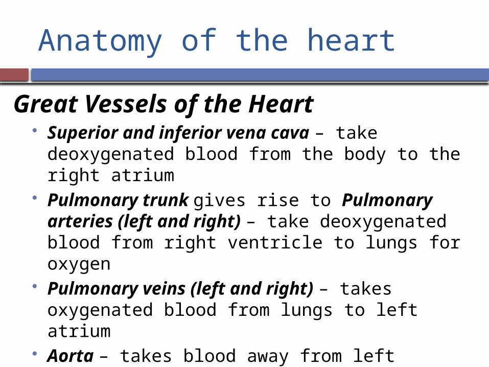
Anatomy of the heart
Great Vessels of the Heart Superior and inferior vena cava – take
deoxygenated blood from the body to the right atrium
Pulmonary trunk gives rise to Pulmonary arteries (left and right) – take deoxygenated blood from right ventricle to lungs for oxygen
Pulmonary veins (left and right) – takes oxygenated blood from lungs to left atrium
Aorta – takes blood away from left ventricle to rest of body

Anatomy of the heart
Cardiac Circulation Coronary arteries branch from the base of the
aorta and encircle the heart at the junction between the atria and ventricles Compressed when ventricles are contracting and fill
when the ventricles are relaxed Cardiac veins drain the myocardium into the
right atrium

Vessels – Anterior View
Superior vena cava
Inferior vena cava
L. Pulmonary artery
L. Pulmonaryveins
R. Pulmonaryveins
aorta
Pulmonary trunkR. Pulmonary artery
Coronary vessels

Vessels – posterior view
Superior vena cava
Inferior vena cava
R. Pulmonary veins
aortaR. Pulmonary arteryL. Pulmonary
arteryL. Pulmonaryveins
Coronary vessels

When the heart beats
The Right side of the heart Deoxygenated blood flows into heart from vena
cava Right atrium Tricuspid valve Right ventricle Pulmonary semilunar valve Pulmonary artery Lungs (to pick up oxygen)

What’s behind a heartbeat
The Left side of the heart Oxygenated blood flows into heart from lungs via
pulmonary veins Left atrium Bicuspid valve (aka mitral valve) Left ventricle Aortic semilunar valve Aorta Body (to deliver oxygen)

Control of Heart Contraction
General info about cardiac muscle Cardiac muscle cells contract spontaneously
and independently, even if all nervous connections are cut
Muscle cells in different areas of the heart have different rythms Atria cells contract at a faster rate than ventricular
cells An intrinsic control system regulates activity

Intrinsic Control System
Intrinsic conduction system Sinoatrial node (SA node) Atrioventricular node (AV node) Bundle of His Purkinje Fibers

Sinoatrial Node (SA node)
The pacemaker of the heart Located in right atrium SA node sends out electrical impulse Impulse spreads over atria, making them
contract Impulse travels to AV node

Atrioventricular node (AV node) Conducting cell group between atria and
ventricle Carries impulse to bundle of His

Bundle of His
Conducting fibers in septum (divider between left and right sides of the heart
Divides into right and left branches to a network of b ranches in ventricles (Purkinje Fibers)

Purkinje Fibers
Impulse shoots along Purkinje fibers causing ventricles to contract

Electrocardiogram (EKG or ECG) Device used to record the electrical
activity of the heart Systole = contraction phase Diastole = relaxation phase
Baseline of EKG is flatline P = atrial contraction QRS = ventricular contraction T = ventricular relaxation