Cardiovascular and Systemic MicrovascularEffects of Anti ... · PDF fileEffects of...
8
PRE-CLINICAL RESEARCH Cardiovascular and Systemic Microvascular Effects of Anti-Vascular Endothelial Growth Factor Therapy for Cancer J. Todd Belcik, BS, RDCS,* Yue Qi, MD,* Beat A. Kaufmann, MD,* Aris Xie, BS,* Sherry Bullens, BA,† Terry K. Morgan, MD, PHD,‡ Susan P. Bagby, MD,§ Ganesh Kolumam, PHD,† Joe Kowalski, BS,† Jon A. Oyer, PHD, Stuart Bunting, PHD,† Jonathan R. Lindner, MD* Portland, Oregon; and South San Francisco, California Objectives This study sought to evaluate the contribution of microvascular functional rarefaction and changes in vascular mechanical properties to the development of hypertension and secondary ventricular remodeling that occurs with anti-vascular endothelial growth factor (VEGF) therapy. Background Hypertension is a common side effect of VEGF inhibitors used in cancer medicine. Methods Mice were treated for 5 weeks with an anti-murine VEGF-A monoclonal antibody, antibody plus ramipril, or sham treatment. Microvascular blood flow (MBF) and blood volume (MBV) were quantified by contrast-enhanced ultra- sound in skeletal muscle, left ventricle (LV), and kidney. Echocardiography and invasive hemodynamics were used to assess ventricular function, dimensions and vascular mechanical properties. Results Ambulatory blood pressure increased gradually over the first 3 weeks of anti-VEGF therapy. Compared with controls, anti-VEGF–treated mice had similar aortic elastic modulus and histological appearance, but a marked increase in ar- terial elastance, indicating increased afterload, and elevated plasma angiotensin II. Increased afterload in treated mice led to concentric LV remodeling and reduced stroke volume without impaired LV contractility determined by LV peak change in pressure over time (dp/dt) and the end-systolic dimension–pressure relation. Anti-VEGF therapy did not alter MBF or MBV in skeletal muscle, myocardium, or kidney; but did produce cortical mesangial glomerulosclero- sis. Ramipril therapy almost entirely prevented the adverse hemodynamic effects, increased afterload, and LV remod- eling in anti-VEGF–treated mice. Conclusions Neither reduced functional microvascular density nor major alterations in arterial mechanical properties are primary causes of hypertension during anti-VEGF therapy. Inhibition of VEGF leads to an afterload mismatch state, increased angiotensin II, and LV remodeling, which are all ameliorated by angiotensin-converting enzyme inhibition. (J Am Coll Cardiol 2012;60:618–25) © 2012 by the American College of Cardiology Foundation Antagonists to vascular endothelial growth factor (VEGF) such as humanized anti-human monoclonal antibodies (mAbs) and small molecule inhibitors of VEGF signaling are commonly used to treat cancers (1). These drugs are associated with a high incidence of hypertension and proteinuria (2–5). Several potential mechanisms for hypertension have been proposed. Sys- temic capillary rarefaction could contribute to hyperten- sion by increasing vascular resistance, although there is still ambiguity regarding whether capillary regression See page 626 during VEGF inhibition is widespread throughout the sys- temic circulation, or confined to specific vulnerable beds such as the thyroid, the trachea, the intestinal villi, and the skin (2,6 –9). Hypertension from VEGF inhibitors could also be secondary to increased vascular tone that occurs from a reduc- tion in VEGF receptor–mediated nitric oxide (NO) produc- From the *Division of Cardiovascular Medicine, Oregon Health & Science Univer- sity, Portland, Oregon; †Department of Tumor Biology and Angiogenesis, Genen- tech Inc., South San Francisco, California; ‡Department of Pathology, Oregon Health & Science University, Portland, Oregon; §Division of Nephrology and Hypertension, Oregon Health & Science University, Portland, Oregon; and the Vollum Institute, Oregon Health & Science University, Portland, Oregon. This study was supported in part by a grant from Genentech Inc. Dr. Kaufmann is supported by a research grant from the Lichtenstein Foundation. Dr. Morgan is supported by the Office of Research on Women’s Health and the National Institute of Child Health and Human Development, Oregon BIRCWH HD043488-08. Dr. Lindner is supported by grants R01-HL-078610, R01-DK-063508, and RC1-HL- 100659 from the National Institutes of Health. All other authors have reported that they have no relationships relevant to the contents of this paper to disclose. Manuscript received November 3, 2011; revised manuscript received February 2, 2012, accepted February 6, 2012. Journal of the American College of Cardiology Vol. 60, No. 7, 2012 © 2012 by the American College of Cardiology Foundation ISSN 0735-1097/$36.00 Published by Elsevier Inc. http://dx.doi.org/10.1016/j.jacc.2012.02.053
Transcript of Cardiovascular and Systemic MicrovascularEffects of Anti ... · PDF fileEffects of...

Journal of the American College of Cardiology Vol. 60, No. 7, 2012© 2012 by the American College of Cardiology Foundation ISSN 0735-1097/$36.00
PRE-CLINICAL RESEARCH
Cardiovascular and Systemic MicrovascularEffects of Anti-Vascular Endothelial Growth FactorTherapy for Cancer
J. Todd Belcik, BS, RDCS,* Yue Qi, MD,* Beat A. Kaufmann, MD,* Aris Xie, BS,*Sherry Bullens, BA,† Terry K. Morgan, MD, PHD,‡ Susan P. Bagby, MD,§ Ganesh Kolumam, PHD,†Joe Kowalski, BS,† Jon A. Oyer, PHD,� Stuart Bunting, PHD,† Jonathan R. Lindner, MD*
Portland, Oregon; and South San Francisco, California
Objectives This study sought to evaluate the contribution of microvascular functional rarefaction and changes in vascularmechanical properties to the development of hypertension and secondary ventricular remodeling that occurswith anti-vascular endothelial growth factor (VEGF) therapy.
Background Hypertension is a common side effect of VEGF inhibitors used in cancer medicine.
Methods Mice were treated for 5 weeks with an anti-murine VEGF-A monoclonal antibody, antibody plus ramipril, or shamtreatment. Microvascular blood flow (MBF) and blood volume (MBV) were quantified by contrast-enhanced ultra-sound in skeletal muscle, left ventricle (LV), and kidney. Echocardiography and invasive hemodynamics wereused to assess ventricular function, dimensions and vascular mechanical properties.
Results Ambulatory blood pressure increased gradually over the first 3 weeks of anti-VEGF therapy. Compared with controls,anti-VEGF–treated mice had similar aortic elastic modulus and histological appearance, but a marked increase in ar-terial elastance, indicating increased afterload, and elevated plasma angiotensin II. Increased afterload in treatedmice led to concentric LV remodeling and reduced stroke volume without impaired LV contractility determined by LVpeak change in pressure over time (dp/dt) and the end-systolic dimension–pressure relation. Anti-VEGF therapy didnot alter MBF or MBV in skeletal muscle, myocardium, or kidney; but did produce cortical mesangial glomerulosclero-sis. Ramipril therapy almost entirely prevented the adverse hemodynamic effects, increased afterload, and LV remod-eling in anti-VEGF–treated mice.
Conclusions Neither reduced functional microvascular density nor major alterations in arterial mechanical properties are primarycauses of hypertension during anti-VEGF therapy. Inhibition of VEGF leads to an afterload mismatch state, increasedangiotensin II, and LV remodeling, which are all ameliorated by angiotensin-converting enzyme inhibition.(J Am Coll Cardiol 2012;60:618–25) © 2012 by the American College of Cardiology Foundation
Published by Elsevier Inc. http://dx.doi.org/10.1016/j.jacc.2012.02.053
Antagonists to vascular endothelial growth factor(VEGF) such as humanized anti-human monoclonalantibodies (mAbs) and small molecule inhibitors of
From the *Division of Cardiovascular Medicine, Oregon Health & Science Univer-sity, Portland, Oregon; †Department of Tumor Biology and Angiogenesis, Genen-tech Inc., South San Francisco, California; ‡Department of Pathology, OregonHealth & Science University, Portland, Oregon; §Division of Nephrology andHypertension, Oregon Health & Science University, Portland, Oregon; and the�Vollum Institute, Oregon Health & Science University, Portland, Oregon. Thisstudy was supported in part by a grant from Genentech Inc. Dr. Kaufmann issupported by a research grant from the Lichtenstein Foundation. Dr. Morgan issupported by the Office of Research on Women’s Health and the National Instituteof Child Health and Human Development, Oregon BIRCWH HD043488-08. Dr.Lindner is supported by grants R01-HL-078610, R01-DK-063508, and RC1-HL-100659 from the National Institutes of Health. All other authors have reported thatthey have no relationships relevant to the contents of this paper to disclose.
Manuscript received November 3, 2011; revised manuscript received February 2,2012, accepted February 6, 2012.
VEGF signaling are commonly used to treat cancers (1).These drugs are associated with a high incidence ofhypertension and proteinuria (2–5). Several potentialmechanisms for hypertension have been proposed. Sys-temic capillary rarefaction could contribute to hyperten-sion by increasing vascular resistance, although there isstill ambiguity regarding whether capillary regression
See page 626
during VEGF inhibition is widespread throughout the sys-temic circulation, or confined to specific vulnerable beds suchas the thyroid, the trachea, the intestinal villi, and the skin(2,6–9). Hypertension from VEGF inhibitors could also besecondary to increased vascular tone that occurs from a reduc-
tion in VEGF receptor–mediated nitric oxide (NO) produc-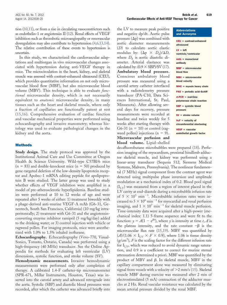
ta(aetk
M
SIH(gttwmiranpctrtEShpdHmt(str
tapa(mwacACpcwteMembwGwMbdstlStdm(
i
t
619JACC Vol. 60, No. 7, 2012 Belcik et al.August 14, 2012:618–25 Cardiovascular Effects of Anti-VEGF Therapy for Cancer
tion (10,11), or from a rise in circulating vasoconstrictors suchas endothelin-1 or angiotensin II (12). Renal effects of VEGFinhibition such as thrombotic microangiopathy or renovasculardysregulation may also contribute to hypertension (4,6,13,14).The relative contribution of these events to hypertension isuncertain.
In this study, we characterized the cardiovascular adap-tations and multiorgan in vivo microvascular changes asso-ciated with hypertension during anti-VEGF therapy inmice. The microcirculation in the heart, kidney, and skeletalmuscle was assessed with contrast-enhanced ultrasound (CEU),which provides quantitative information on not only micro-vascular blood flow (MBF), but also microvascular bloodvolume (MBV). This technique is able to evaluate func-tional microvascular density, which is not necessarilyequivalent to anatomic microvascular density, in manyissues such as the heart and skeletal muscle, where only
fraction of capillaries are functionally patent at rest15,16). Comprehensive evaluation of cardiac functionnd vascular mechanical properties were performed usingchocardiography and invasive manometry; whereas his-ology was used to evaluate pathological changes in theidney and the aorta.
ethods
tudy design. The study protocol was approved by thenstitutional Animal Care and Use Committee at Oregonealth & Science University. Wild-type C57Bl/6 mice
n � 83) and double-knockout mice (n � 50) produced byene-targeted deletion of the low-density lipoprotein recep-or and Apobec-1 mRNA editing peptide for apolipopro-ein B were studied. The latter group was used to studyhether effects of VEGF inhibition were amplified in aodel of pre-atherosclerotic hyperlipidemia. Baseline stud-
es were performed at 10 to 12 weeks of age and wereepeated after 5 weeks of either: 1) treatment biweekly withphage-derived anti-murine VEGF-A mAb (G6-31, Ge-entech, South San Francisco, California) (10 mg/kg intra-eritoneally; 2) treatment with G6-31 and the angiotensin-onverting enzyme inhibitor ramipril (5 mg/kg/day) addedo the drinking water; or 3) control injection with vehicle oragweed pollen. For imaging protocols, mice were anesthe-ized with 1.0% to 1.5% inhaled isoflurane.chocardiography. Echocardiography (Vevo-770, Visual-onics, Toronto, Ontario, Canada) was performed using aigh-frequency (40 MHz) transducer. See the Online Ap-endix for methods for evaluating left ventricular (LV)imensions, systolic function, and stroke volume (SV).emodynamic measurements. Invasive hemodynamiceasurements were performed only after completion of
herapy. A calibrated 1.4-F catheter-tip micromanometerSPR-671, Millar Instruments, Houston, Texas) was in-erted into the carotid artery and advanced retrograde intohe aorta. Systolic (SBP) and diastolic blood pressures were
ecorded, after which the catheter was advanced briefly intohe LV to measure peak positivend negative dp/dt. Aortic pulseressure (�p) was combined withortic diameter measurementsD) to calculate aortic elasticodulus by: (�p � Dd)/�D,here Dd is aortic diastolic di-
meter. Arterial elastance wasalculated by: (0.9 � SBP)/SV (17).mbulatory blood pressure.onscious ambulatory blood
ressure was measured using aarotid artery catheter interfacedith a radiotelemetry pressure
ransducer (PA-C10, Data Sci-nces International, St. Paul,
innesota). After allowing sev-ral days for recovery, pressureeasurements were recorded at
aseline and twice weekly for 5eeks after starting therapy with6-31 (n � 10) or control (rag-eed pollen) injections (n � 9).icrovascular perfusion and
lood volume. Lipid-shelledecafluorobutane microbubbles were prepared (18). Perfu-ion imaging of the myocardium, proximal hindlimb adduc-or skeletal muscle, and kidney was performed using ainear-array transducer (Sequoia 512, Siemens Medicalystems, Malvern, Pennsylvania). The nonlinear fundamen-al (7 MHz) signal component from the contrast agent wasetected using multipulse phase inversion and amplitudeodulation at a mechanical index of 0.2. Blood pool signal
ILV) was measured from a region of interest placed in theLV cavity at end-diastole during a microbubble infusion rateof 5 � 105 min�1. Microbubble infusion rates were in-creased to 5 � 106 min�1 for myocardial and renal perfusionmaging, and 1 � 107 min�1 for skeletal muscle perfusion.
Time-intensity data were acquired after a high-power (me-chanical index: 1.1) 5-frame sequence and were fit to thefunction: y � A(1 � e�t), where y is intensity at time t, A ishe plateau intensity, and the rate constant –� is the
microvascular flux rate (15,19). MBV was quantified by(A)/(1.06 � ILV � F � 0.9), where 1.06 is tissue density(g/cm3), F is the scaling factor for the different infusion ratefor ILV, which was reduced to avoid dynamic range satura-tion, and 0.9 is a coefficient to correct for murine sternalattenuation determined a priori. MBF was quantified by theproduct of MBV and �. In skeletal muscle, MBV in thecapillary compartment alone was measured by eliminatingsignal from vessels with a velocity of �2 mm/s (15). Skeletalmuscle MBF during exercise was measured after 2 min ofelectrostimulated (5 mA) contraction of the adductor mus-cles at 2 Hz. Renal vascular resistance was calculated by the
Abbreviationsand Acronyms
CEU � contrast-enhancedultrasound
LV � leftventricle/ventricular
mAb � monoclonalantibody
MBF � microvascularblood flow
MBV � microvascularblood velocity
MHC � myosin heavy chain
PAS � periodic acid–Schiff
rt-PCR � real-timepolymerase chain reaction
SBP � systolic bloodpressure
SV � stroke volume
VcF � velocity ofcircumferential shortening
VEGF � vascularendothelial growth factor
mean arterial pressure divided by th
e renal MBF.
hncEroecHspts
iCsblccwSaqswssmmm
tagMssknb
R
CILsIsdtcm
swocm
G
620 Belcik et al. JACC Vol. 60, No. 7, 2012Cardiovascular Effects of Anti-VEGF Therapy for Cancer August 14, 2012:618–25
PCR for markers of hypertrophy and remodeling�-myosin heavy chain mRNA. The mRNA for �-myosineavy chain (MHC), atrial natriuretic peptide (ANP), and B-typeatriuretic peptide (BNP) was measured by real-time polymerasehain reaction (rt-PCR) (Online Appendix).ndothelial nitric oxide synthase phosphorylation. Phospho-
ylation of endothelial nitric oxide synthase (eNOS), whichccurs downstream from VEGF receptor activation, wasvaluated by enzyme-linked immunosorbent assay fromardiac muscle samples (Online Appendix).
istology. For renal histology, periodic acid–Schiff (PAS)taining of fixed paraffin-embedded coronal sections waserformed. Sections were evaluated in triplicate blinded toreatment. Glomerular and tubulointerstitial injury werecored as 0 � absent, 1 � mild focal, 2 � moderate
multifocal, or 3 � severe diffuse cortical involvement. Renalmmunohistology was performed using hamster anti-mouseD31 (MAB1398Z, Chemicon/Millipore, Billerica, Mas-
achusetts) primary and Cy3-labeled secondary mAbs. Fi-rin staining was performed with fluorescein isothiocyanate–abeled anti-fibrinogen/fibrin primary mAb (AXL234F, Ac-urate Chemical, Westbury, New York). Skeletal muscle mi-rovascular density was assessed by immunohistochemistryith a Cy3-labeled smooth muscle �-actin mAb (1A4, Sigma,t. Louis, Missouri) and was expressed by the total microvesselrea per muscle section area. Myocardial collagen content wasuantified by the percent area positive for picrosirius redtaining under polarized light. Myocyte cross-sectional areaas measured from 15 to 25 myocytes from �5 separate
ections stained with hematoxylin & eosin. Verhoeff elastintaining of the aorta was performed to evaluate the number andean thickness of elastic lamina and thickness of the tunicaedia averaged from 4 separate radial locations. Elastin frag-entation was scored blinded to treatment as 0 � none, 1 �
minimal, 2 � mild, 3 � moderate, or 4 � severe.Plasma and urine biomarkers. Plasma angiotensin II con-centrations were determined using an enzyme immunoassay(SPI-BIO, Montigny le Bretonneux, France) with an Ell-man’s chromagenic reagent that has a minimum detectable
Echocardiography DataTable 1 Echocardiography Data
Control (n � 18)
Week 0 Week 5
LVIDd, mm 3.4 � 0.2 3.3 � 0.2
LVIDs, mm 2.3 � 0.2 2.4 � 0.3
WTd, mm 0.66 � 0.09 0.68 � 0.09 0
LV mass, mg 66 � 13 66 � 12
Thickening fraction 0.56 � 0.14 0.53 � 0.15 0
SV, �l 50 � 16 40 � 10
VcF, mm/s 0.66 � 0.15 0.60 � 0.17 0
S’, mm/s 15.8 � 4.6 12.9 � 4.7* 1
E’, mm/s 12.3 � 4.2 9.7 � 5.3* 1
Values are mean � SD. *p � 0.05 versus week 0 data; †p � 0.05 versus week 0 data and versus6-31�ramipril with respect to change in value from week 0 to week 5.
E’ � peak early diastolic endocardial velocity; LVID � left ventricular internal dimension; S’ � peak sysWTd � diastolic wall thickness.
concentration of 1 pg/ml. Urinary albumin concentration wasquantified by a competitive enzyme-linked immunosorbentassay (Albuwell-M, Exocell, Philadelphia, Pennsylvania).Statistical methods. Unless otherwise specified, data arepresented as mean � SD. Effects of treatment were assessedwith a paired t test for normally distributed variables. Groupcomparisons were made with 1-way analysis of variancewith t tests for independent samples and Bonferroni correc-ion. Scored histopathology data were evaluated using either
Kruskal-Wallis test or a Mann-Whitney test for inter-roup comparisons. rt-PCR data were compared with aann-Whitney test. A p value of �0.05 was considered
ignificant. Since within each treatment group there were noignificant differences between wild-type and double-nockout mice for any baseline or post-treatment hemody-amic or echocardiographic parameters, data were com-ined for the 2 groups.
esults
ardiovascular and hemodynamic effects of VEGF inhibition.n sham-treated mice, there were no significant changes inV dimensions, mass, or echocardiographic measures of
ystolic function over the 5-week treatment period (Table 1).nhibition of VEGF with G6-31 for 5 weeks produced aignificant increase in LV wall thickness and mass, and aecrease in end-diastolic diameter consistent with concen-ric hypertrophy. The presence of hypertrophy in G6-31ompared with control mice was further supported by largeryocyte cross-sectional area (122 � 15 �m2 vs. 100 �
13 �m2, p � 0.04) and higher ventricular expression of�-MHC, ANP, and BNP mRNA on rtPCR (Online Fig. A),although myocardial collagen area on histology was similar(2.7 � 1.1% vs. 2.8 � 1.3%, p � 0.91; n � 6 each; exampleshown in Online Fig. B). In G6-31–treated animals, thereas also a reduction in thickening fraction, SV, and velocityf circumferential shortening (VcF) (Table 1). All of thehanges in ventricular size and function in G6-31–treatedice were completely attenuated by ramipril co-administration.
G6-31 (n � 18) G6-31�Ramipril (n � 14)
0 Week 5 Week 0 Week 5
0.2 3.1 � 0.3* 3.6 � 0.3 3.5 � 0.4
0.3 2.4 � 0.3 2.5 � 0.3 2.6 � 0.4
0.09 0.79 � 0.06†‡ 0.66 � 0.12 0.68 � 0.08
12 73 � 8* 67 � 13 68 � 13
0.19 0.38 � 0.12* 0.54 � 0.21 0.42 � 0.11
10 31 � 7* 45 � 12 42 � 24
0.13 0.48 � 0.18* 0.54 � 0.12 0.49 � 0.17
3.0 12.3 � 4.7* 15.6 � 2.7 12.2 � 4.9*
5.6 9.7 � 2.7* 13.6 � 5.7 10.3 � 3.3*
control and G6-31�ramipril (corrected for multiple comparisons); ‡p � 0.05 versus control and
Week
3.4 �
2.3 �
.65 �
66 �
.63 �
45 �
.69 �
5.2 �
3.6 �
week 5
tolic endocardial velocity; SV � stroke volume; VcF � velocity of circumferential fiber shortening;
atwsmw3apnGwtmpt3
621JACC Vol. 60, No. 7, 2012 Belcik et al.August 14, 2012:618–25 Cardiovascular Effects of Anti-VEGF Therapy for Cancer
On endocardial tissue Doppler imaging, there was a mildage-dependent decrease in S= and E=, the degree of whichwas not significantly different between treatment groups.
On invasive hemodynamic evaluation, G6-31–treatedmice had higher SBP, diastolic blood pressure, and peakpositive and negative LV dp/dt than either the control orG6-31�ramipril cohort (Table 2). The degree of hyperten-
Hemodynamics and Aortic Mechanical PropertiesTable 2 Hemodynamics and Aortic Mechanical Properties
Control(n � 12)
G6-31(n � 10)
G6-31�Ramipril(n � 9)
SBP, mm Hg 99 � 7 108 � 9* 98 � 6
DBP, mm Hg 65 � 8 76 � 8* 66 � 8
LV�dp/dt, mm Hg/s 9,070 � 1,995 10,508 � 1,407 8,042 � 1,660
LV�dp/dt, mm Hg/s �7,320 � 2,245 �8,624 � 2,369 �7,469 � 2,283
Elastic modulus, KPa 28.0 � 8.3 31.3 � 8.5 29.3 � 10.1
EA, mm Hg/�l 3.4 � 1.0 4.4 � 1.3† 3.7 � 1.7
Values are mean � SD. *p � 0.05 versus control and G6-31�ramipril; †p � 0.05 versus control only.Abbreviations as in Table 1.
Figure 1 Ascending Thoracic Aorta Histology
Mean (�SEM) (A) thickness of the tunica media, (B) number of elastic lamina, anapy. (D) Elastic lamina fragmentation scores, expressed as a proportion for eachascending aorta illustrating normal (top) and mild fragmentation (bottom, arrow)in each group.
sion caused by G6-31 was even more prominent on con-scious ambulatory recordings with a 17 � 11%, 25 � 11%,nd 38 � 11% increase in SBP for weeks 1, 2, and 3 ofherapy, respectively (analysis of variance p � 0.01), afterhich blood pressures stabilized. Ambulatory blood pres-
ures were constant over time in sham-treated mice. Elasticodulus of the aorta was similar between treatment groups,hereas arterial elastance was significantly greater in G6-1–treated mice (Table 2), indicating a marked increase infterload without any major alterations in the mechanicalroperties of the aorta. On aortic histology, medial thick-ess and indices of elastin content were not different in6-31–treated compared with control mice (Fig. 1). Thereas a nonsignificant trend toward more elastin fragmenta-
ion in G6-31–treated mice, although the degree of frag-entation was mild or less in all animals. Post-treatment
lasma angiotensin II concentration was higher in G6-31–reated compared with control mice (47 � 17 pg/ml vs. 78 �6 pg/ml, p � 0.01, n � 12 each), whereas myocardial
elastin layer thickness for control and G6-31–treated mice after 5 weeks of ther-p � NS by Mann-Whitney and chi-square analysis. (E) Elastin staining of theelastic laminae. Scale bar � 20 �m. Four measurements were made for 8 mice
d (C)score.of the
mamtmwTTpn
622 Belcik et al. JACC Vol. 60, No. 7, 2012Cardiovascular Effects of Anti-VEGF Therapy for Cancer August 14, 2012:618–25
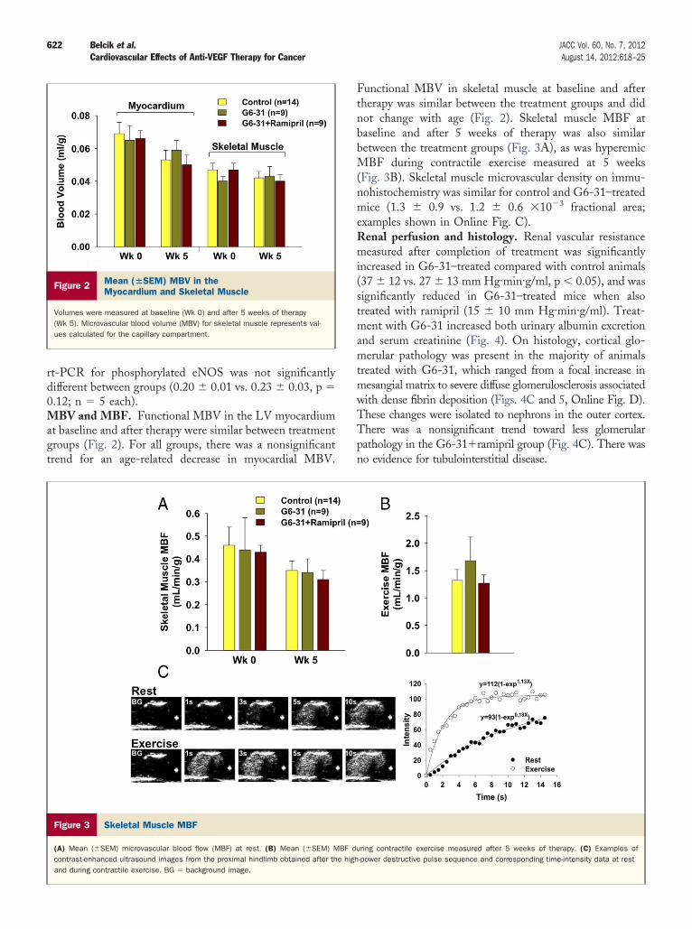
rt-PCR for phosphorylated eNOS was not significantlydifferent between groups (0.20 � 0.01 vs. 0.23 � 0.03, p �0.12; n � 5 each).MBV and MBF. Functional MBV in the LV myocardiumat baseline and after therapy were similar between treatmentgroups (Fig. 2). For all groups, there was a nonsignificanttrend for an age-related decrease in myocardial MBV.
Figure 2 Mean (�SEM) MBV in theMyocardium and Skeletal Muscle
Volumes were measured at baseline (Wk 0) and after 5 weeks of therapy(Wk 5). Microvascular blood volume (MBV) for skeletal muscle represents val-ues calculated for the capillary compartment.
Figure 3 Skeletal Muscle MBF
(A) Mean (�SEM) microvascular blood flow (MBF) at rest. (B) Mean (�SEM) Mcontrast-enhanced ultrasound images from the proximal hindlimb obtained after thand during contractile exercise. BG � background image.
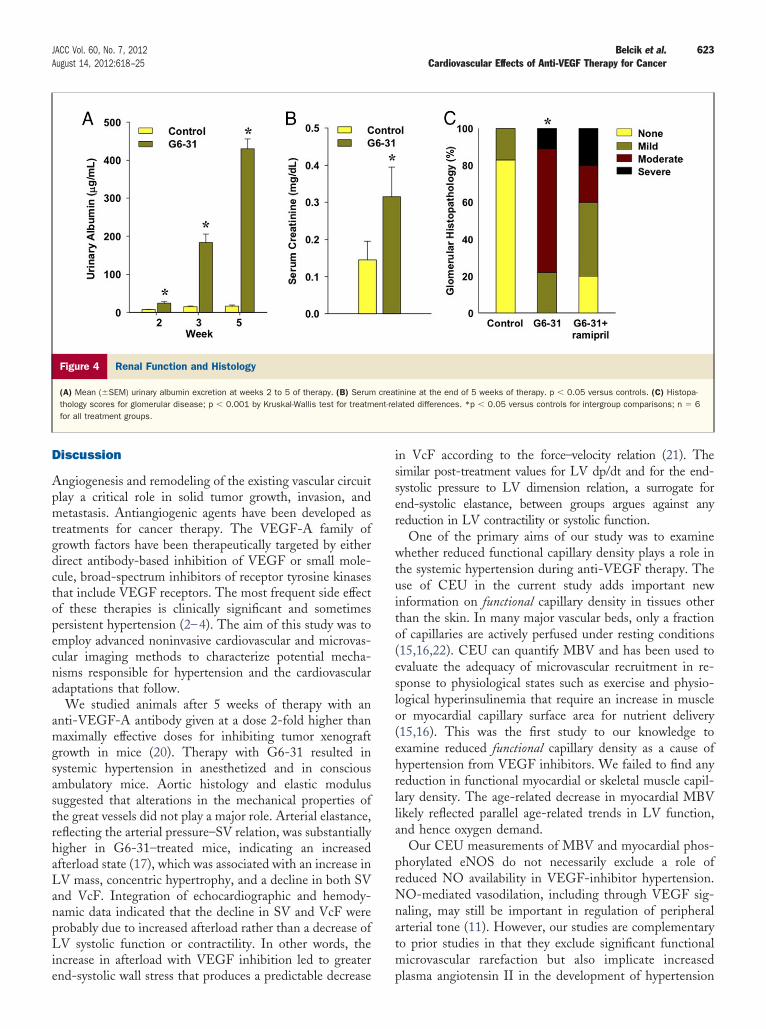
Functional MBV in skeletal muscle at baseline and aftertherapy was similar between the treatment groups and didnot change with age (Fig. 2). Skeletal muscle MBF atbaseline and after 5 weeks of therapy was also similarbetween the treatment groups (Fig. 3A), as was hyperemicMBF during contractile exercise measured at 5 weeks(Fig. 3B). Skeletal muscle microvascular density on immu-nohistochemistry was similar for control and G6-31–treatedmice (1.3 � 0.9 vs. 1.2 � 0.6 �10�3 fractional area;examples shown in Online Fig. C).Renal perfusion and histology. Renal vascular resistancemeasured after completion of treatment was significantlyincreased in G6-31–treated compared with control animals(37 � 12 vs. 27 � 13 mm Hg·min·g/ml, p � 0.05), and wassignificantly reduced in G6-31–treated mice when alsotreated with ramipril (15 � 10 mm Hg·min·g/ml). Treat-
ent with G6-31 increased both urinary albumin excretionnd serum creatinine (Fig. 4). On histology, cortical glo-erular pathology was present in the majority of animals
reated with G6-31, which ranged from a focal increase inesangial matrix to severe diffuse glomerulosclerosis associatedith dense fibrin deposition (Figs. 4C and 5, Online Fig. D).hese changes were isolated to nephrons in the outer cortex.here was a nonsignificant trend toward less glomerularathology in the G6-31�ramipril group (Fig. 4C). There waso evidence for tubulointerstitial disease.
ring contractile exercise measured after 5 weeks of therapy. (C) Examples of-power destructive pulse sequence and corresponding time-intensity data at rest
BF due high
623JACC Vol. 60, No. 7, 2012 Belcik et al.August 14, 2012:618–25 Cardiovascular Effects of Anti-VEGF Therapy for Cancer
Discussion
Angiogenesis and remodeling of the existing vascular circuitplay a critical role in solid tumor growth, invasion, andmetastasis. Antiangiogenic agents have been developed astreatments for cancer therapy. The VEGF-A family ofgrowth factors have been therapeutically targeted by eitherdirect antibody-based inhibition of VEGF or small mole-cule, broad-spectrum inhibitors of receptor tyrosine kinasesthat include VEGF receptors. The most frequent side effectof these therapies is clinically significant and sometimespersistent hypertension (2–4). The aim of this study was toemploy advanced noninvasive cardiovascular and microvas-cular imaging methods to characterize potential mecha-nisms responsible for hypertension and the cardiovascularadaptations that follow.
We studied animals after 5 weeks of therapy with ananti-VEGF-A antibody given at a dose 2-fold higher thanmaximally effective doses for inhibiting tumor xenograftgrowth in mice (20). Therapy with G6-31 resulted insystemic hypertension in anesthetized and in consciousambulatory mice. Aortic histology and elastic modulussuggested that alterations in the mechanical properties ofthe great vessels did not play a major role. Arterial elastance,reflecting the arterial pressure–SV relation, was substantiallyhigher in G6-31–treated mice, indicating an increasedafterload state (17), which was associated with an increase inLV mass, concentric hypertrophy, and a decline in both SVand VcF. Integration of echocardiographic and hemody-namic data indicated that the decline in SV and VcF wereprobably due to increased afterload rather than a decrease ofLV systolic function or contractility. In other words, theincrease in afterload with VEGF inhibition led to greater
Figure 4 Renal Function and Histology
(A) Mean (�SEM) urinary albumin excretion at weeks 2 to 5 of therapy. (B) Serumthology scores for glomerular disease; p � 0.001 by Kruskal-Wallis test for treatmfor all treatment groups.
end-systolic wall stress that produces a predictable decrease
in VcF according to the force–velocity relation (21). Thesimilar post-treatment values for LV dp/dt and for the end-systolic pressure to LV dimension relation, a surrogate forend-systolic elastance, between groups argues against anyreduction in LV contractility or systolic function.
One of the primary aims of our study was to examinewhether reduced functional capillary density plays a role inthe systemic hypertension during anti-VEGF therapy. Theuse of CEU in the current study adds important newinformation on functional capillary density in tissues otherthan the skin. In many major vascular beds, only a fractionof capillaries are actively perfused under resting conditions(15,16,22). CEU can quantify MBV and has been used toevaluate the adequacy of microvascular recruitment in re-sponse to physiological states such as exercise and physio-logical hyperinsulinemia that require an increase in muscleor myocardial capillary surface area for nutrient delivery(15,16). This was the first study to our knowledge toexamine reduced functional capillary density as a cause ofhypertension from VEGF inhibitors. We failed to find anyreduction in functional myocardial or skeletal muscle capil-lary density. The age-related decrease in myocardial MBVlikely reflected parallel age-related trends in LV function,and hence oxygen demand.
Our CEU measurements of MBV and myocardial phos-phorylated eNOS do not necessarily exclude a role ofreduced NO availability in VEGF-inhibitor hypertension.NO-mediated vasodilation, including through VEGF sig-naling, may still be important in regulation of peripheralarterial tone (11). However, our studies are complementaryto prior studies in that they exclude significant functionalmicrovascular rarefaction but also implicate increased
tinine at the end of 5 weeks of therapy. p � 0.05 versus controls. (C) Histopa-lated differences. *p � 0.05 versus controls for intergroup comparisons; n � 6
creaent-re
plasma angiotensin II in the development of hypertension

624 Belcik et al. JACC Vol. 60, No. 7, 2012Cardiovascular Effects of Anti-VEGF Therapy for Cancer August 14, 2012:618–25
and ventricular remodeling during VEGF inhibition. Theangiotensin-converting enzyme inhibitor ramipril had asalutary effect on almost all cardiovascular and hemody-namic derangements in G6-31–treated mice, although thisdoes not necessarily imply that angiotensin II was the onlymediator of hypertension.
With regard to the renal changes, the interaction ofangiotensin and NO in regulating afferent and efferentarteriolar tone, renal blood flow, and glomerular filtrationrate has been well characterized (23). It is feasible that
A
C
E
G
Figure 5 Examples of Histopathology From the Outer Renal Co
(A, C, E, and G) Control mice and (B, D, F, and H) G6-31–treated mice. Examples(E and F), and fibrin staining (G and H representing sections that were directly adglomeruli that were graded as normal (A and C) and severe mesangial change (Bble by CD31 staining (E). Examples of PAS staining for G6-31 mice and fibrin/CD3
altered renal afferent–efferent arteriolar resistance balance
from high plasma angiotensin II, possibly in combinationwith low NO production, may have had a role in thedevelopment of proteinuria and the increase in renal vascu-lar resistance in G6-31–treated mice. Inhibition of VEGFhas also been associated with glomerular endothelial alter-ations and a thrombotic microangiopathy (13,14). Weobserved outer cortical glomerular mesangial proliferationafter G6-31 and diffuse fibrin deposition, which has beenpreviously described with VEGF inhibition (14,24). Therewas no evidence for advanced thrombotic microangiopathy
rom Control and G6-31–Treated Mice
e periodic acid–Schiff (PAS) staining (A to D), endothelial staining with CD31to corresponding CD31 staining sections). The PAS staining examples show). Arrows (G) illustrate the location of nonstaining glomeruli that are clearly visi-rlay images with confocal microscopy are provided in the Online Appendix.
B
D
F
H
rtex F
includjacentand D1 ove
that has also been described, possibly because of study-

etmtg
625JACC Vol. 60, No. 7, 2012 Belcik et al.August 14, 2012:618–25 Cardiovascular Effects of Anti-VEGF Therapy for Cancer
related differences in mechanism and/or duration of VEGFinhibition. Whether the glomerular structural and functionalabnormalities were due to altered afferent/efferent vasoregula-tory balance, direct glomerular effects of VEGF, or simply tothe hypertension is not clear. However, we did observe thattreatment with ramipril had a salutary effect on renal patho-logic changes and especially on renal vascular resistance.Study limitations. First, we did not evaluate the effect ofither dose or duration of anti-VEGF therapy. We believehe 5-week time interval is appropriate to study the deter-inants of hypertension, given the temporal course of hyper-
ension in humans. However, it has been shown that angio-enesis is important in the response to pressure overload (25),
so that longer studies may have revealed progression to systolicfunctional decline associated with a decrease in functionalmyocardial MBV. The dose of G6-31 was based on previousstudies evaluating efficacy of neovascularization in murinetumor models (20). The lack of increased myocardial collagenin the G6-31–treated animals was an unexpected finding if webelieve that angiotensin II may have played a role in LVremodeling during VEGF inhibition. It is possible that thisfinding reflects the short treatment duration.
The most important limitation of this study is that wehave not provided evidence for any 1 mechanism forhypertension during anti-VEGF therapy. We believe thatthe process is likely complex and multifactorial. However,we have shown that antibody-mediated VEGF inhibitionproduces rapid-onset hypertension, increased afterload, andLV remodeling with reduced SV, but without impairmentin LV systolic function or contractility. We have alsoexcluded the pathophysiological contributions from func-tional capillary loss in large microvascular beds or abnormalarterial elastic properties. We have also detected an increasein plasma angiotensin II and that angiotensin-convertingenzyme inhibitors appear to be an effective preventivestrategy against, not only the hypertension, but also theadverse cardiac effects of VEGF inhibition.
Reprint requests and correspondence: Dr. Jonathan R. Lindner,Cardiovascular Division, UHN-62, Oregon Health & ScienceUniversity, 3181 SW Sam Jackson Park Road, Portland, Oregon97239. E-mail: [email protected].
REFERENCES
1. Duda DG, Batchelor TT, Willett CG, Jain RK. VEGF-targetedcancer therapy strategies: current progress, hurdles and future pros-pects. Trends Mol Med 2007;13:223–30.
2. Kamba T, McDonald DM. Mechanisms of adverse effects of anti-VEGF therapy for cancer. Br J Cancer 2007;96:1788–95.
3. Hurwitz H, Fehrenbacher L, Novotny W, et al. Bevacizumab plusirinotecan, fluorouracil, and leucovorin for metastatic colorectal cancer.N Engl J Med 2004;350:2335–42.
4. Shord SS, Bressler LR, Tierney LA, Cuellar S, George A. Under-standing and managing the possible adverse effects associated withbevacizumab. Am J Health Syst Pharm 2009;66:999–1013.
5. Motzer RJ, Rini BI, Bukowski RM, et al. Sunitinib in patients withmetastatic renal cell carcinoma. JAMA 2006;295:2516–24.
6. Kamba T, Tam BY, Hashizume H, et al. VEGF-dependent plasticityof fenestrated capillaries in the normal adult microvasculature. Am JPhysiol Heart Circ Physiol 2006;290:H560–76.
7. Inai T, Mancuso M, Hashizume H, et al. Inhibition of vascularendothelial growth factor (VEGF) signaling in cancer causes loss ofendothelial fenestrations, regression of tumor vessels, and appearanceof basement membrane ghosts. Am J Pathol 2004;165:35–52.
8. Tang K, Breen EC, Gerber HP, Ferrara NM, Wagner PD. Capillaryregression in vascular endothelial growth factor-deficient skeletalmuscle. Physiol Genomics 2004;18:63–9.
9. Steeghs N, Rabelink TJ, op ’t Roodt J, et al. Reversibility of capillarydensity after discontinuation of bevacizumab treatment. Ann Oncol2010;21:1100–5.
10. Horowitz JR, Rivard A, van der Zee R, et al. Vascular endothelialgrowth factor/vascular permeability factor produces nitric oxide-dependent hypotension. Evidence for a maintenance role in quiescentadult endothelium. Arterioscler Thromb Vasc Biol 1997;17:2793–800.
11. Facemire CS, Nixon AB, Griffiths R, Hurwitz H, Coffman TM. Vascularendothelial growth factor receptor 2 controls blood pressure by regulatingnitric oxide synthase expression. Hypertension 2009;54:652–8.
12. Kappers MH, van Esch JH, Sluiter W, Sleijfer S, Danser AH, van denMeiracker AH. Hypertension induced by the tyrosine kinase inhibitorsunitinib is associated with increased circulating endothelin-1 levels.Hypertension 2010;56:675–81.
13. Eremina V, Sood M, Haigh J, et al. Glomerular-specific alterations ofVEGF-A expression lead to distinct congenital and acquired renaldiseases. J Clin Invest 2003;111:707–16.
14. Eremina V, Jefferson JA, Kowalewska J, et al. VEGF inhibition andrenal thrombotic microangiopathy. N Engl J Med 2008;358:1129–36.
15. Dawson D, Vincent MA, Barrett EJ, et al. Vascular recruitment inskeletal muscle during exercise and hyperinsulinemia assessed by contrastultrasound. Am J Physiol Endocrinol Metab 2002;282:E714–20.
16. Le DE, Bin JP, Coggins MP, Wei K, Lindner JR, Kaul S. Relationbetween myocardial oxygen consumption and myocardial blood vol-ume: a study using myocardial contrast echocardiography. J Am SocEchocardiogr 2002;15:857–63.
17. Kelly RP, Ting CT, Yang TM, et al. Effective arterial elastance asindex of arterial vascular load in humans. Circulation 1992;86:513–21.
18. Behm CZ, Kaufmann BA, Carr C, et al. Molecular imaging ofendothelial vascular cell adhesion molecule-1 expression and inflam-matory cell recruitment during vasculogenesis and ischemia-mediatedarteriogenesis. Circulation 2008;117:2902–11.
19. Wei K, Jayaweera AR, Firoozan S, Linka A, Skyba DM, Kaul S.Quantification of myocardial blood flow with ultrasound-induceddestruction of microbubbles administered as a constant venous infu-sion. Circulation 1998;97:473–83.
20. Liang WC, Wu X, Peale FV, et al. Cross-species vascular endothelialgrowth factor (VEGF)-blocking antibodies completely inhibit thegrowth of human tumor xenografts and measure the contribution ofstromal VEGF. J Biol Chem 2006;281:951–61.
21. Glick G, Sonnenblick EH, Braunwald E. Myocardial force-velocityrelations studied in intact unanesthetized man. J Clin Invest 1965;44:978–88.
22. Honig CR, Odoroff CL, Frierson JL. Capillary recruitment in exer-cise: rate, extent, uniformity, and relation to blood flow. Am J Physiol1980;238:H31–42.
23. Patzak A, Persson AE. Angiotensin II-nitric oxide interaction in thekidney. Curr Opin Nephrol Hypertens 2007;16:46–51.
24. Hara A, Wada T, Furuichi K, et al. Blockade of VEGF acceleratesproteinuria, via decrease in nephrin expression in rat crescenticglomerulonephritis. Kidney Int 2006;69:1986–95.
25. Shiojima I, Sato K, Izumiya Y, et al. Disruption of coordinated cardiachypertrophy and angiogenesis contributes to the transition to heartfailure. J Clin Invest 2005;115:2108–18.
Key Words: contrast echocardiography y VEGF y ventricularhypertrophy.
APPENDIX
For an expanded Methods section and supplementary figures,
please see the online version of this paper.