Anti-vascular endothelial growth factor treatment … endothelial growth factor treatment normalizes...
6
Anti-vascular endothelial growth factor treatment normalizes tuberculosis granuloma vasculature and improves small molecule delivery Meenal Datta a,b,1 , Laura E. Via c,1 , Walid S. Kamoun a,1 , Chong Liu a,2 , Wei Chen a,3 , Giorgio Seano a , Danielle M. Weiner c , Daniel Schimel c , Kathleen England c , John D. Martin a,d , Xing Gao a,4 , Lei Xu a , Clifton E. Barry III c,e,5 , and Rakesh K. Jain a,5 a Edwin L. Steele Laboratory for Tumor Biology, Department of Radiation Oncology, Massachusetts General Hospital and Harvard Medical School, Boston, MA 02114; b Department of Chemical and Biological Engineering, Tufts University, Medford, MA 02155; c Tuberculosis Research Section, Laboratory of Clinical Infectious Disease, National Institute of Allergy and Infectious Disease, National Institutes of Health, Bethesda, MD 20892; d Department of Chemical Engineering, Massachusetts Institute of Technology, Cambridge, MA 02139; and e Department of Clinical Laboratory Sciences, Faculty of Health Sciences, University of Cape Town, Rondebosch 7701, South Africa Contributed by Rakesh K. Jain, December 29, 2014 (sent for review November 19, 2014; reviewed by Harold F. Dvorak, Jerrold Ellner, Eric J. Rubin, and David G. Russell) Tuberculosis (TB) causes almost 2 million deaths annually, and an increasing number of patients are resistant to existing therapies. Patients who have TB require lengthy chemotherapy, possibly because of poor penetration of antibiotics into granulomas where the bacilli reside. Granulomas are morphologically similar to solid cancerous tumors in that they contain hypoxic microenvironments and can be highly fibrotic. Here, we show that TB-infected rabbits have impaired small molecule distribution into these disease sites due to a functionally abnormal vasculature, with a low-molecular- weight tracer accumulating only in peripheral regions of granulo- matous lesions. Granuloma-associated vessels are morphologically and spatially heterogeneous, with poor vessel pericyte coverage in both human and experimental rabbit TB granulomas. Moreover, we found enhanced VEGF expression in both species. In tumors, antiangiogenic, specifically anti-VEGF, treatments can “normalize” their vasculature, reducing hypoxia and creating a window of op- portunity for concurrent chemotherapy; thus, we investigated vessel normalization in rabbit TB granulomas. Treatment of TB- infected rabbits with the anti-VEGF antibody bevacizumab signif- icantly decreased the total number of vessels while normalizing those vessels that remained. As a result, hypoxic fractions of these granulomas were reduced and small molecule tracer delivery was increased. These findings demonstrate that bevacizumab treat- ment promotes vascular normalization, improves small molecule delivery, and decreases hypoxia in TB granulomas, thereby pro- viding a potential avenue to improve delivery and efficacy of cur- rent treatment regimens. antiangiogenesis | hypoxia | host-directed therapy | Mycobacterium tuberculosis | rabbit model A s one of the most prevalent infectious diseases in the world today, Mycobacterium tuberculosis (MTB) infects roughly one-third of the global population, resulting in 2 million deaths annually (1). Although current treatment regimens are largely successful in curing the disease (2), they require 6–8 mo of treatment with up to four agents (3), and multidrug-resistant bacterial strains have emerged and proliferated (4). Resistance to front-line therapies necessitates treatment with up to five or six second-line agents that are poorly tolerated, and treatment success is only achieved in 40–70% of patients (5). Failure to cure drug-resistant disease leads to acquisition of further re- sistance with a progressively poorer prognosis for these patients, thus fueling an emerging epidemic of drug-resistant disease that threatens to overwhelm fragile health care systems in developing countries (6). When infected with the tuberculosis (TB) bacilli, the body triggers an immune response that walls off the bacteria in dense cellular masses known as granulomas, or tubercular lesions (7). These abnormal tissue structures, which can vary in size within the same host, are surrounded by fibrous cuffs that serve to contain the MTB bacilli (7, 8). Recent studies have demon- strated a wide variation in the spatial distribution of drugs within TB granulomas, with very few agents able to penetrate the central regions (9). This differential ability of drugs to penetrate TB granulomas has been incorporated into modern TB drug development programs as a criterion for optimizing lead mole- cules and selecting efficacious combinations (10). However, the mechanisms that contribute to this differential penetration of Significance Tuberculosis (TB) is the second most lethal pathogen world- wide. Pulmonary granulomas are a hallmark of this disease. By discovering similarities between granulomas and solid cancer- ous tumors, we identified a novel therapeutic target for TB, the abnormal granuloma-associated vasculature that contributes to the abnormal granuloma microenvironment. We then asked if we could “normalize” granuloma vasculature by blocking VEGF signaling, an approach originally shown to enhance cancer treatment. Our results demonstrate that bevacizumab, a widely prescribed anti-VEGF antibody for cancer and eye diseases, is able to create more structurally and functionally normal granuloma vasculature and improve the delivery of a low-molecular-weight tracer. This effect suggests that vas- cular normalization in combination with anti-TB drugs has the potential to enhance treatment in patients with TB. Author contributions: L.E.V., W.S.K., L.X., C.E.B., and R.K.J. designed research; M.D., L.E.V., W.S.K., C.L., W.C., D.M.W., D.S., K.E., and X.G. performed research; M.D., L.E.V., W.S.K., G.S., and J.D.M. analyzed data; and M.D., L.E.V., L.X., C.E.B., and R.K.J. wrote the paper. Reviewers: H.F.D., Beth Israel Deaconess Medical Center; J.E., Boston University; E.J.R., Harvard School of Public Health; and D.G.R., Cornell University. Conflict of interest statement: R.K.J. received consultant fees from Enlight, Ophthotech, and SynDevRx. R.K.J. owns equity in Enlight, Ophthotech, SynDevRx, and XTuit and serves on the Board of Directors of XTuit and the Boards of Trustees of Tekla Healthcare Invest- ors, Tekla Life Sciences Investors, and the Tekla Healthcare Opportunities Fund. No re- agents or funding from these companies was used in these studies. Freely available online through the PNAS open access option. 1 M.D., L.E.V., and W.S.K. contributed equally to this work. 2 Present address: Department of Critical Care Medicine, Beijing Friendship Hospital, Capital Medical University, Beijing 100050, China. 3 Present address: Department of Pancreato-Biliary Surgery, The First Affiliated Hospital, Sun Yat-sen University, Guangzhou 510080, China. 4 Present address: Department of Oral and Maxillofacial Surgery, Xiangya Hospital, Central South University, Changsha 410008, China. 5 To whom correspondence may be addressed. Email: [email protected] or jain@steele. mgh.harvard.edu. This article contains supporting information online at www.pnas.org/lookup/suppl/doi:10. 1073/pnas.1424563112/-/DCSupplemental. www.pnas.org/cgi/doi/10.1073/pnas.1424563112 PNAS | February 10, 2015 | vol. 112 | no. 6 | 1827–1832 MEDICAL SCIENCES ENGINEERING
Transcript of Anti-vascular endothelial growth factor treatment … endothelial growth factor treatment normalizes...

Anti-vascular endothelial growth factor treatmentnormalizes tuberculosis granuloma vasculature andimproves small molecule deliveryMeenal Dattaa,b,1, Laura E. Viac,1, Walid S. Kamouna,1, Chong Liua,2, Wei Chena,3, Giorgio Seanoa, Danielle M. Weinerc,Daniel Schimelc, Kathleen Englandc, John D. Martina,d, Xing Gaoa,4, Lei Xua, Clifton E. Barry IIIc,e,5, and Rakesh K. Jaina,5
aEdwin L. Steele Laboratory for Tumor Biology, Department of Radiation Oncology, Massachusetts General Hospital and Harvard Medical School, Boston, MA02114; bDepartment of Chemical and Biological Engineering, Tufts University, Medford, MA 02155; cTuberculosis Research Section, Laboratory of ClinicalInfectious Disease, National Institute of Allergy and Infectious Disease, National Institutes of Health, Bethesda, MD 20892; dDepartment of ChemicalEngineering, Massachusetts Institute of Technology, Cambridge, MA 02139; and eDepartment of Clinical Laboratory Sciences, Faculty of Health Sciences,University of Cape Town, Rondebosch 7701, South Africa
Contributed by Rakesh K. Jain, December 29, 2014 (sent for review November 19, 2014; reviewed by Harold F. Dvorak, Jerrold Ellner, Eric J. Rubin, andDavid G. Russell)
Tuberculosis (TB) causes almost 2 million deaths annually, and anincreasing number of patients are resistant to existing therapies.Patients who have TB require lengthy chemotherapy, possiblybecause of poor penetration of antibiotics into granulomas wherethe bacilli reside. Granulomas are morphologically similar to solidcancerous tumors in that they contain hypoxic microenvironmentsand can be highly fibrotic. Here, we show that TB-infected rabbitshave impaired small molecule distribution into these disease sitesdue to a functionally abnormal vasculature, with a low-molecular-weight tracer accumulating only in peripheral regions of granulo-matous lesions. Granuloma-associated vessels are morphologicallyand spatially heterogeneous, with poor vessel pericyte coverage inboth human and experimental rabbit TB granulomas. Moreover,we found enhanced VEGF expression in both species. In tumors,antiangiogenic, specifically anti-VEGF, treatments can “normalize”their vasculature, reducing hypoxia and creating a window of op-portunity for concurrent chemotherapy; thus, we investigatedvessel normalization in rabbit TB granulomas. Treatment of TB-infected rabbits with the anti-VEGF antibody bevacizumab signif-icantly decreased the total number of vessels while normalizingthose vessels that remained. As a result, hypoxic fractions of thesegranulomas were reduced and small molecule tracer delivery wasincreased. These findings demonstrate that bevacizumab treat-ment promotes vascular normalization, improves small moleculedelivery, and decreases hypoxia in TB granulomas, thereby pro-viding a potential avenue to improve delivery and efficacy of cur-rent treatment regimens.
antiangiogenesis | hypoxia | host-directed therapy | Mycobacteriumtuberculosis | rabbit model
As one of the most prevalent infectious diseases in the worldtoday, Mycobacterium tuberculosis (MTB) infects roughly
one-third of the global population, resulting in 2 million deathsannually (1). Although current treatment regimens are largelysuccessful in curing the disease (2), they require 6–8 mo oftreatment with up to four agents (3), and multidrug-resistantbacterial strains have emerged and proliferated (4). Resistanceto front-line therapies necessitates treatment with up to five orsix second-line agents that are poorly tolerated, and treatmentsuccess is only achieved in 40–70% of patients (5). Failure tocure drug-resistant disease leads to acquisition of further re-sistance with a progressively poorer prognosis for these patients,thus fueling an emerging epidemic of drug-resistant disease thatthreatens to overwhelm fragile health care systems in developingcountries (6).When infected with the tuberculosis (TB) bacilli, the body
triggers an immune response that walls off the bacteria in densecellular masses known as granulomas, or tubercular lesions (7).
These abnormal tissue structures, which can vary in size withinthe same host, are surrounded by fibrous cuffs that serve tocontain the MTB bacilli (7, 8). Recent studies have demon-strated a wide variation in the spatial distribution of drugs withinTB granulomas, with very few agents able to penetrate thecentral regions (9). This differential ability of drugs to penetrateTB granulomas has been incorporated into modern TB drugdevelopment programs as a criterion for optimizing lead mole-cules and selecting efficacious combinations (10). However, themechanisms that contribute to this differential penetration of
Significance
Tuberculosis (TB) is the second most lethal pathogen world-wide. Pulmonary granulomas are a hallmark of this disease. Bydiscovering similarities between granulomas and solid cancer-ous tumors, we identified a novel therapeutic target for TB, theabnormal granuloma-associated vasculature that contributesto the abnormal granuloma microenvironment. We then askedif we could “normalize” granuloma vasculature by blockingVEGF signaling, an approach originally shown to enhancecancer treatment. Our results demonstrate that bevacizumab,a widely prescribed anti-VEGF antibody for cancer and eyediseases, is able to create more structurally and functionallynormal granuloma vasculature and improve the delivery ofa low-molecular-weight tracer. This effect suggests that vas-cular normalization in combination with anti-TB drugs has thepotential to enhance treatment in patients with TB.
Author contributions: L.E.V., W.S.K., L.X., C.E.B., and R.K.J. designed research; M.D., L.E.V.,W.S.K., C.L., W.C., D.M.W., D.S., K.E., and X.G. performed research; M.D., L.E.V., W.S.K.,G.S., and J.D.M. analyzed data; and M.D., L.E.V., L.X., C.E.B., and R.K.J. wrote the paper.
Reviewers: H.F.D., Beth Israel Deaconess Medical Center; J.E., Boston University; E.J.R.,Harvard School of Public Health; and D.G.R., Cornell University.
Conflict of interest statement: R.K.J. received consultant fees from Enlight, Ophthotech,and SynDevRx. R.K.J. owns equity in Enlight, Ophthotech, SynDevRx, and XTuit and serveson the Board of Directors of XTuit and the Boards of Trustees of Tekla Healthcare Invest-ors, Tekla Life Sciences Investors, and the Tekla Healthcare Opportunities Fund. No re-agents or funding from these companies was used in these studies.
Freely available online through the PNAS open access option.1M.D., L.E.V., and W.S.K. contributed equally to this work.2Present address: Department of Critical Care Medicine, Beijing Friendship Hospital,Capital Medical University, Beijing 100050, China.
3Present address: Department of Pancreato-Biliary Surgery, The First Affiliated Hospital,Sun Yat-sen University, Guangzhou 510080, China.
4Present address: Department of Oral and Maxillofacial Surgery, Xiangya Hospital,Central South University, Changsha 410008, China.
5To whom correspondence may be addressed. Email: [email protected] or [email protected].
This article contains supporting information online at www.pnas.org/lookup/suppl/doi:10.1073/pnas.1424563112/-/DCSupplemental.
www.pnas.org/cgi/doi/10.1073/pnas.1424563112 PNAS | February 10, 2015 | vol. 112 | no. 6 | 1827–1832
MED
ICALSC
IENCE
SEN
GINEE
RING
drugs are not fully understood, and novel strategies to improveTB drug delivery and efficacy are urgently needed.Following infection with MTB, pulmonary granulomas form in
humans and develop heterogeneous microenvironments, oftenfeaturing hypoxia (i.e., low levels of oxygen) and central necrosis,which are recapitulated in nonhuman primate and rabbit modelsof the disease (11). Large lesions appear to develop their ownvasculature, presumably allowing them to continue to grow (7).However, the morphological and functional characteristics ofgranuloma-associated vessels are largely unknown. In solidtumors, cancer cells can form similar dense tissue masses withabnormal associated vasculature. The physiological abnormali-ties that characterize tumor vessels have been investigated ex-tensively (12, 13). For example, hypoxia, a common feature insolid tumors, stimulates the overproduction of proangiogenicfactors, such as VEGF. Proangiogenic factors enhance the for-mation of new immature, tortuous, and hyperpermeable vessels(12, 14), often with excess endothelial cells, a lack of associatedpericytes (i.e., perivascular cells), and uneven basement mem-branes (15–17). These atypical features result in an impairedblood flow that further compromises delivery of drugs and oxygen(13). Hypoxia also causes immunosuppression, inflammation, andfibrosis, and it can also confer resistance to many drugs (18). Here,we propose that TB granulomas share many characteristics withsolid tumors, namely, that they are associated with abnormal anddysfunctional vasculature that can impair the delivery of smallmolecules, such as oxygen and antibiotics.Because VEGF is a critical growth factor required for new
blood vessel formation (16), anti-VEGF agents were originallydeveloped to block tumor growth by inhibiting blood vesselformation (19). However, bevacizumab, a humanized mono-clonal antibody developed to neutralize human VEGF, failed toimprove survival benefit as a monotherapy but conferred survivalbenefit only in combination with chemotherapy or immuno-therapy (18). A potential explanation for the success of com-bined therapies is that bevacizumab “normalizes” the abnormalvasculature of tumors, resulting in improved delivery of con-currently administered anticancer drugs, as well as alleviation ofhypoxia (13, 15, 18, 20, 21). However, this strategy has not beentested in a TB disease model. In this study we show, for the firsttime to our knowledge, in a rabbit model of TB that treatmentwith bevacizumab normalizes granuloma vasculature, reduceshypoxia, and enhances small molecule delivery during a “windowof normalization,” a transient effect observed in tumors (15, 20).Because anti-VEGF drugs have been approved for both malig-nant and nonmalignant diseases (18), our findings could be rapidlytested in the clinic to enhance TB treatment, shorten treatmentduration, and avert the development of treatment resistance.
ResultsHuman and Rabbit TB Granulomas Express VEGF. First, we examinedVEGF expression levels in histological sections from patients withTB who had received elective lung resection surgery and from ex-perimentally infected rabbits (Fig. 1). Granulomas are character-ized by concentric layers of differing cellular composition, oftenorganized around a central necrotic core (8). The predominant cellsfound in the outer layer are lymphocytes, whereas those cells nearerthe necrotic core are primarily epithelioid macrophages (Fig. 1);these two layers are frequently surrounded by a fibrotic rim (8). Weobserved that VEGF was detectable in the granulomatous regionsof the lung (Fig. 1 A and C) and that it was most highly expressed inthe inner macrophage layer (Fig. 1 B and D) of both human andrabbit granulomas. We examined similarly sized lymphoid aggre-gates in human lungs (Fig. S1 A and B) and rabbit lungs (Fig. S1 Cand D), and found that VEGF expression was minimal in thesestructures, where lymphocytes are the dominant cell type.
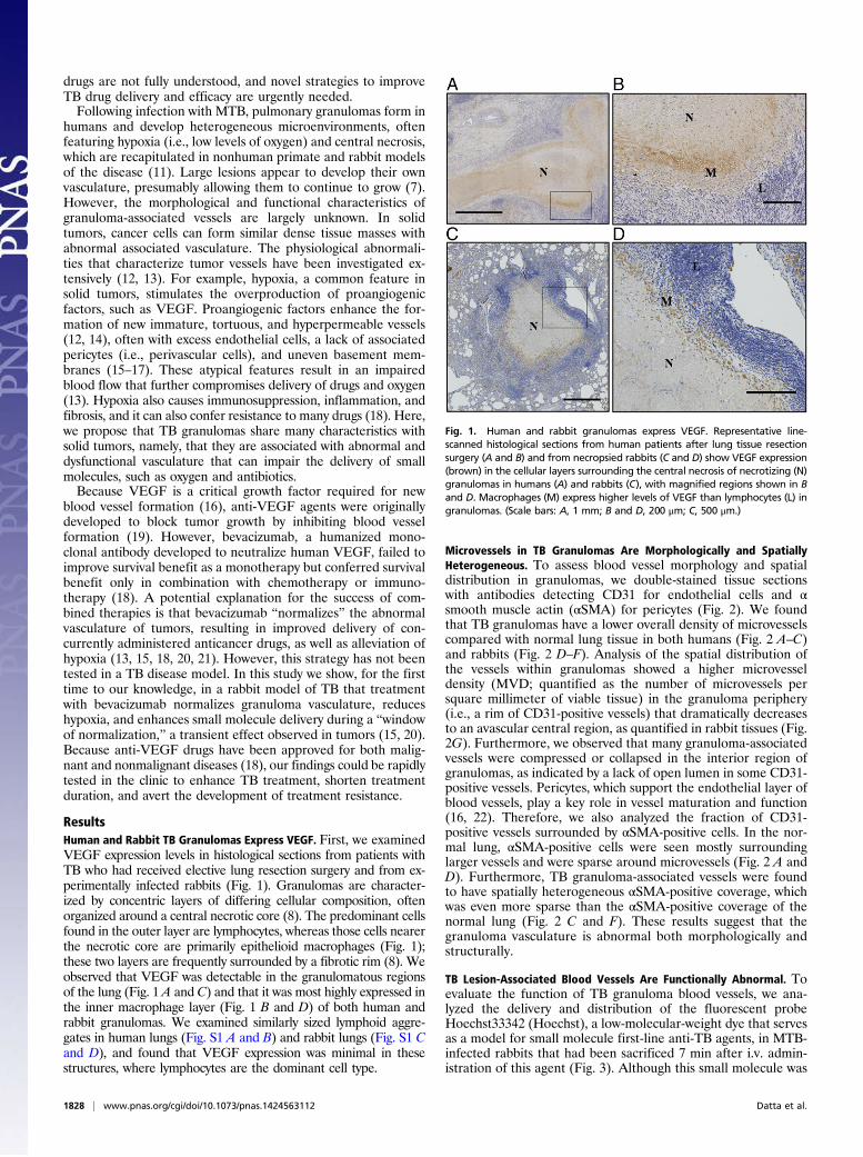
Microvessels in TB Granulomas Are Morphologically and SpatiallyHeterogeneous. To assess blood vessel morphology and spatialdistribution in granulomas, we double-stained tissue sectionswith antibodies detecting CD31 for endothelial cells and αsmooth muscle actin (αSMA) for pericytes (Fig. 2). We foundthat TB granulomas have a lower overall density of microvesselscompared with normal lung tissue in both humans (Fig. 2 A–C)and rabbits (Fig. 2 D–F). Analysis of the spatial distribution ofthe vessels within granulomas showed a higher microvesseldensity (MVD; quantified as the number of microvessels persquare millimeter of viable tissue) in the granuloma periphery(i.e., a rim of CD31-positive vessels) that dramatically decreasesto an avascular central region, as quantified in rabbit tissues (Fig.2G). Furthermore, we observed that many granuloma-associatedvessels were compressed or collapsed in the interior region ofgranulomas, as indicated by a lack of open lumen in some CD31-positive vessels. Pericytes, which support the endothelial layer ofblood vessels, play a key role in vessel maturation and function(16, 22). Therefore, we also analyzed the fraction of CD31-positive vessels surrounded by αSMA-positive cells. In the nor-mal lung, αSMA-positive cells were seen mostly surroundinglarger vessels and were sparse around microvessels (Fig. 2 A andD). Furthermore, TB granuloma-associated vessels were foundto have spatially heterogeneous αSMA-positive coverage, whichwas even more sparse than the αSMA-positive coverage of thenormal lung (Fig. 2 C and F). These results suggest that thegranuloma vasculature is abnormal both morphologically andstructurally.
TB Lesion-Associated Blood Vessels Are Functionally Abnormal. Toevaluate the function of TB granuloma blood vessels, we ana-lyzed the delivery and distribution of the fluorescent probeHoechst33342 (Hoechst), a low-molecular-weight dye that servesas a model for small molecule first-line anti-TB agents, in MTB-infected rabbits that had been sacrificed 7 min after i.v. admin-istration of this agent (Fig. 3). Although this small molecule was
Fig. 1. Human and rabbit granulomas express VEGF. Representative line-scanned histological sections from human patients after lung tissue resectionsurgery (A and B) and from necropsied rabbits (C and D) show VEGF expression(brown) in the cellular layers surrounding the central necrosis of necrotizing (N)granulomas in humans (A) and rabbits (C), with magnified regions shown in Band D. Macrophages (M) express higher levels of VEGF than lymphocytes (L) ingranulomas. (Scale bars: A, 1 mm; B and D, 200 μm; C, 500 μm.)
1828 | www.pnas.org/cgi/doi/10.1073/pnas.1424563112 Datta et al.
well distributed and gave an intense fluorescent signal throughoutnormal lung parenchyma, its intensity was found to be very low ingranulomas and it was heterogeneously distributed (Fig. 3A).Analysis of the spatial distribution of this small molecule showeda statistically significant higher concentration in the periphery ofthe lesion, falling off rapidly toward the central necrotic region(Fig. 3B), correlating with high MVD in this region (Fig. 2G).
Bevacizumab Treatment Reduces Vessel Number and Increases VesselPericyte Coverage and Lumen Area. Because the vasculature in TBgranulomas was found to be structurally and functionally ab-normal, with impaired small molecule delivery, we next treatedTB-infected rabbits with bevacizumab (Avastin; Genentech, Inc.)administered at a dose of 5 mg/kg i.v. once weekly (23). Gran-ulomas were analyzed 3 and 8 d posttreatment from necropsiedrabbit tissues. Granuloma-associated vasculature was assessedusing a custom-built semiautomatic vessel segmentation algorithm(Visiopharm). This program was used to select for endothelialcells, pericytes, and vessel lumen in CD31/αSMA double-stainedrabbit lung tissues. Representative images of vessels in the controland treated groups show the differences seen in vessel morphologywith bevacizumab treatment (Fig. 4A). We found that the vesselarea (i.e., average area of the granuloma-associated vessels) wassignificantly increased 3 and 8 d after bevacizumab treatmentcompared with controls (Fig. 4B). We also found that the numberof vessels per granuloma was reduced 8 d posttreatment comparedwith 3 d posttreatment (Fig. 4C). The MVD featured a decreasingtrend at both 3 and 8 d posttreatment, although this difference wasnot statistically significant (Fig. S2). Furthermore, we found that
the pericyte coverage of granuloma-associated vessels, quantifiedas the average area of pericytes per vessel, was significantly in-creased at both 3 and 8 d posttreatment, and that the vessellumen, quantified as average lumen area per vessel, was alsosignificantly increased 3 d posttreatment compared with thecontrol group (Fig. 4 D and E).
Bevacizumab Treatment Improves Hoechst Delivery and Oxygenationin TB Granulomas. In cancer, normalization of vessels andimprovements in vascular perfusion can enhance the intratumoraldistribution of both low- and high-macromolecular-weight drugs intumors (18, 20, 21). Because bevacizumab treatment normalizedthe structure and morphology of granuloma vasculature, we in-vestigated if small molecule delivery had been improved via nor-malization of vessel function. Hoechst delivery to granulomas wasquantified as an area fraction (Fig. 5). There was a significantincrease in dye delivery to TB lesions 3 d after bevacizumabtreatment compared with control animals (Fig. 5B). Although thelevel of dye penetration was increased 8 d posttreatment, the areafraction was not significantly higher compared with the controls.Of note, bevacizumab treatment did not affect TB granulomavolume, density, or activity, as assessed via [18F]fluorodeoxy-glucose (FDG)-positron emission tomography (PET)/computedtomography (CT) (Fig. S3 A–C).Large TB granulomas are characterized by the presence of
central necrosis surrounded by a rim of hypoxic cells in necro-tizing granulomas (11), as seen in representative images from therabbit model (Fig. 6A). The hypoxic area fraction of the viable(i.e., nonnecrotic) granuloma tissue from treated rabbits was
Fig. 2. TB granuloma blood vessels are morphologically abnormal and have heterogeneous spatial densities. Human resection tissues (A–C) and rabbit tissuesfrom an experimental MTB infection (D–F) labeled with CD31 (brown, open red triangles) and αSMA (pink, closed red triangles) are shown. The MVD is higherin human (A and Inset) and rabbit (D) normal lung tissue adjacent to lesions than in human (B and C) and rabbit (E and F) granulomas. Staining for endothelialcells revealed that in both human (B and C) and rabbit (E and F) granulomas, blood vessels are restricted to the granuloma periphery and tend to be absentfrom the central region, as demonstrated by quantification of MVD (the number of CD31-positive microvessels per square millimeter of viable tissue) in rabbitgranulomas (G). Granuloma vessels are often compressed along the lesion periphery and collapsed within the interior (C and F, white arrows), as indicated bya positive CD31 stain and lack of a visible open vessel lumen, unlike the open, often αSMA-covered vessels in the normal lung (A and D, black arrows). αSMAcostaining with CD31 shows that granuloma-associated blood vessels lack significant pericyte coverage in both human and rabbit tissues. (Scale bars: A, C, D,and E, 200 μm; A, Inset, 20 μm; B, 500 μm; F, 50 μm.)
Datta et al. PNAS | February 10, 2015 | vol. 112 | no. 6 | 1829
MED
ICALSC
IENCE
SEN
GINEE
RING

significantly reduced 3 d posttreatment with bevacizumab (Fig.6B), but the necrotic fraction (Fig. 6C), overall volume (Fig. S3A),and density (Fig. S3C), as assessed by CT, were not different,suggesting a true reduction in the hypoxic fraction. Treatmentwith bevacizumab did not change the bacterial burden of lesionsor the adjacent lung tissue (Fig. S3D).
DiscussionTB treatment in drug-susceptible disease fails due to resistancein as many as 1–5% of patients despite simultaneous treatmentwith four active agents (24). Theoretically, the emergence ofresistance should be very difficult in the face of four agents (25).Recent results using scanning mass spectrometric imaging ofdrug penetration in TB lesions suggest that different drugs havedifferential abilities to diffuse into TB lesions, resulting in dis-crete spatial localization of different agents (9, 10), which sug-gests that drug delivery is limited in regions of TB lesions andthat bacterial populations in specific locations may be subjectedto, effectively, monotherapy. Understanding of the mechanismsthat limit TB drug delivery is urgently needed for the de-velopment of effective treatments. Similarly, cancer treatmentcan be hindered by limited drug delivery (26, 27). Character-ization of the abnormal tumor vasculature has had a profoundinfluence on understanding tumor progression and the limi-tations of treatment. The concept of vascular normalization us-ing judicious doses of antiangiogenic drugs (e.g., bevacizumab)has offered a clinically translatable approach to overcome someof these limitations (13, 18). Therefore, we sought to exploit thesimilarities between blood vessels formed in tumors and bloodvessels formed in TB granulomas to understand the impact ofthese structures on TB disease progression and examine thepotential normalizing effect of antiangiogenic therapy on thestructure and function of granuloma-associated vessels.Granulomas in the rabbit TB model used in this study are
known to harbor regions of hypoxia and necrosis (11). Hypoxia isknown to increase production of proangiogenic cytokines, in-cluding VEGF, in solid tumors (19). Indeed, we found that
VEGF is highly expressed in granulomas compared with normallung tissue in both the rabbit model and samples of humanpatients with TB. TB granulomas, similar to solid tumors, con-tain spatially heterogeneous vessels that are compressed or col-lapsed. These vessels are structurally abnormal, with a lack ofpericyte coverage, and dysfunctional, as demonstrated by poordelivery of Hoechst dye. In solid tumors, the abnormal vascula-ture creates an abnormal tumor microenvironment (i.e., interstitialhypertension, hypoxia, acidosis) that fuels tumor progression andtreatment resistance. Hypoxic tumor cells often show a moreaggressive phenotype with enhanced metastatic potential (28).Furthermore, the hostile microenvironment impairs the functionof antitumor immune cells (29). Importantly, tumor response totherapy is also affected because hypoxic tumor cells often dem-onstrate reduced sensitivity to radiation and chemotherapy (18),and the delivery of systemically administered chemotherapeuticsinto tumors is impeded (18). Previous studies showed that inMTB, hypoxia triggers a set of adaptive transcriptional programsby the bacteria that result phenotypically in dramatic reductionsin drug susceptibility (30).Given these physiological similarities between vasculatures in
TB granulomas and solid cancerous tumors, we next assessed theability of bevacizumab to normalize granuloma-associated vas-culatures. In solid tumors, to reach cancer cells, blood-bornetherapeutic agents must cross vessel walls and diffuse throughthe intervening interstitium (31). The structural and functional
Fig. 3. Delivery of a low-molecular-weight dye into rabbit granulomas is poorin contrast to lung. Small molecule delivery was assessed by analyzing thedistribution of the low-molecular-weight fluorescent probe Hoechst (green)into rabbit granulomas. (A) Representative immunofluorescent confocal mi-croscope image of a rabbit lung shows that Hoechst delivery is reduced ingranulomas (white arrows) compared with the surrounding lung tissue, asindicated by low, heterogeneous distribution of the dye in these areas ofdense nuclei (blue). As seen in a magnified necrotizing granuloma image (B)and via quantitative analysis (C), there is significantly higher delivery of thedye in the periphery of granulomas than in the central regions of the gran-ulomas. a.u., arbitrary units; N, necrosis. *P < 0.05, Student’s t test. (Scale bars:A, 1 mm; B, 500 μm.)
Fig. 4. Bevacizumab treatment normalizes granuloma-associated vesselstructure. (A) Vessel endothelial cells (CD31, brown), associated pericytes(αSMA, pink), and the lumen were quantified in granulomas in tissue sec-tions from rabbits treated with bevacizumab using the semiautomaticVisiopharm software. (Scale bars: 100 μm.). The mean vessel area per granulomawas significantly higher in the treated groups compared with controls (B), andthe mean vessel number per granuloma was significantly reduced betweendays 3 and 8 posttreatment (C). The average pericyte area per vessel was in-creased on both 3 and 8 d posttreatment (D), and the lumen area per vesselwas increased 3 d after treatment with bevacizumab (E). The lumen area wasdecreased 8 d posttreatment compared with controls, although not signifi-cantly (P = 0.06). *P < 0.05; **P < 0.01.
1830 | www.pnas.org/cgi/doi/10.1073/pnas.1424563112 Datta et al.

abnormalities of tumor vasculature hinder drug delivery, and thedelivery and efficacy of blood-borne antitumor agents are im-paired as a result. A number of anti-VEGF agents have beenshown to normalize tumor vessel structure and function in ani-mal models and patients (18). In the hope of applying this con-cept to TB to improve drug delivery, we treated TB-infectedrabbits with bevacizumab, a VEGF-neutralizing antibody. More-over, a recent complimentary study has demonstrated thatinhibiting the VEGF receptor early in infection in a zebrafishmodel of TB reduced pathogenicity and improved treatmentefficacy (32).In the rabbit TB model, bevacizumab treatment did not result
in aggravation of the disease state, evidenced by no significantchange in lesion volume, density, or inflammation, as measuredby FDG uptake, and bacterial load. However, we found thatimmature vessels are pruned by bevacizumab treatment and thatthe remaining vessels are structurally normalized, as indicated byan increase in vessel pericyte coverage. The improved vesselstructure is associated with an increase in average vessel lumenarea. Because the nonpruned vessels are either already associ-ated with or have actively recruited pericytes, the vessel wallsbecome more structurally normal and are better able to resistcompression or collapse. This pruning effect explains why vesselarea is increased 8 d posttreatment despite the reduction invessel number.We found that hypoxic areas in granulomas were significantly
reduced by bevacizumab 3 d posttreatment and that delivery ofthe Hoechst dye was significantly increased at that time point. By8 d posttreatment, however, hypoxia rebounded and the Hoechstdye area was no longer significantly increased. These data sug-gest that by day 8, although the granuloma-associated vessels aremore structurally and functionally normal than vessels in controlgranulomas, the reduction in the number of vessels due to vas-cular pruning cannot sustain the maximal level of small moleculedelivery that was observed 3 d posttreatment. Thus, our datashow that bevacizumab treatment leads to a transient “windowof opportunity” between the start of treatment and 8 d post-treatment, during which the granuloma-associated vasculature is
structurally typical and features improved functionality in termsof oxygen and small molecule delivery, as has been observed intumor studies. Although the Hoechst dye used is an imperfectmodel for TB drugs, our results suggest that permeation of somedrugs will be enhanced in granulomas. The effective concentrationof drug in the compartments bearing bacteria will be based onperfusion into and diffusion out of these lesions, and will need tobe assessed on a drug-by-drug basis to optimize killing of thebacteria. These results suggest that delivery of some drugs intogranulomas would be improved during this window, which wouldbe the optimal time to administer either higher dose therapies orboost drugs given intermittently.In summary, our study suggests that TB granulomas feature
abnormal vasculature and that bevacizumab is transiently able tonormalize granuloma-associated vessel structure and function.Although human disease may have a longer duration of chro-nicity, the lesions are strikingly dynamic (33) and their overallstructure with respect to vasculature is remarkably similar to thelesions in rabbits; therefore, we expect a similar response withantiangiogenic agents in humans as we have seen in this animalmodel. Nonetheless, there may be lesions with differentialstructure resulting from irreversible pathology, such as cavitiesthat were not assessed in this analysis, that would behave dif-ferently in response to antiangiogenic treatment. If administeredin conjunction with anti-TB drugs, antiangiogenic agents, such asbevacizumab, have the potential to improve the delivery of an-timicrobial drugs into granulomas and stimulate sensitivity todrug treatment through improved oxygen delivery into thelesions during the window of normalization. Furthermore, thistype of “host-directed therapy” that targets the abnormal gran-uloma vasculature and reduces hypoxia may lead to a more ro-bust immune response again the bacteria. Through this mechanism,vessel normalization has the potential to reduce the overall dura-tion of TB chemotherapies and possibly avoid localized exposure ofthe bacterium to monotherapy, thereby avoiding the developmentof drug resistance, which is one of the main issues in the globalcontrol of TB.
Fig. 5. Bevacizumab treatment enhances low-molecular-weight dye deliveryin rabbit granulomas. (A) Amount of Hoechst delivered into the granulomawas assessed in immunofluorescent images by quantifying the area fraction ofHoechst (green) of granulomas (dense blue regions), that is, the Hoechst dyearea per viable granuloma area, as shown in the representative images. (Scalebars: 100 μm.) (B) Amount of Hoechst delivered to TB lesions was found to besignificantly higher 3 d after bevacizumab treatment compared with controls(*P = 0.04); by 8 d posttreatment, however, the area fraction was no longersignificantly higher than the area fraction in untreated animals.
Fig. 6. Bevacizumab treatment reduces hypoxia in rabbit granulomas. (A)Immunohistochemical sections of rabbit granulomas were quantified fornecrosis (N) and hypoxia (brown) via pimonidazole staining, as shown inrepresentative images. (Scale bars: 500 μm.) (B) Hypoxic fraction of the viable(nonnecrotic) granuloma tissue area was found to be reduced 3 d post-treatment with bevacizumab (*P = 0.03), but this reduction did not persist.At 8 d posttreatment, the hypoxic fraction is significantly higher than ingranulomas from day 3 (**P = 0.002). (C) Necrotic fraction is also reduced onday 3, but this reduction is not significant.
Datta et al. PNAS | February 10, 2015 | vol. 112 | no. 6 | 1831
MED
ICALSC
IENCE
SEN
GINEE
RING
Materials and MethodsHuman granuloma tissues were obtained from subjects undergoing lung re-section surgery for multidrug-resistant TB, who gave consent for the clinicalprotocol NCT00816426. Female New Zealand White rabbits were infected byaerosol with MTB strain HN878, and were allowed 10–11 wk to develop TBdisease pathology, as described previously (11). Rabbits were treated with5 mg/kg of bevacizumab (Avastin; Genentech, Inc.) by slow intravascular infusion(23) or with a sham infusion of saline. [18F]FDG-PET/CT was used to monitorgranuloma volume, density, and FDG uptake as previously described (11, 34).Rabbits were killed at the described time points, after pimonidazole (11) andHoechst (Sigma–Aldrich) injections, and the lung tissues were collected forhistology. The tissues were assessed for hypoxia (pimonidazole), small moleculedye delivery (Hoechst), endothelial cells (CD31), and pericytes (αSMA). Experi-mental procedures are described in detail in SI Materials and Methods.
ACKNOWLEDGMENTS. We thank Carolyn Smith for outstanding technicalsupport of the immunohistochemistry studies, and Drs. Matija Snuderl andRehka Samuel for their scientific input. We thank all of the participantswho enrolled in the study, as well as the staff of the InternationalTuberculosis Research Center, Asan Medical Center, Pusan National Univer-sity Hospital, and the National Medical Center for their assistance with thiswork. We acknowledge Seokyong Eum for his service as a pathologist forthis study. This study was supported, in part, by Grant P01CA080214 (to R.K.J.)and by grants from the Bill and Melinda Gates Foundation (to R.K.J.), fromthe American Cancer Society (to L.X.), from the Children’s Tumor Foundation(to L.X.), and from the Susan G. Komen Foundation [Grant PDF14301739 (toG.S.)]; through the Grand Challenges in Global Health Program to DouglasYoung, Imperial College (to C.E.B.); by the Intramural Research Program ofthe NIH/National Institute of Allergy and Infectious Diseases (C.E.B.); and bythe Korean Ministry of Health and Welfare.
1. Dye C, et al. (2008) Measuring tuberculosis burden, trends, and the impact of controlprogrammes. Lancet Infect Dis 8(4):233–243.
2. Zumla AI, et al. (2014) New antituberculosis drugs, regimens, and adjunct therapies:Needs, advances, and future prospects. Lancet Infect Dis 14(4):327–340.
3. Zumla A, Nahid P, Cole ST (2013) Advances in the development of new tuberculosisdrugs and treatment regimens. Nat Rev Drug Discov 12(5):388–404.
4. Sakamoto K (2012) The pathology of Mycobacterium tuberculosis infection. VetPathol 49(3):423–439.
5. Falzon D, et al. (2015) Multidrug-resistant tuberculosis around the world: Whatprogress has been made? Eur Respir J 45(1):150–160.
6. Tang S, et al. (2013) Risk factors for poor treatment outcomes in patients with MDR-TB and XDR-TB in China: Retrospective multi-center investigation. PLoS ONE 8(12):e82943.
7. Russell DG, Barry CE, 3rd, Flynn JL (2010) Tuberculosis: What we don’t know can, anddoes, hurt us. Science 328(5980):852–856.
8. Russell DG (2007) Who puts the tubercle in tuberculosis? Nat Rev Microbiol 5(1):39–47.9. Dartois V (2014) The path of anti-tuberculosis drugs: From blood to lesions to my-
cobacterial cells. Nat Rev Microbiol 12(3):159–167.10. Dartois V, Barry CE, 3rd (2013) A medicinal chemists’ guide to the unique difficulties
of lead optimization for tuberculosis. Bioorg Med Chem Lett 23(17):4741–4750.11. Via LE, et al. (2008) Tuberculous granulomas are hypoxic in guinea pigs, rabbits, and
nonhuman primates. Infect Immun 76(6):2333–2340.12. Carmeliet P, Jain RK (2011) Principles and mechanisms of vessel normalization for
cancer and other angiogenic diseases. Nat Rev Drug Discov 10(6):417–427.13. Jain RK (2013) Normalizing tumor microenvironment to treat cancer: Bench to bed-
side to biomarkers. J Clin Oncol 31(17):2205–2218.14. Dvorak HF, Brown LF, Detmar M, Dvorak AM (1995) Vascular permeability factor/
vascular endothelial growth factor, microvascular hyperpermeability, and angio-genesis. Am J Pathol 146(5):1029–1039.
15. Jain RK (2001) Normalizing tumor vasculature with anti-angiogenic therapy: A newparadigm for combination therapy. Nat Med 7(9):987–989.
16. Baluk P, Hashizume H, McDonald DM (2005) Cellular abnormalities of blood vessels astargets in cancer. Curr Opin Genet Dev 15(1):102–111.
17. Tong RT, et al. (2004) Vascular normalization by vascular endothelial growth factorreceptor 2 blockade induces a pressure gradient across the vasculature and improvesdrug penetration in tumors. Cancer Res 64(11):3731–3736.
18. Jain RK (2014) Antiangiogenesis Strategies Revisited: From Starving Tumors to Alle-viating Hypoxia. Cancer Cell 26(5):605–622.
19. Carmeliet P, Jain RK (2000) Angiogenesis in cancer and other diseases. Nature407(6801):249–257.
20. Jain RK (2005) Normalization of tumor vasculature: An emerging concept in anti-angiogenic therapy. Science 307(5706):58–62.
21. Goel S, et al. (2011) Normalization of the vasculature for treatment of cancer andother diseases. Physiol Rev 91(3):1071–1121.
22. Greenberg JI, et al. (2008) A role for VEGF as a negative regulator of pericyte functionand vessel maturation. Nature 456(7223):809–813.
23. Lopez ES, Rizzo MM, Croxatto JO, Mazzolini G, Gallo JE (2011) Suramab, a novelantiangiogenic agent, reduces tumor growth and corneal neovascularization. CancerChemother Pharmacol 67(3):723–728.
24. Ahmad S, Mokaddas E (2014) Current status and future trends in the diagnosis andtreatment of drug-susceptible and multidrug-resistant tuberculosis. J Infect PublicHealth 7(2):75–91.
25. Munck C, Gumpert HK, Wallin AI, Wang HH, Sommer MO (2014) Prediction of re-sistance development against drug combinations by collateral responses to compo-nent drugs. Sci Transl Med 6(262):262ra156.
26. Jain RK (2001) Delivery of molecular and cellular medicine to solid tumors. Adv DrugDeliv Rev 46(1-3):149–168.
27. Chauhan VP, Jain RK (2013) Strategies for advancing cancer nanomedicine. Nat Mater12(11):958–962.
28. DeClerck K, Elble RC (2010) The role of hypoxia and acidosis in promoting metastasisand resistance to chemotherapy. Front Biosci (Landmark Ed) 15:213–225.
29. Huang Y, Goel S, Duda DG, Fukumura D, Jain RK (2013) Vascular normalization as anemerging strategy to enhance cancer immunotherapy. Cancer Res 73(10):2943–2948.
30. Rustad TR, Harrell MI, Liao R, Sherman DR (2008) The enduring hypoxic response ofMycobacterium tuberculosis. PloS One 3(1):e1502.
31. Jain RK, Stylianopoulos T (2010) Delivering nanomedicine to solid tumors. Clin Oncol7(11):653–664.
32. Oehlers SH, et al. (2014) Interception of host angiogenic signalling limits mycobac-terial growth. Nature, 10.1038/nature13967.
33. Coleman MT, et al. (2014) PET/CT imaging reveals a therapeutic response to ox-azolidinones in macaques and humans with tuberculosis. Sci Transl Med 6(265):265ra167.
34. Via LE, et al. (2012) Infection dynamics and response to chemotherapy in a rabbitmodel of tuberculosis using [¹⁸F]2-fluoro-deoxy-D-glucose positron emission tomog-raphy and computed tomography. Antimicrob Agents Chemother 56(8):4391–4402.
1832 | www.pnas.org/cgi/doi/10.1073/pnas.1424563112 Datta et al.