Cardiac diagnostic tests
29
CARDIAC DIAGNOSTIC TESTS Copyright and Owner: POKHREL,BHARAT M.D DOCTOR OF MEDICINE
-
Upload
bharat-pokhrel -
Category
Health & Medicine
-
view
450 -
download
0
Transcript of Cardiac diagnostic tests

CARDIAC DIAGNOSTIC TESTS
Copyright and Owner: POKHREL,BHARAT M.D
DOCTOR OF MEDICINE

ELECTROCARDIOGRAM
(ECG/EKG)
lTo measure:
lAny damage to the heart lHow fast the heart is beating and whether it is
normal or not lThe effect of the drugs or deivices to control the heart (pacemakers)
lThe size and position of the heart chambers

Chest Roentgenogram
lFrontal view is generally Posterior-anterior
l→ For assessing size of: lLeft ventricle
lLeft atrial appendages lPulmonary artery lAorta
lSuperior venacava
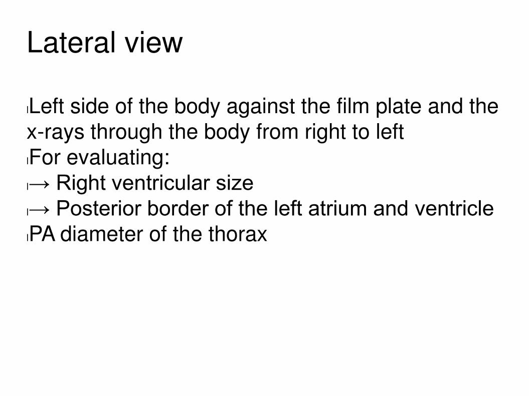
Lateral view
lLeft side of the body against the film plate and the
x-rays through the body from right to left lFor evaluating:
l→ Right ventricular size l→ Posterior border of the left atrium and ventricle lPA diameter of the thorax

Stress Test
lStress is induced by Exercise or drug stimulation.
lMeasure the heart's ability to respond to the external stress and is performed in controlled
clinical environment unless you want to kill the patient with cardiac arrest :) lYou certainly know how it works, if not that's not
quite my problem

Types of stress test
lECG exercise stress testing
l→ a) Treadmill excersie test l→ b) Bicycle exercise test l→ c) Pharmacologic stress test lEchocardiogram exercise stress testing lNuclear stress testing

Nuclear stress testing Treadmill Test
lRadioactive tracer is injected into the veins and small camera called gamma camera detects
the radiation released by tracer to produce computer images of the heart.
lSensitivity: 81%
lSpecificity: 85-95%
lYou challenge the heart with the stress produced by making patient walk in the treadmill and
observe the change in the heart rhythm.
lSensitivity: 73-90%%
lSpecificity: 50-74%


Pharmacological Stress Test
lAgents commonly used:
l→ Adenosine, Dobutamine or Dipyridamole
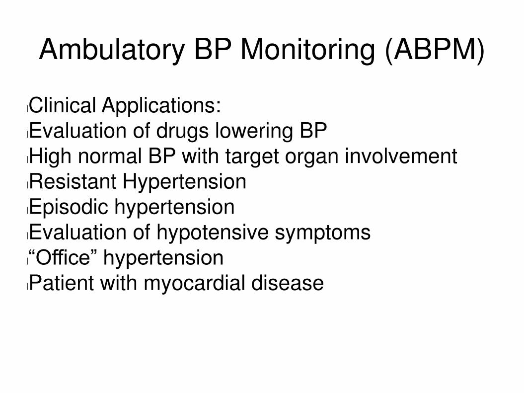
Ambulatory BP Monitoring (ABPM)
lClinical Applications:
lEvaluation of drugs lowering BP lHigh normal BP with target organ involvement
lResistant Hypertension lEpisodic hypertension lEvaluation of hypotensive symptoms
l“Office” hypertension lPatient with myocardial disease

Tilt Table Test
lProcedure often used to diagnose dysautonomia
or syncope lWorks with the change in posture of the body
from lying to standing

Holter Monitor
lPortable recording device worn by the patient
under clothing and attached to the chest via electrode leads and patches to the cord an ECG
continuously. lCLINICAL APPLICATIONS lDiagnosis of cardiac arrythmias
lDiagnosis of suspected ischemia lEvaluation of anti-arrythmic therapy
Arrythmia Event Recorder
lLooping memory monitor is an ECG recording
device about the size of a pager and is attached to the chest with two electrodes with an adhesive
backing that sticks to the skin over a long period of time. lPost Event Monitors are small ECG recording
devices that can be carried in purse or pockets. ECG monitor is held up to the level of chest to make a recording at the time of occurrence of the
symptoms.
Echocardiogram
lUses sound waves to create images of the heart.
lCLINICAL APPLICATIONS: lEstimation of intra-cardiac pressure
lEstimation of intra-cardiac shunts lAssessment of LV function
USES
lMeasure heart size,functions and thickness of
heart muscle lWith doppler technique can measure the blood
flow through the different chambers and measure the pressure gradients across the valves and can actually diagnose stenosis ,regurgitation or
calcification. lVery reliable, non-invasive, no pain or risk
Types of Electrocardiographic
studies
lM-Mod
lTwo dimensional (2D) lDoppler imaging
lContrast
lTransthoracic (Cardiac ultrasound) lTransesophageal (TEE)

Thallium Stress Testing
lAgent: Thallium radioisotope, gamma camera
lCLINICAL APLLICATIONS lDiagnosis of coronary disease
lDetermine extent of diagnosed coronary disease lAssess effectiveness of angioplasty lEvaluates patients with abnormal ECG
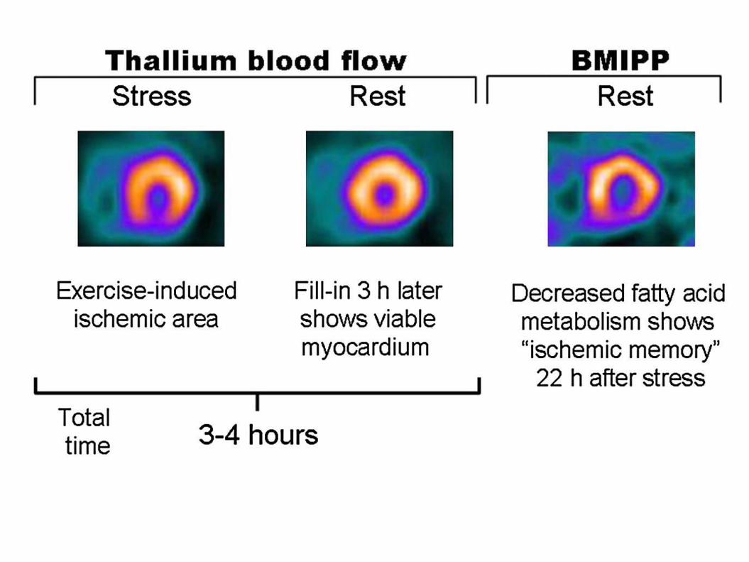
Positron emission tomography
(PET) scan
lAdvances noninvasive method, uses tracer drugs
that emit positively charged particles called positron
lUses gamma camera lProvides information on metabolic functioning rather than the structure of the heart and other
organs.

TYPES
lOne which makes use of tracer called Rubidium-
82 (Rb-82), that measures circulation in the arteries of the heart
lSecond, using sugar like substance called F-18 FDG, distinguish healthy cardiac tissues from damaged ones such as in heart attack.

PET Cardiology Viability Imaging
lAlso calledF-18-FDG PET Viability Imaging,
Cardiac PET FDG viability test

MUGS scans. Radionuclide
angiography (RNA) scans
lPerformed by labelling patient's red blood pool
with radioactive tracer, technetium-99m-petechnetate (Tc-99m) and measuring
radioactivity over anterior part as the radioactive blood flows through the blood vessels and the heart chambers.
Electrophysiology study (EPS)
lHelps doctors understand the nature of abnormal
heart rhythms.

Cardiac Catherization(coronary
angiogram)
lSmall cathether is advanced into and around the
heart through an artery or vein in the groin or arm which helps to measure pressure within heart and
produce angiograms of the coronary arteries, LV and other cardiac structures.

Cardiac MRI
lIt's used to diagnose and assess many diseases
and conditions, including: Coronary heart disease. Damage caused by a heart attack. Heart failure.
Computed Tomography
lUses X rays to produce planar image of the heart
lAssess pericardial and aortic diseases lSpeed of image acquisition limits use due to
cardiac motion

Intravascular Ultrasound (IVUS)
lIntravascular ultrasound (IVUS) is a medical
imaging methodology using a specially designed catheter with a miniaturized ultrasound probe
attached to the distal end of the catheter. The proximal end of the catheter is attached to computerized ultrasound equipment.


References
lDr. Dennis James Torres,Lecture notes
lhttp://www.lorvenheart.com/proIVU.html
lhttps://www.nlm.nih.gov/medlineplus/ency/article/003827.htm