Stress-related illness: Cardiovascular and Psychiatric disorders.
Upload
hester-yorkCategory
view
232download
1
Cardiovascular Disorders
Chapter 18
Pgs 285-308

Overview
• Diagnostic Tests for Cardiovascular Function
• General Treatment Measures for Cardiac Disorders
• Coronary Artery Disease (CAD)– Arteriosclerosis– Atherosclerosis– Myocardial Infarction (MI)
• Cardiac Arrhythmias– Sinus node abnormalities– Atrial conduction
abnormalities– Cardiac arrest
• Congestive Heart Failure (CHF)
• Arterial Diseases– Hypertension
• Shock

Homework
• Due Tuesday Oct 4• Do the following Case Study questions on Pg. 306
– You do not have to type them; Put the answers in your own words!
• Case Study A– a, b, e, g, k, l, m
• Case Study B– a-f
• You may work together– If you work in a group, you can turn in one paper!– You must be present in class to get credit!

Diagnostic Tests for Cardiovascular Function
• ECG– Monitors arrhythmias, MI, infection, pericarditis– Studies conduction activation and systemic abnormalities
• Ausculation– Studies heart sounds using stethoscope
• Exercise stress test– Assess general cardiovascular function– Checks for exercise-induced problems
• Chest X-ray Film– Shows shape, size of heart– Evidence of pulmonary congestion associated with heart failure– Nuclear imaging

Diagnostic Tests
• Cardiac Catheterization– Visualize inside of
heart, measure pressure, assess valve and heart function
– Determine blood flow to and from heart
Diagnostic Tests
• Angiography– Visualization of blood
flow in coronary artery– Obstruction assessed
and treated• Basic catheterization• Balloon angioplasty

Diagnostic Tests
• Doppler Studies– Assessment of blood flow in peripheral vessels– Microphone records sounds of blood flow
• Can detect obstruction
• Blood tests– Assess triglyceride and cholesterol levels– Electrolytes– Hb, hematocrit, cbcs
• Arterial Blood Gas Determination– Essential for pts with shock, MI– Check current oxygen levels, acid-base balance

General Treatment Measures for Cardiac Disorders
• Dietary modification
• Regular exercise program
• Quit smoking
• Drug therapy

Drug Therapy
• Vasodilators (Nitroglycerin)– Provide better balance of oxygen supply and
demand in heart muscle– May cause low bp
• Beta-blockers (Metoprolol or Atenolol)– Treats angina, hypertension, arrhythmias– Blocks beta1-adrenergic receptors in heart
• Prevent epine from increasing heart activity

Drug Therapy• Calcium ion channel blockers
– Block movement of calcium– Decrease heart contraction
• Antiarrhytmatic for excessive atrial activity• Antihypertension and vasodilator
• Digoxin– Treats heart failure– Increases efficiency of heart
• Decreases conduction of impulses and HR• Increases contraction of heart
– Pts must be checked for toxicity• Antihypertensive drugs
– Decrease bp to normal levels– Include:
• Adrenergic blocking agents• Calcium ion blockers• Diuretics• Angiotensin-converting enzyme (ACE) inhibitors
– Used to treat hypertension, CHF, after MI
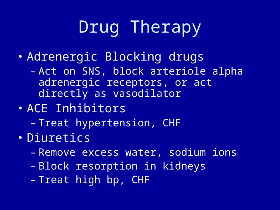
Drug Therapy
• Adrenergic Blocking drugs– Act on SNS, block arteriole alpha adrenergic
receptors, or act directly as vasodilator
• ACE Inhibitors– Treat hypertension, CHF
• Diuretics– Remove excess water, sodium ions– Block resorption in kidneys– Treat high bp, CHF

Drug Therapy
• Anticoagulant– Decrease risk of blood clot formation– ASA decreases platelet adhesion– Block coagulation process
• Cholesterol or lipid reducing drugs– When diet and exercise fail – Decrease LDL and cholesterol

CAD—Arteriosclerosis: Pathophysiology
• General term for all types of arterial changes
• Best for degeneration in small arteries and arterioles
• Loss of elasticity, walls thick and hard, lumen narrows

CAD—Atherosclerosis: Pathophysiology
• Presence of atheromas– Plaques
• Consist of lipids, cells, fibrin, cell debris
– Lipids usually transported with lipoproteins

Lipoproteins and Transport

Atherosclerosis--Pathophysiology
• Analysis of serum lipids:– Total cholesterol, triglycerides, LDL, HDL
• LDL– High cholesterol content– Transports cholesterol liver cells– Dangerous component
• HDL– “good” – Low cholesterol content– Transports cholesterol cells liver

Development of Atheroma

Consequences of Atherosclerosis
Atherosclerosis—Etiology
• Age• Gender• Genetic factors• Obesity, diet high in cholesterol, animal fats• Cigarette smoking• Sedentary life style• Diabetes mellitus• Poorly controlled hypertension• Combo of BC pills and smoking

Atherosclerosis—Diagnostic Tests
• Serum lipid levels
• Exercise stress test
• Radioisotope

Atherosclerosis—Treatment
• Decrease cholesterol and LDL• Decrease sodium ion intake• Control primary disorders• Quit smoking• Oral anticoagulant• Surgical intervention
– Percutaneous transluminal coronary angioplasty (PTCA)
– Cardiac catheterization– Laser beam technology– Coronary artery bypass grafting

CABG

CAD: Myocardial Infarction—Pathophysiology
• Coronary artery completely obstructed– Prolonged ischemia and cell death of myocardium
• Most common cause is atherosclerosis with thrombus
• 3 ways it may develop:– Thrombus obstructs artery– Vasospasm due to partial occlusion– Embolus blocks small branch of coronary artery
• Majority involve L ventricle– Size and location of infarction determine severity of
damage

Myocardial Infarction

MI—Pathophysiology
• Function of myocardium contraction and conduction quickly lost – Oxygen supplies depleted
• 1st 20 minutes critical
• Time Line– 1st 20 min critical– 48 hrs inflammation begins to subside– 7th day necrosis area replaced by fibrous tissue– 6-8 weeks scar forms

MI—Signs and Symptoms
• Pain– Sudden, substernal area– Radiates to L arm and neck– Less severe in females
• Pallor, sweating, nausea, dizziness• Anxiety and fear• Hypotension, rapid and weak pulse (low
CO)• Low grade fever

MI—Diagnostic Tests
• ECG• Serum enzyme and
isoenzyme test• High serum levels of
myosin and troponin• Abnormal electrolytes• Leukocytosis• Arterial blood gases• Pulmonary artery
pressure measure– Determines ventricular
function

MI—Complications
• Arrhythmias– 25% pts sudden death after MI
• Due to ventricular arrhythmias and fibrillation
– Heart block– Premature ventricular contraction (PVCs)
• Cardiogenic shock
• CHF

MI—Treatment
• Rest, oxygen therapy, morphine• Anticoagulant• Drugs• Cardiac rehabilitation• Prognosis depends on site/size of infarct,
presence of collateral circulation, time elapsed before treatment
• Mortality rate in 1st year– 30-40% due to complications, recurrences

Cardiac Arrhythmias
• Alteration in HR or rhythm• ECG monitors
– Holter monitors
• decreases efficiency of heart’s pumping cycle– Slight increase in HR increases CO– Very rapid HR prevents adequate filling in diastole– Very slow HR reduces output to tissues
• Irregular contraction inefficient– Interferes with normal filling/emptying cycle


CA: Sinus Node Abnormalities
• Brachycardia– Regular but slow HR
• Less than 60 beats/min
– Results from vagus nerve stimulation or PNS stimulation
• Tachycardia– Regular rapid HR
• 100-160 beats/min
– SNS stimulation, exercise, fever, compensation for low blood volume


CA: Atrial Conduction Abnormalities
• Premature Atrial Contractions (PAC)– Extra contraction or ectopic beats of atria– Irritable atrial muscle cells outside conduction
pathway• Interfere with timing of next beat
• Atrial flutter– HR 160-350 beats/min– AV node delays conduction
• Slower ventricular rate

Treatment of CA
• Cause should be determined and treated
• Easiest to treat are those due to meds
• SA node problems may require a pacemaker
• Some may require defibrillators


Cardiac Arrest
• Cessation of all activity in the heart• No conduction of impulses (flat line)• May occur b/c:
– Excessive vagal nerve stimulation (decreases heart)
– Drug toxicity– Insufficient oxygen to maintain heart tissue
• Blood flow to heart and brain must be maintained to resuscitate

CHF—Pathophysiology
• Heart unable to pump sufficient blood to meet metabolic needs of body
• Complication• Acute or chronic• Results from
– Problem in heart itself– Increased demands placed on heart– Combo
• One side usually fails 1st

CHF—Pathophysiology
• 1st compensation mechanism to maintain CO– Often aggravates instead of assists– Decreased flow to systemic circ
• Kidneys increase renin, aldosterone secretion• Vasoconstriction (increase afterload) and increased blood vol
(increased preload) = increased work load for heart
– SNS increases HF and periph resistance– Dilatation of heart chambers, myocardium,
hypertrophies


CHF—Pathophysiology
• 2nd effect when heart cannot maintain pumping capability– Decrease in CO or SV
• “forward effect”
– “backup” congestion


CHF—Etiology
• Causes of failure on affected side:– Infarction that impairs pumping ability or
efficiency of conduction system– Valve defects– Congenital heart defects– Coronary artery disease

CHF—Etiology
• Increased demands on heart cause failure– Depends on ventricle most adversely affected– Ex: Hypertension increases diastolic bp
– Requires L ventricle to contract more forcibly to open aortic valve
– Ex: Pulmonary disease– Damages lung caps, increases pulm resistance– Increase work load to R vent

CHF—Signs and Symptoms
• Forward effects– Similar with failure on either side– Decrease blood supply to tissue and general
hypoxia– Fatigue, weakness, dyspnea
(breathlessness), cold intolerance, dizziness
• Compensation mechanism– Indicated by tachycardia, pallor, daytime
oliguira

CHF—Signs and Symptoms
• Systemic backup effects of R-sided failure– Edema in feet, legs– Hepatomegaly, splenomegaly– Ascites– Acute R-sided failure
• Increased pressure on SVC– Flushed face, distended neck veins, headaches, vision
problems

CHF—Diagnostic Tests
• Radiographs
• Catheterization
• Arterial blood gases

CHF—Treatment
• Underlying problem should be treated
• Decrease work load on heart
• Prophylactic measures
• Other methods– Diet – Drugs

Arterial Diseases: Hypertension—Pathophysiology
• Increased bp• Insidious onset, mild symptoms and signs• 3 major categories
– Essential (primary)– Secondary – Malignant
• Can be classified as diastolic or systolic• Develops when bp consistently over 140/90• Diastolic more important


Hypertension—Pathophysiology
• Over long time, high bp damages arterial walls – Sclerosis, decreased lumen– Wall may dilate, tear
• Aneurysm
• Areas most frequently damaged:– Kidneys, brain, retina
• End result of poorly controlled hypertension:– Chronic renal failure– Stroke– Loss of vision– CHF

Hypertension—Etiology
• Increases with age
• Males more freq and severe
• Genetic factors
• High sodium ion intake
• Excessive alcohol
• Obesity
• Prolonged, recurrent stress

Hypertension—Signs and Symptoms
• Asymptomatic in early stages
• Initial signs vague, nonspecific– Fatigue, malaise, morning headache

Hypertension—Treatment
• Treated in sequence of steps– Life style changes– Mild diuretics, ACE inhibitors– One or more drugs added
• Pt compliance is an issue
• Prognosis depends on treating underlying problems and maintaining constant control of bp

Shock (Hypotension)
• Results from decreased circulating blood vol– General hypoxia– Low CO

Classification and Mechanisms of Shock
Type Mechanism
Hypovolemic loss of blood or plasma
Cardiogenic Decreased pumping capability of heart
Anaphylactic Systemic vasodilation due to severe allergic
reaction
Septic Vasodilation due to severe infection
Neurogenic Vasodilation due to loss of SNS and vaso-motor
tone


Shock—Pathophysiology
• Bp decreases when blood vol, heart contraction, or periph resistance fails
• Low CO, microcirculation– = decreased oxygen, nutrients for cells
• Compensation mechanism– SNS, adrenal medulla stimulated– Renin secreted– Increased secretion of ADH– Secretion of glucocorticoids– Acidosis stimulates respiration


Shock—Pathophysiology
• Complications of decompensation of shock– Acute renal failure– Adult respiratory distress syndrome (ARDS)– Hepatic failures– Hemorrhagic ulcers– Infection of septicemia– Decreased cardiac function

Shock—Etiology
• Hypovolemic shock– Loss of blood, plasma
• Burn pts, dehydration
• Cardiogenic shock– Assoc w/ cardiac impairment
• Distributive shock– Blood relocated b/c vasodilation
• Anaphylactic shock• Neurogenic shock
• Septic shock– Severe infection

Shock—Signs and Symptoms
• 1st signs– Shock, thirst, agitation,
restlessness– Often missed
• 2nd signs– Cool, moist, pale skin;
tachycardia; oliguria– Compensation– Vasoconstriction
• Direct effects– Decrease bp and blood
flow– Acidosis
• Prolonged– Decreased responsiveness
in body– Compensated metabolic
acidosis progresses to decompensated
– Acute renal failure– Monitoring

Shock—Treatment
• Primary problem must be treated• Hypovolemic shock
– Whole blood, plasma, electrolytes, bicarbonate required• Anaphylactic shock
– Antihistamines, corticosteroids• Septic
– Antimicrobials, glucocorticoids• Maximize oxygen supply• Epine reinforces heart action and vasoconstriction• Dopamine, dubutamine increase heart function• Good prognosis in early stages• Mortality increases as irreversible shock develops