Cardiac Calcification Following Severe Sepsis: A Case ...
3
Open Access Case Report Akbaş et al., J Anesth Clin Res 2014, 5:5 DOI: 10.4172/2155-6148.1000409 Volume 5 • Issue 5 • 1000409 J Anesth Clin Res ISSN:2155-6148 JACR an open access journal Keywords: Myocardial calcification; Sepsis-related myocardial injury Introduction Sepsis is an important cause of organ failures in the Intensive Care Unit (ICU). All organ systems including myocardium can be injured severely during sepsis [1,2]. Myocardial injury may end up with calcification which is a rare complication of severe sepsis. In this paper, we discuss a patient who developed diffuse leſt ventricular wall calcification during severe systemic sepsis. Case and Method A 36-year-old woman with acute myeloblastic leukemia was admitted to the ICU with septic shock. e patient had leukemia diagnosis about two and a half months ago. She took chemotherapies of cytosine arabinoside (170 mg/day) and idarubicin (20 mg/day) in the week of the diagnosis. Two days aſter chemotherapy, the patient became neutropenic and five days later, she developed a fever. e patient stayed in the hospital until admission to the ICU because of severe neutropenia, thrombocytopenia and unresolved infectious state. Laboratory findings of CRP, procalcitonin, white blood cell count and platelet count studied at the time of ICU admission were 268 mg/L, 97 ng/mL, 200/mm 3 and 2000/mm 3 , respectively. Aſter ICU admission, her first blood culture was positive for Escherichia coli. en, Acinetobacter baumannii was isolated on day 5 and Enterococcus feacium on day 9 from blood cultures. e patient had multiorgan failures of renal, cardiovascular, hepatic and respiratory systems, and critical illness myopathy developed during bacteremic periods. e patient had noradrenalin during the first 3 days of the ICU stay in which a maximum dose of 40 mcg/min was administered for 24 hours. Dopamine was added to noradrenalin during the first day, with a maximum dose of 20 mcg/kg/min for a few hours. Antibiotic treatments were managed according to the antibiograms. e patient benefited from the therapies, became mechanical ventilation-free, and did not have any renal replacement therapy. Daily physiotherapy for critical illness myopathy was continued. e extreme levels of serum creatinine, phosphorus and calcium measured during multiorgan failures were respectively 1.84, 8.1 and 7.7 mg/dL, and calcium- phosphate product was calculated as 68.2 mg 2 /dl 2 . e peak levels of Troponin T and creatine phosphokinase-MB were 47.6 ng/L (normal < 14) and 9.18 IU/L (normal <5) with no significant electrocardiographic changes. e patient started to have fever exceeding 39 celsius on day 15. Pseudomonas aeruginosa was isolated from deep tracheal aspirate on day 20 and antibacterial treatment was modified. Chest and abdomen computed tomographies (CT) were taken on day 25 due to persistent fever. Abdomen CT was normal, but thorax CT showed bilateral pneumonia and diffuse leſt ventricular wall calcification (Figure 1) that was not seen on the previous CT scan from a month ago (Figure 2). Aſter reexamining the chest X-ray taken one day before thorax CT, a vague curvilinear calcification was noticed at the leſt ventricular wall (Figure 3). Previous echocardiography done two and a half months ago was normal, but the new one showed hypokinetic posterior, inferior and septal walls of the leſt ventricle with a 40% ejection fraction. For excluding leukemia-induced fever, a bone marrow biopsy was carried out that showed the disease in remission. e patient also *Corresponding author: Dr. Türkay Akbaş, Department of Internal Medicine and Critical Care, School of Medicine, Marmara University, Unit Fevzi Çakmak Mahallesi, Mimar Sinan Caddesi, No: 41 Ust Kaynarca, Pendik, Istanbul, Turkey, Tel: 090-532-238-31-97; E-mail: [email protected] Received April 25, 2014; Accepted May 27, 2014; Published May 29, 2014 Citation: Akbaş T, Özen M, Adıgüzel C, Çimşit NÇ, Karakurt S (2014) Cardiac Calcification Following Severe Sepsis: A Case Report and Review of the Literature. J Anesth Clin Res 5: 409. doi:10.4172/2155-6148.1000409 Copyright: © 2014 Akbaş T, et al. This is an open-access article distributed under the terms of the Creative Commons Attribution License, which permits unrestricted use, distribution, and reproduction in any medium, provided the original author and source are credited. Cardiac Calcification Following Severe Sepsis: A Case Report and Review of the Literature Türkay Akbaş 1 *, Miraç Özen 2 , Cafer Adıgüzel 3 , Nuri Çağatay Çimşit 4 and Sait Karakurt 5 1 Department of Internal Medicine and Critical Care Unit, Marmara University, Turkey 2 Department of Internal Medicine, Marmara University, School of Medicine, Turkey 3 Department of Haematology, Marmara University, Turkey 4 Department of Radiology, Marmara University, Turkey 5 Department of Pulmonary and Critical Care Medicine, Marmara University, Turkey Abstract There are two types of myocardial calcification: metastatic calcification that is encountered in chronic renal disease patients and dystrophic calcification which develops in injured myocardium. Myocardial infarction, trauma, surgery and myocarditis are the most commonly faced reasons for dystrophic calcification. Here we present a rare case of a patient in whom myocardial calcification was diagnosed after severe sepsis. Figure 1: Computed tomography scan performed one week before the ICU admission showing a normal heart. J o u r n a l o f A n e s t h e s i a & C l i n i c a l R e s e ar c h ISSN: 2155-6148 Journal of Anesthesia & Clinical Research
Transcript of Cardiac Calcification Following Severe Sepsis: A Case ...

Open AccessCase Report
Akbaş et al., J Anesth Clin Res 2014, 5:5 DOI: 10.4172/2155-6148.1000409
Volume 5 • Issue 5 • 1000409J Anesth Clin ResISSN:2155-6148 JACR an open access journal
Keywords: Myocardial calcification; Sepsis-related myocardialinjury
IntroductionSepsis is an important cause of organ failures in the Intensive
Care Unit (ICU). All organ systems including myocardium can be injured severely during sepsis [1,2]. Myocardial injury may end up with calcification which is a rare complication of severe sepsis. In this paper, we discuss a patient who developed diffuse left ventricular wall calcification during severe systemic sepsis.
Case and MethodA 36-year-old woman with acute myeloblastic leukemia was
admitted to the ICU with septic shock. The patient had leukemia diagnosis about two and a half months ago. She took chemotherapies of cytosine arabinoside (170 mg/day) and idarubicin (20 mg/day) in the week of the diagnosis. Two days after chemotherapy, the patient became neutropenic and five days later, she developed a fever. The patient stayed in the hospital until admission to the ICU because of severe neutropenia, thrombocytopenia and unresolved infectious state. Laboratory findings of CRP, procalcitonin, white blood cell count and platelet count studied at the time of ICU admission were 268 mg/L, 97 ng/mL, 200/mm3 and 2000/mm3, respectively. After ICU admission, her first blood culture was positive for Escherichia coli. Then, Acinetobacter baumannii was isolated on day 5 and Enterococcus feacium on day 9 from blood cultures. The patient had multiorgan failures of renal, cardiovascular, hepatic and respiratory systems, and critical illness myopathy developed during bacteremic periods. The patient had noradrenalin during the first 3 days of the ICU stay in which a maximum dose of 40 mcg/min was administered for 24 hours. Dopamine was added to noradrenalin during the first day, with a maximum dose of 20 mcg/kg/min for a few hours. Antibiotic treatments were managed according to the antibiograms. The patient benefited from the therapies, became mechanical ventilation-free, and did not have any renal replacement therapy. Daily physiotherapy for critical illness myopathy was continued. The extreme levels of serum creatinine, phosphorus and calcium measured during multiorgan failures were respectively 1.84, 8.1 and 7.7 mg/dL, and calcium-phosphate product was calculated as 68.2 mg2/dl2. The peak levels of Troponin T and creatine phosphokinase-MB were 47.6 ng/L (normal < 14) and 9.18 IU/L (normal <5) with no significant electrocardiographic changes.
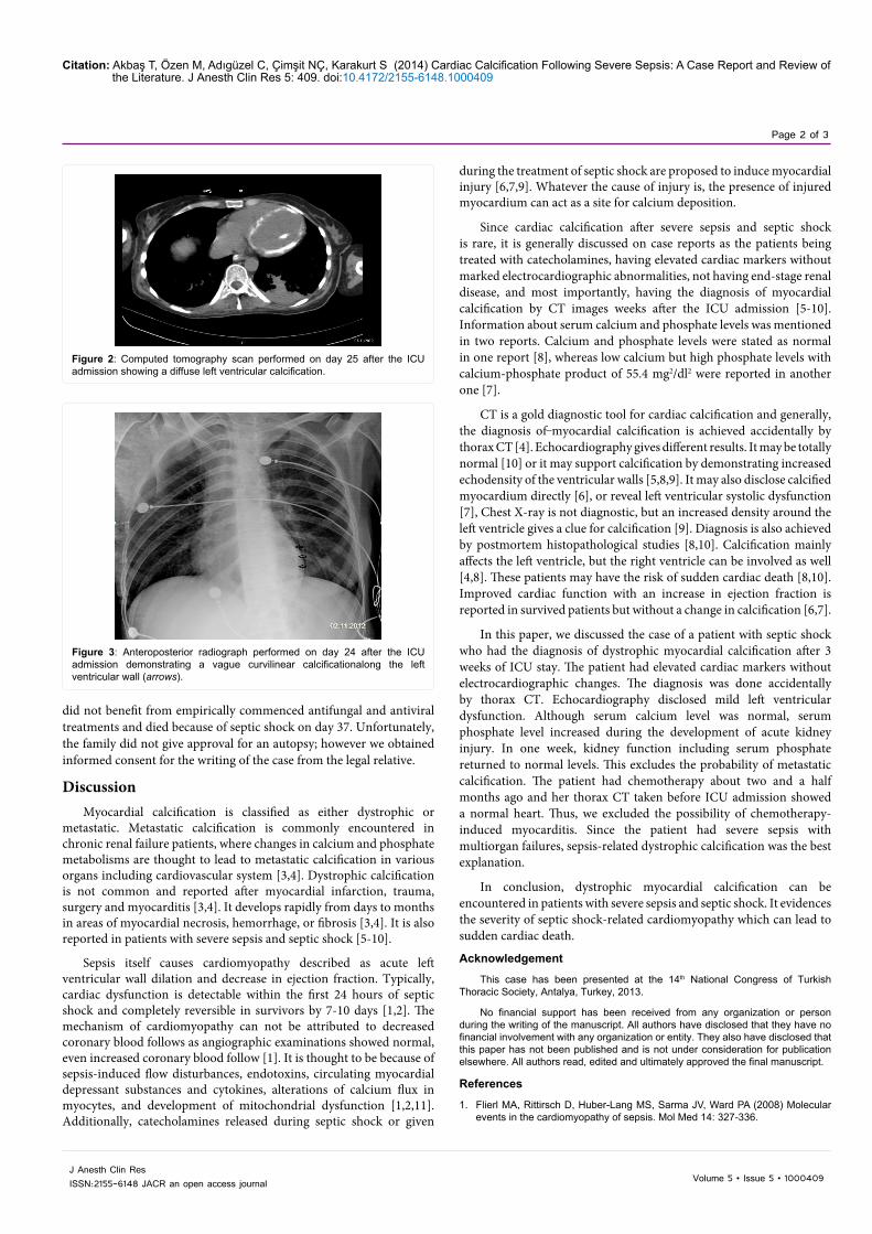
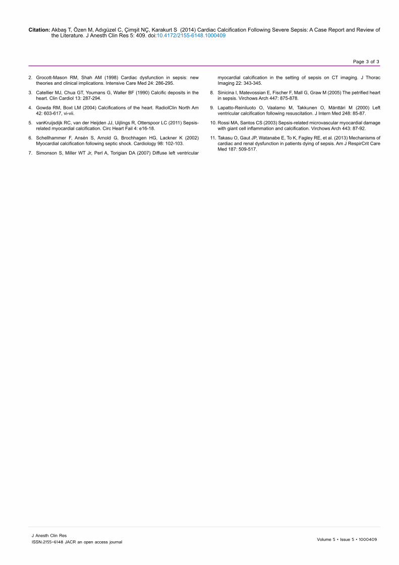
The patient started to have fever exceeding 39 celsius on day 15. Pseudomonas aeruginosa was isolated from deep tracheal aspirate on day 20 and antibacterial treatment was modified. Chest and abdomen computed tomographies (CT) were taken on day 25 due to persistent fever. Abdomen CT was normal, but thorax CT showed bilateral pneumonia and diffuse left ventricular wall calcification (Figure 1) that was not seen on the previous CT scan from a month ago (Figure 2). After reexamining the chest X-ray taken one day before thorax CT, a vague curvilinear calcification was noticed at the left ventricular wall (Figure 3). Previous echocardiography done two and a half months ago was normal, but the new one showed hypokinetic posterior, inferior and septal walls of the left ventricle with a 40% ejection fraction. For excluding leukemia-induced fever, a bone marrow biopsy was carried out that showed the disease in remission. The patient also
*Corresponding author: Dr. Türkay Akbaş, Department of Internal Medicine and Critical Care, School of Medicine, Marmara University, Unit Fevzi Çakmak Mahallesi, Mimar Sinan Caddesi, No: 41 Ust Kaynarca, Pendik, Istanbul, Turkey, Tel: 090-532-238-31-97; E-mail: [email protected]
Received April 25, 2014; Accepted May 27, 2014; Published May 29, 2014
Citation: Akbaş T, Özen M, Adıgüzel C, Çimşit NÇ, Karakurt S (2014) Cardiac Calcification Following Severe Sepsis: A Case Report and Review of the Literature. J Anesth Clin Res 5: 409. doi:10.4172/2155-6148.1000409
Copyright: © 2014 Akbaş T, et al. This is an open-access article distributed under the terms of the Creative Commons Attribution License, which permits unrestricted use, distribution, and reproduction in any medium, provided the original author and source are credited.
Cardiac Calcification Following Severe Sepsis: A Case Report and Review of the LiteratureTürkay Akbaş1*, Miraç Özen2, Cafer Adıgüzel3, Nuri Çağatay Çimşit4 and Sait Karakurt5
1Department of Internal Medicine and Critical Care Unit, Marmara University, Turkey2Department of Internal Medicine, Marmara University, School of Medicine, Turkey3Department of Haematology, Marmara University, Turkey4Department of Radiology, Marmara University, Turkey5Department of Pulmonary and Critical Care Medicine, Marmara University, Turkey
AbstractThere are two types of myocardial calcification: metastatic calcification that is encountered in chronic renal
disease patients and dystrophic calcification which develops in injured myocardium. Myocardial infarction, trauma, surgery and myocarditis are the most commonly faced reasons for dystrophic calcification. Here we present a rare case of a patient in whom myocardial calcification was diagnosed after severe sepsis.
Figure 1: Computed tomography scan performed one week before the ICU admission showing a normal heart.
Jour
nal o
f Ane
sthesia & Clinical Research
ISSN: 2155-6148
Journal of Anesthesia & Clinical Research
Citation: Akbaş T, Özen M, Adıgüzel C, Çimşit NÇ, Karakurt S (2014) Cardiac Calcification Following Severe Sepsis: A Case Report and Review of the Literature. J Anesth Clin Res 5: 409. doi:10.4172/2155-6148.1000409
Page 2 of 3
Volume 5 • Issue 5 • 1000409J Anesth Clin ResISSN:2155-6148 JACR an open access journal
did not benefit from empirically commenced antifungal and antiviral treatments and died because of septic shock on day 37. Unfortunately, the family did not give approval for an autopsy; however we obtained informed consent for the writing of the case from the legal relative.
DiscussionMyocardial calcification is classified as either dystrophic or
metastatic. Metastatic calcification is commonly encountered in chronic renal failure patients, where changes in calcium and phosphate metabolisms are thought to lead to metastatic calcification in various organs including cardiovascular system [3,4]. Dystrophic calcification is not common and reported after myocardial infarction, trauma, surgery and myocarditis [3,4]. It develops rapidly from days to months in areas of myocardial necrosis, hemorrhage, or fibrosis [3,4]. It is also reported in patients with severe sepsis and septic shock [5-10].
Sepsis itself causes cardiomyopathy described as acute left ventricular wall dilation and decrease in ejection fraction. Typically, cardiac dysfunction is detectable within the first 24 hours of septic shock and completely reversible in survivors by 7-10 days [1,2]. The mechanism of cardiomyopathy can not be attributed to decreased coronary blood follows as angiographic examinations showed normal, even increased coronary blood follow [1]. It is thought to be because of sepsis-induced flow disturbances, endotoxins, circulating myocardial depressant substances and cytokines, alterations of calcium flux in myocytes, and development of mitochondrial dysfunction [1,2,11]. Additionally, catecholamines released during septic shock or given
during the treatment of septic shock are proposed to induce myocardial injury [6,7,9]. Whatever the cause of injury is, the presence of injured myocardium can act as a site for calcium deposition.
Since cardiac calcification after severe sepsis and septic shock is rare, it is generally discussed on case reports as the patients being treated with catecholamines, having elevated cardiac markers without marked electrocardiographic abnormalities, not having end-stage renal disease, and most importantly, having the diagnosis of myocardial calcification by CT images weeks after the ICU admission [5-10]. Information about serum calcium and phosphate levels was mentioned in two reports. Calcium and phosphate levels were stated as normal in one report [8], whereas low calcium but high phosphate levels with calcium-phosphate product of 55.4 mg2/dl2 were reported in another one [7].
CT is a gold diagnostic tool for cardiac calcification and generally, the diagnosis of myocardial calcification is achieved accidentally by thorax CT [4]. Echocardiography gives different results. It may be totally normal [10] or it may support calcification by demonstrating increased echodensity of the ventricular walls [5,8,9]. It may also disclose calcified myocardium directly [6], or reveal left ventricular systolic dysfunction [7], Chest X-ray is not diagnostic, but an increased density around the left ventricle gives a clue for calcification [9]. Diagnosis is also achieved by postmortem histopathological studies [8,10]. Calcification mainly affects the left ventricle, but the right ventricle can be involved as well [4,8]. These patients may have the risk of sudden cardiac death [8,10]. Improved cardiac function with an increase in ejection fraction is reported in survived patients but without a change in calcification [6,7].
In this paper, we discussed the case of a patient with septic shock who had the diagnosis of dystrophic myocardial calcification after 3 weeks of ICU stay. The patient had elevated cardiac markers without electrocardiographic changes. The diagnosis was done accidentally by thorax CT. Echocardiography disclosed mild left ventricular dysfunction. Although serum calcium level was normal, serum phosphate level increased during the development of acute kidney injury. In one week, kidney function including serum phosphate returned to normal levels. This excludes the probability of metastatic calcification. The patient had chemotherapy about two and a half months ago and her thorax CT taken before ICU admission showed a normal heart. Thus, we excluded the possibility of chemotherapy-induced myocarditis. Since the patient had severe sepsis with multiorgan failures, sepsis-related dystrophic calcification was the best explanation.
In conclusion, dystrophic myocardial calcification can be encountered in patients with severe sepsis and septic shock. It evidences the severity of septic shock-related cardiomyopathy which can lead to sudden cardiac death.
Acknowledgement
This case has been presented at the 14th National Congress of Turkish Thoracic Society, Antalya, Turkey, 2013.
No financial support has been received from any organization or person during the writing of the manuscript. All authors have disclosed that they have no financial involvement with any organization or entity. They also have disclosed that this paper has not been published and is not under consideration for publication elsewhere. All authors read, edited and ultimately approved the final manuscript.
References
1. Flierl MA, Rittirsch D, Huber-Lang MS, Sarma JV, Ward PA (2008) Molecular events in the cardiomyopathy of sepsis. Mol Med 14: 327-336.
Figure 2: Computed tomography scan performed on day 25 after the ICU admission showing a diffuse left ventricular calcification.
Figure 3: Anteroposterior radiograph performed on day 24 after the ICU admission demonstrating a vague curvilinear calcificationalong the left ventricular wall (arrows).
Citation: Akbaş T, Özen M, Adıgüzel C, Çimşit NÇ, Karakurt S (2014) Cardiac Calcification Following Severe Sepsis: A Case Report and Review of the Literature. J Anesth Clin Res 5: 409. doi:10.4172/2155-6148.1000409
Page 3 of 3
Volume 5 • Issue 5 • 1000409J Anesth Clin ResISSN:2155-6148 JACR an open access journal
2. Grocott-Mason RM, Shah AM (1998) Cardiac dysfunction in sepsis: new theories and clinical implications. Intensive Care Med 24: 286-295.
3. Catellier MJ, Chua GT, Youmans G, Waller BF (1990) Calcific deposits in the heart. Clin Cardiol 13: 287-294.
4. Gowda RM, Boxt LM (2004) Calcifications of the heart. RadiolClin North Am 42: 603-617, vi-vii.
5. vanKruijsdijk RC, van der Heijden JJ, Uijlings R, Otterspoor LC (2011) Sepsis-related myocardial calcification. Circ Heart Fail 4: e16-18.
6. Schellhammer F, Ansén S, Arnold G, Brochhagen HG, Lackner K (2002) Myocardial calcification following septic shock. Cardiology 98: 102-103.
7. Simonson S, Miller WT Jr, Perl A, Torigian DA (2007) Diffuse left ventricular
myocardial calcification in the setting of sepsis on CT imaging. J Thorac Imaging 22: 343-345.
8. Sinicina I, Matevossian E, Fischer F, Mall G, Graw M (2005) The petrified heart in sepsis. Virchows Arch 447: 875-878.
9. Lapatto-Reiniluoto O, Vaalamo M, Takkunen O, Mänttäri M (2000) Left ventricular calcification following resuscitation. J Intern Med 248: 85-87.
10. Rossi MA, Santos CS (2003) Sepsis-related microvascular myocardial damage with giant cell inflammation and calcification. Virchows Arch 443: 87-92.
11. Takasu O, Gaut JP, Watanabe E, To K, Fagley RE, et al. (2013) Mechanisms of cardiac and renal dysfunction in patients dying of sepsis. Am J RespirCrit Care Med 187: 509-517.