Extraosseous Calcification - JCI
8
Extraosseous Calcification EVIDENCE FOR ABNORMAL PYROPHOSPHATE METABOLISM IN UREMIA ALLEN C. ALFREY, CLIVE C. SOLOMONS, JENNY CiRciuLLo, and NANCY L. MILLER From the Renal Section, Denver Veterans Administration Hospital, the Division of Renal Medicine, and Department of Pediatrics, University of Colorado Medical Center, Denver, Colorado 80220 A B STR A C T The inorganic constituents and crystal- line features of extraosseous calcium-phosphate deposits obtained from dialyzed uremic and hypercalcemic pa- tients were studied. Visceral calcification (heart, lung, and kidney) in hypercalcemic patients exhibited either an amorphous or apatitic X-ray diffraction pattern. Uremic visceral calcification consistently gave an amorphous diffraction pattern. Although the calcium content of uremic and hypercalcemic visceral deposits was similar, other inorganic constituents were different. The mean pyrophosphate was 11±11.8 and magnesium 4.91±3.86 mg/g in the uremic group as compared to 0.92+0.24 and 1.36±1.26 mg/g in the hypercalcemic group (P < 0.025). After incineration hypercalcemic visceral deposits hav- ing an amorphous diffraction pattern were found to gen- erate pyrophosphate supporting the presence of brushite in these deposits. The small amount of pyrophosphate in apatitic deposits from both uremic and hypercalcemic pa- tients actually decreased after incineration and the pyro- phosphate content of uremic visceral deposits was un- changed by incineration. It is concluded that in hypercalcemic patients the initial visceral deposit is brushite which is subsequently transformed to apatite. Arterial and tumoral calcium- phosphate deposits in uremic patients were also apatite. Uremic visceral calcium-phosphate deposits are an unique mineral high in magnesium with approximately 30% of the phosphorus present as pyrophosphate. The high pyrophosphate content of these deposits could alter their crystalline structure and prevent the transformation to apatite. The infrared features, high magnesium con- tent of the deposit, and resistance of pyrophosphate in the deposit to hydrolysis by pyrophosphatase suggests Received for publication 8 August 1975 and in revised form 29 October 1975. that the pyrophosphate may be deposited as the mag- nesium salt. INTRODUCTION Three distinct clinical types of extraosseous calcifications are found in uremic patients: (a) calcification in median size arteries, (b) periarticular (tumoral) calcification, and (c) visceral calcification (heart, lung, and kidney). The fact that severe visceral calcification can occur in patients who are free of arterial and tumoral calcification as well as the converse, where severe arterial or tumoral calcification occurs in patients who are free of visceral calcification strongly suggests that the etiology of these deposits is different. Massry et al. (1) have previously suggested that the increased calcium content in uremic patients' skin is a consequence of the hyperparathyroid state. However, Contiguglia et al. (2) were unable to show any relationship between parathyroid size and the development of pulmonary, myocardial, and skeletal mus- cle calcification. Although it is widely accepted that tumoral calcification can be prevented or cured (3, 4) by phosphate depletion it is unknown whether such treat- ment has any effect on visceral calcification. In spite of the routine use of phosphate-binding gels and maintain- ing serum phosphate at 7 mg/100 ml or less, a prospec- tive study showed that approximately 50% of dialysis patients developed visceral calcification (5) whereas none developed arterial or tumoral deposits during the study period. The chemical composition and crystalline nature of uremic visceral calcification has also been found to be different from arterial and periarticular calcium-phos- phate deposits (6). An unexpected finding was that uremic visceral calcium-phosphate deposits uniformly The Journal of Clinical Investigation Volume 57 March 1976 692-699 692
Transcript of Extraosseous Calcification - JCI

Extraosseous Calcification
EVIDENCEFORABNORMALPYROPHOSPHATE
METABOLISMIN UREMIA
ALLEN C. ALFREY, CLIVE C. SOLOMONS,JENNYCiRciuLLo, andNANCYL. MILLER
From the Renal Section, Denver Veterans Administration Hospital, theDivision of Renal Medicine, and Department of Pediatrics, University ofColorado Medical Center, Denver, Colorado 80220
A B STRA C T The inorganic constituents and crystal-line features of extraosseous calcium-phosphate depositsobtained from dialyzed uremic and hypercalcemic pa-tients were studied. Visceral calcification (heart, lung, andkidney) in hypercalcemic patients exhibited either anamorphous or apatitic X-ray diffraction pattern. Uremicvisceral calcification consistently gave an amorphousdiffraction pattern. Although the calcium content ofuremic and hypercalcemic visceral deposits was similar,other inorganic constituents were different. The meanpyrophosphate was 11±11.8 and magnesium 4.91±3.86mg/g in the uremic group as compared to 0.92+0.24 and1.36±1.26 mg/g in the hypercalcemic group (P < 0.025).After incineration hypercalcemic visceral deposits hav-ing an amorphous diffraction pattern were found to gen-erate pyrophosphate supporting the presence of brushitein these deposits. The small amount of pyrophosphate inapatitic deposits from both uremic and hypercalcemic pa-tients actually decreased after incineration and the pyro-phosphate content of uremic visceral deposits was un-changed by incineration.
It is concluded that in hypercalcemic patients theinitial visceral deposit is brushite which is subsequentlytransformed to apatite. Arterial and tumoral calcium-phosphate deposits in uremic patients were also apatite.Uremic visceral calcium-phosphate deposits are anunique mineral high in magnesium with approximately30% of the phosphorus present as pyrophosphate. Thehigh pyrophosphate content of these deposits could altertheir crystalline structure and prevent the transformationto apatite. The infrared features, high magnesium con-tent of the deposit, and resistance of pyrophosphate inthe deposit to hydrolysis by pyrophosphatase suggests
Received for publication 8 August 1975 and in revisedform 29 October 1975.
that the pyrophosphate may be deposited as the mag-nesium salt.
INTRODUCTION
Three distinct clinical types of extraosseous calcificationsare found in uremic patients: (a) calcification in mediansize arteries, (b) periarticular (tumoral) calcification,and (c) visceral calcification (heart, lung, and kidney).The fact that severe visceral calcification can occur inpatients who are free of arterial and tumoral calcificationas well as the converse, where severe arterial or tumoralcalcification occurs in patients who are free of visceralcalcification strongly suggests that the etiology of thesedeposits is different. Massry et al. (1) have previouslysuggested that the increased calcium content in uremicpatients' skin is a consequence of the hyperparathyroidstate. However, Contiguglia et al. (2) were unable toshow any relationship between parathyroid size and thedevelopment of pulmonary, myocardial, and skeletal mus-cle calcification. Although it is widely accepted thattumoral calcification can be prevented or cured (3, 4) byphosphate depletion it is unknown whether such treat-ment has any effect on visceral calcification. In spite ofthe routine use of phosphate-binding gels and maintain-ing serum phosphate at 7 mg/100 ml or less, a prospec-tive study showed that approximately 50% of dialysispatients developed visceral calcification (5) whereasnone developed arterial or tumoral deposits during thestudy period.
The chemical composition and crystalline nature ofuremic visceral calcification has also been found to bedifferent from arterial and periarticular calcium-phos-phate deposits (6). An unexpected finding was thaturemic visceral calcium-phosphate deposits uniformly
The Journal of Clinical Investigation Volume 57 March 1976 692-699692
exhibited an amorphous X-ray diffraction pattern (6).It has previously been felt that the initial extraosseouscalcium-phosphate deposit is brushite (CaHPO4 2H20).Since brushite is stable at a pH of 6.2 but is rapidlytransformed to apatite at a pH of 6.9 or higher, it hasbeen stated that apatite [Calo(PO4)e8 (OH)2] is the onlysolid phase of calcium and phosphate that can form underphysiological conditions (7). This would suggest thatan inhibitor or stabilizer such as pyrophosphate (P207) 1is present in uremic visceral calcification which preventsthe transformation of these deposits to apatite.
The present study was carried out to fully characterizethe inorganic constituents of uremic visceral, tumoral,and arterial calcification. To determine if the crystallinecharacteristics of visceral calcification are primarily de-termined by the matrix within which it is formed, vis-ceral calcification obtained from hypercalcemic patientswas also studied to compare with the uremic deposits.
METHODS
Tissues (heart, lung, kidney, skeletal muscle, dura, arterial,and periarticular deposits) were obtained from 11 dialyzeduremic patients previously shown to have calcifications and3 patients who had hypercalcemia. The etiology of the hy-percalcemia was different in all of the latter three patients.Two patients had acute hypercalcemia shortly before deathwhich was secondary to multiple myeloma in one and rhab-domyolysis in the second. The third patient had long-stand-ing undiagnosed hyperparathyroidism. At time of deaththese patients had normal or only moderately reduced renalfunction. Tissues for comparison were obtained from twocontrol subjects who had experienced sudden death andthree dialyzed uremic patients without calcification.
Tissues were kept in a frozen state until analyzed. Thetissues were then dried, ground into a fine powder, and ex-tracted four times with 2 ml of ether-petroleum ether (1/1vol/vol). For chemical analysis the dry defatted tissue wasdigested by boiling in 1 ml nitric acid for 1 h. The resultingclear liquid was diluted to 20 ml with deionized water. Cal-cium, magnesium, and phosphate were determined by previ-ously reported methods (2).
Five tissues with heavy calcification obtained from uremicand hypercalcemic patients were dried at room temperatureand analyzed for multiple trace elements using an X-rayfluorescence technique (8).
For P2O7 determination, 25 mg of the finely ground dry,defatted tissue, or tissue after ashing at 550°C was placedin 2 ml of a 15%o EDTA solution with pH adjusted to 7.4for 12 h and P207 determinations were performed on thesupernate. The EDTA extraction of calcium and magnesiumfrom the dry tissue has been shown to be complete as de-termined by ashing and analyzing the residue. 1 ml of theEDTA solution was applied to a resin column (Bio-RadAG 1 X 10, 100-200 mesh, chloride form, Bio-Rad Labora-tories, Richmond, Calif.) and the orthophosphate fractioneluted with 100 ml of 0.1 mol/liter potassium chloride. TheP201 was then eluted with 1.1 mol/liter hydrochloric acid.The first 6 ml of the acid was discarded and subsequent4-10 ml-fractions collected. Concentrated HCl, 0.3 ml, wasadded to the first three fractions. The proteins were eluted
'Abbrezaation used in this paper: P907, pyrophosphate.
in the first 30 ml of the KCl fraction and did not contami-nate the orthophosphate or P207 peaks. The hydrochloricacid fractions were hydrolyzed at 1000-1200C for 60 minbefore colorimetric analysis for phosphate was performed.The phosphate was determined by a modification of thestandard method of Chen et al. (9). Recovery of P207 withthis method has been consistently in the range of 95-105%oas determined with TMP-labeled P207. The specific activity ofeach fraction in the P207 peak has been found not to differsignificantly from unity over a wide range of P207 concen-trations and amounts of labels used. Extraction of phospho-lipids with a chloroform-methanol mixture did not alter theP207 values and the addition of pure lecithin, sphingomyelin,and phosphatidylethanolamine likewise had no effect on theP207 recoveries. Treatment with yeast pyrophosphatase(EC 3.6.1.1) completely removed the P207 peak. To showthat nucleotides were not responsible for falsely increasingP207 levels, tissue samples high in P207 were pretreatedwith charcoal. This did not alter the P207 determination.This method for determining P207 has been used success-fully by two other investigators (10, 11). P207 and ortho-phosphate content of the various tissue is reported as milli-gram P for each compound.
For infrared analysis 1 mg of dry sample was mixed with300 mg KBr and pressed into a clear disk using 12 tons
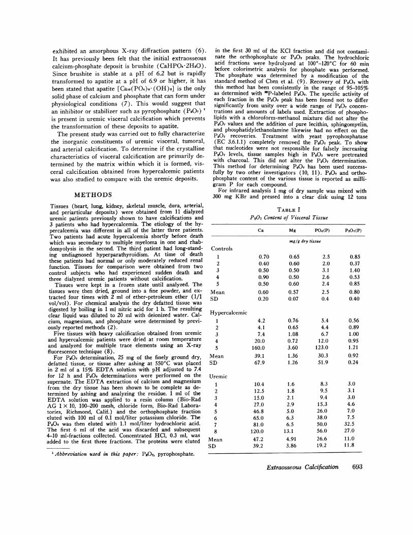
TABLE IP207 Content of Visceral Tissue
Ca Mg P04(P) P207(P)
mg/g dry tissue
Controls1 0.70 0.65 2.5 0.852 0.40 0.60 2.0 0.373 0.50 0.50 3.1 1.404 0.90 0.50 2.6 0.535 0.50 0.60 2.4 0.85
Mean 0.60 0.57 2.5 0.80SD 0.20 0.07 0.4 0.40
Hypercalcemic1 4.2 0.76 5.4 0.562 4.1 0.65 4.4 0.893 7.4 1.08 6.7 1.004 20.0 0.72 12.0 0.955 160.0 3.60 123.0 1.21
Mean 39.1 1.36 30.3 0.92SD 67.9 1.26 51.9 0.24
Uremic1 10.4 1.6 8.3 3.02 12.5 1.8 9.5 3.13 15.0 2.1 9.4 3.04 27.0 2.9 15.3 4.65 46.8 5.0 26.0 7.06 65.0 6.3 38.0 7.57 81.0 6.5 50.0 32.58 120.0 13.1 56.0 27.0
Mean 47.2 4.91 26.6 11.0SD 39.2 3.86 19.2 11.8
Extraosseous Calcification 693

Uremic Visceral Calcification
Hypercalcemic Visceral Calcification
40 36 32 28 24 20Degrees 2e
FIGURE 1 X-ray diffraction patterns obtained on untreated hypercalcemic and uremic visceralcalcium-phosphate deposits. Both tissues were extensively calcified. No diffraction peaks(amorphous patterns) are present in the uremic deposit. Apatitic peaks are present in thehypercalcemic deposit.
pound per square inch in a vacuum. The sample was thenmounted in a Perkin-Elmer Infrared Spectrophotometer(Perkin-Elmer Ultek Inc., Palo Alto, Calif.) and scanned
from 4,000 to 600 cm-' (2-15 gm).Powder X-ray diffraction studies were carried out with
a Norelco diffractometer (North American Philips Co.,Inc., New York) equipped with a graphite crystal mono-chrometer. The 2-e-diffraction range covered was 12'-60'at a scanning speed of 10 min. Patterns were interpretedby comparing them with the American Society of TestingMaterials index cards.
The brushite content of the various deposits was estimatedby determining the P04 and P207 content of the depositsbefore and after incineration at 550'C. Although Greenfieldet al. (12) have recently criticized the use of the pyrolyticmethod for estimating HP04 content of carbonate-containingapatites, their studies would suggest that this method isvalid for estimating the HP04 content of amorphous cal-cium-phosphate deposits.
RESULTS
X-ray diffraction studies were carried out on all tissues.The dialyzed uremic visceral calcification, and the threetissues with the lowest calcium content obtained from thehypercalcemic group all gave an amorphous X-ray dif-fraction pattern (Fig. 1). An apatitic diffraction patternwas found in the two tissues with the highest calciumcontent in the hypercalcemic group (Fig. 1) and in thearterial and periarticular deposits obtained from uremicpatients.
The calcium, magnesium, orthophosphate, and P207content of the dry tissues are shown in Table I. Thecalcium and orthophosphate content was significantlyhigher in the uremic visceral calcification than the con-trol group but not different from the hypercalcemicgroup (Table I). The mean P207 was 10.96 and mag-nesium 4.91 mg/g in the uremic visceral calcification as
compared to 0.92 and 1.36 mg/g in the hypercalcemicgroup (P < 0.025). There were not enough tissues inthe uremic, arterial, or periarticular groups for statisticalcomparison. However, these deposits were low in bothmagnesium and P207.
The thermochemical properties of the various depositswere studied to more fully characterize their crystallinestructure. The brushite content of the dry deposits was
estimated by determining orthophosphate and P207content before and after incineration. The results are
given in Table II. In the hypercalcemic visceral calcifi-cation, which gave an amorphous X-ray diffraction pat-tern, there was a significantly higher P207 and lowerP0, content after incineration supporting the presence ofbrushite in these deposits. In contrast, the P207 contentdid not significantly change in the uremic visceral cal-cium-phosphate deposits after incineration. In the tissuesdemonstrating an apatitic diffraction pattern, with theexception of one hypercalcemic visceral deposit, the P207content actually fell after incineration (Table II).
694 A. C. Alfrey, C. C. Solomons, J. Ciricillo, and N. L. Miller

TABLE I IEffect of Incinceration on P207 Generation
X-ray diffraction P207(P) P04(P)pattern
Dry tissue Before After 550'C Before After 550'C
mg/g dry tissue
AmorphousHypercalcemic
1 0.56 3.0 5.4 3.42 1.00 5.0 6.7 5.63 0.89 5.7 4.4 2.8
Uremic1 7.0 6.5 26 312 3.0 3.0 8.3 11.33 4.6 5.6 15.3 25.9
ApatiticHypercalcemic
1 0.95 2.3 12 152 1.71 0.28 123 125
Uremic1 3.5 0.4 109 1262 0.64 0.20 115 1173 1.36 1.20 108 111
The postincineration X-ray diffraction patterns werealso different in the three groups. In the hypercalcemicgroup demonstrating an amorphous X-ray diffractionpattern before incineration, the major peaks of P207were found after incineration (Fig. 2). In the uremicvisceral calcification group (after incineration) majorpeaks of whitlockite and minor peaks of P207 were re-peatedly found. In tissues which had an apatitic patternonly apatite peaks were found after incineration. Thesestudies give support for the validity of the chemicalmethod for determining P207 in that those tissues withhigh P207 content also demonstrated X-ray diffractionpatterns of P207.
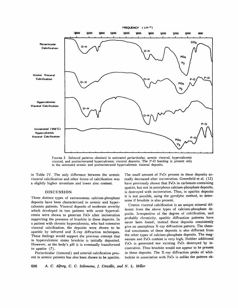
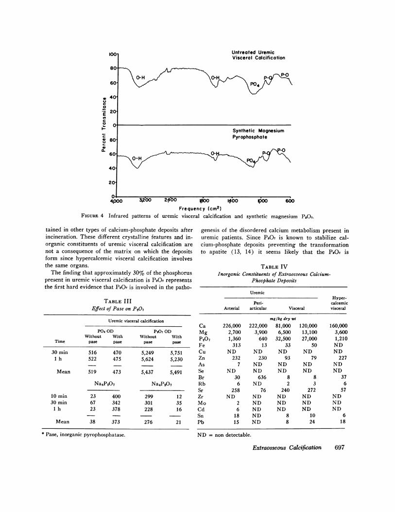
Additional confirmation for the presence of P207 in theuntreated uremic visceral calcification and incineratedhypercalcemic visceral calcification is the demonstrationof P-0 bondings with infrared techniques in only thesetwo groups of tissues (Fig. 3). The similarity betweenthe infrared pattern of uremic visceral calcification andmagnesium P207 is striking suggesting that the P207might be deposited as the magnesium salt (Fig. 4).
To determine if the P207 present in uremic visceralcalcification was resistant to pyrophosphatase the drydeposit was incubated with 30 U of yeast inorganic pyro-phosphatase in a Tris buffer solution at pH 7.2. The re-sults are given in Table III. Although the enzyme wasshown to be capable of hydrolyzing Na4P207 the P207 inthe uremic deposit was totally resistant to this enzyme.
Uremic Visceral
Calcification
35 33 31 29 27 25
Degrees 20FIGURE 2 Postincineration X-ray diffraction patterns ob-tained in uremic periarticular and visceral deposits and hy-percalcemic visceral deposits. Apatitic peaks are markedwith an A, whitlockitic with a W, and pyrophosphate withP207.
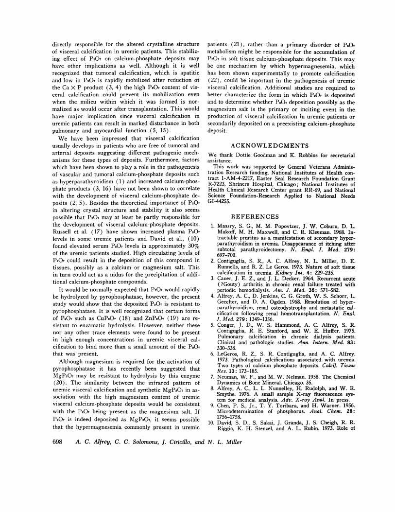
To determine if other inorganic constituents were dif-ferent in the various types of calcium-phosphate depositsa trace element profile using an X-ray fluorescencemethod was obtained on five tissues. Results are shown
Extraosseous Calcification 695
FREQUENCY (Cm-1)12003,200 20 OO 240 OO 100 poo 14OO go2o lpOO 800
Periarticular0
Calcif icat ion 0-H
Uremic VisceralCalcification | \A1
Hypercalcemic \,Visceral Calcification | \ \ ,
Incinerated ( 550C)__Hypercalcemic
Visceral Calcification
FIGURE 3 Infrared patterns obtained in untreated periarticular, uremic visceral, hypercalcemicvisceral, and postincinerated hypercalcemic visceral deposits. The P-O bonding is present onlyin the untreated uremic and postincinerated hypercalcemic visceral deposits.
in Table IV. The only difference between the uremicvisceral calcification and other forms of calcification wasa slightly higher strontium and lower zinc content.
DISCUSSIONThree distinct types of extraosseous calcium-phosphatedeposits have been characterized in uremic and hyper-calcemic patients. Visceral deposits of moderate severitywhich developed in two patients with acute hypercal-cemia were shown to generate P207 after incinerationsupporting the presence of brushite in these deposits. Ina patient with chronic hypercalcemia, who had extensivevisceral calcification, the deposits were shown to beapatitic by infrared and X-ray diffraction techniques.These findings would support the previous concept thatin hypercalcemic states brushite is initially deposited.However, at the body's pH it is eventually transformedto apatite (7).
Periarticular (tumoral) and arterial calcification pres-ent in uremic patients has also been shown to be apatitic.
The small amount of P207 present in these deposits ac-tually decreased after incineration. Greenfield et al. (12)have previously shown that P207 in carbonate-containingapatite, but not in amorphous calcium-phosphate deposits,is destroyed with incineration. Thus, in apatitic depositsit is not possible, using the pyrolytic method, to deter-mine if brushite is also present.
Uremic visceral calcification is an unique mineral dif-ferent from the above types of calcium-phosphate de-posits. Irrespective of the degree of calcification, andprobably chronicity, apatitic diffraction patterns havenever been found; instead these deposits consistentlygive an amorphous X-ray diffraction pattern. The chem-ical consituents of these deposits is also different fromthe other types of calcium-phosphate deposits. The mag-nesium and P207 content is very high. Neither additionalP207 is generated nor existing P207 destroyed by in-cineration. Thus brushite would not appear to be presentin these deposits. The X-ray diffraction peaks of whit-lockite in association with P207 is unlike the pattern ob-
696 A. C. Alfrey, C. C. Solomons, J. Ciricillo, and N. L. Miller
Untreated UremicVisceral Calcification
0-H 0~~ ~~~~-HPD P-004
Synthetic MagnesiumPyrophosphate
0- -0-HH P
4,00 300 2,400 l0o 14,00 p00 600Frequency (cm2)
Infrared patterns of uremic visceral calcification and synthetic magnesium P207.
tained in other types of calcium-phosphate deposits afterincineration. These different crystalline features and in-organic constituents of uremic visceral calcification arenot a consequence of the matrix on which the depositsform since hypercalcemic visceral calcification involvesthe same organs.
The finding that approximately 30% of the phosphoruspresent in uremic visceral calcification is P207 representsthe first hard evidence that P207 is involved in the patho-
TABLE IIIEffect of Pase on P207
Uremic visceral calcification
P04 OD P207 ODWithout With Without With
Time pase pase pase pase
30 min 516 470 5,249 5,7511 h 522 475 5,624 5,230
Mean 519 473 5,437 5,491
Na4P207 Na4P207
10 min 23 400 299 1230 min 67 342 301 35
1 h 23 378 228 16
Mean 38 373 276 21
* Pase, inorganic pyrophosphatase.
genesis of the disordered calcium metabolism present inuremic patients. Since P207 is known to stabilize cal-cium-phosphate deposits preventing the transformationto apatite (13, 14) it seems likely that the P207 is
TABLE IVInorganic Constituents of Extraosseous Calcium-
Phosphate Deposits
UremicHyper-
Peri- calcemicArterial articular Visceral visceral
mg/kg dry wt
Ca 226,000 222,000 81,000 120,000 160,000Mg 2,700 3,900 6,500 13,100 3,600P207 1,360 640 32,500 27,000 1,210Fe 313 13 33 50 NDCu ND ND ND ND NDZn 232 230 93 79 227As 7 ND ND ND NDSe ND ND ND ND NDBr 30 636 8 8 37Rb 6 ND 2 3 6Sr 258 76 240 272 57Zr ND ND ND ND NDMo 2 ND ND ND NDCd 6 ND ND ND NDSn 18 ND 8 10 6Pb 15 ND 8 24 18
ND=non detectable.
Extraosseous Calcification 697
100
80
60
,, 40c0
200Ca
CsOb-
80
0.60
40
20
FIGURE 4
directly responsible for the altered crystalline structureof visceral calcification in uremic patients. This stabiliz-ing effect of P207 on calcium-phosphate deposits mayhave other implications as well. Although it is wellrecognized that tumoral calcification, which is apatiticand low in P207 is rapidly mobilized after reduction ofthe Ca X P product (3, 4) the high P207 content of vis-ceral calcification could prevent its mobilization evenwhen the milieu within which it was formed is nor-malized as would occur after transplantation. This wouldhave major implication since visceral calcification inuremic patients can result in marked disturbance in bothpulmonary and myocardial function (5, 15).
We have been impressed that visceral calcificationusually develops in patients who are free of tumoral andarterial deposits suggesting different pathogenic mech-anisms for these types of deposits. Furthermore, factorswhich have been shown to play a role in the pathogenesisof vascular and tumoral calcium-phosphate deposits suchas hyperparathyroidism (1) and increased calcium-phos-phate products (3, 16) have not been shown to correlatewith the development of visceral calcium-phosphate de-posits (2, 5). Besides the theoretical importance of P207in altering crystal structure and stability it also seemspossible that P207 may at least be partly responsible forthe development of visceral calcium-phosphate deposits.Russell et al. (17) have shown increased plasma P207levels in some uremic patients and David et al., (10)found elevated serum P207 levels in approximately 30%of the uremic patients studied. High circulating levels ofP207 could result in the deposition of this compound intissues, possibly as a calcium or magnesium salt. Thisin turn could act as a nidus for the precipitation of addi-tional calcium-phosphate compounds.
It would be normally expected that P207 would rapidlybe hydrolyzed by pyrophosphatase, however, the presentstudy would show that the deposited P207 is resistant topyrophosphatase. It is well recognized that certain formsof P207 such as CuP207 (18) and ZnP207 (19) are re-sistant to enzamatic hydrolysis. However, neither thesenor any other trace elements were found to be presentin high enough concentrations in uremic visceral cal-cification to bind more than a small amount of the P207that was present.
Although magnesium is required for the activation ofpyrophosphatase it has recently been suggested thatMgP207 may be resistant to hydrolysis by this enzyme(20). The similarity between the infrared pattern ofuremic visceral calcification and synthetic MgP207 in as-sociation with the high magnesium content of uremicvisceral calcium-phosphate deposits would be consistentwith the P207 being present as the magnesium salt. IfP207 is indeed deposited as MgP207, it seems possiblethat the hypermagnesemia commonly present in uremic
patients (21), rather than a primary disorder of PS07metabolism might be responsible for the accumulation ofP207 in soft tissue calcium-phosphate deposits. This maybe one mechanism by which hypermagnesemia, whichhas been shown experimentally to promote calcification(22), could be important in the pathogenesis of uremicvisceral calcification. Additional studies are required tobetter characterize the form in which P207 is depositedand to determine whether P207 deposition possibly as themagnesium salt is the primary or inciting event in theproduction of visceral calcification in uremic patients orsecondarily deposited on a preexisting calcium-phosphatedeposit.
ACKNOWLEDGMENTSWe thank Dottie Goodman and K. Robbins for secretarialassistance.
This work was supported by General Veterans Adminis-tration Research funding, National Institutes of Health con-tract 1-AM-4-2217, Easter Seal Research Foundation GrantR-7223, Shriners Hospital, Chicago; National Institutes ofHealth Clinical Research Center grant RR-69, and NationalScience Foundation-Research Applied to National NeedsGI-44255.
REFERENCES1. Massry, S. G., M. M. Popovtzer, J. W. Coburn, D. L.
Makoff, M. H. Maxwell, and C. R. Kleeman. 1968. In-tractable pruritus as a manifestation of secondary hyper-parathyroidism in uremia. Disappearance of itching aftersubtotal parathyroidectomy. N. Engl. J. Med. 279:697-700.
2. Contiguglia, S. R., A. C. Alfrey, N. L. Miller, D. E.Runnells, and R. Z. Le Geros. 1973. Nature of soft tissuecalcification in uremia. Kidney Int. 4: 229-235.
3. Caner, J. E. Z., and J. L. Decker. 1964. Recurrent acute(?Gouty) arthritis in chronic renal failure treated withperiodic hemodialysis. Am. J. Med. 36: 571-582.
4. Alfrey, A. C., D. Jenkins, C. G. Groth, W. S. Schorr, L.Gecelter, and D. A. Ogden. 1968. Resolution of hyper-parathyroidism, renal osteodystrophy and metastatic cal-cification following renal homotransplantation. N. Engl.J. Med. 279: 1349-1356.
5. Conger, J. D., W. S. Hammond, A. C. Alfrey, S. R.Contiguglia, R. E. Stanford, and W. E. Huffer. 1975.Pulmonary calcification in chronic dialysis patients.Clinical and pathologic studies. Ann. Intern. Med. 83:330-336.
6. LeGeros, R. Z., S. R. Contiguglia, and A. C. Alfrey.1973. Pathological calcifications associated with uremia.Two types of calcium phosphate deposits. Calcif. TissueRes. 13: 173-185.
7. Neuman, W. F., and M. W. Nelman. 1958. The ChemicalDynamics of Bone Mineral. Chicago. 35.
8. Alfrey, A. C., L. L. Nunnelley, H. Rudolph, and W. R.Smythe. 1976. A small sample X-ray fluorescence sys-tem for medical analysis. Adv. X-ray Anal. In press.
9. Chen, P. S., Jr., T. Y. Toribara, and H. Warner. 1956.Microdetermination of phosphorus. Anal. Chem. 28:1756-1758.
10. David, S. D., S. Sakai, J. Granda, J. S. Cheigh, R. R.Riggio, K. H. Stenzel, and A. L. Rubin. 1973. Role of
698 A. C. Alfrey, C. C. Solomons, J. Ciricillo, and N. L. Miller

pyrophosphate in renal osteodystrophy. Trans. Am. Soc.Artif. Intern. Organs. 19: 440-445.
11. Armstrong, D., D. Van Warner, and C. C. Solomons.1975. Serum pyrophosphate levels in osteogenesis im-perfecta. J. Clin. Chem. 21: 104-108.
12. Greenfield, D. J., J. D. Termine, and E. D. Eanes. 1974.A chemical study of apatites prepared by hydrolysis ofamorphous calcium phosphates in carbonate-containingaqueous solutions. Calcif. Tissue Res. 14: 131-138.
13. Francis, M. D. 1969. The inhibition of calcium hydroxy-apatite crystal growth by polyphosphonates and poly-phosphates. Calicf. Tissue Res. 3: 151-162.
14. Russell, R. G. G., R. C. Muhlbauer, S. Bisaz, D. A.Williams, and H. Fleisch. 1970. The influence of pyro-phosphate, condensed phosphates, phosphonates and otherphosphate compounds on the dissolution of hydroxyapa-tite in vitro and on bone resorption induced by para-thyroid hormone in tissue culture and in thyropara-thyroidectomized rats. Calcif. Tissue Res. 6: 183-196.
15. Terman, D. S., A. C. Alfrey, W. S. Hammond, T. Don-nedlinger, D. A. Ogden, and J. H. Holmes. 1971. Cardiaccalcification in uremia. A clinical, biochemical and patho-logic study. Am. J. Med. 50: 744-755.
16. Katz, A. I., C. L. Hampers, and J. P. Merrill. 1969.Secondary hyperparathyroidism and renal osteodystrophy
in chronic renal failure. Analysis of 195 patients, withobservations on the effect of chronic dialysis, kidneytransplantation, and subtotal parathyroidectomy. Medi-cine (Baltimore). 48: 333-374.
17. Russell, R. G. G., S. Bisaz, and H. Fleisch. 1969. Pyro-phosphate and diphosphonates in calcium metabolism andtheir possible role in renal failure. Arch. Intern. Med.124: 571-577.
18. W6ltgens, J. H. M., S. L. Bonting, and 0. L. Bijvoet.1973. Inorganic pyrophosphatase in mineralizing hamstermolars. III. Influence of diphosphonates. Calcif. TissueRes. 13: 151-157.
19. W6ltgens, J., and W. Ahsmann. 1970. Determination oforthophosphate in the presience of inorganic pyrophos-phate in the assay of inorganic pyrophosphatase activity.Anal. Biochim. 35: 526-529.
20. Cathala, G., and C. Brunel. 1973. L'Activite pyrophos-phatasique de la phosphatase alcaline du cerveau. Bio-chim. Biophys. Acta. 315: 73-82.
21. Alfrey, A. C., N. L. Miller, and D. Butkus. 1974. Evalu-ation of body magnesium stores. J. Lab. Clin. Med. 84:153-162.
22. Whittier, F. C., and R. M. Freeman. 1971. Potentiationof metastatic calcification in vitamin D-treated rats bymagnesium. Am. J. Physiol. 220: 209-212.
Extraosseous Calcification 699