Carbon monoxide intoxication: An updated revie monoxide intoxication: An updated review ... In World...
9
Carbon monoxide intoxication: An updated review Leon D. Prockop ⁎ , Rossitza I. Chichkova Department of Neurology, College of Medicine, University of South Florida, USA Available online 27 August 2007 Abstract Carbon monoxide (CO), a highly toxic gas produced by incomplete combustion of hydrocarbons, is a relatively common cause of human injury. Human toxicity is often overlooked because CO is tasteless and odorless and its clinical symptoms and signs are non specific. The brain and the heart may be severely affected after CO exposure with carboxyhemoglobin (COHb) levels exceeding 20%. Damage occurs because the affinity of hemoglobin for CO is 210 times higher than for O 2 . Hypoxic brain damage predominates in the cerebral cortex, cerebral white matter and basal ganglia, especially in the globus pallidus. Diagnosis requires clinical acumen and a high index of suspicion, combined with epidemiological data, clinical examination, analysis of ambient air CO and patient COHb levels; also required are cardiology evaluation including ECG as well as neurological evaluation including brain imaging (CT and/or MRI, MR spectroscopy), and neuropsychological testing. Although immediate O 2 breathing is sometimes an adequate treatment, hyperbaric oxygen therapy (HBO) is favored. Subsequently, only symptomatic therapy is available for the long-term sequelae of CO poisoning. © 2007 Published by Elsevier B.V. Keywords: Carbon monoxide (CO); Carboxyhemoglobin (COHb); Hyperbaric oxygen (HBO); Brain damage; Cardiac damage; Neurotoxicity; CT; MRI 1. Introduction Carbon monoxide (CO) intoxication is one of the most common types of poisoning in the modern world, and the leading cause of death by poisoning in the Unites States. CO is a tasteless, odorless, non-irritating but highly toxic gas. Because of these properties and because it lacks a unique clinical signature, CO is difficult to detect and can mimic other common disorders. Therefore, the true incidence of CO poisoning is unknown and many cases probably go unrecog- nized. CO has been termed “the unnoticed poison of the 21st century” [1]. An environmental CO exposure is suggested when more than one person and animals are affected; when there is a history of fire, presence of fireplace or combustion appliances, or with occupational exposure; and by the occurrence of symptoms in relation to a possible exposure [2]. CO is a by-product of the incomplete combustion of hydrocarbons. Common sources include motor vehicle exhaust gases in a poorly ventilated garage or in areas in close proximity to garages; combustion appliances, e.g. heating units, in which partial combustion of oils, coal, wood, kerosene and other fuels generate CO. A common scenario is that of a heating unit used only occasionally and not well maintained. Retrograde flow can occur in residential, occupational or institutional settings in the presence of pressure problems, chimney or equipment malfunction. CO poisoning with immediate deaths may occur during a building fire or from fuel powered generators and heaters, especially in poorly ventilated spaces [3]. The latter causes are frequently reported during winter storms, hurricanes, earthquakes or other disasters after a power outage has occurred. There are also endogenous sources of CO, such as during the heme degradation to bile pigments, catalyzed by heme oxygenases [4]. Constitutive and inducible isoforms (HO-l, HO-2) of the enzyme are known. Endogenously produced CO serves as a signaling molecule involved in multiple cellular functions, such as inflammation, proliferation, and apoptosis. CO, like nitric oxide, is a recently defined gaseous neurotransmitter in the central nervous system (CNS). 2. History The ancient Greeks and Romans used CO to execute criminals. The deaths of two Byzantine emperors was related Journal of the Neurological Sciences 262 (2007) 122 – 130 www.elsevier.com/locate/jns ⁎ Corresponding author. Box 55, 12901 Bruce B. Downs Blvd., Tampa, FL 33612, USA. E-mail address: [email protected] (L.D. Prockop). 0022-510X/$ - see front matter © 2007 Published by Elsevier B.V. doi:10.1016/j.jns.2007.06.037
Transcript of Carbon monoxide intoxication: An updated revie monoxide intoxication: An updated review ... In World...

nces 262 (2007) 122–130www.elsevier.com/locate/jns
Journal of the Neurological Scie
Carbon monoxide intoxication: An updated review
Leon D. Prockop⁎, Rossitza I. Chichkova
Department of Neurology, College of Medicine, University of South Florida, USA
Available online 27 August 2007
Abstract
Carbon monoxide (CO), a highly toxic gas produced by incomplete combustion of hydrocarbons, is a relatively common cause of humaninjury. Human toxicity is often overlooked because CO is tasteless and odorless and its clinical symptoms and signs are non specific. Thebrain and the heart may be severely affected after CO exposure with carboxyhemoglobin (COHb) levels exceeding 20%. Damage occursbecause the affinity of hemoglobin for CO is 210 times higher than for O2. Hypoxic brain damage predominates in the cerebral cortex,cerebral white matter and basal ganglia, especially in the globus pallidus. Diagnosis requires clinical acumen and a high index of suspicion,combined with epidemiological data, clinical examination, analysis of ambient air CO and patient COHb levels; also required are cardiologyevaluation including ECG as well as neurological evaluation including brain imaging (CT and/or MRI, MR spectroscopy), andneuropsychological testing. Although immediate O2 breathing is sometimes an adequate treatment, hyperbaric oxygen therapy (HBO) isfavored. Subsequently, only symptomatic therapy is available for the long-term sequelae of CO poisoning.© 2007 Published by Elsevier B.V.
Keywords: Carbon monoxide (CO); Carboxyhemoglobin (COHb); Hyperbaric oxygen (HBO); Brain damage; Cardiac damage; Neurotoxicity; CT; MRI
1. Introduction
Carbon monoxide (CO) intoxication is one of the mostcommon types of poisoning in the modern world, and theleading cause of death by poisoning in the Unites States. CO isa tasteless, odorless, non-irritating but highly toxic gas.Because of these properties and because it lacks a uniqueclinical signature, CO is difficult to detect and can mimic othercommon disorders. Therefore, the true incidence of COpoisoning is unknown and many cases probably go unrecog-nized. CO has been termed “the unnoticed poison of the 21stcentury” [1]. An environmental CO exposure is suggestedwhen more than one person and animals are affected; whenthere is a history of fire, presence of fireplace or combustionappliances, or with occupational exposure; and by theoccurrence of symptoms in relation to a possible exposure [2].
CO is a by-product of the incomplete combustion ofhydrocarbons. Common sources include motor vehicleexhaust gases in a poorly ventilated garage or in areas inclose proximity to garages; combustion appliances, e.g.
⁎ Corresponding author. Box 55, 12901 Bruce B. Downs Blvd., Tampa,FL 33612, USA.
E-mail address: [email protected] (L.D. Prockop).
0022-510X/$ - see front matter © 2007 Published by Elsevier B.V.doi:10.1016/j.jns.2007.06.037
heating units, in which partial combustion of oils, coal,wood, kerosene and other fuels generate CO. A commonscenario is that of a heating unit used only occasionally and notwell maintained. Retrograde flow can occur in residential,occupational or institutional settings in the presence ofpressure problems, chimney or equipment malfunction. COpoisoning with immediate deaths may occur during a buildingfire or from fuel powered generators and heaters, especially inpoorly ventilated spaces [3]. The latter causes are frequentlyreported duringwinter storms, hurricanes, earthquakes or otherdisasters after a power outage has occurred.
There are also endogenous sources of CO, such as duringthe heme degradation to bile pigments, catalyzed by hemeoxygenases [4]. Constitutive and inducible isoforms (HO-l,HO-2) of the enzyme are known. Endogenously producedCO serves as a signaling molecule involved in multiplecellular functions, such as inflammation, proliferation, andapoptosis. CO, like nitric oxide, is a recently defined gaseousneurotransmitter in the central nervous system (CNS).
2. History
The ancient Greeks and Romans used CO to executecriminals. The deaths of two Byzantine emperors was related
123L.D. Prockop, R.I. Chichkova / Journal of the Neurological Sciences 262 (2007) 122–130
to CO produced by the burning of coal in braziers, the usualmethod of indoor heating during that epoch [5]. CO was firstprepared by the French chemist de Lassone in 1776. Becauseit burned with a blue flame, he mistakenly thought it to behydrogen. In 1880, William Cruikshank identified it as acompound containing carbon and oxygen. In the middle ofthe 19th century, Claude Bernard recognized that CO causeshypoxia by interaction with hemoglobin (Hb). He poisoneddogs with the gas, noticing the scarlet appearance of theirblood. Towards the end of the 19th century, Haldane demon-strated that a high partial pressure of O2 can counteract theinteraction between Hb and CO despite the high affinity forthis interaction. In World War II, wood was used widely asfuel and caused many cases of acute or chronic CO poison-ing [6].
3. Biochemistry, physiopathology, and pathology
CO binds rapidly to Hb, leading to the formation ofcarboxyhemoglobin (COHb). The oxygen carrying capacity ofthe blood decreases, causing tissue hypoxia. COHb is red whichexplains the “cherry-like” discoloration of victims. CO diffusesfrom the alveoli to the blood in the pulmonary capillaries acrossthe alveolo-capillary membrane that is composed of pulmonaryepithelium, the capillary endothelium and their fused basementmembrane. CO is taken up by the Hb at such a high rate that thepartial pressure of CO in the capillaries stays very low.Therefore, the CO transfer is diffusion-limited. The affinity ofhemoglobin for CO is 210 times its affinity for O2. CO easilydisplaces oxygen from hemoglobin. On the other hand, COHbliberates CO very slowly. In the presence of COHb, thedissociation curve of the remaining HbO2 shifts to the leftfurther decreasing the amount of the oxygen released. Theamount ofCOHb formed depends on the duration of exposure toCO, the concentration of CO in the inspired air and the alveolarventilation. Even though CO is toxic to the cytochromes, thismechanism plays a minor role in clinical CO poisoning, sincethe amount of CO required to poison cytochromes is 1000 timeshigher than the lethal dose. CO binds to intracellular myoglobinin the myocardium and impairs the oxygen supply to themitochondria. This negatively affects the oxidative phosphor-ylation and consequently, the energy source of heart muscle.Patients with underlying cardiac conditions are at risk for deathfrom arrhythmias and fatal heart attacks can occur. However,chest pain as a symptom of myocardial ischemia can occurwithout underlying coronary artery disease. For example,2 weeks after accidental exposure to CO, 34% of a group ofSwiss soldiers had chest pain [7].
Henry et al. [8] studied mortality risk in patients withmoderate to severe CO poisoning. In those at low risk forcardiovascular diseases, 37% suffered acute myocardial injuryand 38% were dead within 7.6 years. The mortality rate wasthree times higher than the US expected mortality by age andsex [8].
After CO exposure, angina attacks, arrhythmias, andincreased level of cardiac enzymes frequently occur. This has
led to a search for morphological changes that could beattributed to CO, especially because the myocardium bindsmore CO than skeletal muscle. Ultramicroscopic lesionshave been reported but the relative roles of general tissuehypoxia and specific CO toxicity are unknown [9]. Inaddition to COHb, the binding of CO to cytochromes is alsosignificant and is thought to be responsible for cytotoxicity.Combined ultra structural and cytochemical studies haveenabled differentiation between toxic, hypoxic, and mixedalteration. The marked decrease in cytochrome oxidase inexperimental studies suggests a direct toxic effect [9].
Myocardial injury with ischemic ECG changes andelevated cardiac biomarkers were found in 37% of 230patients with moderate to severe CO poisoning with 5% in-hospital mortality [10]. Therefore, patients admitted to thehospital with CO poisoning should have a baseline ECG andserial cardiac enzymes.
Recent investigations suggest other mechanisms of CO-mediated toxicity. One hypothesis is that CO-induced tissuehypoxia may be followed by reoxygenation injury to theCNS. Hyperoxygenation facilitates the production of partial-ly reduced oxygen species, which in turn can oxidizeessential proteins and nucleic acids, resulting in typicalreperfusion injury. In addition, CO exposure has been shownto cause lipid peroxygenation, i.e. degradation of unsaturatedfatty acids leading to reversible demyelination of CNS lipids.CO exposure also creates substantial oxidative stress on cells,with production of oxygen radicals resulting from theconversion of xanthine dehydrogenase to xanthine oxidase[11]. Myocardial fiber necrosis was described in a 26 year oldpatient with accidental CO poisoning and blood concentra-tion of COHb of 46.6% [12]. Electron microscopy of leftventricular biopsies of a 25 year old woman with functionalevidence of cardiac failure after acute CO poisoning andotherwise normal myocardial perfusion showed slightultrastructural changes in the myocytes, large glycogendeposits and swollen mitochondria. The above changes havebeen though to be signs of impaired energymetabolism of themyocardial cells [13]. In rat heart, CO causes vasodilatationand increased coronary flow that are not mediated by simplehypoxia [14]. CO exposure in the fetal period in rats causesmyocyte hyperplasia and cardiomegaly. This cellular re-sponse is sustained through the early neonatal period inanimals exposed to CO both in utero and in the post-partum[15]. Although hemorrhages and areas of necrosis in theheart, mostly in the septum and the papillary muscles weredescribed with CO poisoning as early as 1865 [16] only fewhuman cases of acute, fatal CO intoxication, with small fociof coagulation necrosis have been reported [17].
Disturbances of brain function predominate in acute COintoxication. Delayed neurological effects also occur [18].Tissue hypoxia is the end result of intoxication with CO andwith many other physical and chemical agents. Some brainregions are sensitive to hypoxic damage including the cerebralcortex, particularly its second and third layers; the whitematter, the basal nuclei, and Purkinje cells of the cerebellum.
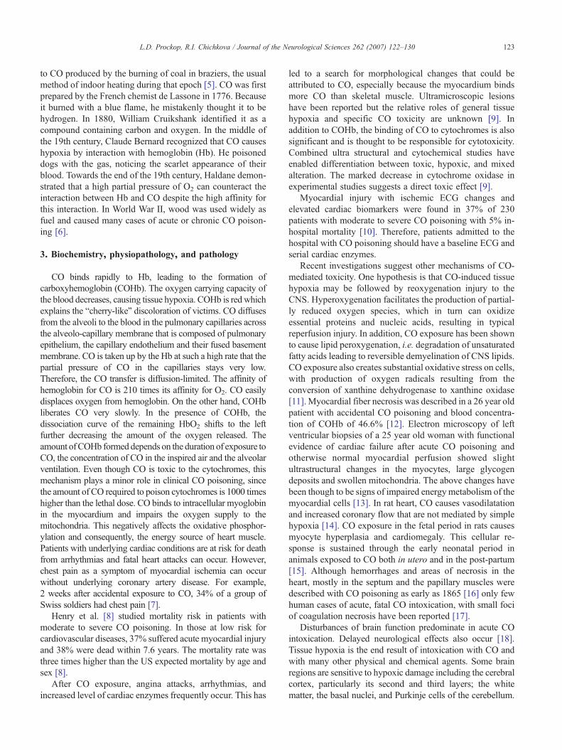
Fig. 1. Brain CT and MRI scans in the same patient. (A) CT findings. The arrows show low-density areas. (B) T2-weighted MRI cortical areas are more affectedthan the subcortical areas from Murata et al. [20].
124 L.D. Prockop, R.I. Chichkova / Journal of the Neurological Sciences 262 (2007) 122–130
Attempts have been made to relate this “selective vulnerabil-ity” to the cause of the hypoxia, but the nature and distributionof the lesions appear to depend on the severity, suddenness,and duration of the oxygen deprivation, as well as on itsmechanism (hypoxemia or ischemia) rather than on its cause.Regions with relatively poor vascularization and “watershed”areas between two sources of blood supply, such as the globuspallidus, may be more vulnerable, especially during periods ofhypotension. The effects of hypoxia on the brain, therefore, donot reflect it cause and neither the character of the lesions northe areas affected are regarded as pathognomonic for CO.
The neuropathology of CO toxicity has been well describedin postmortem studies [19] and includes, in acute cases,petechial hemorrhages of the white matter involving inparticular the corpus callosum; in cases surviving more than48 h there is multifocal necrosis involving globus pallidus,hippocampus, pars reticularis of the substantia nigra, laminarnecrosis of the cortex and loss of Purkinje cells in the
cerebellum, along with white matter lesions. The typicalpallidum lesions are well-defined, bilateral globus pallidusmacroscopic infarctions, usually asymmetrical, extendinganteriorly, superiorly, or into the internal capsule. Occasionally,only a small linear focus of necrosis is found at the junction ofthe internal capsule and the internal nucleus of the globuspallidus. CO intoxication usually spares the hypothalamus,walls of the third ventricle, thalamus, striatum, and brainstem.Myelin damage is constant and ranges from discrete, perivas-cular foci in the corpus callosum, the internal–external capsuleand the optic tracts usually seen in comatose patients who diedwithin 1 week, to extensive periventricular demyelination andaxonal destruction observed in comatose subjects with longersurvival, sometimes leading to formation of plaques ofdemyelination. A distinct constellation of brain and MRIabnormalities appears premortem and in those surviving anexposure. It includes globus pallidus lesions, white matterchanges, and diffuse low-density lesions throughout the brain.

Fig. 2. T1-weightedMRI. High-signal regions are seen in the globus pallidusbilaterally (arrows) from Murata et al. [20].
Table 1Signs and symptoms of carbon monoxide intoxication
1. Headache 8. Difficulty in coordinating2. Dizziness 9. Difficulty in breathing3. Irritability 10. Chest pain4. Confusion/memory loss 11. Cerebral edema5. Disorientation 12. Convulsions/seizures6. Nausea and vomiting 13. Coma7. Nausea and vomiting 14. Death
125L.D. Prockop, R.I. Chichkova / Journal of the Neurological Sciences 262 (2007) 122–130
In general, CT and MRI neuroimaging findings reflect theneuropathologic changes described by Lapresle and Fardeau[19] (Figs. 1–3). Previously unreported neuropathologicalfindings include the MRI findings of thalamic lesions [22].
4. Clinical findings
Because of their high metabolic rate, the brain and theheart are most susceptible to CO toxicity. The clinical
Fig. 3. (A) Proton density brain MRI, transaxial view, shows bilateral hemorrhageet al. [21].
symptoms of CO poisoning are often non-specific and canmimic a variety of common disorders. The severity rangesfrom mild flu-like symptoms to coma and death. About 50%of exposed people may develop weakness, nausea, confu-sion, and shortness of breath. Less frequently, abdominalpain, visual changes, chest pain and loss of consciousnessoccur. Tachycardia and tachypnea develop to compensatefrom cellular hypoxia and cardiac output increases initially.Responses to cellular hypoxia vary depending on thepremorbid condition of victims; those with underlying lungand heart disease will have little tolerance to even mildhypoxia. Hypoxia leads to increased intracranial pressureand cerebral edema which is partly responsible for decreasedlevel of consciousness, seizures and coma. The classiccherry-red discoloration of the skin and cyanosis are rarelyseen [19]. Varying degrees of cognitive impairment havebeen reported [23].
Table 1 lists some of the symptoms and signs of COintoxication. Headache is one of the most commonpresenting features of CO poisoning: it occurs in 84% ofthe victims [24] and has been described as predominantly
in the globus pallidus. (B) T1-weighted brain MRI, coronal view from Tom

126 L.D. Prockop, R.I. Chichkova / Journal of the Neurological Sciences 262 (2007) 122–130
frontal, dull, sharp, continuous, throbbing, and intermittentin patients with a mean COHb level of 21.3% (± 9.3%).There is no clear correlation between pain intensity andCOHb levels [25]. Some have reported tightness across theforehead at COHb levels of 10–20%, throbbing in thetemples at 20–30%, and severe headache at 30–40% [26].Headaches, generalized weakness, fatigue and sleepiness,are part of the vague symptomatology observed in subjectswith COHb levels below 20%. Headache is a frequentcomplaint not only with acute but with chronic COpoisoning. Dizziness is a frequent companion of headacheand can be seen in about 92% of CO victims of COpoisoning. In one report, 76% of 38 victims reportedweakness with COHb levels N 30–40% [3].
A delayed neuropsychiatric syndrome may occur inpatients from 3 days up to 240 days after acute CO exposure.Even those victims without neurological and psychiatricsymptoms immediately after an exposure accident maydemonstrate features of delayed impairment ranging fromsubtle abnormalities such as personality changes or mildcognitive deficit to severe dementia, psychosis, Parkinson-ism, incontinence or other [18,27].
CO encephalopathy may cause a number of behavioralfunctional impairments including alterations in attention,executive function, verbal fluency, motor abilities, visuos-patial skills, learning, short-term memory, and mood andsocial adjustment. Formal neuropsychological testing usual-ly confirms these impairments [28].
5. Diagnosis
Diagnosis of CO poisoning requires a high level ofsuspicion. Epidemiological history with information aboutother affected individuals or pets as well as circumstancessuggestive of possible exposure is of paramount importance.
Ambient air CO levels should be obtained as soon aspossible after the exposure. Because the half-life of COHb is4–5 h, a victim's COHb level should also be obtained assoon as possible. Normal level for non-smokers is b 2% andfor smokers 5–13%. The Expert Panel on Air QualityStandard of the World Health Organization (WHO) in 1994reported that blood COHb levels between 2.5% and 4%decrease in the short-term maximal exercise duration inyoung healthy men. Decrease exercise duration because ofincreased chest pain and in patients with ischemic heartoccurred at levels from 2.7% to 4.1%. Levels between 2%and 20% can cause effects on visual perception levels,audition, motor, and sensory-motor functions, and behavior.Therefore, ambient air CO levels which produce bloodCOHb levels below 2.5% are recommended. The COHblevels depend not only upon the CO level in ambient air butalso on the duration of exposure. According to WHOguidelines, exposures to levels of ambient air CO2 in partsper million (ppm) should conform to the following durationsof exposure: 87.1 ppm (100 mg/m3) for 15 min; 52.3 ppm(60 mg/m3) for 30 min; 26.1 ppm (30 mg/m3) for 60 min;
8.7 ppm (10 mg/m3) for 8 min. [29]. Any exposure toambient air with CO levels greater than 100 ppm isdangerous to human health [28,30].
Physicians who deal with CO intoxication should beaware that pulse oximetry is a colorimetric method,unreliable for the diagnosis of CO intoxication since it cannot distinguish oxyhemoglobin from COHb. Therefore,pulse oximeters overestimate arterial oxygenation in patientswith severe CO poisoning. Accurate assessment of arterialoxygenation in patients with severe CO poisoning cancurrently be performed only by analysis of arterial bloodwith a laboratory CO-oximetry. High-flow oxygen should beadministered to all patients suspected of significant COexposure until direct measurement of CO levels can beperformed, regardless of pulse oximetry readings [31].
For clinical purposes, automated spectrophotometer CO-oximeter device are recommended. Spectrophotometersmeasures light intensity as a function of color and candifferentiate the different wavelengths of oxyhemoglobin andCOHb with an acceptable accuracy for COHb saturationlevels above 5%; they can simultaneously estimate total Hband percentages of oxyhemoglobin and COHb. Gas chroma-tography, a more sensitive method, can be utilized for low-level exposure and for post-mortem blood samples [32].
Cardiac function must be monitored closely by ECG, 2D-ECHO, and cardiac enzymes. The heart, like the brain, isvery sensitive to hypoxic injury. Patients with underlyingcardiac disorders, whose reserves are impaired at baseline,are at higher risk compared to normal individuals. Cardiacarrest and sudden cardiac death can be expected. Chest pain,due to myocardial ischemia or infarction is a consequence ofdecreased oxygen supply to the cardiac muscle. Features ofischemia as well as other abnormalities, such as tachycardia,bradycardia, atrial and ventricular fibrillation, prematureventricular contractions, conduction abnormalities andothers can be easily detected on ECG. Non-invasive devicesthat can be used to screen firefighters and victims canestimate the COHb levels in the exhaled alveolar breath havebeen suggested [31]. A non-invasive, high-resolutionmethod of measuring COHb fraction using expiratory gasanalysis in patients without evidence for pulmonary edemaor atelectasis has been found to have accuracy equivalent tothat of CO-oximetry [33].
Very high levels of S100B protein, a structural astroglialprotein, have been found in patients who died from COpoisoning; elevated levels occur in unconscious patients, andnormal levels in those without loss of consciousness. It wasproposed that the S100B protein levels could be used as abiochemical marker of brain injury in CO poisoning [34].However, Rasmussen et al. [35] failed to find significantincrease in blood concentrations of neuron-specific enolaseand of S-100 protein there was no correlation with level ofconsciousness in CO poisoning [35]. Scintigraphy studies ofthe heart with 99mTc have been proposed as method ofchoice for evaluation of heart injury in patients after acuteCO intoxication [36].

127L.D. Prockop, R.I. Chichkova / Journal of the Neurological Sciences 262 (2007) 122–130
Diffusion weighted images (DWI) in brain MRI showwhite matter high signal intensities consistent with restricteddiffusion in acute CO poisoning. Follow-up MRI performed16 days later reveals disappearance of white matter lesions,suggesting the white matter can be more sensitive than graymatter to hypoxia in the acute phase [37].
T2-weighted brain MRI shows increased signal intensitybilaterally in the putamen and the caudate nucleus, as well ashigh signal intensity in the globus pallidus [38]. Initially,unilateral low attenuation areas in the right putamen, globuspallidus and thalamuswere observed in theCTof a patient afterCO exposure, followed by transient bilateral appearance oflesions on subsequent CTexamination.Hemorrhagic infarctionof the right putamen, and ischemic lesions in both thalami werevisualized onMRI 2weeks later [39]. Diffusion-weightedMRIin a case of COpoisoning revealed pallidoreticular damage anddelayed leukoencephalopathy characterized by restricted waterdiffusion pattern in the early stage. DWI brain MRI imaging ismore sensitive than brain CT and is useful for earlyidentification of the effects of acute CO poisoning [40].
Brain MRI changes after CO poisoning are variable andreflect the neuropathological lesions. Most unconsciouspatients present with abnormalities of globus pallidus or theentire lentiform nucleus (globus pallidus and putamen),putamen alone, caudate nucleus, thalamus, periventricularand subcortical white matter, cerebral cortex hippocampusand cerebellum. Brain MRI may appear to be normal in somevictims who have suffered CO brain damage [41]. Previouslyunreported brain MRI findings in CO poisoning includedbilateral diffuse high signal in the centrum semiovale andbilateral high intensity lesions in the anterior thalami [42].Extensive bilateral cerebellar white matter signal change,with sparing of the overlying cortex, consistent withdemyelination was reported 6 years after CO poisoning [43].
In a study of patients with severe CO intoxication, comaon admission and normobaric 100% oxygen, persistentchanges on the MRI were found 1 to 10 years after exposure,independently of the neuropsychiatric findings. T2-weightedand FLAIR images showed bilateral symmetric hyperinten-sity of the white matter, more often involving the centrumsemiovale, with relative sparing of the temporal lobes andanterior parts of the frontal lobes, along with atrophy of thecerebral cortex, cerebellar hemispheres, vermis, corpuscallosum as well as T1-hypointensities and T2 and FLAIRhyperintensities in globus pallidus [44].
Kim and colleagues studied the delayed effects of CO onthe cerebral white mater 25–95 days after the exposure withinitial recovery followed by relapse of neuropsychiatricsymptoms. T2-weighted imaged, DWI and FLAIRsequences demonstrate bilateral, diffuse and confluentlesions in the periventricular white matter and centrumsemiovale, more prominent changes were present in thefrontal lobes than elsewhere [45,46]. The effects of COpoisoning in acute stages can be evaluated by DWIsequences on brain MRI. A restricted water diffusion patternin globus pallidus and substantia nigra can be seen [47].
Cerebral edema occurs early. Clinical status and outcomecorrelate with diffuse white matter changes [48]. Long-term(25 years) after CO exposure, MRI has demonstratedsymmetrical globus pallidus and white matter changes inmost patients. Temporal, parietal and occipital lobes areusually affected with asymmetrical cortical and subcorticallesions [49,50].
Magnetic resonance spectroscopy (MRS) examines brainmetabolites. The major resonances of MRS are N-acetylaspartate (NAA), choline (Cho), and creatine (Cr). NAA islocated within neurons and is a specific neuronal and axonalmarker. Choline is part of the membrane constituentphosphatidylcholine. Based on previous studies of demye-linating brains, Cho increases are due to an increase inphosphatidylcholine due to demyelination or gliosis. NAAdecreases in demyelinated white matter presumably repre-sent axonal and neuronal loss [51]. MRS provides evidencefor CO-induced brain damage including decreased NAAfound in the basal ganglia and elsewhere [23].
Proton magnetic resonance spectroscopy (1H MRS) is anon-invasive method that can provide biochemical informa-tion about brain tissues. In early CO poisoning 1H MRSstudies showed a persistent increase in choline related toprogressive demyelination. In irreversibly injury, lactateappears and NAA decreases [52] 1H MRS studies of frontallobe white matter revealed increase in the choline-containingcompounds, and reductions of NAA in all cases [53].Normalization of the findings was found in a subclinicalcase. In two cases with akinetic mutism increased lactate wasnoted to persist. These results indicate the 1H MRS is auseful indicator in the clinical evaluation of patients with theinterval form of CO poisoning when compared to MRI, EEGand N-isopropyl–p123idoamphetamine SPECT [54].
Kamada et al. [55] reported that MRS in patients withdelayed sequelae of carbonmonoxide (CO) exposure preciselyreflects the severity of symptoms. With severe clinicalpresentation, marked lowering of NAA/Cr ratio and slightlyincreased Cho/Cr ratio is notedwith subsequent return ofNAAand Cho/Cr ratio to normal with clinical improvement. ProtonMRS appears to be superior to conventional radiologicalexaminations in CO poisoning [55].
Brain CT, MRI and MRS as well as neuropsychologicaltesting are useful tools in diagnosis of CO toxicity and itsseverity. In addition, positron emission tomography (PET)and single photon emission tomography (SPECT) mayprovide additional information [56,57].
6. Treatment
Tissue hypoxia is the major outcome of CO intoxication:therefore based on chemical and pathophysiological data, O2 isthe “natural antidote” [58]. Since the clinical signs andsymptoms ofCO toxicity are nonspecific, all suspected victimsshould be treated withO2 inhalation immediately after blood isdrawn for COHb content. Furthermore, there is wide variationin individual responses to similar levels of CO exposure,
128 L.D. Prockop, R.I. Chichkova / Journal of the Neurological Sciences 262 (2007) 122–130
ranging from death to a Parkinsonian syndrome to mild ormoderate intellectual impairment [23]. Therefore, immediatelyafter securing the airway and adequate ventilation, adminis-tration of normobaric oxygen (NBO) is the corner stone oftherapy, reducing the half-life of COHb from a mean of 5 h(range, 2–7 h) to about 1 h. Hyperbaric oxygen therapy (HBO)at 2.5 atm reduces it to 20 min and also has other benefits, atleast in animal models. For example in rat brains, it preventslipid peroxidation and leukocyte adherence to brain micro-vascular endothelium while accelerating regeneration ofinactivated cytochrome oxidase. Therefore, usually at 2.5 to3 ATA for 90 to 120 min, it is considered the treatment ofchoice for those who present with syncope, coma, or seizure,and focal neurological deficit or COHb N 25% (15% inpregnancy) [59–63].
In theory, NBO should be the treatment of the lastseverely poisoned patients, reserving HBO for severeintoxications. However, there are problems with this policy:1) COHb levels do not correlate with the clinical severity ofCO poisoning. 2) There is not universally accepted severityscale of CO poisoning, although loss of consciousness andneurologic deficits generally indicate severe poisoning. 3)All victims of CO poisoning are at risk for delayedneuropsychological sequelae. Therefore, in general thefollowing approach is appropriate: 1) Patients with presumedCO poisoning should be placed on 100% oxygen. 2) Patientswith severe poisoning must receive HBO regardless ofCOHb level. 3) Pregnant women must be treated with HBOirrespective of signs and symptoms. 4) In patients with lesserdegrees of poisoning, careful evaluation is advised beforedeciding that 100% NBO for N 6 h is the adequate therapy[58]. Administration of more than one course of HBO isthose who remain in coma remains controversial.
There are a number of practical considerations since notall treatment facilities, e.g. hospital ERs, can measure COHband/or administer HBO. For example, in one recent study,only 44% of acute care hospitals had the capability ofmeasuring COHb [64].
HBO is 100% oxygen at two to three times the atmosphericpressure at sea level. The oxygen tension in the arteriesincreases to about 2000 mm Hg and that of the tissues — toalmost 400 mm Hg. The pressure is expressed in multiples ofthe atmospheric pressure, which is 1 at sea level. At sea level,the blood oxygen concentration is 0.3 ml/dl. At 100% oxygenat ambient (normobaric) pressure, the amount of the dissolvedoxygen in the blood increases five fold to 1.5 ml/dl. At 3 atm,the dissolved-oxygen content reaches 6 ml/dl. HBO decreasesthe bubble formation in the blood and replaces inert gaseswith oxygen, which is rapidly taken up and utilized by thetissues. HBO can be bactericidal, bacteriostatic or suppresstoxin production increasing tissues resistance against infec-tions. HBO is more effective that normobaric oxygen inpromoting collagen formation and angiogenesis and thus canfacilitate wound healing. HBO inhibits neutrophil adherenceto the walls of the ischemic vessels, which decreased the freeradical production, vasoconstriction and tissue destruction.
HBO is commonly delivered in a monoplace chamber, orless often in a multi-occupant chamber. The duration of asingle treatment for CO poisoning is about 45 min. HBOwith oxygen pressures of up to 3 atm for a maximum of120 min is safe. Adverse effects include reversible myopia,cataract, tracheobronchial symptoms, self-limited seizuresand barotraumas to the middle ear, cranial sinuses, rarelyteeth or lungs. Claustrophobia can be an issue in monoplacechambers. Despite the conflicting results from the literatureregarding the effect of the HBO versus normobaric oxygen,Tibbles and Edelsberg [63] concluded that patients withsevere carbon monoxide poisoning should receive at leastone HBO treatment at 2.5 to 3.0 atm because this therapy isthe fastest method of treatment of the potentially reversiblelife-threatening effects.
The treatment of a patient with CO poisoning should notbe based solely on the COHb levels. The clinical manifesta-tions, COHb levels and, importantly, the patient's underlyingmedical history should be taken into account. In patients withsuspected CO poisoning, 100% oxygen should be givenimmediately by a mask. The goal is to raise the PaO2 levels,decrease the half-life of CO, and facilitate its dissociationfrom hemoglobin, thus allowing oxygen to attach to the freedbinding sites. Strict bed rest should be provided, since itdecreases oxygen demand and consumption. Patients withrespiratory distress and decreased level of consciousnessshould be intubated and ventilated. Chest radiograph, bloodlactate levels and arterial blood gases should be performed inthe emergency department.
Headache improved prior to hyperbaric oxygen treatmentin 72%, resolving entirely in 21% of those with residualheadache, pain improved with hyperbaric oxygen in 97%,resolving entirely in 44% [25]. Even though the deaths fromCO poisoning have decreased in the United States in therecent years, the total burden, including fatal and non-fatalcases has not significantly changed [65].
Juurlink et al. [66] analyzed available data from sixrandomized controlled trials involving non-pregnant adultsacutely poisoned with CO. At one month follow-up aftertreatment, symptoms possible related to carbon monoxidepoisoning were present in 34.2% of those treated with HBO,compared with 37.2% treated with NBO. They find notevidence that unselected use of HBO in the treatment ofacute CO poisoning reduces the frequency of neurologicalsymptoms at 1 month. Because of insufficient evidence, theyrecommend further research for defining the role of HBO intreatment of carbon monoxide poisoning.
Five years later, the same group examines the evidencefor the effectiveness of the HBO for prevention ofneurological sequelae in patients with acute carbon monox-ide poisoning. Four out of six trials find no benefit of HBOfor the reduction of neurologic sequelae, while two other donot. The authors conclude that the existing randomized trialshave not been able to establish reduction of neurologicalsequelae with the administration of HBO to patients withcarbon monoxide poisoning [68–71]. In a recent study,

129L.D. Prockop, R.I. Chichkova / Journal of the Neurological Sciences 262 (2007) 122–130
Weaver et al. [72] concluded that HBO is indicated forpatients with acute CO poisoning who are ≥ 36 years of age,or have exposure intervals ≥ 24 h, or in patients with loss ofconsciousness, or with higher COHb levels.
Close monitoring of the serum pH and lactic acid levels isrequired, since the anaerobic metabolism in the presence oftissue hypoxia generates lactic acidosis. Acidosis below pH of7.15 should be treatedwith sodiumbicarbonate. Caution has tobe exercised with the administration of sodium bicarbonatebecause carbon dioxide, a by-product of its metabolism, couldlead to respiratory acidosis and has to be eliminated by properventilation.
Because prevention is the best treatment, our societyshould be on high alert in attempt to prevent cases of COpoisoning.
References
[1] Proceedings Satellite Meeting IUTOX VIIIth Internationazl Congressof Toxicology. “Carbon monoxide: the unnoticed poison of the 21stCentury” 1998. Université de Bourgogne, France.
[2] Wikipedia contributors. Carbon Monoxide. Wikipedia, The free ency-clopedia;http://en.wikipedia.org/windex.php?title=Carbon_monoxide.
[3] Abelsohn A, Sanborn MD, Jessiman BJ, Weir E. Identifying andmanaging adverse environmental health effects: 6. Carbon monoxidepoisoning. CMAJ 2002;166(13):1685–90.
[4] Ryter SW, Alam J, Choi AM. Heme oxygenase-1/carbon monoxide:from basic science to therapeutic applications. Physiol Rev 2006;86(2):583–650.
[5] Lascaratos JG, Marketos SG. The carbon monoxide poisoning of twoByzantine emperors. J Toxicol Clin Toxicol 1998;36(1-2):103–7.
[6] Tvedt B, Kjuus H. Chronic CO poisoning. Use of generator gas duringthe Second World War and recent research. Tidsskr Nor Laegeforen1997;117(17):2454–7.
[7] Henz S, Maeder M. Prospective study of accidental carbon monoxidepoisoning in 38 Swiss soldiers. Swiss Med Wkly 2005;135(27-28):398–408.
[8] Henry CR, Satran D, Lindgren B, Adkinson C, Nicholson CI, HenryTD. Myocardial injury and long-term mortality following moderateto severe carbon monoxide poisoning. JAMA 2006;295(4):398–402.
[9] Somogyi E, Balogh I, Rubanyi G, Sotonyi P, Szegedi L. New findingsconcerning the pathogenesis of acute carbon monoxide (CO)poisoning. Am J Forensic Med Pathol 1981;2(1):31–9.
[10] Satran D, Henry CR, Adkinson C, Nicholson CI, Bracha Y, Henry TD.Cardiovascular manifestations of moderate to severe carbon monoxidepoisoning. J Am Coll Cardiol 2005;3;45(9):1513–6.
[11] Ernst A, Zibrak JD. Current concepts: carbon monoxide poisoning. NEngl J Med 1998;339(22):1603–8.
[12] Kumazawa T, Watanabe-Suzuki K, Seno H, Ishii A, Suzuki O. Acurious autopsy case of accidental carbon monoxide poisoning in amotor vehicle. Leg Med 2000;2(3):181–5 [Tokyo].
[13] Tritapepe L, Macchiarelli G, Rocco M, Scopinaro F, Schillaci O,Martuscelli E, et al. Functional and ultra structural evidence ofmyocardial stunning after acute carbon monoxide poisoning. Crit CareMed 1998;26(4):797–801.
[14] Lin H, McGrath JJ. Responses of the working rat heart to carbonmonoxide. Physiol Behav 1989;46(1):81-4 2813558.
[15] Clubb Jr FJ, Penney DG, Baylerian MS, Bishop SP. Cardiomegaly dueto myocyte hyperplasia in prenatal rats exposed to 200 ppm carbonmonoxide. J Mol Cell Cardiol 1986;18(5):477–86.
[16] Anderson RF, Allensworth DC, DeGroot WJ. Myocardial toxicityfrom carbon monoxide poisoning. Ann Intern Med 1967;67(6):1172–82.
[17] Fineschi V, Agricola E, Baroldi G, Bruni G, Cerretani D, Mondillo S, etal. Myocardial findings in fatal carbon monoxide poisoning: a humanand experiment morphometric study. Int J Legal Med 2000;113(5):276–82.
[18] Choi S. Delayed neurological sequelae in carbon monoxide intoxica-tion. Arch Neurol 2000;57:1214–8.
[19] Lapresle J, Fardeau M. The central nervous system and carbonmonoxide poisoning II. Anatomical study of brain lesions followingintoxication with carbon monoxide (22 cases). In: Bour H, LedinghamIM, editors. Carbon monoxide poisoning, vol. 24. Prog Brain Res;1967. p. 31–74.
[20] Murata S, Asaba H, Hiraishi K, et al. Magnetic resonance imagingfindings on carbon monoxide intoxication. J Neuroimaging1993;3:128–31.
[21] Tom T, Abedon S, Clark RI, Wong W. Neuroimaging characteristics incarbon monoxide toxicity. J Neuroimaging 1996;6:161–6.
[22] Ernst A, Zibrak JD. Carbon monoxide poisoning. N Engl J Med1998;339(22):1603–8.
[23] Prockop LD. Carbon monoxide brain toxicity: clinical, magneticresonance imaging, magnetic resonance spectroscopy, and neuropsy-chological effects in 9 people. J Neuroimaging 2005;15(2):144–9.
[24] Handa PK, Tai DY. Carbon monoxide poisoning: a five year review atTan Tock Seng Hospital, Singapore. Ann Acad Med Singapore2005;34(10):611–4.
[25] Hampson NB, Hampson LA. Characteristics of headache associatedwith acute carbon monoxide poisoning. Headache 2002;42(3):220–3.
[26] Choi IS. Carbon monoxide poisoning: systemic manifestations andcomplication. J Korean Med Sci 2001;16(3):253–61.
[27] MimuraK,HaradaM, Sumiyoshi S, et al. Long-terms follow-up study onsequelae of carbon monoxide poisoning: serial investigation 33 yearsafter poisoning. Seishin Shinkeigaku Zasshi 1999;101:592–618.
[28] White RF, Feldman RG, Proctor SP. Neurobehavioral effects of toxicexposure. In: White RF, editor. Clinical syndrome in adultneuropsychology. The practitioner's handbook. New York: Elsevier;1992. p. 1–51.
[29] Department of the Environment. Expert panel on air quality standard.London: HMSO; 1994.
[30] Townsend CL, Maynard RI. Effects on health of prolonged exposure tolow concentrations of carbon monoxide. Occup Environ Med 2002;59(10):708–11.
[31] Bozeman WP, Hampson NB. Pulse oximetry in CO poisoning —additional data. Chest 2000 Jan; 117(1):295-6 (Hampson NB Pulseoximetry in severe carbon monoxide poisoning. Chest 1998;114(4):1036-41.
[32] Widdop B. Analysis of carbon monoxide. Ann Clin Biochem 2002;39(pt 4):378–91.
[33] Sawano M, Mato T, Tsutsumi H. Bedside red cell volumetry by low-dose carboxyhaemoglobin dilution using expiratory gas analysis. Br JAnaesth 2006;96(2):186–94.
[34] Brvar M, Mozina H, Osredkar J, Mozina M, Noe M, Brucan A, et al.S100B protein in carbon monoxide poisoning: a pilot study.Resuscitation 2004;61(3):357–60.
[35] Rasmussen LS, Poulsen MG, Christiansen M, Jansen EC. Biochemicalmarkers from brain damage after carbon monoxide poisoning. ActaAnaesthesiol Scand 2004;48(4):469–73.
[36] Hubalewska-Hola A, Pach D, Pach J, Sowa-Staszczak A, Winnik L,Huszno B. Clinical and scintigraphic (99mTc-MIBI SPECT) heartevaluation in young acutely carbon monoxide poisoned patents. PrzeglLek 2003;60(4):226–32.
[37] Sener RN. Acute carbon monoxide poisoning: diffusion MR imagingfindings. AJNR Am J Neuroradiol 2003;24(7):1475–7.
[38] Ferrier D, Wallace CJ, Fletcher WA, Fong TC. Magnetic resonancefeatures in carbon monoxide poisoning. Can Assoc Radiol J 1994;45(6):466–8.
[39] Schils F, Cabay JE, Flandroy P, Dondelinger RF. Unusual CT and MRIappearance of carbon monoxide poisoning. JBR-BTR 1999;82(1):13–5.

130 L.D. Prockop, R.I. Chichkova / Journal of the Neurological Sciences 262 (2007) 122–130
[40] Kinoshita T, Sugihara S, Matsusue E, Fujii S, Ametani M, Ogawa T.Pallidoreticular damage in acute carbon monoxide poisoning:diffusion-weighted MR imaging findings. AJNR Am J Neuroradiol2005;26(7):1845–8.
[41] O'Donnell P, Buxton PJ, Pitkin A, Jarvis LJ. The magnetic resonanceimaging appearances of the brain in acute carbon monoxide poisoning.Clin Radiol 2000;55(4):273–80.
[42] Tuchman RF, Moser FG, Moshe SL. Carbon monoxide poisoning:bilateral lesions in the thalamus on MR imaging of the brain. PediatrRadiol 1990;20(6):478–9.
[43] Mascalchi M, Petruzi P, Zampa V. MRI of cerebellar white matterdamage due to carbon monoxide poisoning: case report. Neuroradi-ology 1996;38(Suppl 1):S73–4.
[44] Durak AC, Coskun A, Yikilmaz A, Erdogan F, Mavili E, Guven M.Magnetic resonance imaging findings in chronic carbon monoxideintoxication. Acta Radiol 2005;46(3):322–7.
[45] Kim JH, Chang KH, Song IC, Kim KH, Kwon BJ, Kim HC, et al.Delayed encephalopathy of acute carbon monoxide intoxication:diffusivity of cerebral white lesions. AJNR Am J Neuroradiol 2003.
[46] Kawada N, Ochiai N, Kuzuhara S. Diffusion MRI in acute carbonmonoxide poisoning. Intern Med 2004;43(7):639–40.
[47] Kinoshita T, Sugihara S, Matsusue E, Fujii S, Ametani M, Ogawa T.Pallidoreticular damage in acute carbon monoxide poisoning:diffusion-weighted MR imaging findings. AJNR Am J Neuroradiol2005;26(7):1845–8.
[48] Schils F, Cabay JE, Flandroy P, Dondelinger RF. Unusual CT and MRIappearance of carbon monoxide poisoning. JBR-BTR 1999;83(1):13–5.
[49] Inagaki T, Ishino H, Seno H, Umegae N, Aoyama T. A long-termfollow-up study of serial magnetic resonance images in patients withdelayed encephalopathy after acute carbon monoxide poisoning.Psychiatry Clin Neurosci 1997;51(6):4213.
[50] Prockop LD, Naidu KA. Brain CT and MRI findings after carbonmonoxide toxicity. J Neuroimaging 1999 Jul;9(3):175–81.
[51] Van Zijl PCM, Barker PB. Magnetic resonance spectroscopy andspectroscopic imaging for the study of brain metabolism. Ann NYAcad Sci 1997;820:75–96.
[52] Murata T, Itoh S, Koshino Y, et al. Serial proton magnetic resonancespectroscopy in a patient with the interval form of carbon monoxidepoisoning. J Neurol Neurosurg Psychiatry 1995;58:100–3.
[53] Murata T, Itoh S, Koshino Y, et al. Serial proton magnetic resonancespectroscopy in a patient with the interval form of carbon monoxidepoisoning. J Neurol Neurosurg Psychiatry 1995;58:100–3.
[54] Murata T, Kimura H, Kado H, Omori M, Onizuka J, Takahashi T, et al.Neuronal damage in the interval form of carbon monoxide determinedby serial diffusion weighted magnetic resonance imaging plus 1H-magnetic resonance spectroscopy. J Neurol Neurosurg Psychiatry2001;71(2):250–3.
[55] Kamada K, Houkin K, Aoki T, Koiwa M, Kashiwaba T, Iwasaki Y, et al.Cerebral metabolic changed in delayed carbon monoxide sequelaestudied by proton MR spectroscopy. Neuroradiology 1994;36(2):104–6.
[56] Tengvar C, Johansson B, Sorensen J. Frontal lobe and cingulatedcortical metabolic dysfunction in acquired akinetic mutism: a PETstudy of the interval form of carbon monoxide poisoning. Brain Inj2004;18(6):615–25.
[57] Gale SD, Hoskins RO, Weaver LK, Bigler ED, Booth EJ, Blatter DD.MRI, quantitative MRI, SPECT, and neuropsychological findingsfollowing carbon monoxide poisoning. Brain Inj 1999;13(4):229–43.
[58] Elkharrat D. Indications of normobaric and hyperbaric oxygen therapyin acute CO intoxication. Proceedings satellite meeting IUTOX VIIIthinternational congress of toxicology Dijon France July 3–4; 1998.
[59] Tibbles PM, Perotta PL. Treatment of carbon monoxide poisoning: acritical review of human outcome studies comparing normobaricoxygen with hyperbaric oxygen. Ann Emerg Med 1994;24:269–76.
[60] Weaver LK, Hopkins RO, Larson-Lorh V, Howe S, Haberstock D.Double blind, controlled, prospective, randomized clinical trial (RCT)in patients with acute carbon monoxide (CO) poisoning: outcome ofpatients tested with normobaric oxygen or hyperbaric oxygen (HBO2),an interim report. Undersea Hyperb Med 1995;22(suppl):14.
[61] Thom SR, Taber RI, Mediguren II, Clark JM, Hardy KR, Fisher AB.Delayed neuropsychological sequelae after carbon monoxide poison-ing: prevention by treatment with hyperbaric oxygen. Ann emerg Med1995;25:479–86.
[62] Van Meter KW, Weiss L, Harch PE, et al. Should the pressure be off oron with use of oxygen in the treatment of carbon monoxide poisonedpatients. Ann Emerg Med 1994;24:283–8.
[63] Tibbles PM, Edelsberg JS. Hyperbaric-oxygen therapy. N Engl J Med1996;334:1642–8.
[64] Hampson NB, Scott KL, Zmaeff JL. Carboxyhemoglobin measure-ment by hospitals: Implications for the diagnosis of carbon monoxidepoisoning. J Emerg Med 2006 Jul;31(1):13–6.
[65] Hampson NB. Trends in the incidence of carbon monoxide poisoningin the United States. Am J Emerg Med 2005;23(7):838–41.
[66] Juurlink DN, Stanbrook MB, McGuigan MA. Hyperbaric oxygen forcarbon monoxide poisoning. Cochrane Database Syst Rev 2000(2):CD002041.
[67] Juurlink DN, Buckley NA, Stanbrook MB, Isbister GK, Bennett M,McGuigan MA. Hyperbaric oxygen for carbon monoxide poisoning.Cochrane Database Syst Rev 2005;(1):CD002041 Review.
[68] Muller NG, Gruber O. High-resolution magnetic resonance imagingreveals symmetric bitemporal cortical necrosis after carbon monoxideintoxication. J Neuroimaging 2001;11(3):322–5.
[69] Kesler SR, Hopkins RO, Blatter DD, Edge-Both H, Bigler ED. Verbalmemory deficits associated with fornix atrophy in carbon monoxidepoisoning. J Int Neuropsychol Soc 2001;7(5):640–6.
[70] Sohn YH, Jeong Y, Kim HS, Im JH, Kim JS. The brain lesionresponsible for parkinsonism after carbon monoxide poisoning. ArchNeurol 2000;57(8):1214–8.
[71] Weaver LK, Valentine KJ, Hopkins RO. Carbon monoxide poisoning:risk factors for cognitive sequelae and the role of hyperbaric oxygen.Am J Respir Crit Care Med 2007 May 11 [Epub ahead of print].