Capstone Project Ryla Best VETT215 Spring 2015
25
Ryla Best Spring 2015 CAPSTONE PROJECT
Transcript of Capstone Project Ryla Best VETT215 Spring 2015

Ryla BestSpring 2015
CAPSTONE PROJECT
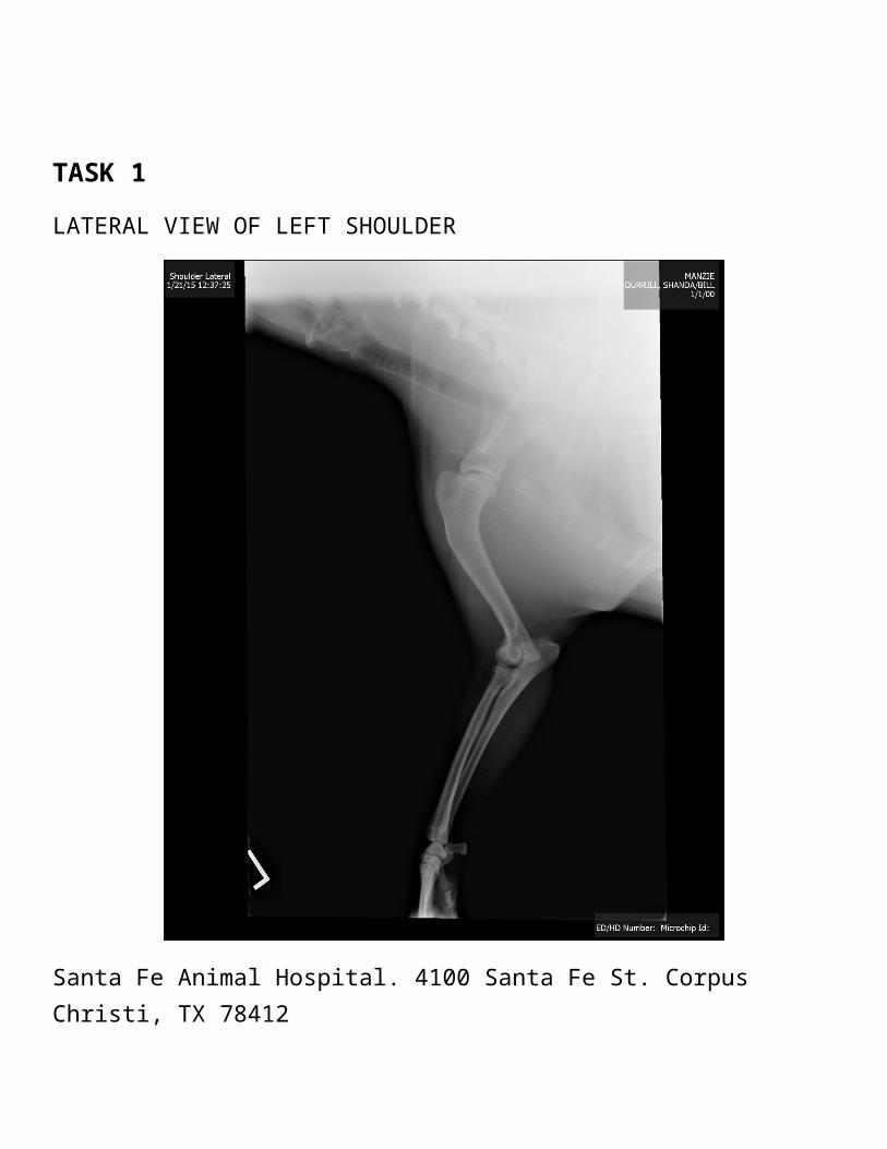
TASK 1
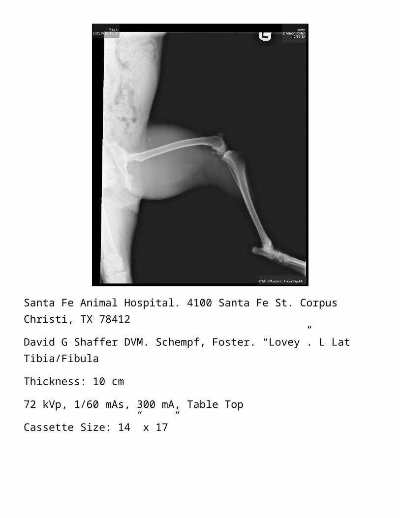
LATERAL VIEW OF LEFT SHOULDER
Santa Fe Animal Hospital. 4100 Santa Fe St. Corpus Christi, TX 78412
David G Shaffer DVM. Durrill, Shanda/Bill. “Manzie”. L Lat Shoulder
Thickness: 9 cm
70 kVp, 1/60 mAs, 300 mA, Table Top
Cassette Size: 14” x 17”

CRITIQUE TASK 1 POSITIONING CRITIQUE
Positioning is good with the humerus laterally in the middle. The bulldog was under sedation since she’s wiggly and has stubby legs.
LANDMARK CRITIQUE
The humerus and scapula are visible as well as the radius, ulna, and carpus. You can also see the spine and esophagus.
COLLIMATION CRITIQUE
I probably didn’t need to include the radius, ulna, and carpus. I should of focused more on the humerus and scapula and exclude about an inch from the top of the image.
EXPOSURE FACTOR CRITIQUE
The exposure choices were good. The whole left foot was pretty well defined, even though not all of it was needed in the shot. I think the scapula would have been more pronounced if the collimation was better.
OVERALL QUALITY
I think it’s an okay image but collimation could be better. I would give it a 7.
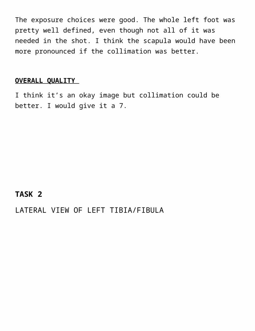
TASK 2
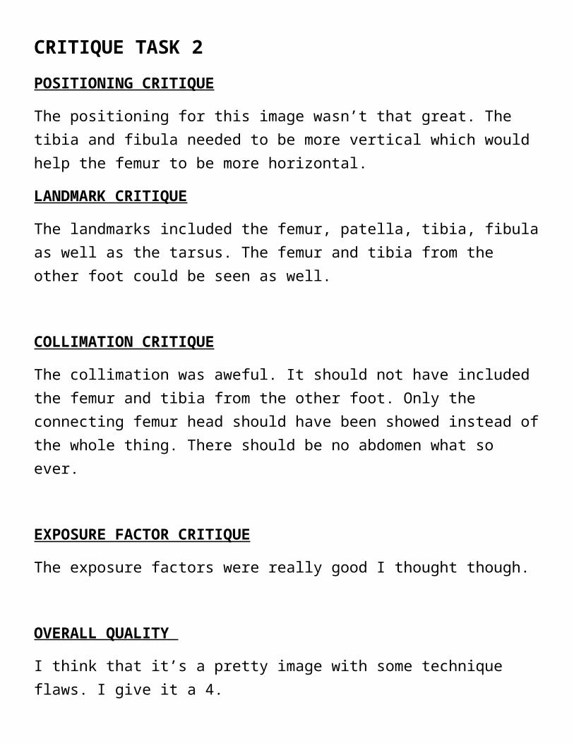
LATERAL VIEW OF LEFT TIBIA/FIBULA
Santa Fe Animal Hospital. 4100 Santa Fe St. Corpus Christi, TX 78412
David G Shaffer DVM. Schempf, Foster. “Lovey”. L Lat Tibia/Fibula
Thickness: 10 cm
72 kVp, 1/60 mAs, 300 mA, Table Top
Cassette Size: 14” x 17”
CRITIQUE TASK 2 POSITIONING CRITIQUE
The positioning for this image wasn’t that great. The tibia and fibula needed to be more vertical which would help the femur to be more horizontal.
LANDMARK CRITIQUE
The landmarks included the femur, patella, tibia, fibula as well as the tarsus. The femur and tibia from the other foot could be seen as well.
COLLIMATION CRITIQUE
The collimation was aweful. It should not have included the femur and tibia from the other foot. Only the connecting femur head should have been showed instead of the whole thing. There should be no abdomen what so ever.
EXPOSURE FACTOR CRITIQUE
The exposure factors were really good I thought though.
OVERALL QUALITY
I think that it’s a pretty image with some technique flaws. I give it a 4.
TASK 3
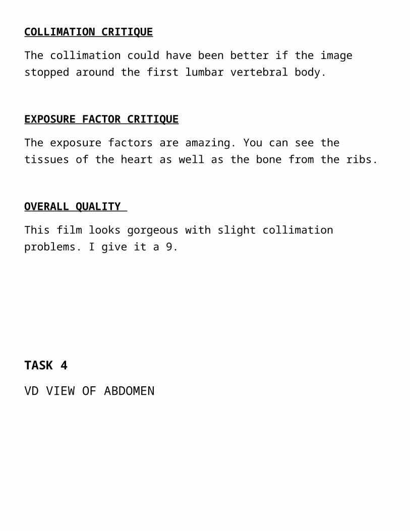
VD VIEW OF THORAX
Santa Fe Animal Hospital. 4100 Santa Fe St. Corpus Christi, TX 78412
David G Shaffer DVM. Shamel, Deborah. “Annabell”. VD Thorax
Thickness: 3 cm
64 kVp, 1/60 mAs, 300 mA, Table Top
Cassette Size: 14” x 17”

CRITIQUE TASK 3 POSITIONING CRITIQUE
The positioning for this image is pretty good. The thorax is pretty well vertical. The dog was not sedated and she allowed us to hold on to her legs as she was in dorsal recumbency.
LANDMARK CRITIQUE
The landmarks visible include the thoracic inlet, the heart, sternum, ribs, diaphragm, vertebrae and an elevated trachea. The pelvis and femurs could also be seen as well as the humerus and scapula.
COLLIMATION CRITIQUE
The collimation could have been better if the image stopped around the first lumbar vertebral body.
EXPOSURE FACTOR CRITIQUE
The exposure factors are amazing. You can see the tissues of the heart as well as the bone from the ribs.
OVERALL QUALITY
This film looks gorgeous with slight collimation problems. I give it a 9.

TASK 4
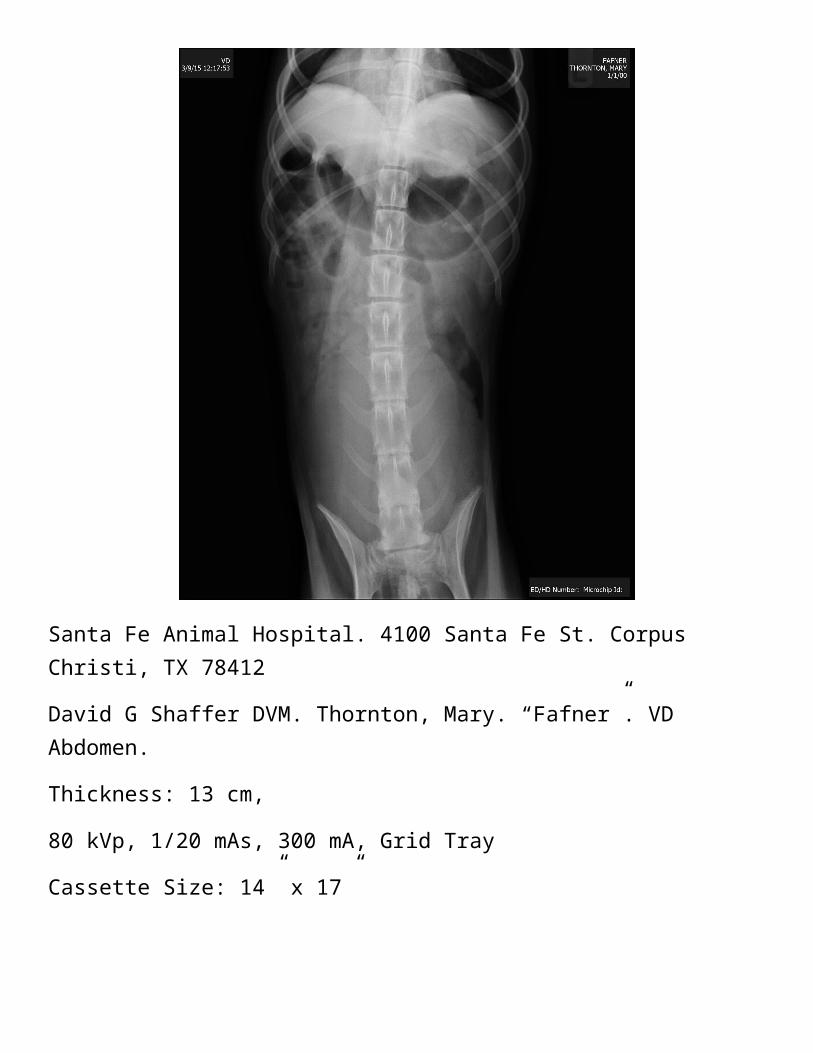
VD VIEW OF ABDOMEN
Santa Fe Animal Hospital. 4100 Santa Fe St. Corpus Christi, TX 78412
David G Shaffer DVM. Thornton, Mary. “Fafner”. VD Abdomen.
Thickness: 13 cm,
80 kVp, 1/20 mAs, 300 mA, Grid Tray
Cassette Size: 14” x 17”
CRITIQUE TASK 4 POSITIONING CRITIQUE
The positioning could have been a little better. The spine was slightly curved so the whole image was slightly curved. The dog was not sedated which might be why it was harder to get him straighten out.
LANDMARK CRITIQUE
Sternum and spine are nicely superimposed. The pelvis was crooked. The stomach and bowel was filled with gas. The liver looked small. The small intestines and colon was also visible.
COLLIMATION CRITIQUE
The collimation was okay. There is some dead space on the outside of the image but the borders of the sternum and pelvis was accurate minus the positioning.
EXPOSURE FACTOR CRITIQUE
The exposure factors were okay. The bones seemed more pronounced than the tissues. Maybe if the kVp was decreased by a little you could see more of a contrast but it was kind of hard because of the build up gas in the abdomen.
OVERALL QUALITY
Overall this image was okay but the positioning and collimation could have been a little better. I would give it an 7.
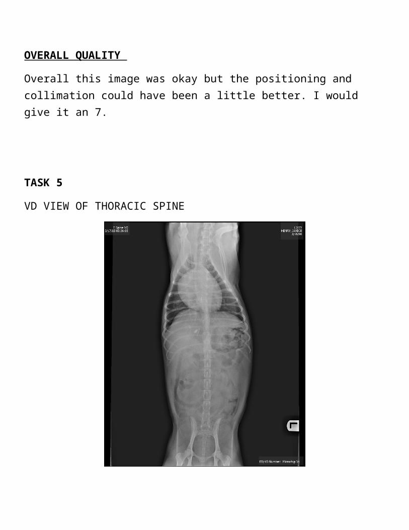
TASK 5
VD VIEW OF THORACIC SPINE
Santa Fe Animal Hospital. 4100 Santa Fe St. Corpus Christi, TX 78412
David G Shaffer DVM. Henry, Janice. “Lizzy”. VD T Spine.
Thickness: 11 cm
76 kVp, 1/60 mAs, 300 mA, Table Top
Cassette Size: 14” x 17”

CRITIQUE TASK 5 POSITIONING CRITIQUE
The positioning for this image was pretty good. The spine overall is straight but the thoracic spine is slightly curved.
LANDMARK CRITIQUE
The entire spine is very visible except where the heart is that is covering up part of the T spine. The sternum, pelvis and femurs are visible as well as the heart, lung field, diaphragm, abdomen, humerus, scapula, and intestines.
COLLIMATION CRITIQUE
The collimation could be better by ending at the last rib and getting rid of some of the dead space around the image.
EXPOSURE FACTOR CRITIQUE
The exposure factors are alright. I would increase the kVp by a little to bring out more of the bone structure visibility. Overall the tissue to bone ratio is good.
OVERALL QUALITY
Overall a good image minus the slight collimation concern. I would give it a 8.
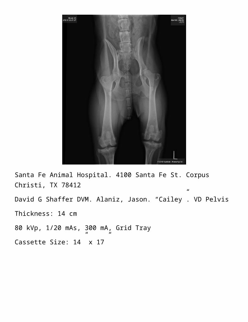
TASK 6
VD VIEW OF PELVIS
Santa Fe Animal Hospital. 4100 Santa Fe St. Corpus Christi, TX 78412
David G Shaffer DVM. Alaniz, Jason. “Cailey”. VD Pelvis
Thickness: 14 cm
80 kVp, 1/20 mAs, 300 mA, Grid Tray
Cassette Size: 14” x 17”

CRITIQUE TASK 6 POSITIONING CRITIQUE
The positioning of this image was okay. The femurs were straight but the pelvis looked crooked. The hip was extended.
LANDMARK CRITIQUE
The view includes lumbar spine, all the pelvis (crest and wings of ilium, obturator foramens, pubis, ischium, pelvic symphysis), acetabulum, head and greater trochanter or femurs, body of femur, patellae , tibia, fibula, and chronic stifle joint problems.
COLLIMATION CRITIQUE
The collimation isn’t too bad but it needed to start from the crest of the ilium to the distal patella. There was a little more of the lumbar vertebrae and tibia which didn’t really need to be seen. Some background dead space was also present in the image.
EXPOSURE FACTOR CRITIQUE
The exposure factors were pretty good. You could see the contrast of tissue from the abdomen as well as the pelvis bone. The bones seemed to stand out perfectly.
OVERALL QUALITY
Overall, I think the image was not bad. The pelvis just needed to be a little more vertical. I would give it an 8.
TASK 7
GI SERIESThickness: 24 cm
90 kVp, 1/20 mAs, 300 mA, Grid Tray
Cassette Size: 14” x 17”
LATERAL ABDOMEN
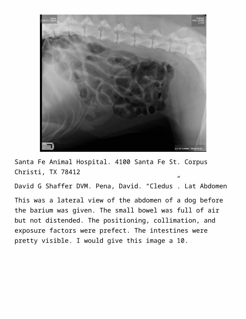
Santa Fe Animal Hospital. 4100 Santa Fe St. Corpus Christi, TX 78412
David G Shaffer DVM. Pena, David. “Cledus”. Lat Abdomen
This was a lateral view of the abdomen of a dog before the barium was given. The small bowel was full of air but not distended. The positioning, collimation, and exposure factors were prefect. The intestines were pretty visible. I would give this image a 10.
Santa Fe Animal Hospital. 4100 Santa Fe St. Corpus Christi, TX 78412
David G Shaffer DVM. Pena, David. “Cledus”. Lat Abdomen
The barium was given and the image was taken about 2 hours later. The contrast was collecting in the colon. Some small intestine lining roughening was present. The positioning, collimation, and exposure factors were prefect. The intestines were visible as well as the bright barium highlighting the abdomen beautifully. I would give this image a 10.

VD ABDOMEN
Santa Fe Animal Hospital. 4100 Santa Fe St. Corpus Christi, TX 78412
David G Shaffer DVM. Pena, David. “Cledus”. VD Abdomen
The barium was given and the image was taken about 4:13pm. This was taken about the same time as the above image but from a VD point of view. The frog leg positioning, collimation, and exposure factors were prefect. You can see the barium go through the intestines down the abdomen. I would give this image a 10.
LATERAL ABDOMEN
Santa Fe Animal Hospital. 4100 Santa Fe St. Corpus Christi, TX 78412
David G Shaffer DVM. Pena, David. “Cledus”. Lat Abdomen
The image was taken the next day about 9:28am in the lateral view. The contrast was mostly in the small intestine. Looks like there was some roughening of the intestine lining. The positioning, collimation, and exposure factors were prefect. I would give this image a 10.
Santa Fe Animal Hospital. 4100 Santa Fe St. Corpus Christi, TX 78412
David G Shaffer DVM. Pena, David. “Cledus”. Lat Abdomen
This image was taken the day after the pervious image at about 10:27am. The contrast is in the colon. There might be a foreign body in the intestine that would cause the irritation of the intestinal lining. The positioning, collimation, and exposure factors were prefect. I would give this image a 10.

Santa Fe Animal Hospital. 4100 Santa Fe St. Corpus Christi, TX 78412
David G Shaffer DVM. Pena, David. “Cledus”. Lat Abdomen
The image was taken the next day about 10:25am in the lateral view. Most of the contrast was still in the colon. Some of the contrast remained in the “irritating” looking segment of the small intestine. The collimation, and exposure factors were prefect. I would give this image a 10.
OVERALL CRITIQUE TASK 7 POSITIONING CRITIQUE
The positioning for these images were perfect. The very large bloodhound was sedated which made him easier to position.

LANDMARK CRITIQUE
The landmarks that were visible on VD are the sternum, diaphragm, stomach, duodenum, liver, spleen, small intestines, colon and urinary bladder. The ribs and vertebrae were also included. The Lateral view includes the ribs, sternum, and The femur and wings of the ilium appears on some of the images. The stomach, liver, spleen and small intestines are visible as well.
COLLIMATION CRITIQUE
The overall collimation was really good.
EXPOSURE FACTOR CRITIQUE
The exposure factors were excellent on all images.
OVERALL QUALITY
I really enjoyed all these images from the amazing contrasting details to the story of where the barium travels in the abdomen of the dog. I would rate this overall a 10.