Cancer Tumor and Stem Cell Biology Research · Tumor and Stem Cell Biology...
13
Tumor and Stem Cell Biology RNA Trafficking by Acute Myelogenous Leukemia Exosomes Jianya Huan 1,4 , Noah I. Hornick 1,4 , Matthew J. Shurtleff 1 , Amy M. Skinner 1,4 , Natalya A. Goloviznina 1,4 , Charles T. Roberts Jr 1,2,3,5 , and Peter Kurre 1,2,4 Abstract Extrinsic signaling cues in the microenvironment of acute myelogenous leukemia (AML) contribute to disease progression and therapy resistance. Yet, it remains unknown how the bone marrow niche in which AML arises is subverted to support leukemic persistence at the expense of homeostatic function. Exosomes are cell membrane–derived vesicles carrying protein and RNA cargoes that have emerged as mediators of cell–cell communication. In this study, we examined the role of exosomes in developing the AML niche of the bone marrow microenvironment, investigating their biogenesis with a focus on RNA trafficking. We found that both primary AML and AML cell lines released exosome-sized vesicles that entered bystander cells. These exosomes were enriched for several coding and noncoding RNAs relevant to AML pathogenesis. Furthermore, their uptake by bone marrow stromal cells altered their secretion of growth factors. Proof-of- concept studies provided additional evidence for the canonical functions of the transferred RNA. Taken together, our findings revealed that AML exosome trafficking alters the proliferative, angiogenic, and migratory responses of cocultured stromal and hematopoietic progenitor cell lines, helping explain how the microenvironmental niche becomes reprogrammed during invasion of the bone marrow by AML. Cancer Res; 73(2); 918–29. Ó2012 AACR. Introduction Although the majority of patients with acute myelogenous leukemia (AML) achieve remission, nearly half will die from disease relapse. The contribution of microenvironmental cues to the survival, spread, and relapse of AML in the marrow are increasingly recognized as crucial in the process of leukemic evolution (1, 2). The bone marrow is structurally and functionally specialized to sustain lifelong hemato- poietic and immune function; its infiltration by leukemia is associated with the acquisition of unique properties that make the bone marrow a sanctuary for persistent disease (3, 4). Even as differences emerge that distinguish the leukemic microenvironment from the homeostatic hemato- poietic stem cell (HSC) niche, the mechanisms whereby malignant cells reprogram the niche remain to be clarified (4–6). Instances of extrinsic resistance are illustrated in the treatment of high-risk AML associated with internal tandem duplication (ITD) mutations in FMS-like tyrosine kinase 3 (FLT3), in which specific inhibitors eliminate cir- culating blasts without eradicating leukemic cells in the bone marrow (7). Conversely, FLT3-ITD pos AML cells are sen- sitized to the tyrosine kinase inhibitor sorafenib by inhibition of chemokine receptor-4 (CXCR4)-SDF-1a niche signaling with bone marrow stromal cells (2). Although integrin-, chemokine-, and cytokine-mediated adhesive and paracrine interactions all contribute to drug resistance, none of these fully address how marrow invasion by leukemia cells results in niche remodeling (6). Conventional intercellular signaling relies on direct cell–cell contact or the action of secreted molecules, but more recent studies indicate an additional, evolutionarily conserved mech- anism whereby cells communicate via the exchange of extra- cellular vesicles carrying protein and RNA cargo (8, 9). As a unique consequence of cell–cell vesicle trafficking, the cyto- plasmic delivery of mRNA, miRNA, or protein can circum- vent transcriptional controls. Vesicle trafficking of receptors between cells, for example, may transcend niche signaling that is based on conventional restrictions of cell-specific ligand or receptor expression and modulate growth factor signaling cascades. Vesicles ranging in size from 30 to 100 nm (exo- somes) to 100 to 1,000 nm (broadly termed microvesicles) have been detected in the urine and plasma of patients with diverse malignancies (10). However, detailed studies of vesicle biology in AML have not been reported (11, 12). The results presented here show the release of exosome- sized vesicles by AML cells and illustrate how vesicle trafficking alters gene expression, protein secretion, and behavior of Authors' Affiliations: Departments of 1 Pediatrics, 2 Cell & Developmental Biology, and 3 Medicine; 4 Pap e Family Pediatric Research Institute, Oregon Health & Science University, Portland; and 5 Oregon National Primate Research Center, Beaverton, Oregon Note: Supplementary data for this article are available at Cancer Research Online (http://cancerres.aacrjournals.org/). Prior presentation: This work was presented, in part, at the 2011 meeting of the American Society of Hematology. Corresponding Author: Peter Kurre, Department of Pediatrics and Pap e Family Pediatric Research Institute, Oregon Health & Science University, 3181 SW Sam Jackson Park Rd., Portland, OR 97239. Phone: 503-494- 7655; Fax: 503-494-0714; E-mail: [email protected] doi: 10.1158/0008-5472.CAN-12-2184 Ó2012 American Association for Cancer Research. Cancer Research Cancer Res; 73(2) January 15, 2013 918 on June 16, 2020. © 2013 American Association for Cancer Research. cancerres.aacrjournals.org Downloaded from Published OnlineFirst November 13, 2012; DOI: 10.1158/0008-5472.CAN-12-2184
Transcript of Cancer Tumor and Stem Cell Biology Research · Tumor and Stem Cell Biology...

Tumor and Stem Cell Biology
RNA Trafficking by AcuteMyelogenous Leukemia Exosomes
Jianya Huan1,4, Noah I. Hornick1,4, Matthew J. Shurtleff1, Amy M. Skinner1,4, Natalya A. Goloviznina1,4,Charles T. Roberts Jr1,2,3,5, and Peter Kurre1,2,4
AbstractExtrinsic signaling cues in the microenvironment of acute myelogenous leukemia (AML) contribute to
disease progression and therapy resistance. Yet, it remains unknown how the bone marrow niche in whichAML arises is subverted to support leukemic persistence at the expense of homeostatic function. Exosomesare cell membrane–derived vesicles carrying protein and RNA cargoes that have emerged as mediators ofcell–cell communication. In this study, we examined the role of exosomes in developing the AML niche of thebone marrow microenvironment, investigating their biogenesis with a focus on RNA trafficking. We foundthat both primary AML and AML cell lines released exosome-sized vesicles that entered bystander cells.These exosomes were enriched for several coding and noncoding RNAs relevant to AML pathogenesis.Furthermore, their uptake by bone marrow stromal cells altered their secretion of growth factors. Proof-of-concept studies provided additional evidence for the canonical functions of the transferred RNA. Takentogether, our findings revealed that AML exosome trafficking alters the proliferative, angiogenic, andmigratory responses of cocultured stromal and hematopoietic progenitor cell lines, helping explain howthe microenvironmental niche becomes reprogrammed during invasion of the bone marrow by AML. CancerRes; 73(2); 918–29. �2012 AACR.
IntroductionAlthough the majority of patients with acute myelogenous
leukemia (AML) achieve remission, nearly half will die fromdisease relapse. The contribution of microenvironmentalcues to the survival, spread, and relapse of AML in themarrow are increasingly recognized as crucial in the processof leukemic evolution (1, 2). The bone marrow is structurallyand functionally specialized to sustain lifelong hemato-poietic and immune function; its infiltration by leukemiais associated with the acquisition of unique properties thatmake the bone marrow a sanctuary for persistent disease(3, 4). Even as differences emerge that distinguish theleukemic microenvironment from the homeostatic hemato-poietic stem cell (HSC) niche, the mechanisms wherebymalignant cells reprogram the niche remain to be clarified(4–6). Instances of extrinsic resistance are illustrated in
the treatment of high-risk AML associated with internaltandem duplication (ITD) mutations in FMS-like tyrosinekinase 3 (FLT3), in which specific inhibitors eliminate cir-culating blasts without eradicating leukemic cells in thebone marrow (7). Conversely, FLT3-ITDpos AML cells are sen-sitized to the tyrosine kinase inhibitor sorafenib by inhibitionof chemokine receptor-4 (CXCR4)-SDF-1a niche signalingwith bone marrow stromal cells (2). Although integrin-,chemokine-, and cytokine-mediated adhesive and paracrineinteractions all contribute to drug resistance, none of thesefully address how marrow invasion by leukemia cells resultsin niche remodeling (6).
Conventional intercellular signaling relies on direct cell–cellcontact or the action of secreted molecules, but more recentstudies indicate an additional, evolutionarily conserved mech-anism whereby cells communicate via the exchange of extra-cellular vesicles carrying protein and RNA cargo (8, 9). As aunique consequence of cell–cell vesicle trafficking, the cyto-plasmic delivery of mRNA, miRNA, or protein can circum-vent transcriptional controls. Vesicle trafficking of receptorsbetween cells, for example, may transcend niche signaling thatis based on conventional restrictions of cell-specific ligand orreceptor expression and modulate growth factor signalingcascades. Vesicles ranging in size from 30 to 100 nm (exo-somes) to 100 to 1,000 nm (broadly termedmicrovesicles) havebeen detected in the urine and plasma of patients with diversemalignancies (10). However, detailed studies of vesicle biologyin AML have not been reported (11, 12).
The results presented here show the release of exosome-sized vesicles byAMLcells and illustrate howvesicle traffickingalters gene expression, protein secretion, and behavior of
Authors' Affiliations: Departments of 1Pediatrics, 2Cell & DevelopmentalBiology, and 3Medicine; 4Pap�e Family Pediatric Research Institute, OregonHealth & Science University, Portland; and 5Oregon National PrimateResearch Center, Beaverton, Oregon
Note: Supplementary data for this article are available at Cancer ResearchOnline (http://cancerres.aacrjournals.org/).
Prior presentation: This work was presented, in part, at the 2011meeting ofthe American Society of Hematology.
Corresponding Author: Peter Kurre, Department of Pediatrics and Pap�eFamily Pediatric Research Institute, Oregon Health & Science University,3181 SW Sam Jackson Park Rd., Portland, OR 97239. Phone: 503-494-7655; Fax: 503-494-0714; E-mail: [email protected]
doi: 10.1158/0008-5472.CAN-12-2184
�2012 American Association for Cancer Research.
CancerResearch
Cancer Res; 73(2) January 15, 2013918
on June 16, 2020. © 2013 American Association for Cancer Research. cancerres.aacrjournals.org Downloaded from
Published OnlineFirst November 13, 2012; DOI: 10.1158/0008-5472.CAN-12-2184
bystander cells. We show that vesicles contain diverse RNAspecies, including mRNAs and miRNAs relevant to AML bio-logy and with broad biomarker potential. In aggregate, ourwork indicates that trafficking of AML exosomes elicits broadfunctional changes in bystander cells.
Materials and MethodsCell lines and cell cultureAML cell lines (HEL, HL-60, Molm-14, and U937) were
provided by Dr. Jeffrey (Oregon Health & Science University)Tyner and cultured in RPMI (Invitrogen) with 10% vesicle-freeFBS. Vesicle-free FBS was produced by centrifugation of FBS(Gemini Bio-Products) at 100,000 � g for 2.5 hours. Patientsamples (with >90% leukemic blasts) were obtained underOregon Health & Science University-Institutional ReviewBoard (OHSU-IRB; Portland, OR)–approved protocol fromtreatment-na€�ve patients. Primary cells were cultured inEGM-2 media (Lonza). OP9 cells, from Dr. William H. Fleming(Oregon Health & Science University), were cultured ina-minimum essential media (Invitrogen) with 20% FBS and60 mmol/L 2-mercaptoethanol. Igf-1r knockout (R�) mouseembryonic fibroblasts and R� cells expressing human insulin-like growth factor (IGF)-IR cDNA (termed Rþ), provided byDrs. Briony Forbes and Douglas Yee, respectively, were cultur-ed in Dulbecco's Modified Eagle's Medium (Invitrogen) with10% FBS (13). Cryopreserved human CD34þ-enriched bonemarrow cells (StemCell Technologies) were cultured in serum-free expansion media with the StemSpan CC110 cytokinecocktail for 5 to 7 days. All cultures contained 50 mg/mLPen/Strep (Invitrogen). Cell lines were obtained from reliablesources without additional authentification beyond what isshown in the content of the experiments described.
Vesicle preparation and stainingVesicles were isolated from cell lines and primary
AML cells after 48 to 72 hours culture via centrifugation at300 � g for 10 minutes. The supernatant was sequentiallycentrifuged at 2,000 � g for 20 minutes, at 10,000 � g for20 minutes, and at 100,000 � g for 2 hours. The resultingpelletwaswashedwith PBS and then centrifuged at 100,000� gfor 2 hours. For sucrose gradient density purification, thepellet from the first 100,000 � g spin was resuspended in200 mL PBS by shaking for 4 hours at 4�C. The suspension wastransferred to a sucrose step gradient (8%/15%/30%/45%/60%) and centrifuged at 150,000 � g for 90 minutes. The30%/45% interface was harvested, diluted 10-fold with PBS,and the ultracentrifugation was repeated. The pellet was re-suspended in 200 mL of PBS (methods adapted from ref. 8).For PKH26 (Sigma) staining, exosomes were isolated with thefollowing changes: following the first 100,000 � g centrifuga-tion, the pellet was resuspended in 1 mL PKH26 membranedye-diluent C (Sigma) by shaking for 30 minutes at 4�C, alongwith a control aliquot containing diluent only.
Vesicle transfer assaysTranswell experiments were carried out in 6-well plates
using 0.4-mm pore size inserts (Corning). Target cells were
seeded at 2� 104 cells per well. A total of 1 to 2� 106 cells wereadded into the transwell insert and cocultured for 48 hours.Media collected from vesicle preparations after the 10,000 � gspin was termed vesicle-rich media (VRM). Two millilitersVRM were cocultured with 2 � 104 target cells per well in6-well plate. Target cells were incubated with the vesiclepellets resuspended in RPMI or PBS for 48 hours in 2 mL ofmedia, then washed twice. For real-time PCR (RT-PCR), RNAwas extracted using either the RNeasy (Qiagen) or miRNeasykit (Qiagen). Transwell cocultures with CD34þ cells were notconducted because of culture media incompatibility and thelimited availability of cells.
Fluorescence microscopyCells were exposed to 25 mL of PKH26-stained, sucrose-
purified exosomes or PKH26-stained diluent C for 2 hours,followed bywash and fixation in 4% paraformaldehyde (Sigma)before treatment with Fluoromount G (Southern Biotech).Fluorescence microscopy was conducted with an OlympusIX71 microscope and DeltaVision SoftWoRx, using a �601.4NA oil lens. Z-stacks were acquired every 0.2 mm for thecomplete depth of the cells. A reference bright-field image wascaptured from the center of each Z-stack. Particle quantifica-tion was conducted using ImarisCell (Bitplane, Inc.). For live-cell imaging, cells were plated on poly-L-lysine (Sigma)–coatedchamber wells, treated with 5 mg/mL Hoechst 33342 and2.5 mmol/L N-Rh-PE (Avanti Polar Lipids) for 1.5 hours fol-lowed by wash and resuspension in RPMImedia with 9% horseserum (Invitrogen) and 9% FBS (Gemini Bioproducts). Imageswere acquired every 2.5 minutes for 2 hours, at 37�C and5% CO2. For colocalization studies, cells were treated with5 mmol/L N-Rh-PE for 1 hour, fixed for 15 minutes, permea-bilized with NET (NaCl þ EDTA þ Tris) buffer, cytospun andblocked with 2% FBS for 30 minutes at 4�C. Fixation precludedN-Rh-PE polarization seen during live cell imaging. Permea-bilized cells were stained with anti-human CD63 hybridomasupernatant and anti-human Tsg101 antibodies (Abcam).AlexaFluor488 anti-mouse immunoglobulin G (IgG) and Alex-aFluor647 anti-rabbit IgG (Invitrogen) were used as the sec-ondary antibodies. Cells were stained with 40,6-diamidino-2-phenylindole (DAPI; Invitrogen).
Transmission electron microscopyExosome preparations (10 mL) were deposited onto UV-ac-
tivated carbon formvar 400-mesh copper grids (Ted Pella01822-F) for 3 minutes, rinsed and stained in filtered 1.33%uranyl acetate, and then air-dried. Samples were imaged at100 kV on a Philips CM120 transmission electron micro-scope. The size distribution of exosomes was determinedby averaging the maximum and minimum diameters of atleast 100 vesicles imaged at �37,000 magnification.
Dynamic light scatterLight-scattering experiments were conducted in a DynaPro
molecular sizing instrument (Protein Solutions). A sample inPBS buffer was loaded into a quartz cuvette and analyzedwith a 488 nm laser. Fifty spectra were collected at 25�C toestimate diffusion coefficient and relative polydispersity. Data
AML Exosomes
www.aacrjournals.org Cancer Res; 73(2) January 15, 2013 919
on June 16, 2020. © 2013 American Association for Cancer Research. cancerres.aacrjournals.org Downloaded from
Published OnlineFirst November 13, 2012; DOI: 10.1158/0008-5472.CAN-12-2184

were analyzed with Dynamics software V.5.25.44 (ProteinSolutions) and vesicle size was measured as the mean hydro-dynamic radius.
RNA analysis and qRT-PCRRNA was extracted using miRNeasy (Qiagen) or RNeasy
(Qiagen) kits and quantified using a Nanodrop 2000c(Thermo). RNA integrity was measured using the AgilentBioanalyzer Pico Chip or small RNA Chip (Agilent). For RT-PCR, RNAs were converted into cDNA using the SuperScript IIIFirst Strand Synthesis Kit (Invitrogen) with oligo-dT primingfollowed by PCR. See Supplementary Table S1 for primersequences. For quantitative PCR (qPCR), human IGF-IR pri-mers/probe (forward primer: AACTTTCTCCCTCATCGGCCG;reverse primer: GTGTGTCGCCAGCGTGTCT; probe: FAM-CGCTGATTCCTCGTGTCCGGAGG), mouse VEGF, mousec-fos, humanGAPDH, andmouseGAPDH (Applied Biosystems)were used. Relative quantification was calculated using DDCTalgorithm with glyceraldehyde-3-phosphate dehydrogenase(GAPDH) as the endogenous control. For miRNA quantitation,TaqMan Assay Kits (Applied Biosystems) were used for reversetranscription and qRT-PCR. For PCR array analysis, reversetranscription was conducted using the RT2 First Strand (Qia-gen) Kit. PCR arrays (SABiosciences) were run according tomanufacturer's instructions.
Flow-cytometry analysisFor IGF-IR detection, target R� murine embryonic fibro-
blasts (MEF) were trypsinized after coculture, washed, andresuspended in staining buffer (1% bovine serum albumin inPBS, w/v). Cells were stained with mouse anti-human IGF-IR-PE antibody (BioLegend) or CXCR4-APC antibody(eBioscience) � 30 minutes at 4�C, washed twice, propidiumiodide (PI) was added and cells analyzed with a FACSCalibur(BD Biosciences). Data were analyzed using FlowJo software(Tree Star).
Transwell migration and cell proliferationBa/F3 cells were cultured for 48 hours in the presence or
absence of VRM by plating 5 � 105 cells in 2 mL Ba/F3media with 4 mL of either vesicle-rich or base media. Ba/F3cells were washed and plated on 8-mm transwell inserts inplates containing either media alone or SDF-1a gradient.After 2 hours, transwells were removed, migrated cells werewashed and counted using a Guava PCA cytometer (Milli-pore). For proliferation assays, 5 � 104 R� MEFs werecocultured with 1 � 106 HL-60 cells in 0.4-mm transwellsfor 48 hours, after which R� MEFs were counted. IGF-IRinhibitor picropodophyllin (PPP; CalBiochem) was added at100 nmol/L before coculture.
Western blottingProtein lysates were generated using RIPA buffer with
protease inhibitors (Thermo Scientific) and protein concen-trations quantified by BCAProtein Assay (Pierce). Lysates wereloaded on 4% to 15% SDS-PAGE gels (Bio-Rad) for transfer andPonceau S stain. IGF-IR was detected using rabbit anti-humanIGF-IRb (Cell Signaling Technology) and anti-rabbit IgG-HRP
(Thermo Scientific). Actin was detected using mouse anti-human pan-actin (Millipore) and anti-mouse IgG-HRP (Jack-son ImmunoResearch Laboratories). Chemiluminescence wasdetected by the Supersignal West Pico Chemiluminescentsubstrate (Pierce).
ResultsAML cells release exosomes
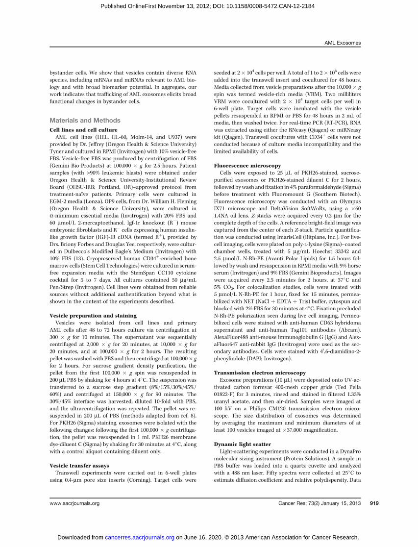
To systematically examine AML vesicle release, we usedseveral AML cell lines and patient cells. Primary AML samplesphenotyped by flow-cytometry showed more than 90% of cellsexpressing leukemia-associated antigens (data not shown).Vesicles were isolated from culture media using differentialcentrifugation and sucrose gradient purification (Fig. 1A andB). Transmission electron microscopy (TEM) revealed thatboth HL-60 and primary AML cells released vesicles primarilybetween 30 and 100 nm in diameter (Fig. 1C), the size rangeof exosomes (9). To exclude detectionmethod- or purification-protocol bias, we conducted dynamic light scattering (DLS)measurements comparing HL-60 vesicle preparations purifiedby sucrose gradient versus differential centrifugation (captur-ing larger vesicles; Fig. 1D). Results mirrored the TEM size anddistribution profile without significant method specific bias.To show direct exosome release from AML cells, we imagedprimary AML cells labeled with N-Rh-PE, a marker dye routedfrom the endocytic compartment to the multivesicular body(MVB) and released within exosomes (14). Representativetime-lapse images from stained primary AML blasts showthe polarized aggregation of dye-labeled MVB's at the plasmamembrane before release of the exosome content, represen-tative of 53% of cells in 3 primary AML samples (Fig. 1E;ref. 15). The canonical N-Rh-PE trafficking and vesicle buddingprovide further evidence that primary AML cells release exo-somes. When we conducted immunofluorescent imagingfor exosomal markers Tsg101 and CD63 (16, 17), the N-Rh-PE–stained compartment in primary AML cells colocalizedwith CD63 in more than 90%, with Tsg101 in 70%, and withboth proteins in 29% of cells (Fig. 1F). Together, these experi-ments provide strong complementary evidence that AMLcell lines and primary AML cells give rise to exosomes. Here-after, we use the term "exosomes" specifically and "vesicles"generically.
Exosomes traffic mRNA between AML and stromal cellsVesicles transfer protein and RNA between cells (18),
leading us to investigate the trafficking of AML exosomesto bystander cells. To determine exosome entry, GFP-expres-sing murine OP9 bone marrow stromal cells were exposedto PKH26-stained exosomes from HL-60 cells (19). Fluores-cence microscopy images revealed rapid and significantuptake of PKH26-stained exosomes as early as 15 minutesand increasing until 2 hours after initial exposure (Fig.2A–F), at which time their number per cell reached a plateau(data not shown). To ascertain the transfer of specific RNAtranscripts via AML exosomes, we cultured leukemia cellsacross a 0.4-mm transwell membrane from OP9 cells. After48 hours of coculture, OP9 cells were harvested, RNA was
Huan et al.
Cancer Res; 73(2) January 15, 2013 Cancer Research920
on June 16, 2020. © 2013 American Association for Cancer Research. cancerres.aacrjournals.org Downloaded from
Published OnlineFirst November 13, 2012; DOI: 10.1158/0008-5472.CAN-12-2184
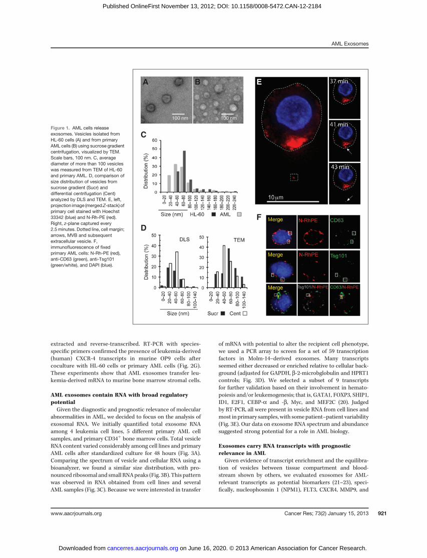
extracted and reverse-transcribed. RT-PCR with species-specific primers confirmed the presence of leukemia-derived(human) CXCR-4 transcripts in murine OP9 cells aftercoculture with HL-60 cells or primary AML cells (Fig. 2G).These experiments show that AML exosomes transfer leu-kemia-derived mRNA to murine bone marrow stromal cells.
AML exosomes contain RNA with broad regulatorypotentialGiven the diagnostic and prognostic relevance of molecular
abnormalities in AML, we decided to focus on the analysis ofexosomal RNA. We initially quantified total exosome RNAamong 4 leukemia cell lines, 5 different primary AML cellsamples, and primary CD34þ bone marrow cells. Total vesicleRNA content varied considerably among cell lines and primaryAML cells after standardized culture for 48 hours (Fig. 3A).Comparing the spectrum of vesicle and cellular RNA using abioanalyzer, we found a similar size distribution, with pro-nounced ribosomal and small RNApeaks (Fig. 3B). This patternwas observed in RNA obtained from cell lines and severalAML samples (Fig. 3C). Because we were interested in transfer
of mRNA with potential to alter the recipient cell phenotype,we used a PCR array to screen for a set of 59 transcriptionfactors in Molm-14–derived exosomes. Many transcriptsseemed either decreased or enriched relative to cellular back-ground (adjusted for GAPDH, b-2-microbglobulin and HPRT1controls; Fig. 3D). We selected a subset of 9 transcriptsfor further validation based on their involvement in hemato-poiesis and/or leukemogenesis; that is, GATA1, FOXP3, SHIP1,ID1, E2F1, CEBP-a and -b, Myc, and MEF2C (20). Judgedby RT-PCR, all were present in vesicle RNA from cell lines andmost in primary samples, with somepatient–patient variability(Fig. 3E). Our data on exosome RNA spectrum and abundancesuggested strong potential for a role in AML biology.
Exosomes carry RNA transcripts with prognosticrelevance in AML
Given evidence of transcript enrichment and the equilibra-tion of vesicles between tissue compartment and blood-stream shown by others, we evaluated exosomes for AML-relevant transcripts as potential biomarkers (21–23), speci-fically, nucleophosmin 1 (NPM1), FLT3, CXCR4, MMP9, and
Figure 1. AML cells releaseexosomes. Vesicles isolated fromHL-60 cells (A) and from primaryAML cells (B) using sucrose gradientcentrifugation, visualized by TEM.Scale bars, 100 nm. C, averagediameter of more than 100 vesicleswas measured from TEM of HL-60and primary AML. D, comparison ofsize distribution of vesicles fromsucrose gradient (Sucr) anddifferential centrifugation (Cent)analyzed by DLS and TEM. E, left,projection image (mergedZ-stack) ofprimary cell stained with Hoechst33342 (blue) and N-Rh-PE (red).Right, z-plane captured every2.5 minutes. Dotted line, cell margin;arrows, MVB and subsequentextracellular vesicle. F,immunofluorescence of fixedprimary AML cells: N-Rh-PE (red),anti-CD63 (green), anti-Tsg101(green/white), and DAPI (blue).
A
C
DF
B E
AML Exosomes
www.aacrjournals.org Cancer Res; 73(2) January 15, 2013 921
on June 16, 2020. © 2013 American Association for Cancer Research. cancerres.aacrjournals.org Downloaded from
Published OnlineFirst November 13, 2012; DOI: 10.1158/0008-5472.CAN-12-2184
IGF-IR (21, 24, 25). RT-PCR analysis revealed the presenceof all 5 candidate transcripts in exosomes from patient AMLblasts, primary CD34þ bone marrow cells, and leukemia celllines (Fig. 4A and B and Table 1). Indeed, exosome RNAreproduced the FLT3 allelic diversity (wt vs. ITD tran-scripts), including multiple unique ITDs present in the bulk
cell population from which they were derived (Fig. 4A and B;ref. 26). Similarly, we were able to detect transcripts bearingmutations in exon 12 of NPM1 (confirmed by sequencingand capillary gel electrophoresis) in matched exosomal andcellular RNA (Fig. 4A). We next determined relative IGF-IRtranscript levels in RNA from AML patient cells and vesicles
Figure 2. AML exosomes are takenup by bone marrow stromal cells.PGK-GFP OP9 cells were exposedto PKH-26–labeled HL-60exosomes (A, C, and E) and PKH-26–labeled diluent C (B and D) for 2hours. A and B, differentialinterference contrast (D DIC)images. C and D, single z-slicesshowing labeled exosomes (red). E,Z-stack in the xy-, xz-, andyz-planes. Scale bars, 10 mm. F,quantification of labeled exosomeswithin cells. N, number of cellsanalyzed; error bars, SEM; P <0.006 by Student t test. G, RT-PCRfor human CXCR4 in murine OP9safter 48 hours coculture orexposure to vesicles from HL-60 orprimary AML. Transfer conditions:0.4-mm transwell (Transwell), VRMexposure, purified vesicleexposure (vesicles). Data arerepresentative of at least3 independent experiments. NTC,non template control.
Huan et al.
Cancer Res; 73(2) January 15, 2013 Cancer Research922
on June 16, 2020. © 2013 American Association for Cancer Research. cancerres.aacrjournals.org Downloaded from
Published OnlineFirst November 13, 2012; DOI: 10.1158/0008-5472.CAN-12-2184

Figure 3. AML exosomes contain unique RNA profiles. A, quantification of AML or control vesicle RNA. BM: CD34þ bone marrow progenitors. Data representat least 3 individual experiments. B and C, bioanalyzer electropherograms of RNA from cells and vesicles of AML cell lines and primary samples. D,transcription factor array comparison of exosomal RNA from Molm-14 cells, expressed as fold change in vesicles versus cells. E, RT-PCR for selecttranscription factors in RNA from primary AML cells and vesicles from 4 patients. Data represent at least 3 individual experiments.
AML Exosomes
www.aacrjournals.org Cancer Res; 73(2) January 15, 2013 923
on June 16, 2020. © 2013 American Association for Cancer Research. cancerres.aacrjournals.org Downloaded from
Published OnlineFirst November 13, 2012; DOI: 10.1158/0008-5472.CAN-12-2184

(24, 25, 27). When normalized to GAPDH, IGF-IR mRNAlevels differed substantially between primary AML samples(28). Yet, expression in both cellular and exosomal RNA wasgenerally 2- to 5-fold higher in AML samples than in CD34þ
progenitors (Fig. 4C). A direct comparison of IGF-IR mRNAlevels confirmed exosome enrichment over matched cellularRNA, by up to 1,000-fold in 2 of the samples tested (Fig. 4D).This relative enrichment of IGF-IR mRNA (normalized toGAPDH) seemed to be specific for leukemia-derived vesicles,as primary CD34þ bone marrow vesicles exhibited IGF-IRmRNA levels proportional to those found in cells (Fig. 4D).
Transfer of IGF-IR mRNA modulates cellular responsesin Igf-Ir-deficient cells
To show proof-of-principle for the specific functional con-sequences of AML exosome trafficking to bystander cells, weset up contact-independent cocultures of HL-60 or Molm-14donor cells and (murine) OP9 target cells. Under all 3 exper-imental modalities (transwell coculture, vesicle-rich mediaexposure, and purified vesicle exposure), human-specificIGF-IR transcripts were detected in OP9 recipient cells
(Fig. 5A). To determine if AML vesicle-derived IGF-IR con-tributes to target cell signaling after transfer, we coculturedleukemia cells across a transwell from Igf-Ir knockout mouseembryonic fibroblasts (R�MEF; refs. 13, 29). In flow-cytometryanalysis, R� MEFs cocultured with HL-60, Molm-14, or U937cells expressed human IGF-IR, although at a level less thanthat of R� cells stably transfected with human IGF-IR cDNA(termed Rþ; Fig. 5B). Protein lysates from AML cells andvesicles revealed broad differences in banding profile, reflect-ing the disparity between vesicle and cellular protein con-tent that includes conventional controls, such as actin andGAPDH, or tubulin (data not shown). Even when loading 3-foldthe amount of vesicle than cell protein lysate (reflected inthe Ponceau S stain), significantly less actin was detected invesicles. IGF-IR protein, as well, was present only at very lowlevels in AML exosomes by Western blot analysis, althoughit was readily detected in both WT HL-60 cells and HL-60overexpressing IGF-IR (Fig. 5C; Supplementary Fig. S1A andS1B; ref. 30). Allowing for small amounts of IGF-IR proteinin vesicles, these results nonetheless suggest that transferredhuman IGF-IR mRNA may be successfully translated and
Figure 4. Leukemia-relevanttranscripts in AML exosomes.RT-PCR for AML-relatedtranscripts in cells and vesiclesfrom primary AML samples (A) andAML cell lines (B). C, qRT-PCR forIGF-IR in primary AML cellular andvesicle RNA, normalized to CD34þ
bone marrow cells and vesicles.GAPDH was used as theendogenous control. D, foldenrichment of IGF-IR mRNA invesicles versus cells, normalized toCD34þ cells/vesicles.
Huan et al.
Cancer Res; 73(2) January 15, 2013 Cancer Research924
on June 16, 2020. © 2013 American Association for Cancer Research. cancerres.aacrjournals.org Downloaded from
Published OnlineFirst November 13, 2012; DOI: 10.1158/0008-5472.CAN-12-2184

trafficked to the cell surface of R� MEFs after coculture withAML cells. This observation is corroborated at the functionallevel by measuring rates of IGF-I–stimulated proliferationin R� MEFs (13). In experiments using transwell coculturesof HL-60 with R� MEFs, we observed increased prolifera-tion in cocultured R� MEFs compared with R� MEFs alone(Fig. 5D). This proliferative response was specifically abro-gated in response to the IGF-IR–specific inhibitor PPP, whichsimilarly decreased proliferation in Rþ MEFs, indicating thatthe altered proliferation kinetics resulted from the transfer ofIGF-IR mRNA. In addition to its mitogenic activity, IGF-IRsignaling has been shown to upregulate VEGF expression(31, 32). When we assessed the effect of coculture on VEGFexpression in target cells, we found that R� MEF cells exhi-bited increased VEGF transcript levels by qRT-PCR followingcoculture with HL-60 cells (Fig. 5E). As another canonical IGFsignaling response, we observed upregulation of the earlyresponse gene c-fos under these conditions, although this didnot reach statistical significance (Fig. 5F; ref. 33). Our experi-ments show that exosomal transfer of IGF-IR can modulateproliferative signaling in bystander cells and promote expres-sion of VEGF. Exosomal induction of transcripts promotingangiogenesis in bystander cells is further corroborated in theexposure of OP9 cells to Molm-14 coculture (SupplementaryFig. S2A–S2C) and Molm-14 vesicles (Supplementary Fig. S3Aand S3B). We observed substantial changes in growth fac-tor secretion into OP9 culture supernatant using commercial(murine) arrays. Despite apparent differences in secretion
resulting from the presence of soluble factors under cocultureconditions versus concentrated vesicle exposure, several rel-evant proteins whose secretion was upregulated after cocul-ture (angiopoietin 1, endothelin 1, CXCL4, and FGF-2) werealso elevated following treatment with purified vesicles. VEGFwas consistently elevated after both treatments, whereassecreted osteopontin (OPN) and SDF-1 were decreased aftervesicle treatment despite an increase following coculture.To validate these results (and discrepancies), we investigatedseveral of these targets at the mRNA level with qRT-PCR,and found that while SDF-1 and VEGF mRNA tracked withsecreted protein levels, OPN transcription had increased aftervesicle exposure without changes in its secretion. These resultsillustrate hat physiologic vesicle transfer occurs in the contextof other secreted effectors, which act in concert to influencesurrounding cells.
miRNA enriched in AML exosomes regulates the biologicfunction of neighboring cells
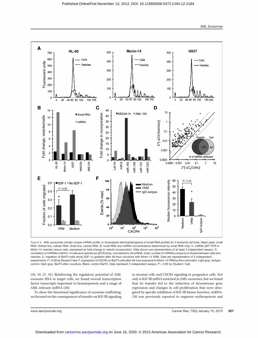
Our initial bioanalyzer data suggested the enrichment ofmiRNA in exosomes (Fig. 3B and C; ref. 34). The RNA size rangebetween 0 and 150 nts was therefore further evaluated usingthe small RNA bioanalyzer chip. Results confirmed abun-dant representation of small RNAs (defined as 0–150 nts) andmiRNAs (40–80 nts; Fig. 6A). Remarkably, when comparingthe calculated concentrations, we found 2-fold enrichment ofsmall RNAs and 5- to 13-fold greater gains among miRNAsin vesicles versus parental cells across several cell lines
Table 1. Transcripts detected in AML exosomes
Sample ID Sample source FLT-3 NPM1 IGF-IR CXCR4 MMP9
AML10128 LA ITD þ þ ND NDAML10226 LA ITD þ þ ND NDAML10831 PB WT þ þ ND NDAML11009 PB � þ þ ND NDAML11105 PB � � þ ND NDAML11254 PB ITD þ þ þ þAML11261 PB ITD þ þ þ þAML11376 PB ITD þ þ þ þAML11378 PB ITD þ þ þ þAML11423 PB WT þ þ þ þAML12073 PB WT þ þ þ þAML12035 BM WT þ þ þ þ
NL-1 BM WT þ þ þ þNL-2 BM WT þ þ þ þ
HL-60 Cell line WT þ þ þ þMolm-14 Cell line ITD þ þ þ þU937 Cell line WT þ þ þ þHEL Cell line WT þ þ þ þAbbreviations: LA, leukapheresis cells; ND, not done; NL, normal bone marrow cells; PB, peripheral blood cells; þ, detected; �, notdetected.
AML Exosomes
www.aacrjournals.org Cancer Res; 73(2) January 15, 2013 925
on June 16, 2020. © 2013 American Association for Cancer Research. cancerres.aacrjournals.org Downloaded from
Published OnlineFirst November 13, 2012; DOI: 10.1158/0008-5472.CAN-12-2184

(Fig. 6B). We conducted qRT-PCR assays to quantify specificmiRNAs inMolm-14 cells and vesicles, including Let-7a, miR-9,miR-99b, miR-150, miR-155, miR-191, and miR-223. Using thenoncoding small nuclear U6 RNA for normalization, weobserved significant incorporation in vesicles, ranging from
2- to near 40-fold enrichment compared with cells among thisset of miRNA (Fig. 6C). When we conducted a qRT-PCR arraystudy to profile 234 human miRNAs, our screen detectedmiRNAs in both Molm-14 cellular and exosomal RNA(Fig. 6D). Many miRNA were enriched in vesicles, indicatedby data point skewing toward the y-axis, with approximately83% of miRNAs detected in both cells and vesicles. AmongmiRNA represented by the array, 40 were excluded fromexosomes, whereas 1 miRNA (miR-146a) was present at adetectable level only in exosomes.
One functional consequence of leukemic invasion of theniche is the displacement of hematopoietic stem and progen-itor cells from the bone marrow microenvironment throughaltered migration and retention (4). The CXCR4/SDF-1a sig-naling axis plays a crucial role in this, and CXCR4 itselfwas recently shown to be a target of miR-150 (4, 35). Wetherefore examined the effect of Molm-14 exosomes (enrichedin miRNA-150) on migration of Ba/F3 cells, a conditionallySDF-1a–responsive murine B-precursor cell line (36). Remark-ably, Ba/F3 cells cultured for 48 hours in the presence ofMolm-14–derived, vesicle-rich media and placed on 8-mm Transwellgrids showed significantly decreasedmigration toward SDF-1a(Fig. 6E; ref. 2). Moreover, vesicle exposure specifically reducedthe surface expression of CXCR4, the cognate SDF-1a receptor,by 50% as judged by median fluorescence intensity (Fig. 6F).Taken together, our results reveal that AML vesicles altertranscriptional responses, protein secretion, and migration inbystander cells.
DiscussionA specialized AML stromal niche is thought to account, in
part, for the evasion of cells from therapy-induced apoptosisand is implicated in relapse (1–3). Conceptually, niche con-version requires the successive displacement and suppressionof hematopoietic function and establishment of conditionsconducive to leukemic spread. The mechanisms underlyingthis process, however, remain to be clarified (1, 4). On thebasis of the established potential for exosomes to mediatecell-to-cell communication (8, 18, 37), and increasing evidenceof their relevance in cancer (17, 28, 38), we investigated arole for exosomes in modulating signaling between cells inthe AML microenvironment.
Classes of cell membrane–derived vesicles produced bydifferent cell types can be distinguished on the basis of size,density, and characteristic marker proteins (39). Our studiesprovide compelling evidence that AML cells produce primarilyexosome-sized vesicles, capable of rapid entry and cargotransfer to bystander cells. Given the importance of molecularabnormalities in AML risk stratification, we decided to exam-ine exosomal RNA content, and observed a broad spectrum,including small RNA. Profiling the mRNA content of thesevesicles revealed the presence of transcripts relevant to AMLprognosis (FLT3-ITD, NPM1), treatment (FLT3-ITD, IGF-IR,CXCR4), and niche function (IGF-IR, CXCR4, MMP9; Table 1;refs. 21, 27, 40). Other experimental systems have shown thatthe direct cytoplasmic transfer of RNA between cells canhave significant and lasting impact on cellular phenotypes
Figure 5. AML exosomes transfer leukemia-specific transcripts andmodulate target cell function. A, RT-PCR for human IGF-IR mRNA inmurine OP9s after 48 hours coculture or exposure to vesicles fromHL-60or MOLM-14. Transfer conditions: 0.4-mm transwell (Transwell), VRMexposure, purified vesicle exposure (vesicles). Data are representative ofat least 5 independent experiments. B, flow-cytometry analysis of humanIGF-IR expression in R�MEFs after 48 hours coculturewith U937, HL-60,or Molm-14. Light gray, R� MEFs; dark gray, R� MEFs after coculture;black, Rþ MEFs. C, top: Ponceau S stain of HL-60 cell, HL60 vesiclelysate, and cell lysate from IGF-IR overexpressing HL-60 (positivecontrol). Bottom, Western blot analysis for IGF-IR and actin. Top andbottom, 10-mg cellular and 30-mg vesicle protein were loaded. D,proliferation of R�MEFs after coculturewithHL-60s. R�MEFs, RþMEFs,or R� MEFs/HL-60 cells were cultured with or without PPP for 48 hours.E and F, relative expression of VEGF and c-fos transcripts in R� MEFscocultured with HL-60 cells for 12 hours and in Rþ MEFs or R� MEFsalone. Error bars, SEM. P < 0.05 by Student t test.
Huan et al.
Cancer Res; 73(2) January 15, 2013 Cancer Research926
on June 16, 2020. © 2013 American Association for Cancer Research. cancerres.aacrjournals.org Downloaded from
Published OnlineFirst November 13, 2012; DOI: 10.1158/0008-5472.CAN-12-2184
(18, 19, 37, 41). Reinforcing the regulatory potential of AMLexosome RNA in target cells, we found several transcriptionfactor transcripts important to hematopoiesis and a range ofAML-relevant miRNA (20).To show the functional significance of exosome trafficking,
we focused on the consequences of transfer on IGF-IR signaling
in stromal cells and CXCR4 signaling in progenitor cells. Notonly is IGF-IRmRNA enriched in AML exosomes, but we foundthat its transfer led to the induction of downstream geneexpression and changes in cell proliferation that were abro-gated by specific inhibition of IGF-IR kinase function. miRNA-150 was previously reported to suppress erythropoiesis and
Figure 6. AML exosomes contain unique miRNA profile. A, bioanalyzer electropherograms of small RNA profiles for 3 leukemia cell lines. Major peak, smallRNA. Dotted line, cellular RNA. Solid line, vesicle RNA. B, small RNA and miRNA concentrations determined by small RNA chip. C, miRNA qRT-PCR inMolm-14 vesicles versus cells, expressed as fold-change in vesicle incorporation. Data shown are representative of at least 3 independent assays. D,correlation ofmiRNAs inMolm-14 cells and vesicles by qPCR array, normalized toU6 snRNA. Inset, number ofmiRNAs unique to or shared between cells andvesicles. E, migration of Ba/F3 cells along SDF-1a gradient after 48-hour coculture with Molm-14 VRM. Data are representative of 3 independentexperiments.P < 0.05 by Student t test. F, expression of CXCR4 onBa/F3 cells after 48-hour exposure toMolm-14 VRMby flowcytometry. Light gray, isotypecontrol. Dark gray, Ba/F3 after coculture. Black, control Ba/F3. Data represent 3 independent assays, P < 0.05 by Student t test.
AML Exosomes
www.aacrjournals.org Cancer Res; 73(2) January 15, 2013 927
on June 16, 2020. © 2013 American Association for Cancer Research. cancerres.aacrjournals.org Downloaded from
Published OnlineFirst November 13, 2012; DOI: 10.1158/0008-5472.CAN-12-2184

target CXCR4 transcripts for degradation (35). Although bothmiR-150 and CXCR4 mRNA are present in AML exosomes,miR-150 is highly enriched therein, and exosome transfer toBa/F3 progenitor cells was associated with a loss of CXCR4surface expression and consequent decrease in cell migrationtoward SDF-1a. Several caveats remain: we did not titratevesicle doses and it is possible that some of these effects willreveal exosome dose–response kinetics. At the same time, ourdata caution against the direct extrapolation of vesicle effectsto biologic response, as both the cell coculture and VRMtreatment provide further physiologic modulation via solublefactors, illustrated in Supplementary Figs. S2 and S3. This isto be expected, as the exosome transfer of a receptor, forexample, would be expected to result in differential signalingevents in the presence of media containing the cognate ligand.The diverse exosome cargo highlights the inherent challengein assigning a specific functional outcome to the transfer ofan individual exosomal protein or RNA transcript in thecontext of many others. The presence of prognostically rele-vant mRNA in AML-derived exosomes implies their potentialdevelopment as a minimally invasive AML biomarker plat-form. Here, it will be important to see the extent to whichspecific AML exosome content will track with diagnosticparameters and change during the course of treatment.
Taken together, our results show that exosome traffickingby AML cells can alter the function of bystander cells. Ourobservations show the complexity of cell–cell communicationand emphasize the potential impact of exosome traffickingon AML physiology. Coding and noncoding RNAs are keydeterminants of the cellular phenotype, and in vitro RNAtransfer can produce experimental phenotype conversion
(37, 41). The transfer of transcription factor and miRNA fromAML to bystander cells presents a compelling paradigm forcell–cell communication in the leukemic niche.
Disclosure of Potential Conflicts of InterestNo potential conflicts of interest were disclosed.
Authors' ContributionsConception and design: J. Huan, C.T. Roberts Jr, P. KurreDevelopment of methodology: J. Huan, P. KurreAcquisition of data (provided animals, acquired and managed patients,provided facilities, etc.): J. Huan, N.I. Hornick, A.M. Skinner, N.A. GolovizninaAnalysis and interpretation of data (e.g., statistical analysis, biostatistics,computational analysis): J. Huan, N.I. Hornick, N.A. Goloviznina, C.T. RobertsJr, P. KurreWriting, review, and/or revision of the manuscript: J. Huan, N.I. Hornick,A.M. Skinner, C.T. Roberts Jr, P. KurreAdministrative, technical, or material support (i.e., reporting or orga-nizing data, constructing databases): J. Huan, A.M. Skinner, P. KurreStudy supervision: P. Kurre
AcknowledgmentsThe authors thank experimental contributions by Dr. Thomas Russell. The
authors also thank Drs. Jeffrey Tyner (Oregon Health & Science University),Briony Forbes (University of Adelaide), and Douglas Yee (University of Minesota)for generous provision of cell lines.
Grant SupportThis study was supported by the Friends of Doernbecher and Hyundai Hope
on Wheels Foundations.The costs of publication of this article were defrayed in part by the payment
of page charges. This article must therefore be hereby marked advertisementin accordance with 18 U.S.C. Section 1734 solely to indicate this fact.
Received May 31, 2012; revised October 12, 2012; accepted November 5, 2012;published OnlineFirst November 13, 2012.
References1. Ayala F, Dewar R, Kieran M, Kalluri R. Contribution of bone microen-
vironment to leukemogenesis and leukemia progression. Leukemia2009;23:2233–41.
2. Zeng Z, Shi YX, Samudio IJ, Wang RY, Ling X, Frolova O, et al.Targeting the leukemia microenvironment by CXCR4 inhibition over-comes resistance to kinase inhibitors and chemotherapy in AML.Blood 2009;113:6215–24.
3. Ishikawa F, Yoshida S, Saito Y, Hijikata A, Kitamura H, Tanaka S, et al.Chemotherapy-resistant human AML stem cells home to and engraftwithin the bone-marrow endosteal region. Nat Biotechnol 2007;25:1315–21.
4. Colmone A, Amorim M, Pontier AL, Wang S, Jablonski E, Sipkins DA.Leukemic cells create bonemarrow niches that disrupt the behavior ofnormal hematopoietic progenitor cells. Science 2008;322:1861–5.
5. Hatfield K, Ryningen A, Corbascio M, Bruserud O. Microvascularendothelial cells increase proliferation and inhibit apoptosis of nativehuman acute myelogenous leukemia blasts. Int J Cancer 2006;119:2313–21.
6. Ryningen A, Wergeland L, Glenjen N, Gjertsen BT, Bruserud O. In vitrocrosstalk between fibroblasts and native human acute myelogenousleukemia (AML) blasts via local cytokine networks results in increasedproliferation and decreased apoptosis of AML cells as well asincreased levels of proangiogenic Interleukin 8. Leuk Res 2005;29:185–96.
7. Kindler T, Lipka DB, Fischer T. FLT3 as a therapeutic targetin AML: still challenging after all these years. Blood 2010;116:5089–102.
8. Valadi H, Ekstrom K, Bossios A, Sjostrand M, Lee JJ, Lotvall JO.Exosome-mediated transfer of mRNAs and microRNAs is a novel
mechanism of genetic exchange between cells. Nat Cell Biol 2007;9:654–9.
9. Simons M, Raposo G. Exosomes–vesicular carriers for intercellularcommunication. Curr Opin Cell Biol 2009;21:575–81.
10. Lee TH, D'Asti E, Magnus N, Al-Nedawi K, Meehan B, Rak J. Micro-vesicles as mediators of intercellular communication in cancer-theemerging science of cellular 'debris'. Semin Immunopathol 2011;33:455–67.
11. Fiegl M, Samudio I, Clise-Dwyer K, Burks JK, Mnjoyan Z, AndreeffM. CXCR4 expression and biologic activity in acute myeloid leu-kemia are dependent on oxygen partial pressure. Blood 2009;113:1504–12.
12. Szczepanski MJ, Szajnik M, Welsh A, Whiteside TL, Boyiadzis M.Blast-derived microvesicles in sera from patients with acute myeloidleukemia suppress natural killer cell function via membrane-asso-ciated transforming growth factor-beta1. Haematologica 2011;96:1302–9.
13. Sell C, Dumenil G, Deveaud C, Miura M, Coppola D, DeAngelis T, et al.Effect of a null mutation of the insulin-like growth factor I receptor geneon growth and transformation of mouse embryo fibroblasts. Mol CellBiol 1994;14:3604–12.
14. Willem J, ter Beest M, Scherphof G, Hoekstra D. A non-exchangeablefluorescent phospholipid analog as a membrane traffic marker of theendocytic pathway. Eur J Cell Biol 1990;53:173–84.
15. Shen B, Wu N, Yang JM, Gould SJ. Protein targeting to exosomes/microvesicles by plasma membrane anchors. J Biol Chem 2011;286:14383–95.
16. Pols MS, Klumperman J. Trafficking and function of the tetraspaninCD63. Exp Cell Res 2009;315:1584–92.
Huan et al.
Cancer Res; 73(2) January 15, 2013 Cancer Research928
on June 16, 2020. © 2013 American Association for Cancer Research. cancerres.aacrjournals.org Downloaded from
Published OnlineFirst November 13, 2012; DOI: 10.1158/0008-5472.CAN-12-2184

17. Logozzi M, De Milito A, Lugini L, Borghi M, Calabro L, Spada M, et al.High levels of exosomes expressing CD63 and caveolin-1 in plasma ofmelanoma patients. PLoS ONE 2009;4:e5219.
18. Al-Nedawi K, Meehan B, Micallef J, Lhotak V, May L, Guha A, et al.Intercellular transfer of the oncogenic receptor EGFRvIII by microve-sicles derived from tumour cells. Nat Cell Biol 2008;10:619–24.
19. Deregibus MC, Cantaluppi V, Calogero R, Lo Iacono M, Tetta C,Biancone L, et al. Endothelial progenitor cell derived microvesiclesactivate an angiogenic program in endothelial cells by a horizontaltransfer of mRNA. Blood 2007;110:2440–8.
20. Orkin SH, Zon LI. Hematopoiesis: an evolving paradigm for stem cellbiology. Cell 2008;132:631–44.
21. Bacher U, Schnittger S, Haferlach T. Molecular genetics in acutemyeloid leukemia. Curr Opin Oncol 2010;22:646–55.
22. Marcucci G, Haferlach T, Dohner H. Molecular genetics of adult acutemyeloid leukemia: prognostic and therapeutic implications. J ClinOncol 2011;29:475–86.
23. Gercel-Taylor C AS, Tullis RH, Kesimer M, Taylor DD. Nanoparticleanalysis of circulating cell-derived vesicles in ovarian cancer patients.Anal Biochem 2012;428:44–53.
24. KangHJ, Lee JW,KhoSH,KimMJ,SeoYJ,KimH, et al. High transcriptlevel of FLT3 associated with high risk of relapse in pediatric acutemyeloid leukemia. J Korean Med Sci 2010;25:841–5.
25. Doepfner KT, Boller D, Arcaro A. Targeting receptor tyrosine kinasesignaling in acute myeloid leukemia. Crit Rev Oncol Hematol 2007;63:215–30.
26. Cheung AM, Chow HC, Kwong YL, Liang R, Leung AY. FLT3/internaltandem duplication subclones in acute myeloid leukemia differ in theirengraftment potential in NOD/SCID mice. Leuk Res 2010;34:119–22.
27. Chapuis N, Tamburini J, Cornillet-Lefebvre P, Gillot L, Bardet V, Will-ems L, et al. Autocrine IGF-1/IGF-1R signaling is responsible forconstitutivePI3K/Akt activation in acutemyeloid leukemia: therapeuticvalue of neutralizing anti-IGF-1R antibody. Haematologica 2010;95:415–23.
28. Keller S, Ridinger J, Rupp AK, Janssen JW, Altevogt P. Body fluidderived exosomes as a novel template for clinical diagnostics. J TranslMed 2011;9:86.
29. Coppola D, Ferber A, Miura M, Sell C, D'Ambrosio C, Rubin R, et al. Afunctional insulin-like growth factor I receptor is required for themitogenic and transforming activities of the epidermal growth factorreceptor. Mol Cell Biol 1994;14:4588–95.
30. Atay S, Gercel-Taylor C, Kesimer M, Taylor DD. Morphologic andproteomic characterization of exosomes released by cultured extra-villous trophoblast cells. Exp Cell Res 2011;317:1192–202.
31. Mitsiades CS, Mitsiades NS, McMullan CJ, Poulaki V, Shringarpure R,Akiyama M, et al. Inhibition of the insulin-like growth factor receptor-1tyrosine kinase activity as a therapeutic strategy for multiple myeloma,other hematologic malignancies, and solid tumors. Cancer Cell2004;5:221–30.
32. Rajski M, Zanetti-Dallenbach R, Vogel B, Herrmann R, Rochlitz C,Buess M. IGF-I induced genes in stromal fibroblasts predict theclinical outcome of breast and lung cancer patients. BMC Med2010;8:1.
33. Muller R, Bravo R, Burckhardt J, Curran T. Induction of c-fos gene andprotein by growth factors precedes activation of c-myc. Nature1984;312:716–20.
34. Chen TS, Lai RC, Lee MM, Choo AB, Lee CN, Lim SK. Mesenchymalstem cell secretes microparticles enriched in pre-microRNAs. NucleicAcids Res 2010;38:215–24.
35. Tano N, Kim HW, Ashraf M. microRNA-150 regulates mobilization andmigration of bone marrow-derived mononuclear cells by targetingCxcr4. PLoS ONE 2011;6:e23114.
36. Jacobi A, Thieme S, Lehmann R, Ugarte F, Malech HL, Koch S, et al.Impact of CXCR4 inhibition on FLT3-ITD-positive human AML blasts.Exp Hematol 2010;38:180–90.
37. Ratajczak J, Miekus K, Kucia M, Zhang J, Reca R, Dvorak P, et al.Embryonic stem cell-derived microvesicles reprogram hematopoieticprogenitors: evidence for horizontal transfer of mRNA and proteindelivery. Leukemia 2006;20:847–56.
38. Ghosh AK, Secreto CR, Knox TR, Ding W, Mukhopadhyay D, Kay NE.Circulating microvesicles in B-cell chronic lymphocytic leukemia canstimulate marrow stromal cells: implications for disease progression.Blood 2010;115:1755–64.
39. TheryC, Zitvogel L, Amigorena S. Exosomes: composition, biogenesisand function. Nat Rev Immunol 2002;2:569–79.
40. Olsnes AM, Hatfield KJ, Bruserud O. The chemokine system and itscontribution to leukemogenesis and treatment responsiveness inpatients with acute myeloid leukemia. J BUON 2009;14(Suppl 1):S131–40.
41. Del Tatto M, Ng T, Aliotta JM, Colvin GA, Dooner MS, Berz D, et al.Marrow cell genetic phenotype change induced by human lung cancercells. Exp Hematol 2011;39:1072–80.
AML Exosomes
www.aacrjournals.org Cancer Res; 73(2) January 15, 2013 929
on June 16, 2020. © 2013 American Association for Cancer Research. cancerres.aacrjournals.org Downloaded from
Published OnlineFirst November 13, 2012; DOI: 10.1158/0008-5472.CAN-12-2184

2013;73:918-929. Published OnlineFirst November 13, 2012.Cancer Res Jianya Huan, Noah I. Hornick, Matthew J. Shurtleff, et al. RNA Trafficking by Acute Myelogenous Leukemia Exosomes
Updated version
10.1158/0008-5472.CAN-12-2184doi:
Access the most recent version of this article at:
Material
Supplementary
http://cancerres.aacrjournals.org/content/suppl/2012/11/12/0008-5472.CAN-12-2184.DC1
Access the most recent supplemental material at:
Cited articles
http://cancerres.aacrjournals.org/content/73/2/918.full#ref-list-1
This article cites 41 articles, 12 of which you can access for free at:
Citing articles
http://cancerres.aacrjournals.org/content/73/2/918.full#related-urls
This article has been cited by 21 HighWire-hosted articles. Access the articles at:
E-mail alerts related to this article or journal.Sign up to receive free email-alerts
Subscriptions
Reprints and
To order reprints of this article or to subscribe to the journal, contact the AACR Publications Department at
Permissions
Rightslink site. Click on "Request Permissions" which will take you to the Copyright Clearance Center's (CCC)
.http://cancerres.aacrjournals.org/content/73/2/918To request permission to re-use all or part of this article, use this link
on June 16, 2020. © 2013 American Association for Cancer Research. cancerres.aacrjournals.org Downloaded from
Published OnlineFirst November 13, 2012; DOI: 10.1158/0008-5472.CAN-12-2184