Glioblastoma stem cells: lessons from the tumor hierarchy ...
20
REVIEW Glioblastoma stem cells: lessons from the tumor hierarchy in a lethal cancer Ryan C. Gimple, 1,2 Shruti Bhargava, 1 Deobrat Dixit, 1 and Jeremy N. Rich 1,3 1 Division of Regenerative Medicine, Department of Medicine, University of California at San Diego, La Jolla, California 92037, USA; 2 Department of Pathology, Case Western Reserve University, Cleveland, Ohio 44106, USA; 3 Department of Neurosciences, University of California at San Diego School of Medicine, La Jolla, California 92037, USA Glioblastoma ranks among the most lethal of all human cancers. Glioblastomas display striking cellular heteroge- neity, with stem-like glioblastoma stem cells (GSCs) at the apex. Although the original identification of GSCs dates back more than a decade, the purification and char- acterization of GSCs remains challenging. Despite these challenges, the evidence that GSCs play important roles in tumor growth and response to therapy has grown. Like normal stem cells, GSCs are functionally defined and distinguished from their differentiated tumor progeny at core transcriptional, epigenetic, and metabolic regula- tory levels, suggesting that no single therapeutic modality will be universally effective against a heterogenous GSC population. Glioblastomas induce a systemic immuno- suppression with mixed responses to oncoimmunologic modalities, suggesting the potential for augmentation of response with a deeper consideration of GSCs. Unfortu- nately, the GSC literature has been complicated by fre- quent use of inferior cell lines and a lack of proper functional analyses. Collectively, glioblastoma offers a re- liable cancer to study cancer stem cells to better model the human disease and inform improved biologic under- standing and design of novel therapeutics. Tumors are not homogeneous masses of neoplastic cells; rather, they contain ecosystems with diverse neoplastic populations and recruited supportive stroma. Numerous efforts to model the complexity of neoplastic populations have included not only integration of the tumor microen- vironment but also efforts to explain heterogeneous tu- mor cells based on genetic and epigenetic diversity. The cancer stem cell hypothesis represents one element of nongenetic complexity in cancer biology that leverages the similarities between tumorigenesis and both develop- ment and wound responses, processes in which normal tissue stem and progenitor cells serve critical roles. The cancer stem cell hypothesis holds that tumors mimic nor- mal tissues with hierarchically arranged and dynamically regulated populations of cells, with stem-like cells at the apex that display regenerative potential and the capacity to recapitulate the entire functional diversity present within the original tumor. The underpinnings of cancer stem cell modeling date back to early functional studies of cancer, which showed that the injection of a single leukemic cell into mice could produce a lethal leukemia in as little as 2 wk (Furth et al. 1937). In the modern era, the cancer stem cell model was revitalized by Dick and coworkers (Lapidot et al. 1994), who identified a subset of patient-derived leukemia cells able to traffic to the bone marrow of immunodeficient mice, with sustained proliferation and maintenance of the original leukemic cell phenotypes. Leukemia-initiat- ing cells differentiate in vivo and possess self-renewal properties (Bonnet and Dick 1997). These landmark stud- ies kicked off the hunt for cancer stem cells in additional tumor types, including breast cancer (Al-Hajj et al. 2003), brain cancer (Hemmati et al. 2003; Singh et al. 2003, 2004), prostate cancer (Collins et al. 2005), colorectal can- cer (O’Brien et al. 2007; Ricci-Vitiani et al. 2007), and pan- creatic cancer (Li et al. 2007). Amid the rapid pace of identification of novel cancer stem cells in a variety of cancer types, challenges to the cancer stem cell model have been raised: (1) the relevance of the cancer stem cell model to inform our understanding of the disease state and guide therapeutic development, (2) the cell of or- igin of cancer stem cells in many tumor types, and (3) the liberal use of the term “cancer stem cell” without strict adherence to the required functional definition. Clinical relevance of glioblastoma stem cells (GSCs) Glioblastoma is the most common of all primary malig- nant central nervous system (CNS) tumors, with a dismal 5-yr survival rate of only 5% and a median survival of <15 mo (Stupp et al. 2009; Ostrom et al. 2015). [Keywords: brain tumor; cancer stem cell; glioblastoma; glioblastoma stem cell; tumor-initiating cell] Corresponding author: [email protected] Article is online at http://www.genesdev.org/cgi/doi/10.1101/gad.324301. 119. © 2019 Gimple et al. This article is distributed exclusively by Cold Spring Harbor Laboratory Press for the first six months after the full-issue publication date (see http://genesdev.cshlp.org/site/misc/terms.xhtml). After six months, it is available under a Creative Commons License (Attri- bution-NonCommercial 4.0 International), as described at http://creative- commons.org/licenses/by-nc/4.0/. GENES & DEVELOPMENT 33:591–609 Published by Cold Spring Harbor Laboratory Press; ISSN 0890-9369/19; www.genesdev.org 591 Cold Spring Harbor Laboratory Press on April 6, 2022 - Published by genesdev.cshlp.org Downloaded from
Transcript of Glioblastoma stem cells: lessons from the tumor hierarchy ...
REVIEW
Glioblastoma stem cells: lessons fromthe tumor hierarchy in a lethal cancerRyan C. Gimple,1,2 Shruti Bhargava,1 Deobrat Dixit,1 and Jeremy N. Rich1,3
1Division of Regenerative Medicine, Department of Medicine, University of California at San Diego, La Jolla, California 92037,USA; 2Department of Pathology, Case Western Reserve University, Cleveland, Ohio 44106, USA; 3Department of Neurosciences,University of California at San Diego School of Medicine, La Jolla, California 92037, USA
Glioblastoma ranks among the most lethal of all humancancers. Glioblastomas display striking cellular heteroge-neity, with stem-like glioblastoma stem cells (GSCs) atthe apex. Although the original identification of GSCsdates back more than a decade, the purification and char-acterization of GSCs remains challenging. Despite thesechallenges, the evidence that GSCs play important rolesin tumor growth and response to therapy has grown.Like normal stem cells, GSCs are functionally definedand distinguished from their differentiated tumor progenyat core transcriptional, epigenetic, and metabolic regula-tory levels, suggesting that no single therapeuticmodalitywill be universally effective against a heterogenous GSCpopulation. Glioblastomas induce a systemic immuno-suppression with mixed responses to oncoimmunologicmodalities, suggesting the potential for augmentation ofresponse with a deeper consideration of GSCs. Unfortu-nately, the GSC literature has been complicated by fre-quent use of inferior cell lines and a lack of properfunctional analyses. Collectively, glioblastoma offers a re-liable cancer to study cancer stem cells to better modelthe human disease and inform improved biologic under-standing and design of novel therapeutics.
Tumors are not homogeneous masses of neoplastic cells;rather, they contain ecosystems with diverse neoplasticpopulations and recruited supportive stroma. Numerousefforts to model the complexity of neoplastic populationshave included not only integration of the tumor microen-vironment but also efforts to explain heterogeneous tu-mor cells based on genetic and epigenetic diversity. Thecancer stem cell hypothesis represents one element ofnongenetic complexity in cancer biology that leveragesthe similarities between tumorigenesis and both develop-ment and wound responses, processes in which normaltissue stem and progenitor cells serve critical roles. Thecancer stem cell hypothesis holds that tumorsmimic nor-
mal tissues with hierarchically arranged and dynamicallyregulated populations of cells, with stem-like cells at theapex that display regenerative potential and the capacityto recapitulate the entire functional diversity presentwithin the original tumor.The underpinnings of cancer stem cell modeling date
back to early functional studies of cancer, which showedthat the injection of a single leukemic cell intomice couldproduce a lethal leukemia in as little as 2 wk (Furth et al.1937). In the modern era, the cancer stem cell model wasrevitalized by Dick and coworkers (Lapidot et al. 1994),who identified a subset of patient-derived leukemia cellsable to traffic to the bone marrow of immunodeficientmice, with sustained proliferation and maintenance ofthe original leukemic cell phenotypes. Leukemia-initiat-ing cells differentiate in vivo and possess self-renewalproperties (Bonnet and Dick 1997). These landmark stud-ies kicked off the hunt for cancer stem cells in additionaltumor types, including breast cancer (Al-Hajj et al. 2003),brain cancer (Hemmati et al. 2003; Singh et al. 2003,2004), prostate cancer (Collins et al. 2005), colorectal can-cer (O’Brien et al. 2007; Ricci-Vitiani et al. 2007), and pan-creatic cancer (Li et al. 2007). Amid the rapid pace ofidentification of novel cancer stem cells in a variety ofcancer types, challenges to the cancer stem cell modelhave been raised: (1) the relevance of the cancer stemcell model to inform our understanding of the diseasestate and guide therapeutic development, (2) the cell of or-igin of cancer stem cells in many tumor types, and (3) theliberal use of the term “cancer stem cell” without strictadherence to the required functional definition.
Clinical relevance of glioblastoma stem cells (GSCs)
Glioblastoma is the most common of all primary malig-nant central nervous system (CNS) tumors, with a dismal5-yr survival rate of only 5% and a median survival of<15 mo (Stupp et al. 2009; Ostrom et al. 2015).
[Keywords: brain tumor; cancer stem cell; glioblastoma; glioblastomastem cell; tumor-initiating cell]Corresponding author: [email protected] is online at http://www.genesdev.org/cgi/doi/10.1101/gad.324301.119.
© 2019 Gimple et al. This article is distributed exclusively by ColdSpring Harbor Laboratory Press for the first six months after the full-issuepublication date (see http://genesdev.cshlp.org/site/misc/terms.xhtml).After six months, it is available under a Creative Commons License (Attri-bution-NonCommercial 4.0 International), as described at http://creative-commons.org/licenses/by-nc/4.0/.
GENES & DEVELOPMENT 33:591–609 Published by Cold Spring Harbor Laboratory Press; ISSN 0890-9369/19; www.genesdev.org 591
Cold Spring Harbor Laboratory Press on April 6, 2022 - Published by genesdev.cshlp.orgDownloaded from
Glioblastomas are classified by the presence of charac-teristic mutations, which often provide prognostic in-formation, and also into three main tumor-intrinsictranscriptional subtypes: proneural, classical, and mesen-chymal, although significant intratumoral heterogeneityexists (Wang et al. 2017a). Current standard of care in-cludes gross total surgical resection followed by concur-rent treatment with radiotherapy and temozolomide, acytotoxic chemotherapy (Stupp et al. 2005, 2009). Tu-mor-treating fields (TTFs) represent a new therapeuticmodality that extends overall patient survival from 16 to20.9 mo when used in combination with radiotherapyand chemotherapy (Stupp et al. 2017).
Major contributors to the poor prognosis of glioblasto-ma patients include a high degree of intratumoral cellularheterogeneity and plasticity, the infiltrative and migrato-ry nature of glioblastoma cells, and a high rate of recur-rence. Recurrent tumors are frequently evolutionarilydivergent from the original tumor, with distinct driversand sensitivities, limiting the informative capacity of ini-tial biopsies when treating recurrent disease (Kim et al.2015b). Many of these features can be modeled throughthe lens of the cancer stem cell hypothesis. Functionally
definedGSCs have been identified in human brain tumors(Singh et al. 2003, 2004) and play important roles in medi-ating therapeutic resistance through supporting radiore-sistance (Bao et al. 2006a), chemoresistance (Liu et al.2006; Chen et al. 2012), angiogenesis (Bao et al. 2006b;Cheng et al. 2013), invasion (Wakimoto et al. 2009), andrecurrence (Chen et al. 2012). Both in vitro and in vivo ob-servations support the existence of subpopulations of hu-man tumor cells that express stemness-related markers,are capable of initiating tumors, and recapitulate tumorheterogeneity when injected orthotopically into mice(Singh et al. 2004; Lee et al. 2006). Thus, gaining a deeperunderstanding of the underlying molecular processes thatdrive cancer stem cell maintenance, plasticity, and resil-iency will enhance our ability to selectively target and ab-late these tumor-initiating and -propagating populations.
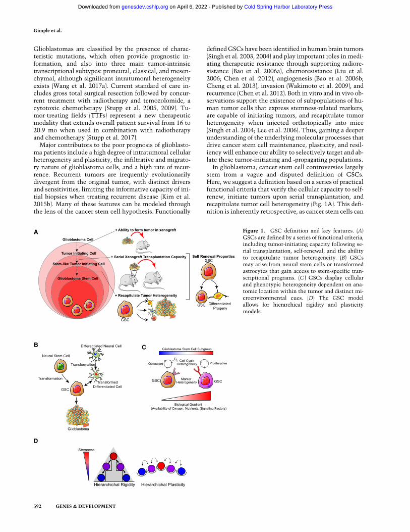
In glioblastoma, cancer stem cell controversies largelystem from a vague and disputed definition of GSCs.Here, we suggest a definition based on a series of practicalfunctional criteria that verify the cellular capacity to self-renew, initiate tumors upon serial transplantation, andrecapitulate tumor cell heterogeneity (Fig. 1A). This defi-nition is inherently retrospective, as cancer stem cells can
C
D
B
A Figure 1. GSC definition and key features. (A)GSCs are defined by a series of functional criteria,including tumor-initiating capacity following se-rial transplantation, self-renewal, and the abilityto recapitulate tumor heterogeneity. (B) GSCsmay arise from neural stem cells or transformedastrocytes that gain access to stem-specific tran-scriptional programs. (C ) GSCs display cellularand phenotypic heterogeneity dependent on ana-tomic location within the tumor and distinct mi-croenvironmental cues. (D) The GSC modelallows for hierarchical rigidity and plasticitymodels.
Gimple et al.
592 GENES & DEVELOPMENT
Cold Spring Harbor Laboratory Press on April 6, 2022 - Published by genesdev.cshlp.orgDownloaded from
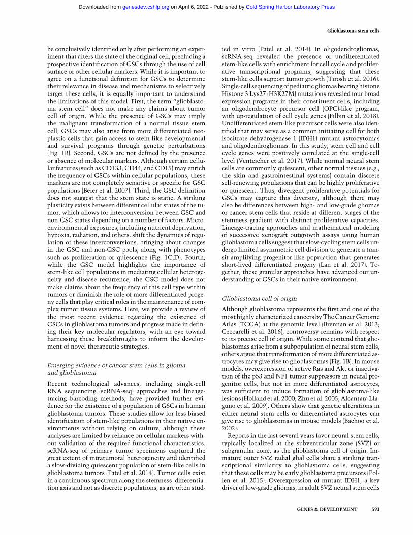
be conclusively identified only after performing an exper-iment that alters the state of the original cell, precluding aprospective identification of GSCs through the use of cellsurface or other cellular markers. While it is important toagree on a functional definition for GSCs to determinetheir relevance in disease and mechanisms to selectivelytarget these cells, it is equally important to understandthe limitations of this model. First, the term “glioblasto-ma stem cell” does not make any claims about tumorcell of origin. While the presence of GSCs may implythe malignant transformation of a normal tissue stemcell, GSCs may also arise from more differentiated neo-plastic cells that gain access to stem-like developmentaland survival programs through genetic perturbations(Fig. 1B). Second, GSCs are not defined by the presenceor absence of molecular markers. Although certain cellu-lar features (such as CD133, CD44, and CD15) may enrichthe frequency of GSCs within cellular populations, thesemarkers are not completely sensitive or specific for GSCpopulations (Beier et al. 2007). Third, the GSC definitiondoes not suggest that the stem state is static. A strikingplasticity exists between different cellular states of the tu-mor, which allows for interconversion between GSC andnon-GSC states depending on a number of factors. Micro-environmental exposures, including nutrient deprivation,hypoxia, radiation, and others, shift the dynamics of regu-lation of these interconversions, bringing about changesin the GSC and non-GSC pools, along with phenotypessuch as proliferation or quiescence (Fig. 1C,D). Fourth,while the GSC model highlights the importance ofstem-like cell populations in mediating cellular heteroge-neity and disease recurrence, the GSC model does notmake claims about the frequency of this cell type withintumors or diminish the role of more differentiated proge-ny cells that play critical roles in themaintenance of com-plex tumor tissue systems. Here, we provide a review ofthe most recent evidence regarding the existence ofGSCs in glioblastoma tumors and progress made in defin-ing their key molecular regulators, with an eye towardharnessing these breakthroughs to inform the develop-ment of novel therapeutic strategies.
Emerging evidence of cancer stem cells in gliomaand glioblastoma
Recent technological advances, including single-cellRNA sequencing (scRNA-seq) approaches and lineage-tracing barcoding methods, have provided further evi-dence for the existence of a population of GSCs in humanglioblastoma tumors. These studies allow for less biasedidentification of stem-like populations in their native en-vironments without relying on culture, although theseanalyses are limited by reliance on cellular markers with-out validation of the required functional characteristics.scRNA-seq of primary tumor specimens captured thegreat extent of intratumoral heterogeneity and identifieda slow-dividing quiescent population of stem-like cells inglioblastoma tumors (Patel et al. 2014). Tumor cells existin a continuous spectrum along the stemness–differentia-tion axis and not as discrete populations, as are often stud-
ied in vitro (Patel et al. 2014). In oligodendrogliomas,scRNA-seq revealed the presence of undifferentiatedstem-like cells with enrichment for cell cycle and prolifer-ative transcriptional programs, suggesting that thesestem-like cells support tumor growth (Tirosh et al. 2016).Single-cell sequencingof pediatric gliomasbearinghistoneHistone 3 Lys27 (H3K27M)mutations revealed four broadexpression programs in their constituent cells, includingan oligodendrocyte precursor cell (OPC)-like program,with up-regulation of cell cycle genes (Filbin et al. 2018).Undifferentiated stem-like precursor cells were also iden-tified that may serve as a common initiating cell for bothisocitrate dehydrogenase 1 (IDH1) mutant astrocytomasand oligodendrogliomas. In this study, stem cell and cellcycle genes were positively correlated at the single-celllevel (Venteicher et al. 2017). While normal neural stemcells are commonly quiescent, other normal tissues (e.g.,the skin and gastrointestinal systems) contain discreteself-renewing populations that can be highly proliferativeor quiescent. Thus, divergent proliferative potentials forGSCs may capture this diversity, although there mayalso be differences between high- and low-grade gliomasor cancer stem cells that reside at different stages of thestemness gradient with distinct proliferative capacities.Lineage-tracing approaches and mathematical modelingof successive xenograft outgrowth assays using humanglioblastomacells suggest that slow-cycling stem cells un-dergo limited asymmetric cell division to generate a tran-sit-amplifying progenitor-like population that generatesshort-lived differentiated progeny (Lan et al. 2017). To-gether, these granular approaches have advanced our un-derstanding of GSCs in their native environment.
Glioblastoma cell of origin
Although glioblastoma represents the first and one of themosthighly characterized cancers byTheCancerGenomeAtlas (TCGA) at the genomic level (Brennan et al. 2013;Ceccarelli et al. 2016), controversy remains with respectto its precise cell of origin. While some contend that glio-blastomas arise from a subpopulation of neural stem cells,others argue that transformation ofmore differentiated as-trocytes may give rise to glioblastomas (Fig. 1B). In mousemodels, overexpression of active Ras and Akt or inactiva-tion of the p53 and NF1 tumor suppressors in neural pro-genitor cells, but not in more differentiated astrocytes,was sufficient to induce formation of glioblastoma-likelesions (Holland et al. 2000; Zhu et al. 2005; Alcantara Lla-guno et al. 2009). Others show that genetic alterations ineither neural stem cells or differentiated astrocytes cangive rise to glioblastomas in mouse models (Bachoo et al.2002).Reports in the last several years favor neural stem cells,
typically localized at the subventricular zone (SVZ) orsubgranular zone, as the glioblastoma cell of origin. Im-mature outer SVZ radial glial cells share a striking tran-scriptional similarity to glioblastoma cells, suggestingthat these cells may be early glioblastoma precursors (Pol-len et al. 2015). Overexpression of mutant IDH1, a keydriver of low-grade gliomas, in adult SVZneural stemcells
Glioblastoma stem cells
GENES & DEVELOPMENT 593
Cold Spring Harbor Laboratory Press on April 6, 2022 - Published by genesdev.cshlp.orgDownloaded from
induces progenitor cell hyperplasia and tumor-like nod-ules consistent with early gliomagenesis in a mouse mod-el (Bardella et al. 2016). Deletion of the tumor suppressorsNF1, TP53, and PTEN in either neural or oligodendroglialprecursor cells induces glioblastoma-like lesions in amouse model. Disruption of identical tumor suppressorgenes in two distinct progenitor cell populations gaverise to distinct diseases, with neural precursor-derived tu-mors demonstrating more aggressive phenotypes thantheir oligodendrocyte precursor-derived counterparts(Alcantara Llaguno et al. 2015). PTENmutations in neuralstem cells are also able to induce neoplastic transforma-tion, while the same mutations in mesenchymal stemcells do not (Duan et al. 2015). These data suggest thatthe unique underlyingmolecular features of different neu-ral progenitor cell populations poise particular popula-tions for tumorigenesis and endow the resulting tumorwith distinct functional properties, highlighting the im-portance of understanding the cell of origin.
Compelling evidence from a series of high-throughputsequencingandmousemodeling approacheshas suggestedthat astrocyte-like stem cells from the astrocytic ribbonlayer of the SVZ are the glioblastoma cell of origin, furthersubstantiating prior reports (Lee et al. 2018).Whole-exomeand single-cell sequencing analyses frommatched humanglioblastoma tissue and normal SVZ tissue revealed that(1) approximately half of patients contained shared muta-tions between tumor andSVZ tissue, (2)most somaticmu-tations and copy number alterations were tumor-private,and (3) single-cell clones from SVZ tissue with mutationsshared with the tumor tissue lacked tumor-private muta-tions. Mousemodeling data showed that induction of mu-tations within the SVZ promoted tumor formation andmigration from the SVZ, while the same mutations didnot lead to tumor initiationwhen induced incortical astro-cytes (Lee et al. 2018). These recent reports highlight theimportance of cellular background in the establishmentof glioblastomas and suggest that neural precursor popula-tions are uniquely situated for malignant transformationfollowing the appropriate genetic insults. Taken together,these studies support a cancer stem cell model in gliomaswhereby a small population of GSCs derived from trans-formed SVZ-derived neural stem cells acts to maintaintumor heterogeneity, although controversy remains re-garding the applicabilityofmousemodeling data to thehu-man disease. In the following sections, we review thefield’s progress in understanding the defining molecularcharacteristics and potential targetable vulnerabilities ofthis stem cell population.
Modeling glioblastoma: isolation, enrichment,and propagation of GSCs
Strategies to isolate and enrich cancer stem cells are basedon the methods used to isolate their normal counterparts:tissue stem cells. These methods involve using specificcellular markers and growing cells in defined suspensioncultures to distinguish between GSCs and other nonstemtumor cells. Strategies to identify GSCs using cellularmarkers rely on the sensitivity and specificity of cell
surface factors to enrich for stem-like populations. How-ever, the spectrum of GSC markers has an extensiveoverlap with those used for identification of neuralstem cells. These include intracellular proteins (SOX2,OLIG2, MYC, and NESTIN) and the cell surface markers(CD133, L1CAM, CD44, and A2B5) (Brescia et al. 2012).As we understand the biology of GSCs in greater depth,we should re-evaluate these current methods. The neuro-sphere formation assay depends on self-renewal propertiesof GSCs, which allows for growth within defined nonad-herent medium conditions. Caveats with this method in-clude the inability to reflect the precise number of cellswith in vivo tumor formation capacity and the inabilityto detect quiescent stem cells. An alternative to neuro-sphere cultures is two-dimensional adherent culture ofGSCs on poly-L-lysine/laminin-coated plates, which re-duces cellular differentiation (Lee et al. 2006). However,both of these methods fail to properly model the interac-tion of GSCs with the various other cell types that existin vivo.
Recently, three-dimensional GSC organoid culture sys-tems have been developed, which more faithfully recapit-ulate in vivo tumor growth, cellular heterogeneity, andhypoxic gradients (Hubert et al. 2016). CRISPR–Cas9 ge-nomic editing can also be used to generate glioblastomaorganoid models. Introduction of the HRasG12V alleleinto the TP53 locus in cells of cerebral organoids drove in-vasive phenotypes both in the organoid model and whenorthotopically xenografted in immunodeficient micewith transcriptional similarities to mesenchymal tumors(Ogawaet al. 2018). Somatic in vivoCRISPR/Cas9-mediat-ed genomic editing approaches have also been applied tostudy glioblastomas in mouse models (Oldrini et al.2018) and have allowed for the performance of large-scalescreens in vivo (Chow et al. 2017). These organoid and an-imal systems may better model in vivo environments toimprove our understanding of GSCs in their appropriatephysiological context. Despite these advances, newmeth-ods for the prospective isolation and propagation of GSCsare required. The ideal in vitro assay should accuratelyassess the tumor formation ability of GSCs, be largelyindependent of growthmediated by high amounts of exog-enous growth factors, incorporate various cellular interac-tions, and recapitulate the tumor microenvironment.Furthermore, efforts should be made to develop modelsthat reflect the diversemutational spectrum of glioblasto-ma to more completely understand the role of geneticbackground in cellular dependencies and therapeutic re-sponse (Table 1).
Glioma stem cell epigenetics: interface betweenenvironment and cellular response
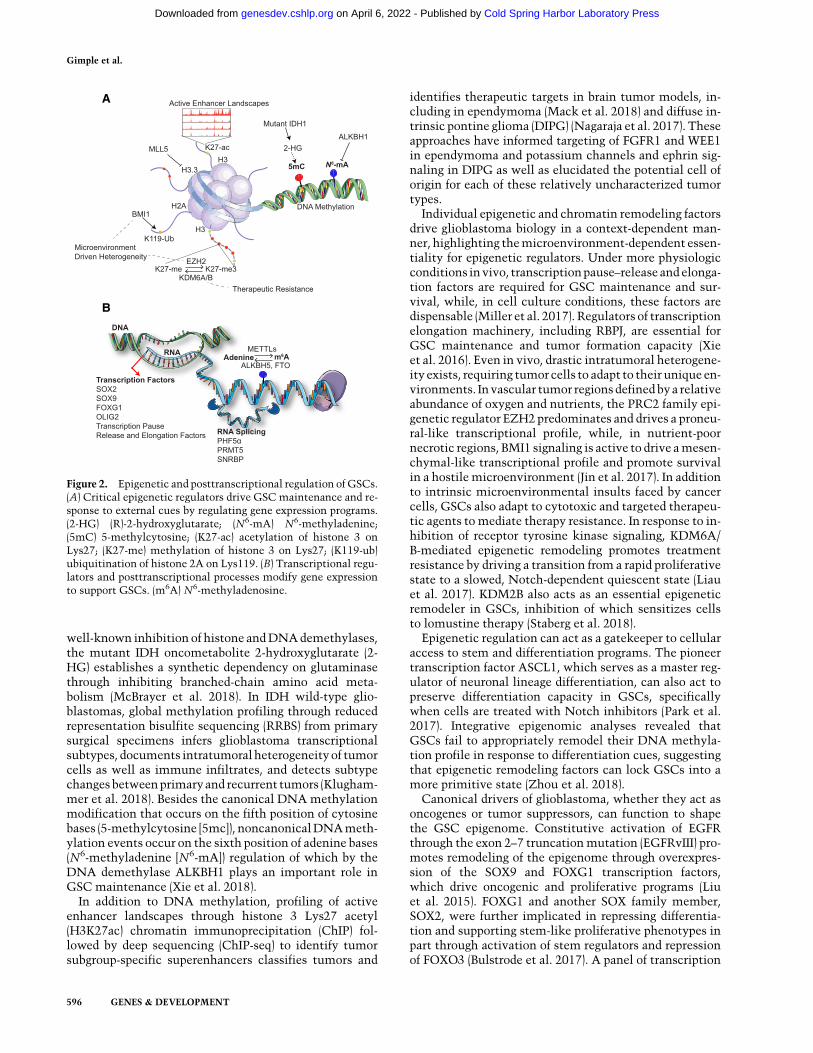
While nearly every cell in the human body contains anidentical genomic blueprint, epigenomic regulation ofthis genomic code allows for the generation of cell anddevelopmental stage-dependent transcriptional programsthrough opening relevant genomic regions while seques-tering others (Fig. 2A). Appropriate epigenetic regulationis critical for the maintenance of GSCs and serves to
Gimple et al.
594 GENES & DEVELOPMENT
Cold Spring Harbor Laboratory Press on April 6, 2022 - Published by genesdev.cshlp.orgDownloaded from
integrate information from numerous cellular inputs. Thepresumed initiating event in low-grade gliomas (alongwith other cancers) is mutation of IDH1 or IDH2, whichgenerates widespread epigenomic dysregulation throughgeneration of the glioma-CpG island methylator pheno-type (G-CIMP) hypermethylator phenotype (Turcan et al.
2012). The epigenetic alterations induced by IDHmutationinvitroarepartially reversible, contribute to transcriptionalremodeling and deposition of histone modifications at spe-cific genomic loci, promote the emergence of a CD24+
stem-like population, and contribute to increased genomicinstability (Turcan et al. 2018). In addition to the more
Table 1. Advantages and limitations of GSC and glioblastoma models
GSC modelingmethods Advantages Limitations References
Sorting using cellularmarkers (CD133,CD15, and others)
Prospective identification ofputative stem populationsis possible
Individual stem markers arefrequently disputed; no functionalcriteria are used; may deplete GSCheterogeneity; integrity of surfacemarkers may be affected duringsingle-cell dissociation
Galli et al. 2004; Singh et al. 2004;Bidlingmaier et al. 2008; Gilbertand Ross 2009; Son et al. 2009;Wan et al. 2010
In vitro neurosphereculture
Uses a functional assay forself-renewal capacity;high throughput
Inability to determine tumorformation capacity, identifyquiescent stem populations, andmodel diverse cellular interactions;reliance on artificial andunphysiological medium conditionswith loss of tumor heterogeneity;contain populations of stem anddifferentiated cells; not ideal forassays in which homogeneity isrequired; percentage of enrichedGSCs will vary with every spheredepending on size, passage, andtechnique used in propagation andculture medium
Lee et al. 2006; Wan et al. 2010;Pastrana et al. 2011
Two-dimensionaladherent culture onpoly-L-lysine/laminin plates
May reduce differentiationcompared withneurosphere culture; idealfor assays in whichhomogeneity is required;high throughput
Inability to determine tumorformation capacity, identifyquiescent stem populations, andmodel diverse cellular interactions;reliance on artificial andunphysiological medium conditionswith loss of tumor heterogeneity
Pollard et al. 2009
Three-dimensionalGSC organoidculture systems andbiomaterial scaffolds
Recapitulate in vivoenvironment with higherfidelity than in vitrosystems; model cellularand stromal interactions;model hypoxic and otherbiologic gradients; modelmulticellular andmicroenvironmentalinteractions
Decreased throughput compared withtwo-dimensional methods; complexprocedure for initiation andmaintenance; lack certain cellularinteractions, including withvasculature, microglia, and others
Hubert et al. 2016; Bian et al. 2018;Ogawa et al. 2018; Langer et al.2019
Genetically engineeredand syngeneic mousemodeling approaches
Allow for studies of tumorinitiation and progressionwith ability to modelcellular and immuneinteractions in a native invivo environment
Systems to generate murine tumorsdo not fully recapitulatetumorigenesis in humans; mousetumors may be fundamentallydifferent from the human disease;expensive and labor-intensive
Holland et al. 2000; Zhu et al.2005; Bardella et al. 2016; Miyaiet al. 2017; Oldrini et al. 2018;Hambardzumyan et al. 2009
Patient-derivedxenograft methods
Allow for studies of humancancers with an ability tomodel cellularinteractions in a morephysiologic in vivoenvironment
Require immediate generationfollowing tumor surgical resection;inability to assess mechanisms oftumor initiation or model adaptiveimmune interactions; serialpassaging may depleteheterogeneity and induce genomicalterations; expensive and labor-intensive
Singh et al. 2004; Hidalgo et al.2014; Ben-David et al. 2017; Junget al. 2018
Glioblastoma stem cells
GENES & DEVELOPMENT 595
Cold Spring Harbor Laboratory Press on April 6, 2022 - Published by genesdev.cshlp.orgDownloaded from
well-known inhibition of histone andDNAdemethylases,the mutant IDH oncometabolite 2-hydroxyglutarate (2-HG) establishes a synthetic dependency on glutaminasethrough inhibiting branched-chain amino acid meta-bolism (McBrayer et al. 2018). In IDH wild-type glio-blastomas, global methylation profiling through reducedrepresentation bisulfite sequencing (RRBS) from primarysurgical specimens infers glioblastoma transcriptionalsubtypes, documents intratumoral heterogeneityof tumorcells as well as immune infiltrates, and detects subtypechangesbetweenprimaryand recurrent tumors (Klugham-mer et al. 2018). Besides the canonical DNA methylationmodification that occurs on the fifth position of cytosinebases (5-methylcytosine [5mc]), noncanonicalDNAmeth-ylation events occur on the sixth position of adenine bases(N6-methyladenine [N6-mA]) regulation of which by theDNA demethylase ALKBH1 plays an important role inGSC maintenance (Xie et al. 2018).
In addition to DNA methylation, profiling of activeenhancer landscapes through histone 3 Lys27 acetyl(H3K27ac) chromatin immunoprecipitation (ChIP) fol-lowed by deep sequencing (ChIP-seq) to identify tumorsubgroup-specific superenhancers classifies tumors and
identifies therapeutic targets in brain tumor models, in-cluding in ependymoma (Mack et al. 2018) and diffuse in-trinsic pontine glioma (DIPG) (Nagaraja et al. 2017). Theseapproaches have informed targeting of FGFR1 and WEE1in ependymoma and potassium channels and ephrin sig-naling in DIPG as well as elucidated the potential cell oforigin for each of these relatively uncharacterized tumortypes.
Individual epigenetic and chromatin remodeling factorsdrive glioblastoma biology in a context-dependent man-ner, highlighting themicroenvironment-dependent essen-tiality for epigenetic regulators. Under more physiologicconditions invivo, transcriptionpause–release andelonga-tion factors are required for GSC maintenance and sur-vival, while, in cell culture conditions, these factors aredispensable (Miller et al. 2017). Regulators of transcriptionelongation machinery, including RBPJ, are essential forGSC maintenance and tumor formation capacity (Xieet al. 2016). Even in vivo, drastic intratumoral heterogene-ity exists, requiring tumorcells to adapt to their unique en-vironments. Invascular tumor regionsdefinedbya relativeabundance of oxygen and nutrients, the PRC2 family epi-genetic regulator EZH2 predominates and drives a proneu-ral-like transcriptional profile, while, in nutrient-poornecrotic regions, BMI1 signaling is active to drive amesen-chymal-like transcriptional profile and promote survivalin a hostile microenvironment (Jin et al. 2017). In additionto intrinsic microenvironmental insults faced by cancercells, GSCs also adapt to cytotoxic and targeted therapeu-tic agents to mediate therapy resistance. In response to in-hibition of receptor tyrosine kinase signaling, KDM6A/B-mediated epigenetic remodeling promotes treatmentresistance by driving a transition from a rapid proliferativestate to a slowed, Notch-dependent quiescent state (Liauet al. 2017). KDM2B also acts as an essential epigeneticremodeler in GSCs, inhibition of which sensitizes cellsto lomustine therapy (Staberg et al. 2018).
Epigenetic regulation can act as a gatekeeper to cellularaccess to stem and differentiation programs. The pioneertranscription factor ASCL1, which serves as a master reg-ulator of neuronal lineage differentiation, can also act topreserve differentiation capacity in GSCs, specificallywhen cells are treated with Notch inhibitors (Park et al.2017). Integrative epigenomic analyses revealed thatGSCs fail to appropriately remodel their DNA methyla-tion profile in response to differentiation cues, suggestingthat epigenetic remodeling factors can lock GSCs into amore primitive state (Zhou et al. 2018).
Canonical drivers of glioblastoma, whether they act asoncogenes or tumor suppressors, can function to shapethe GSC epigenome. Constitutive activation of EGFRthrough the exon 2–7 truncationmutation (EGFRvIII) pro-motes remodeling of the epigenome through overexpres-sion of the SOX9 and FOXG1 transcription factors,which drive oncogenic and proliferative programs (Liuet al. 2015). FOXG1 and another SOX family member,SOX2, were further implicated in repressing differentia-tion and supporting stem-like proliferative phenotypes inpart through activation of stem regulators and repressionof FOXO3 (Bulstrode et al. 2017). A panel of transcription
A
B
Figure 2. Epigenetic and posttranscriptional regulation of GSCs.(A) Critical epigenetic regulators drive GSC maintenance and re-sponse to external cues by regulating gene expression programs.(2-HG) (R)-2-hydroxyglutarate; (N6-mA) N6-methyladenine;(5mC) 5-methylcytosine; (K27-ac) acetylation of histone 3 onLys27; (K27-me) methylation of histone 3 on Lys27; (K119-ub)ubiquitination of histone 2A on Lys119. (B) Transcriptional regu-lators and posttranscriptional processes modify gene expressionto support GSCs. (m6A) N6-methyladenosine.
Gimple et al.
596 GENES & DEVELOPMENT
Cold Spring Harbor Laboratory Press on April 6, 2022 - Published by genesdev.cshlp.orgDownloaded from
factors, including SOX2, OLIG2, and ZEB1, transformsastrocytes into tumor-initiating cells even in the absenceof oncogenic driver mutations, suggesting that alteredepigenetic landscapes can drive tumor formation (Singhet al. 2017). Another significantly mutated gene in glio-blastoma, PTEN, shapes the glioblastoma epigenomethrough nonenzymatic interactions with the chromatinregulator DAXX, which regulates histone H3.3 genomiclocalization (Benitez et al. 2017). Histone H3.3 levels canalso be controlled by the chromatin regulatory elementMLL5,which is critical for themaintenanceof genomic ar-chitecture required for persistence in a stemcell state (Gal-lo et al. 2015).
Posttranscriptional regulation: RNAs as driversof GSC biology
In the previous section, we discussed howGSCs fine-tunetheir genetic and epigenetic programs to regulate gene ex-pression. Another complex node of gene regulation existsat the posttranscriptional level, including regulation ofRNAmaturation, cellular localization, stability, and alter-native splicing of transcripts. These mechanisms contrib-ute to the effective translation of transcripts intofunctional proteins, which ultimately carry out most cel-lular functions (Fig. 2B). Many of these processes are regu-lated by RNA-binding proteins (RBPs), a class of deeplyconserved and highly abundant proteins, which form ribo-nucleoprotein complexes with transcripts to facilitatetheir functions (Hentze et al. 2018). In glioblastoma,many RBPs are expressed at high levels in patient tumorsand portend poor prognosis, including SNRBP, a splicingfactor that regulates RNA processing and DNA repairpathways (Correa et al. 2016). RNAi screens of patient-derived GSCs identified the splicing factor PHF5α as aselective dependency in GSCs but not in nontransformedcontrols, such as fibroblasts, astrocytes, and neural stemcells (Hubert et al. 2013). MYC overexpression in GSCsis associated with increased sensitivity to splicing inhibi-tion (Hubert et al. 2013). Splicing factors can also be pro-vided in the form of vesicular secretions from apoptoticcells that promote proliferation and therapy resistance inneighboring cells within a tumor (Pavlyukov et al. 2018).Glioma cells also rely on fine regulation of splicing factors,coordinated by PRMT5, which is essential for preventinginclusion of “detained introns” into transcripts, allowingfor their efficient translation (Braun et al. 2017).In eukaryotes, posttranscriptional messenger RNA
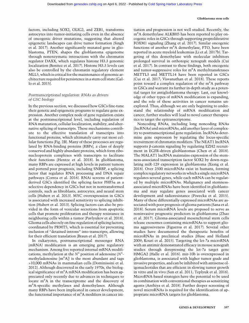
(mRNA) modification is an emerging gene regulatorymechanism. Among five reported internal mRNAmodifi-cations, methylation at the N6 position of adenosine (N6-methyladenosine [m6A]) is the most abundant and tags>10,000 mRNAs in mammalian cells (Dominissini et al.2012). Although discovered in the early 1970s, the biolog-ical significance of m6AmRNAmodification has been ap-preciated only recently due to advances in techniques tolocate m6A in the transcriptome and the discovery ofm6A-specific methylases and demethylases. Althoughmany RBPs have been implicated in cancer development,the functional importance of m6Amodifiers in cancer ini-
tiation and progression is not well studied. Recently, them6A demethylase ALKBH5 has been reported to play on-cogenic roles in GSCs through supporting proproliferativeFOXM1 signaling (Zhang et al. 2017). Similar oncogenicfunctions of another m6A demethylase, FTO, have beenreported in acutemyeloid leukemias (Li et al. 2017b). Tar-geting of this demethylase with molecular inhibitorsprolonged survival in orthotopic xenograft models (Cuiet al. 2017). In contrast to these findings, both oncogenicand tumor-suppressive roles for m6A methyltransferasesMETTL3 and METTL14 have been reported in GSCs(Cui et al. 2017; Visvanathan et al. 2018). These reportspoint toward a complex regulation of the m6A pathwayin GSCs and warrant its further in-depth study as a poten-tial target for antiglioblastoma therapy. Last, our knowl-edge of other types of mRNA modification is expanding,and the role of these activities in cancer remains un-explored. Thus, although we are only beginning to under-stand the relationship of mRNA modification andcancer, further studies will lead to novel cancer therapeu-tics to target the epitranscriptome.Noncoding RNAs, including long noncoding RNAs
(lncRNAs) andmicroRNAs, add another layer of complex-ity to posttranscriptional gene regulation. lncRNAs deter-mine gene expression by regulating the locus-specificrecruitment of chromatinmodifiers. TheNEAT1 lncRNAsupports β-catenin signaling by regulating EZH2 recruit-ment in EGFR-driven glioblastomas (Chen et al. 2018).The MALAT1 lncRNAmaintains expression of the stem-ness-associated transcription factor SOX2 by down-regu-lating miR-129 expression in glioblastoma (Xiong et al.2018). Over 2500 microRNAs exist in humans, formingcomplex regulatorynetworks inwhich a singlemicroRNAregulates several genes, while eachmRNA can be regulat-ed by multiple microRNAs. Malignancy and stemness-associated microRNAs have been identified in glioblasto-ma and may regulate genes associated with cancerdevelopment and radioresistance (Piwecka et al. 2015).Many of these differentially expressed microRNAs are as-sociatedwith poor prognosis of glioma patients (Sana et al.2018). Serum microRNA levels are proposed to serve asnoninvasive prognostic predictors in glioblastoma (Zhaoet al. 2017). Glioma-associated mesenchymal stem cellsrelease exosomes containing microRNAs to support glio-ma aggressiveness (Figueroa et al. 2017). Several otherstudies have documented the therapeutic benefits ofmicroRNAs in preclinical studies (Huse and Holland2009; Kouri et al. 2015). Targeting the let-7a microRNAwith an antimir demonstrated efficacy inmouse xenograftstudies through derepressing the let-7a target geneHMGA2 (Halle et al. 2016). mir-10b is overexpressed inglioblastoma, is associated with higher tumor grade andinvasive properties, and canbe inhibitedwith antisense ol-igonucleotides that are efficient in slowing tumor growthin vitro and in vivo (Sun et al. 2011; Teplyuk et al. 2016).MicroRNA-based strategies have the potential to be usedin combinationwith conventional therapies as sensitizingagents (Anthiya et al. 2018). Further deeper screening ofnovel microRNAs is required for the identification of ap-propriate microRNA targets for glioblastoma.
Glioblastoma stem cells
GENES & DEVELOPMENT 597
Cold Spring Harbor Laboratory Press on April 6, 2022 - Published by genesdev.cshlp.orgDownloaded from
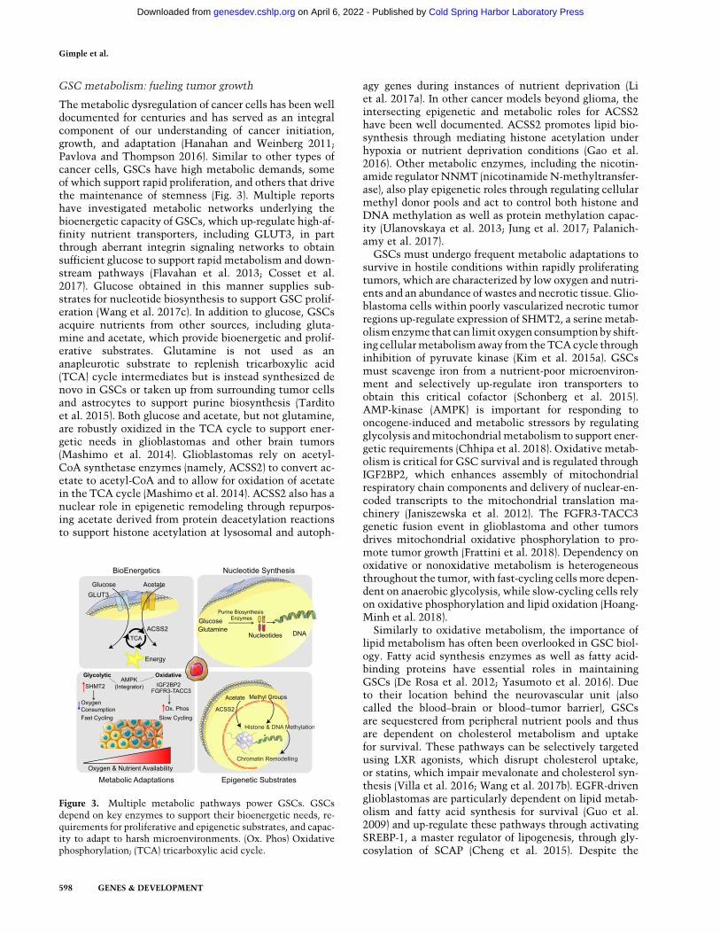
GSC metabolism: fueling tumor growth
The metabolic dysregulation of cancer cells has been welldocumented for centuries and has served as an integralcomponent of our understanding of cancer initiation,growth, and adaptation (Hanahan and Weinberg 2011;Pavlova and Thompson 2016). Similar to other types ofcancer cells, GSCs have high metabolic demands, someof which support rapid proliferation, and others that drivethe maintenance of stemness (Fig. 3). Multiple reportshave investigated metabolic networks underlying thebioenergetic capacity of GSCs, which up-regulate high-af-finity nutrient transporters, including GLUT3, in partthrough aberrant integrin signaling networks to obtainsufficient glucose to support rapid metabolism and down-stream pathways (Flavahan et al. 2013; Cosset et al.2017). Glucose obtained in this manner supplies sub-strates for nucleotide biosynthesis to support GSC prolif-eration (Wang et al. 2017c). In addition to glucose, GSCsacquire nutrients from other sources, including gluta-mine and acetate, which provide bioenergetic and prolif-erative substrates. Glutamine is not used as ananapleurotic substrate to replenish tricarboxylic acid(TCA) cycle intermediates but is instead synthesized denovo in GSCs or taken up from surrounding tumor cellsand astrocytes to support purine biosynthesis (Tarditoet al. 2015). Both glucose and acetate, but not glutamine,are robustly oxidized in the TCA cycle to support ener-getic needs in glioblastomas and other brain tumors(Mashimo et al. 2014). Glioblastomas rely on acetyl-CoA synthetase enzymes (namely, ACSS2) to convert ac-etate to acetyl-CoA and to allow for oxidation of acetatein the TCA cycle (Mashimo et al. 2014). ACSS2 also has anuclear role in epigenetic remodeling through repurpos-ing acetate derived from protein deacetylation reactionsto support histone acetylation at lysosomal and autoph-
agy genes during instances of nutrient deprivation (Liet al. 2017a). In other cancer models beyond glioma, theintersecting epigenetic and metabolic roles for ACSS2have been well documented. ACSS2 promotes lipid bio-synthesis through mediating histone acetylation underhypoxia or nutrient deprivation conditions (Gao et al.2016). Other metabolic enzymes, including the nicotin-amide regulator NNMT (nicotinamide N-methyltransfer-ase), also play epigenetic roles through regulating cellularmethyl donor pools and act to control both histone andDNA methylation as well as protein methylation capac-ity (Ulanovskaya et al. 2013; Jung et al. 2017; Palanich-amy et al. 2017).
GSCs must undergo frequent metabolic adaptations tosurvive in hostile conditions within rapidly proliferatingtumors, which are characterized by low oxygen and nutri-ents and an abundance of wastes and necrotic tissue. Glio-blastoma cells within poorly vascularized necrotic tumorregions up-regulate expression of SHMT2, a serinemetab-olismenzyme that can limit oxygen consumptionby shift-ing cellularmetabolism away from theTCAcycle throughinhibition of pyruvate kinase (Kim et al. 2015a). GSCsmust scavenge iron from a nutrient-poor microenviron-ment and selectively up-regulate iron transporters toobtain this critical cofactor (Schonberg et al. 2015).AMP-kinase (AMPK) is important for responding tooncogene-induced and metabolic stressors by regulatingglycolysis andmitochondrialmetabolism to support ener-getic requirements (Chhipa et al. 2018). Oxidative metab-olism is critical for GSC survival and is regulated throughIGF2BP2, which enhances assembly of mitochondrialrespiratory chain components and delivery of nuclear-en-coded transcripts to the mitochondrial translation ma-chinery (Janiszewska et al. 2012). The FGFR3-TACC3genetic fusion event in glioblastoma and other tumorsdrives mitochondrial oxidative phosphorylation to pro-mote tumor growth (Frattini et al. 2018). Dependency onoxidative or nonoxidative metabolism is heterogeneousthroughout the tumor, with fast-cycling cellsmore depen-dent on anaerobic glycolysis, while slow-cycling cells relyon oxidative phosphorylation and lipid oxidation (Hoang-Minh et al. 2018).
Similarly to oxidative metabolism, the importance oflipid metabolism has often been overlooked in GSC biol-ogy. Fatty acid synthesis enzymes as well as fatty acid-binding proteins have essential roles in maintainingGSCs (De Rosa et al. 2012; Yasumoto et al. 2016). Dueto their location behind the neurovascular unit (alsocalled the blood–brain or blood–tumor barrier), GSCsare sequestered from peripheral nutrient pools and thusare dependent on cholesterol metabolism and uptakefor survival. These pathways can be selectively targetedusing LXR agonists, which disrupt cholesterol uptake,or statins, which impair mevalonate and cholesterol syn-thesis (Villa et al. 2016; Wang et al. 2017b). EGFR-drivenglioblastomas are particularly dependent on lipid metab-olism and fatty acid synthesis for survival (Guo et al.2009) and up-regulate these pathways through activatingSREBP-1, a master regulator of lipogenesis, through gly-cosylation of SCAP (Cheng et al. 2015). Despite the
Figure 3. Multiple metabolic pathways power GSCs. GSCsdepend on key enzymes to support their bioenergetic needs, re-quirements for proliferative and epigenetic substrates, and capac-ity to adapt to harsh microenvironments. (Ox. Phos) Oxidativephosphorylation; (TCA) tricarboxylic acid cycle.
Gimple et al.
598 GENES & DEVELOPMENT
Cold Spring Harbor Laboratory Press on April 6, 2022 - Published by genesdev.cshlp.orgDownloaded from
findings discussed here, the roles of key metabolic en-zymes and the utilization of their respective substratesare certain to be context-dependent. Caution should beused when interpreting results of metabolic profilingstudies that are conducted in artificial cell culturesystems replete with serum and under high-oxygenconditions.
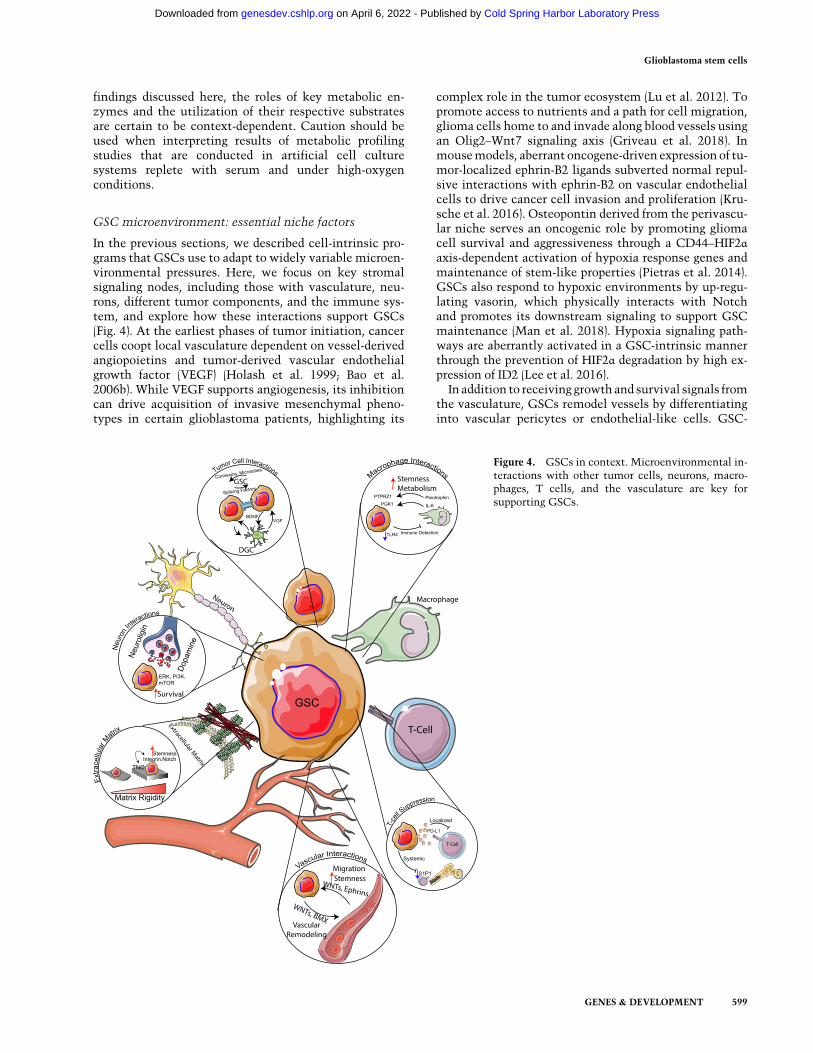
GSC microenvironment: essential niche factors
In the previous sections, we described cell-intrinsic pro-grams that GSCs use to adapt to widely variable microen-vironmental pressures. Here, we focus on key stromalsignaling nodes, including those with vasculature, neu-rons, different tumor components, and the immune sys-tem, and explore how these interactions support GSCs(Fig. 4). At the earliest phases of tumor initiation, cancercells coopt local vasculature dependent on vessel-derivedangiopoietins and tumor-derived vascular endothelialgrowth factor (VEGF) (Holash et al. 1999; Bao et al.2006b). While VEGF supports angiogenesis, its inhibitioncan drive acquisition of invasive mesenchymal pheno-types in certain glioblastoma patients, highlighting its
complex role in the tumor ecosystem (Lu et al. 2012). Topromote access to nutrients and a path for cell migration,glioma cells home to and invade along blood vessels usingan Olig2–Wnt7 signaling axis (Griveau et al. 2018). Inmousemodels, aberrant oncogene-driven expression of tu-mor-localized ephrin-B2 ligands subverted normal repul-sive interactions with ephrin-B2 on vascular endothelialcells to drive cancer cell invasion and proliferation (Kru-sche et al. 2016). Osteopontin derived from the perivascu-lar niche serves an oncogenic role by promoting gliomacell survival and aggressiveness through a CD44–HIF2αaxis-dependent activation of hypoxia response genes andmaintenance of stem-like properties (Pietras et al. 2014).GSCs also respond to hypoxic environments by up-regu-lating vasorin, which physically interacts with Notchand promotes its downstream signaling to support GSCmaintenance (Man et al. 2018). Hypoxia signaling path-ways are aberrantly activated in a GSC-intrinsic mannerthrough the prevention of HIF2α degradation by high ex-pression of ID2 (Lee et al. 2016).In addition to receiving growth and survival signals from
the vasculature, GSCs remodel vessels by differentiatinginto vascular pericytes or endothelial-like cells. GSC-
Figure 4. GSCs in context. Microenvironmental in-teractions with other tumor cells, neurons, macro-phages, T cells, and the vasculature are key forsupporting GSCs.
Glioblastoma stem cells
GENES & DEVELOPMENT 599
Cold Spring Harbor Laboratory Press on April 6, 2022 - Published by genesdev.cshlp.orgDownloaded from
derived pericytes depend primarily on TGF-β and BMX-ty-rosine kinase activity to shape their microenvironmentandmaintain the integrity of the blood–tumor barrier—in-teractions that can be targeted for therapeutic benefit(Cheng et al. 2013; Zhou et al. 2017; Shi et al. 2018).GSC-derived endothelial-like cells rely onWNT5A signal-ing topromote cellular lineage infidelity and acquisition ofendothelial-like phenotypes, which promotes tumor neo-vascularization and invasion (Hu et al. 2016).
Signals derived fromneuronal components of the tumormicroenvironment are similarly critical for maintainingGSCs. Using optogenetic control of neuronal activity inxenograft models, neuronal stimulation was found to pro-mote glioma growth via the solublemitogen neuroligin-3,which is liberated from neurons (as well as OPCs) byADAM10 sheddases and functions to support FAK andPI3K–mTOR signaling (Venkatesh et al. 2015, 2017). Dop-amine signaling, which presumably originates from neu-rons, maintains GSCs, as inhibition of the dopaminereceptor DRD4 inhibits GSC growth and stem propertiesthrough disrupting autophagy and ERK–mTOR signalingpathways (Dolma et al. 2016). Neural precursor cells inthe SVZ provide chemoattractant and proinvasive signalsto DIPG cells through secretion of pleiotrophin, whichmaintainsRhoAandROCKsignaling (Qin et al. 2017). Jag-ged, a NOTCH ligand expressed along axons, supportsGSC invasion along unmyelinated white matter tractsthrough up-regulating SOX9 and SOX2 in GSCs (Wanget al. 2019). Furthermore, GSCs adapt to conditions out-side of their typical environmental niche. Deletion oftheRBPQki promotes stemness by down-regulating endo-lysosome-mediated receptor degradation, allowing forlimited signaling factors to stimulate stem signaling path-ways, including Wnt and Notch signaling (Shingu et al.2017).
Interactions between tumor and stromal componentsare also important for maintaining GSCs. In IDH mutantgliomas, high expression of the extracellular matrixglycoprotein tenascin C (TNC) supports increased extra-cellular matrix stiffness, which increases mechanosignal-ing through the oncogenic FAK pathway (Miroshnikovaet al. 2016). In glioblastoma, TNC supports the mainte-nance of stemness through activating Notch signalingthrough binding to cell surface integrins (Sarkar et al.2017). Extracellular matrix stiffness driven by high levelsof glycoproteins in mesenchymal subsets of glioblasto-mas promotes stemness through integrin mechanosignal-ing pathways (Barnes et al. 2018). Integrin signaling,particularly mediated by integrin α7, maintains GSCproliferation and invasiveness, serving as a therapeutictarget (Haas et al. 2017). Additionally, glioma-associatedmesenchymal stem cells, a nontumorigenic stromalcomponent of human glioblastomas, support GSCsthrough an IL-6/STAT3 axis (Hossain et al. 2015) andsecretion of microRNA-containing exosomes (Figueroaet al. 2017).
Tumor tissues are composed of a heterogenous popula-tion of tumor cells with distinct functional properties.Transcriptomic and genomic mapping efforts of histolog-ically defined anatomic regions of glioblastoma tumors
provide a resource to begin to functionally characterizethis intratumoral heterogeneity (Puchalski et al. 2018).Intercellular communication plays important roles inthe maintenance of the complex cellular systems thatcompose a tumor. In vivo multiphoton microscopy ofglioblastomas in xenograft hosts reveals multicellular an-atomic networks composed of “tumor microtubes” con-necting glioblastoma cells. These microtubes permitlong-range coordinated communication between cellsvia connexins, providing tracts for invasion, and are re-quired for maintenance of the tumor cell network follow-ing therapeutic interventions (Osswald et al. 2015).Functional connexin-mediated gap junctions are es-sential for GSCs (Hitomi et al. 2015). Communicationvia secreted factors is also important, as differentiatedglioblastoma cells produce BDNF to support the growthand survival of their stem cell counterparts (Wang et al.2018).
The critical role of the immune system in regulating tu-mor biology represents one of the most rapidly evolvingmicroenvironmental dependencies. Immune editing ofdeveloping tumors by both innate and adaptive arms ofthe immune system drives the evolution of GSCs asthey evade immunosurveillance, while signaling fromGSCs can shape their local immune environment. Tu-mor-associated macrophages (TAMs) stimulate GSCmaintenance and tumorigenicity through a pleiotro-phin–PTPRZ1 signaling axis (Shi et al. 2017) and promoteglycolytic metabolism through an IL6–PGK1 axis (Zhanget al. 2018). Inhibition of this GSC–TAM cross-talk hasbeen explored as a therapeutic avenue through targetingCSF-1R on macrophages (Pyonteck et al. 2013; Quailet al. 2016). GSCs down-regulate expression of innate im-mune sensors (namely, the toll-like receptor TLR4) to pre-vent negative modulation of stem-like properties by theimmune system (Alvarado et al. 2017) and recruit TAMsthat enhance tumor growth and block tumor rejection(Zhou et al. 2015; Otvos et al. 2016). Engineered mousemodels of low-grade IDH mutant gliomas demonstratelower immune infiltration and leukocyte chemotaxisthan in IDHwild-typemodels, suggesting that differentialimmune activation between low- and high-grade tumorsmay partially explain the prognostic disparity betweenthese two diseases (Amankulor et al. 2017). Glioblasto-mas, along with other cancers localized to the brain, me-diate profound systemic T-cell deficiency, with T cellsaccumulating within bone marrow and unable to trafficto the site of the tumor due to loss of cell surface S1P1(Chongsathidkiet et al. 2018). Glioblastomas also generateboth local and systemic immunosuppression by inducingM2 macrophage polarization and Th2 reactivity that im-pairs antitumor responses (Prosniak et al. 2013; Harshyneet al. 2016). GSCs evade T-cell killing by secretion of ex-tracellular vesicles containing the T-cell checkpoint mol-ecule PD-L1 (Ricklefs et al. 2018). GSC-derived exosomesmay suppress T cells through monocyte maturation(Domenis et al. 2017). Thus, GSCs use a number of mech-anisms to avoid immune-mediated destruction and derivegrowth benefit from factors derived from the immunemicroenvironment.
Gimple et al.
600 GENES & DEVELOPMENT
Cold Spring Harbor Laboratory Press on April 6, 2022 - Published by genesdev.cshlp.orgDownloaded from
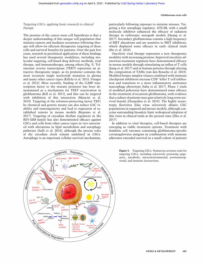
Targeting GSCs: applying basic research to clinicaltherapy
The promise of the cancer stem cell hypothesis is that adeeper understanding of this unique cell population thatinitiates tumors and mediates recurrence following ther-apy will allow for efficient therapeutic targeting of thesecells and survival benefits for patients. Over the past fewyears, research in preclinical application of these findingshas used several therapeutic modalities, including mo-lecular targeting, cell-based drug delivery methods, viraltherapy, and immunotherapy, among others (Fig. 5). Tel-omerase reverse transcriptase (TERT) represents an at-tractive therapeutic target, as its promoter contains themost recurrent single nucleotide mutation in gliomaand many other cancer types (Killela et al. 2013; Vinagreet al. 2013). More recently, binding of the GABP tran-scription factor to the mutant promoter has been de-monstrated as a mechanism for TERT reactivation inglioblastoma (Bell et al. 2015), and this can be targetedwith inhibition of this interaction (Mancini et al.2018). Targeting of the telomere-protecting factor TRF1by chemical and genetic means can also reduce GSC vi-ability and tumorigenicity and lead to regression of es-tablished tumors in mouse models (Bejarano et al.2017). Targeting of circadian rhythm regulators in theREV-ERB family has also demonstrated efficacy againstGSCs and cells from other cancer types in vivo associat-ed with alterations in lipid metabolism and autophagypathways (Sulli et al. 2018), although the precise rolesof the circadian clock remain undefined in GSCs.Autophagy is an important cellular survival mechanism,
particularly following exposure to extreme stresses. Tar-geting a key autophagy regulator, ATG4B, with a smallmolecule inhibitor enhanced the efficacy of radiationtherapy in orthotopic xenograft models (Huang et al.2017). Secondary glioblastomas contain a high frequencyof MET alterations and are sensitive to MET inhibitors,which displayed some efficacy in early clinical trials(Hu et al. 2018).Oncolytic viral therapy represents a new therapeutic
modalitywith increasing promise. Improved oncolytic ad-enovirus treatment regimens have demonstrated efficacyin mouse models through stimulating an influx of T cells(Jiang et al. 2017) and in human patients through alteringthe composition of TAMs (van den Bossche et al. 2018).Modified herpes simplex viruses combined with immunecheckpoint inhibition increase CD8+ killer T-cell infiltra-tion and transition to a more inflammatory antitumormacrophage phenotype (Saha et al. 2017). Phase 1 trialsof modified poliovirus have demonstrated some efficacyin the treatment of recurrent glioblastoma, with evidencethat a subset of patientsmay gain relatively long-term sur-vival benefit (Desjardins et al. 2018). The highly neuro-tropic flavivirus Zika virus selectively ablates GSCpopulations in organoid andmousemodels, although con-cerns surrounding biosafety limit widespread adoption ofthis virus in clinical trials at the present time (Zhu et al.2017).In addition to viral therapies, cell-based therapies are
emerging as viable treatment options. Treatment withdendritic cell vaccines containing glioblastoma-specificcytomegalovirus antigens in combination with immuneadjuvants extended survival in a small cohort of patients
Figure 5. Targeting GSCs. Numerous avenues exist fortargeting GSCs, including selectively poisoning epige-netic, metabolic, microenvironmental, posttranscrip-tional, and immune interactions.
Glioblastoma stem cells
GENES & DEVELOPMENT 601
Cold Spring Harbor Laboratory Press on April 6, 2022 - Published by genesdev.cshlp.orgDownloaded from
(Mitchell et al. 2015). Treatment with autologous tumorlysate-containing dendritic cells may also be a feasible ap-proach (Prins et al. 2011). In two early clinical trials, ad-ministration of personalized vaccines containing tumorneoepitopes elicited strong T-cell immunologic responseswith evidence of immunologic memory and tumor-infil-trating capacity (Hilf et al. 2019; Keskin et al. 2019).Neural stem-like cells engineered from autologous pa-tient-derived fibroblasts home to glioblastoma tumorsand deliver cytotoxic compounds to tumor cells in mousemodels (Bagó et al. 2017) and have been investigated inearly phase clinical trials (Portnow et al. 2017). Chimericantigen receptor (CAR) T-cell therapy against tumor-spe-cific targets, including IL13RA2 (Brown et al. 2016) andEGFRvIII (O’Rourke et al. 2017), display some efficacy inpatients with recurrent glioblastomawithoutmajor limit-ing toxicities in phase 1 clinical trials. In preclinical stud-ies, CAR natural killer (NK) cells targeting EGFR appearto also prolong survival in xenograft-bearing mouse glio-blastoma models (Han et al. 2015). While immune check-point inhibitors targeting CTLA-4 and the PD-1/PD-L1axis demonstrate remarkable efficacy in other cancers,their role in glioblastoma remains to be determined pend-ing completion of clinical trials. Thus, utilization of in-nate and adaptive immune responses to target GSCs andglioblastoma remains an active and fruitful area of ongo-ing investigation.
Emerging directions and GSC themes
Over the past 5 yr, progress has been made in understand-ing the epigenetic, metabolic, microenvironmental, anddevelopmental underpinnings of GSCs. These findingshave been driven by new technological advances, includ-ing the rise of single-cell genomics, which has allowedfor an unprecedented characterization of tumor heteroge-neity at the epigenomic and transcriptomic levels. Theemergence of CRISPR–Cas9 genome-editing tools and de-rivatives has enhanced our ability to precisely perturb ourcellular models and read out effects in a high-throughputmanner. Thus, the burden of screening large sets of genesor genomic regions in a variety of cellular contexts hasbeen dramatically reduced. The role of the immune sys-tem as a critical regulator of tumor biology and an essen-tial component of therapeutic intervention has come intosharper focus. Immune checkpoint inhibition and cell-based therapies, including CAR-T cells, must be investi-gated further in the coming years.
However, despite the deluge of basic science publica-tions purporting to uncover the next greatest moleculartarget for glioblastoma therapy, few targets have been ef-fectively translated into clinical care. Glioblastoma pa-tient survival has increased only marginally since theaddition of temozolomide and radiation, and the diseaseremains uniformly fatal. Many obstacles remain that arebeyond the scope of this review, including scientific repro-ducibility issues, poorly aligned incentive structures thatdo not always reward the most high-impact investigation,and the immense cost and time required to translate a po-tential target into clinical practice (Kaelin 2017). We fo-
cused on several outstanding challenges here, whichrevolve around a single major issue: our current inabilityto effectively model the heterogenous patient disease.First, a large proportion of glioblastoma studies continueto use cell lines that have been cultured in artificial condi-tions. These experiments almost certainly fail to recapit-ulate the patient disease, as one prominently used model,U87MG, has an unknown origin (Allen et al. 2016). Sec-ond, even if low-passage patient-derived xenograft modelsare used for study, clonal selection following in vitro cul-ture rapidly depletes cellular heterogeneity. This preventsthe field from effectively modeling tumor heterogeneityandmay explainwhy certain therapies performwell in rel-atively homogenous tumor models but fail in real-worldheterogenous tumors in patients. Third, in vitro studiesare frequently conducted in hyperoxic, hyperglycemic,and otherwise nonphysiologic conditions that are devoidof normal cell–cell interactions. Increased use of organoidmodel systems and other tools to perform high-through-put and high-fidelity tumor modeling will be essentialto overcome this challenge. Fourth, modeling of the spe-cialized immune–tumor cell interactions has been limit-ed by the field’s inability to study the effects of a humanimmune system on human tumors in a model organism.These challenges, while difficult, are not insurmountableand must be addressed to ensure that basic science re-search can be relevant beyond the benchtop and informpatient management.
Acknowledgments
We apologize to the authors of the many outstanding publica-tions not referenced here due to space restrictions. Figures wereprepared in part using images from Servier Medical Art byServier (https://smart.servier.com),which is licensed under aCre-ative Commons Attribution 3.0 Unported License (https://creativecommons.org/licenses/by/3.0). This work was support-ed by grants provided by the National Institutes of Health(CA217065 to R.C.G. and CA197718, CA154130, CA169117,CA171652, NS087913, NS089272, and NS103434 to J.N.R.).
References
Alcantara Llaguno S, Chen J, Kwon CH, Jackson EL, Li Y, BurnsDK, Alvarez-Buylla A, Parada LF. 2009. Malignant astrocyto-mas originate from neural stem/progenitor cells in a somatictumor suppressor mouse model. Cancer Cell 15: 45–56.doi:10.1016/j.ccr.2008.12.006
Alcantara Llaguno SR, Wang Z, Sun D, Chen J, Xu J, Kim E,Hatanpaa KJ, Raisanen JM, Burns DK, Johnson JE, et al.2015. Adult lineage-restricted CNS progenitors specify dis-tinct glioblastoma subtypes. Cancer Cell 28: 429–440. doi:10.1016/j.ccell.2015.09.007
Al-Hajj M, Wicha MS, Benito-Hernandez A, Morrison SJ, ClarkeMF. 2003. Prospective identification of tumorigenic breastcancer cells. Proc Natl Acad Sci 100: 3983–3988. doi:10.1073/pnas.0530291100
Allen M, Bjerke M, Edlund H, Nelander S, Westermark B. 2016.Origin of the U87MG glioma cell line: good news and badnews. Sci Transl Med 8: 354re353. doi:10.1126/scitranslmed.aaf6853
Gimple et al.
602 GENES & DEVELOPMENT
Cold Spring Harbor Laboratory Press on April 6, 2022 - Published by genesdev.cshlp.orgDownloaded from
Alvarado AG, Thiagarajan PS, Mulkearns-Hubert EE, Silver DJ,Hale JS, Alban TJ, Turaga SM, Jarrar A, Reizes O, LongworthMS, et al. 2017. Glioblastoma cancer stem cells evade innateimmune suppression of self-renewal through reduced TLR4expression. Cell Stem Cell 20: 450–461.e4. doi:10.1016/j.stem.2016.12.001
AmankulorNM,KimY,Arora S, Kargl J, Szulzewsky F,HankeM,Margineantu DH, Rao A, Bolouri H, Delrow J, et al. 2017.Mu-tant IDH1 regulates the tumor-associated immune system ingliomas.Genes Dev 31: 774–786. doi:10.1101/gad.294991.116
Anthiya S, Griveau A, Loussouarn C, Baril P, Garnett M, IssartelJP, Garcion E. 2018. MicroRNA-based drugs for brain tumors.Trends Cancer 4: 222–238. doi:10.1016/j.trecan.2017.12.008
Bachoo RM, Maher EA, Ligon KL, Sharpless NE, Chan SS, YouMJ, Tang Y, DeFrances J, Stover E, Weissleder R, et al. 2002.Epidermal growth factor receptor and Ink4a/Arf: convergentmechanisms governing terminal differentiation and transfor-mation along the neural stem cell to astrocyte axis. CancerCell 1: 269–277. doi:10.1016/S1535-6108(02)00046-6
Bagó JR, Okolie O, Dumitru R, Ewend MG, Parker JS, Werff RV,Underhill TM, Schmid RS,Miller CR, Hingtgen SD. 2017. Tu-mor-homing cytotoxic human induced neural stem cells forcancer therapy. Sci Transl Med 9: eaah6510. doi:10.1126/scitranslmed.aah6510
Bao S, Wu Q, McLendon RE, Hao Y, Shi Q, Hjelmeland AB, Dew-hirst MW, Bigner DD, Rich JN. 2006a. Glioma stem cells pro-mote radioresistance by preferential activation of the DNAdamage response. Nature 444: 756–760. doi:10.1038/nature05236
Bao S, Wu Q, Sathornsumetee S, Hao Y, Li Z, Hjelmeland AB, ShiQ, McLendon RE, Bigner DD, Rich JN. 2006b. Stem cell-likeglioma cells promote tumor angiogenesis through vascular en-dothelial growth factor. Cancer Res 66: 7843–7848. doi:10.1158/0008-5472.CAN-06-1010
Bardella C, Al-DalahmahO, Krell D, Brazauskas P, Al-Qahtani K,TomkovaM, Adam J, Serres S, LockstoneH, Freeman-Mills L,et al. 2016. Expression of Idh1(R132H) in the murine subven-tricular zone stem cell niche recapitulates features of earlygliomagenesis. Cancer Cell 30: 578–594. doi:10.1016/j.ccell.2016.08.017
Barnes JM, Kaushik S, Bainer RO, Sa JK, Woods EC, Kai F,Przybyla L, Lee M, Lee HW, Tung JC, et al. 2018. A tension-mediated glycocalyx-integrin feedback loop promotes mesen-chymal-like glioblastoma.NatCell Biol 20: 1203–1214. doi:10.1038/s41556-018-0183-3
Beier D, Hau P, Proescholdt M, Lohmeier A, Wischhusen J, Oef-ner PJ, Aigner L, Brawanski A, Bogdahn U, Beier CP. 2007.CD133+ and CD133− glioblastoma-derived cancer stem cellsshow differential growth characteristics and molecular pro-files. Cancer Res 67: 4010–4015. doi:10.1158/0008-5472.CAN-06-4180
Bejarano L, Schuhmacher AJ, MéndezM,Megías D, Blanco-Apar-icio C,Martínez S, Pastor J, SquatritoM, BlascoMA. 2017. In-hibition of TRF1 telomere protein impairs tumor initiationand progression in glioblastoma mouse models and patient-derived xenografts. Cancer Cell 32: 590–607.e4. doi:10.1016/j.ccell.2017.10.006
Bell RJ, Rube HT, Kreig A, Mancini A, Fouse SD, Nagarajan RP,Choi S, Hong C, He D, Pekmezci M, et al. 2015. Cancer.The transcription factor GABP selectively binds and activatesthe mutant TERT promoter in cancer. Science 348: 1036–1039. doi:10.1126/science.aab0015
Ben-David U, Ha G, Tseng YY, Greenwald NF, Oh C, Shih J,McFarland JM, Wong B, Boehm JS, Beroukhim R, et al. 2017.
Patient-derived xenografts undergo mouse-specific tumorevolution. Nat Genet 49: 1567–1575. doi:10.1038/ng.3967
Benitez JA, Ma J, D’Antonio M, Boyer A, Camargo MF, Zanca C,Kelly S, Khodadadi-Jamayran A, Jameson NM, Andersen M,et al. 2017. PTEN regulates glioblastoma oncogenesis throughchromatin-associated complexes of DAXX and histone H3.3.Nat Commun 8: 15223. doi:10.1038/ncomms15223
Bian S, RepicM,GuoZ, Kavirayani A, BurkardT, Bagley JA, Krau-ditsch C, Knoblich JA. 2018. Genetically engineered cerebralorganoids model brain tumor formation. Nat Methods 15:631–639. doi:10.1038/s41592-018-0070-7
Bidlingmaier S, Zhu X, Liu B. 2008. The utility and limitations ofglycosylated human CD133 epitopes in defining cancer stemcells. J Mol Med (Berl) 86: 1025–1032. doi:10.1007/s00109-008-0357-8
Bonnet D, Dick JE. 1997. Human acutemyeloid leukemia is orga-nized as a hierarchy that originates from a primitive hemato-poietic cell. Nat Med 3: 730–737. doi:10.1038/nm0797-730
Braun CJ, Stanciu M, Boutz PL, Patterson JC, Calligaris D, Higu-chi F, Neupane R, Fenoglio S, Cahill DP, Wakimoto H, et al.2017. Coordinated splicing of regulatory detained intronswithin oncogenic transcripts creates an exploitable vulnera-bility in malignant glioma. Cancer Cell 32: 411–426.e11.doi:10.1016/j.ccell.2017.08.018
Brennan CW, Verhaak RG, McKenna A, Campos B, NoushmehrH, Salama SR, Zheng S, Chakravarty D, Sanborn JZ, BermanSH, et al. 2013. The somatic genomic landscape of glioblasto-ma. Cell 155: 462–477. doi:10.1016/j.cell.2013.09.034
Brescia P, Richichi C, Pelicci G. 2012. Current strategies for iden-tification of glioma stem cells: adequate or unsatisfactory? JOncol 2012: 376894. doi:10.1155/2012/376894
Brown CE, Alizadeh D, Starr R, Weng L, Wagner JR, Naranjo A,Ostberg JR, Blanchard MS, Kilpatrick J, Simpson J, et al.2016. Regression of glioblastoma after chimeric antigen recep-tor T-cell therapy.N Engl J Med 375: 2561–2569. doi:10.1056/NEJMoa1610497
Bulstrode H, Johnstone E, Marques-Torrejon MA, Ferguson KM,Bressan RB, Blin C, Grant V, Gogolok S, Gangoso E, GagricaS, et al. 2017. Elevated FOXG1 and SOX2 in glioblastoma en-forces neural stem cell identity through transcriptional con-trol of cell cycle and epigenetic regulators. Genes Dev 31:757–773. doi:10.1101/gad.293027.116
Ceccarelli M, Barthel FP, Malta TM, Sabedot TS, Salama SR,Murray BA,Morozova O, Newton Y, Radenbaugh A, PagnottaSM, et al. 2016. Molecular profiling reveals biologically dis-crete subsets and pathways of progression in diffuse glioma.Cell 164: 550–563. doi:10.1016/j.cell.2015.12.028
Chen J, Li Y, Yu TS,McKay RM, Burns DK, Kernie SG, Parada LF.2012. A restricted cell population propagates glioblastomagrowth after chemotherapy. Nature 488: 522–526. doi:10.1038/nature11287
ChenQ, Cai J, WangQ,WangY, LiuM, Yang J, Zhou J, KangC, LiM, Jiang C. 2018. Long noncoding RNA NEAT1, regulated bythe EGFR pathway, contributes to glioblastoma progressionthrough the WNT/β-catenin pathway by scaffolding EZH2.Clin Cancer Res 24: 684–695. doi:10.1158/1078-0432.CCR-17-0605
Cheng L, Huang Z, Zhou W, Wu Q, Donnola S, Liu JK, Fang X,Sloan AE, Mao Y, Lathia JD, et al. 2013. Glioblastoma stemcells generate vascular pericytes to support vessel functionand tumor growth. Cell 153: 139–152. doi:10.1016/j.cell.2013.02.021
Cheng C, Ru P, Geng F, Liu J, Yoo JY, Wu X, Cheng X, Euthine V,Hu P, Guo JY, et al. 2015. Glucose-mediated N-glycosylation
Glioblastoma stem cells
GENES & DEVELOPMENT 603
Cold Spring Harbor Laboratory Press on April 6, 2022 - Published by genesdev.cshlp.orgDownloaded from
of SCAP is essential for SREBP-1 activation and tumor growth.Cancer Cell 28: 569–581. doi:10.1016/j.ccell.2015.09.021
Chhipa RR, Fan Q, Anderson J, Muraleedharan R, Huang Y, Cir-aolo G, Chen X, Waclaw R, Chow LM, Khuchua Z, et al.2018. AMP kinase promotes glioblastoma bioenergetics andtumour growth. Nat Cell Biol 20: 823–835. doi:10.1038/s41556-018-0126-z
Chongsathidkiet P, Jackson C, Koyama S, Loebel F, Cui X, FarberSH, Woroniecka K, Elsamadicy AA, Dechant CA, KemenyHR, et al. 2018. Sequestration of T cells in bone marrow inthe setting of glioblastoma and other intracranial tumors.Nat Med 24: 1459–1468. doi:10.1038/s41591-018-0135-2
Chow RD, Guzman CD, Wang G, Schmidt F, Youngblood MW,Ye L, Errami Y, Dong MB, Martinez MA, Zhang S, et al.2017. AAV-mediated direct in vivo CRISPR screen identifiesfunctional suppressors in glioblastoma. Nat Neurosci 20:1329–1341. doi:10.1038/nn.4620
CollinsAT, Berry PA,HydeC, StowerMJ,MaitlandNJ. 2005. Pro-spective identification of tumorigenic prostate cancer stemcells. Cancer Res 65: 10946–10951. doi:10.1158/0008-5472.CAN-05-2018
Correa BR, de Araujo PR, Qiao M, Burns SC, Chen C, Schlegel R,Agarwal S, Galante PA, Penalva LO. 2016. Functional geno-mics analyses of RNA-binding proteins reveal the splicing reg-ulator SNRPB as an oncogenic candidate in glioblastoma.Genome Biol 17: 125. doi:10.1186/s13059-016-0990-4
Cosset É, Ilmjärv S,Dutoit V, Elliott K, vonSchalschaT, CamargoMF, Reiss A, Moroishi T, Seguin L, Gomez G, et al. 2017.Glut3 addiction is a druggable vulnerability for a molecularlydefined subpopulation of glioblastoma. Cancer Cell 32: 856–868.e5. doi:10.1016/j.ccell.2017.10.016
CuiQ, ShiH, Ye P, Li L, QuQ, SunG, SunG, LuZ,HuangY, YangCG, et al. 2017. m6A RNA methylation regulates the self-re-newal and tumorigenesis of glioblastoma stem cells. CellRep 18: 2622–2634. doi:10.1016/j.celrep.2017.02.059
De Rosa A, Pellegatta S, Rossi M, Tunici P, Magnoni L, SperanzaMC, Malusa F, Miragliotta V, Mori E, Finocchiaro G, et al.2012. A radial glia gene marker, fatty acid binding protein 7(FABP7), is involved in proliferation and invasion of glioblasto-ma cells. PLoS One 7: e52113. doi:10.1371/journal.pone.0052113
Desjardins A, Gromeier M, Herndon JE II, Beaubier N, BolognesiDP, Friedman AH, Friedman HS, McSherry F, Muscat AM,Nair S, et al. 2018. Recurrent glioblastoma treated with re-combinant poliovirus. N Engl J Med 379: 150–161. doi:10.1056/NEJMoa1716435
Dolma S, Selvadurai HJ, LanX, Lee L, KushidaM, Voisin V,Whet-stone H, So M, Aviv T, Park N, et al. 2016. Inhibition of dop-amine receptorD4 impedes autophagic flux, proliferation, andsurvival of glioblastoma stem cells. Cancer Cell 29: 859–873.doi:10.1016/j.ccell.2016.05.002
Domenis R, Cesselli D, Toffoletto B, Bourkoula E, Caponnetto F,Manini I, BeltramiAP, Ius T, SkrapM,Di Loreto C, et al. 2017.Systemic T cells immunosuppression of glioma stem cell-de-rived exosomes is mediated by monocytic myeloid-derivedsuppressor cells. PLoS One 12: e0169932. doi:10.1371/journal.pone.0169932
Dominissini D, Moshitch-Moshkovitz S, Schwartz S, Salmon-Divon M, Ungar L, Osenberg S, Cesarkas K, Jacob-Hirsch J,Amariglio N, Kupiec M, et al. 2012. Topology of the humanand mouse m6A RNA methylomes revealed by m6A-seq.Nature 485: 201–206. doi:10.1038/nature11112
Duan S, Yuan G, Liu X, Ren R, Li J, ZhangW,Wu J, Xu X, Fu L, LiY, et al. 2015. PTEN deficiency reprogrammes human neural
stem cells towards a glioblastoma stem cell-like phenotype.Nat Commun 6: 10068. doi:10.1038/ncomms10068
Figueroa J, Phillips LM, Shahar T, Hossain A, Gumin J, Kim H,Bean AJ, Calin GA, Fueyo J, Walters ET, et al. 2017. Exosomesfrom glioma-associated mesenchymal stem cells increase thetumorigenicity of glioma stem-like cells via transfer of miR-1587. Cancer Res 77: 5808–5819. doi:10.1158/0008-5472.CAN-16-2524
Filbin MG, Tirosh I, Hovestadt V, ShawML, Escalante LE, Math-ewson ND, Neftel C, Frank N, Pelton K, Hebert CM, et al.2018. Developmental and oncogenic programs in H3K27Mgliomas dissected by single-cell RNA-seq. Science 360: 331–335. doi:10.1126/science.aao4750
FlavahanWA,WuQ,HitomiM, RahimN, KimY, SloanAE,WeilRJ, Nakano I, Sarkaria JN, Stringer BW, et al. 2013. Brain tu-mor initiating cells adapt to restricted nutrition through pref-erential glucose uptake. Nat Neurosci 16: 1373–1382. doi:10.1038/nn.3510
Frattini V, Pagnotta SM, Tala FJ, Russo MV, Lee SB, Garofano L,Zhang J, Shi P, Lewis G, et al. 2018. A metabolic function ofFGFR3-TACC3 gene fusions in cancer. Nature 553: 222–227.doi:10.1038/nature25171
Furth J, Kahn M, Breedis C. 1937. The transmission of leukemiaof mice with a single cell. Am J Cancer 31: 276–282.
Galli R, Binda E, Orfanelli U, Cipelletti B, Gritti A, De Vitis S,Fiocco R, Foroni C, Dimeco F, Vescovi A. 2004. Isolationand characterization of tumorigenic, stem-like neural precur-sors from human glioblastoma. Cancer Res 64: 7011–7021.doi:10.1158/0008-5472.CAN-04-1364
Gallo M, Coutinho FJ, Vanner RJ, Gayden T, Mack SC, MurisonA, Remke M, Li R, Takayama N, Desai K, et al. 2015. MLL5orchestrates a cancer self-renewal state by repressing the his-tone variant H3.3 and globally reorganizing chromatin. Can-cer Cell 28: 715–729. doi:10.1016/j.ccell.2015.10.005
Gao X, Lin SH, Ren F, Li JT, Chen JJ, Yao CB, Yang HB, Jiang SX,Yan GQ, Wang D, et al. 2016. Acetate functions as an epige-netic metabolite to promote lipid synthesis under hypoxia.Nat Commun 7: 11960. doi:10.1038/ncomms11960
Gilbert CA, Ross AH. 2009. Cancer stem cells: cell culture,mark-ers, and targets for new therapies. J Cell Biochem 108: 1031–1038. doi:10.1002/jcb.22350
Griveau A, Seano G, Shelton SJ, Kupp R, Jahangiri A, Obernier K,Krishnan S, Lindberg OR, Yuen TJ, Tien AC, et al. 2018. A gli-al signature and Wnt7 signaling regulate glioma-vascular in-teractions and tumor microenvironment. Cancer Cell 33:874–889.e7. doi:10.1016/j.ccell.2018.03.020
Guo D, Prins RM, Dang J, Kuga D, Iwanami A, Soto H, Lin KY,Huang TT, Akhavan D, HockMB, et al. 2009. EGFR signalingthrough an Akt-SREBP-1-dependent, rapamycin-resistantpathway sensitizes glioblastomas to antilipogenic therapy.Sci Signal 2: ra82. doi:10.1126/scisignal.2000446
Haas TL, SciutoMR, Brunetto L, Valvo C, SignoreM, FioriME, diMartino S, Giannetti S, Morgante L, Boe A, et al. 2017. Integ-rin α7 is a functional marker and potential therapeutic targetin glioblastoma. Cell Stem Cell 21: 35–50.e9. doi:10.1016/j.stem.2017.04.009
Halle B, Marcusson EG, Aaberg-Jessen C, Jensen SS, Meyer M,Schulz MK, Andersen C, Kristensen BW. 2016. Convection-enhanced delivery of an anti-miR is well-tolerated, preservesanti-miR stability and causes efficient target de-repression: aproof of concept. J Neurooncol 126: 47–55. doi:10.1007/s11060-015-1947-2
HambardzumyanD, AmankulorNM,HelmyKY, BecherOJ, Hol-land EC. 2009.Modeling adult gliomas using RCAS/t-va tech-nology. Transl Oncol 2: 89–95. doi:10.1593/tlo.09100
Gimple et al.
604 GENES & DEVELOPMENT
Cold Spring Harbor Laboratory Press on April 6, 2022 - Published by genesdev.cshlp.orgDownloaded from
Han J, Chu J, Keung Chan W, Zhang J, Wang Y, Cohen JB, VictorA, Meisen WH, Kim SH, Grandi P, et al. 2015. CAR-engineered NK cells targeting wild-type EGFR and EGFRvIIIenhance killing of glioblastoma and patient-derived glioblas-toma stem cells. Sci Rep 5: 11483. doi:10.1038/srep11483
Hanahan D, Weinberg RA. 2011. Hallmarks of cancer: the nextgeneration. Cell 144: 646–674. doi:10.1016/j.cell.2011.02.013
Harshyne LA, Nasca BJ, Kenyon LC, Andrews DW, Hooper DC.2016. Serum exosomes and cytokines promote a T-helpercell type 2 environment in the peripheral blood of glioblasto-ma patients. Neuro Oncol 18: 206–215. doi:10.1093/neuonc/nov107
Hemmati HD, Nakano I, Lazareff JA, Masterman-Smith M,Geschwind DH, Bronner-Fraser M, Kornblum HI. 2003.Cancerous stem cells can arise from pediatric brain tumors.Proc Natl Acad Sci 100: 15178–15183. doi:10.1073/pnas.2036535100
HentzeMW, Castello A, Schwarzl T, Preiss T. 2018. A brave newworld of RNA-binding proteins. Nat Rev Mol Cell Biol 19:327–341. doi:10.1038/nrm.2017.130
Hidalgo M, Amant F, Biankin AV, Budinska E, Byrne AT, CaldasC, Clarke RB, de Jong S, Jonkers J, Maelandsmo GM, et al.2014. Patient-derived xenograft models: an emerging platformfor translational cancer research. Cancer Discov 4: 998–1013.doi:10.1158/2159-8290.CD-14-0001
Hilf N, Kuttruff-Coqui S, Frenzel K, Bukur V, Stevanovic S, Gout-tefangeas C, Platten M, Tabatabai G, Dutoit V, van der BurgSH, et al. 2019. Actively personalized vaccination trial fornewly diagnosed glioblastoma. Nature 565: 240–245. doi:10.1038/s41586-018-0810-y
Hitomi M, Deleyrolle LP, Mulkearns-Hubert EE, Jarrar A, Li M,Sinyuk M, Otvos B, Brunet S, Flavahan WA, Hubert CG,et al. 2015. Differential connexin function enhances self-re-newal in glioblastoma. Cell Rep 11: 1031–1042. doi:10.1016/j.celrep.2015.04.021
Hoang-Minh LB, Siebzehnrubl FA, Yang C, Suzuki-Hatano S,Dajac K, Loche T, Andrews N, Schmoll Massari M, Patel J,Amin K, et al. 2018. Infiltrative and drug-resistant slow-cycling cells support metabolic heterogeneity in glioblasto-ma. EMBO J 37: e98772. doi:10.15252/embj.201798772
Holash J, Maisonpierre PC, Compton D, Boland P, Alexander CR,Zagzag D, Yancopoulos GD, Wiegand SJ. 1999. Vessel coop-tion, regression, and growth in tumorsmediated by angiopoie-tins and VEGF. Science 284: 1994–1998. doi:10.1126/science.284.5422.1994
HollandEC,Celestino J, DaiC, Schaefer L, SawayaRE, FullerGN.2000. Combined activation of Ras and Akt in neural progeni-tors induces glioblastoma formation in mice. Nat Genet 25:55–57. doi:10.1038/75596
Hossain A, Gumin J, Gao F, Figueroa J, Shinojima N, Takezaki T,Priebe W, Villarreal D, Kang SG, Joyce C, et al. 2015. Mesen-chymal stem cells isolated from human gliomas increase pro-liferation andmaintain stemness of glioma stem cells throughthe IL-6/gp130/STAT3 pathway. Stem Cells 33: 2400–2415.doi:10.1002/stem.2053
Hu B, Wang Q, Wang YA, Hua S, Sauvé CG, Ong D, Lan ZD,Chang Q, Ho YW, Monasterio MM, et al. 2016. Epigenetic ac-tivation of WNT5A drives glioblastoma stem cell differentia-tion and invasive growth. Cell 167: 1281–1295.e18. doi:10.1016/j.cell.2016.10.039
HuH,MuQ, BaoZ, ChenY, LiuY, Chen J,WangK,WangZ,NamY, Jiang B, et al. 2018. Mutational landscape of secondary glio-blastoma guides MET-targeted trial in brain tumor. Cell 175:1665–1678.e18. doi:10.1016/j.cell.2018.09.038
Huang T, Kim CK, Alvarez AA, Pangeni RP, Wan X, Song X, ShiT, Yang Y, Sastry N, Horbinski CM, et al. 2017. MST4 phos-phorylation of ATG4B regulates autophagic activity, tumori-genicity, and radioresistance in glioblastoma. Cancer Cell32: 840–855.e8. doi:10.1016/j.ccell.2017.11.005
Hubert CG, Bradley RK, Ding Y, Toledo CM, Herman J, Skutt-Kakaria K, Girard EJ, Davison J, Berndt J, Corrin P, et al.2013. Genome-wide RNAi screens in human brain tumor iso-lates reveal a novel viability requirement for PHF5A. GenesDev 27: 1032–1045. doi:10.1101/gad.212548.112
Hubert CG, Rivera M, Spangler LC, Wu Q, Mack SC, Prager BC,CouceM,McLendon RE, Sloan AE, Rich JN. 2016. A three-di-mensional organoid culture system derived from human glio-blastomas recapitulates the hypoxic gradients and cancerstem cell heterogeneity of tumors found in vivo. Cancer Res76: 2465–2477. doi:10.1158/0008-5472.CAN-15-2402
Huse JT,Holland EC. 2009. Yin and yang: cancer-implicatedmiR-NAs that have it both ways. Cell Cycle 8: 3611–3612. doi:10.4161/cc.8.22.9893
Janiszewska M, Suva ML, Riggi N, Houtkooper RH, Auwerx J,Clement-Schatlo V, Radovanovic I, Rheinbay E, Provero P,Stamenkovic I. 2012. Imp2 controls oxidative phosphoryla-tion and is crucial for preserving glioblastoma cancer stemcells. Genes Dev 26: 1926–1944. doi:10.1101/gad.188292.112
Jiang H, Rivera-Molina Y, Gomez-Manzano C, Clise-Dwyer K,Bover L, Vence LM, Yuan Y, Lang FF, Toniatti C, HossainMB, et al. 2017. Oncolytic adenovirus and tumor-targetingimmune modulatory therapy improve autologous cancer vac-cination. Cancer Res 77: 3894–3907. doi:10.1158/0008-5472.CAN-17-0468
Jin X, Kim LJY, Wu Q, Wallace LC, Prager BC, Sanvoranart T,Gimple RC, Wang X, Mack SC, Miller TE, et al. 2017. Target-ing glioma stem cells through combined BMI1 and EZH2 inhi-bition. Nat Med 23: 1352–1361. doi:10.1038/nm.4415
Jung J, Kim LJY, Wang X, Wu Q, Sanvoranart T, Hubert CG,Prager BC, Wallace LC, Jin X, Mack SC, et al. 2017. Nicotin-amide metabolism regulates glioblastoma stem cell mainte-nance. JCI insight 2: 90019. doi:10.1172/jci.insight.90019
Jung J, Seol HS, Chang S. 2018. The generation and application ofpatient-derived xenograft model for cancer research. CancerRes Treat 50: 1–10. doi:10.4143/crt.2017.307
Kaelin WG Jr. 2017. Common pitfalls in preclinical cancer targetvalidation. Nat Rev Cancer 17: 425–440. doi:10.1038/nrc.2017.32
Keskin DB, Anandappa AJ, Sun J, Tirosh I, Mathewson ND, Li S,OliveiraG,Giobbie-Hurder A, Felt K, Gjini E, et al. 2019.Neo-antigen vaccine generates intratumoral T cell responses inphase Ib glioblastoma trial. Nature 565: 234–239. doi:10.1038/s41586-018-0792-9
Killela PJ, Reitman ZJ, Jiao Y, Bettegowda C, Agrawal N, DiazLA Jr, Friedman AH, Friedman H, Gallia GL, Giovanella BC,et al. 2013. TERT promoter mutations occur frequently in gli-omas and a subset of tumors derived from cells with low ratesof self-renewal. Proc Natl Acad Sci 110: 6021–6026. doi:10.1073/pnas.1303607110
Kim D, Fiske BP, Birsoy K, Freinkman E, Kami K, Possemato RL,Chudnovsky Y, PacoldME, ChenWW,Cantor JR, et al. 2015a.SHMT2drives gliomacell survival in ischaemia but imposes adependence on glycine clearance.Nature 520: 363–367. doi:10.1038/nature14363
Kim J, Lee IH, Cho HJ, Park CK, Jung YS, Kim Y, Nam SH, KimBS, JohnsonMD, KongDS, et al. 2015b. Spatiotemporal evolu-tion of the primary glioblastoma genome. Cancer Cell 28:318–328. doi:10.1016/j.ccell.2015.07.013
Glioblastoma stem cells
GENES & DEVELOPMENT 605
Cold Spring Harbor Laboratory Press on April 6, 2022 - Published by genesdev.cshlp.orgDownloaded from
Klughammer J, Kiesel B, Roetzer T, Fortelny N, Nemc A, Nen-ning KH, Furtner J, Sheffield NC, Datlinger P, Peter N, et al.2018. The DNA methylation landscape of glioblastoma dis-ease progression shows extensive heterogeneity in time andspace. Nat Med 24: 1611–1624. doi:10.1038/s41591-018-0156-x
Kouri FM, Hurley LA, Daniel WL, Day ES, Hua Y, Hao L, PengCY, Merkel TJ, Queisser MA, Ritner C, et al. 2015. miR-182integrates apoptosis, growth, and differentiation programs inglioblastoma. Genes Dev 29: 732–745. doi:10.1101/gad.257394.114
Krusche B, Ottone C, Clements MP, Johnstone ER, Goetsch K,Lieven H, Mota SG, Singh P, Khadayate S, Ashraf A, et al.2016. EphrinB2 drives perivascular invasion and proliferationof glioblastoma stem-like cells. eLife 5: e14845. doi:10.7554/eLife.14845
Lan X, Jörg DJ, Cavalli FMG, Richards LM, Nguyen LV, VannerRJ, Guilhamon P, Lee L, Kushida MM, Pellacani D, et al.2017. Fate mapping of human glioblastoma reveals an invari-ant stem cell hierarchy. Nature 549: 227–232. doi:10.1038/nature23666
Langer EM, Allen-Petersen BL, King SM, Kendsersky ND, Turn-idgeMA, Kuziel GM, Riggers R, SamathamR, Amery TS, Jac-ques SL, et al. 2019.Modeling tumor phenotypes in vitrowiththree-dimensional bioprinting. Cell Rep 26: 608–623.e6.doi:10.1016/j.celrep.2018.12.090
Lapidot T, Sirard C, Vormoor J, Murdoch B, Hoang T, Caceres-Cortes J, Minden M, Paterson B, Caligiuri MA, Dick JE.1994. A cell initiating human acute myeloid leukaemia aftertransplantation into SCID mice. Nature 367: 645–648.doi:10.1038/367645a0
Lee J, Kotliarova S, Kotliarov Y, Li A, Su Q, Donin NM, PastorinoS, Purow BW, Christopher N, Zhang W, et al. 2006. Tumorstem cells derived from glioblastomas cultured in bFGF andEGF more closely mirror the phenotype and genotype of pri-mary tumors than do serum-cultured cell lines. Cancer Cell9: 391–403. doi:10.1016/j.ccr.2006.03.030
Lee SB, Frattini V, Bansal M, Castano AM, Sherman D, Hutchin-son K, Bruce JN, Califano A, Liu G, Cardozo T, et al. 2016. AnID2-dependent mechanism for VHL inactivation in cancer.Nature 529: 172–177. doi:10.1038/nature16475
Lee JH, Lee JE, Kahng JY, Kim SH, Park JS, Yoon SJ, Um JY, KimWK, Lee JK, Park J, et al. 2018. Human glioblastoma arisesfrom subventricular zone cells with low-level driver muta-tions. Nature 560: 243–247. doi:10.1038/s41586-018-0389-3
Li C, Heidt DG, Dalerba P, Burant CF, Zhang L, Adsay V, WichaM, Clarke MF, Simeone DM. 2007. Identification of pancreat-ic cancer stem cells. Cancer Res 67: 1030–1037. doi:10.1158/0008-5472.CAN-06-2030
Li X, Yu W, Qian X, Xia Y, Zheng Y, Lee JH, Li W, Lyu J, Rao G,Zhang X, et al. 2017a. Nucleus-translocated ACSS2 promotesgene transcription for lysosomal biogenesis and autophagy.Mol Cell 66: 684–697.e9. doi:10.1016/j.molcel.2017.04.026
Li Z,WengH, Su R,Weng X, Zuo Z, Li C, HuangH, NachtergaeleS, Dong L, Hu C, et al. 2017b. FTO plays an oncogenic role inacute myeloid leukemia as a N6-methyladenosine RNAdemethylase. Cancer Cell 31: 127–141. doi:10.1016/j.ccell.2016.11.017
Liau BB, Sievers C, Donohue LK, Gillespie SM, Flavahan WA,Miller TE, Venteicher AS, Hebert CH, Carey CD, Rodig SJ,et al. 2017. Adaptive chromatin remodeling drives glioblasto-ma stem cell plasticity and drug tolerance. Cell Stem Cell 20:233–246.e7. doi:10.1016/j.stem.2016.11.003
LiuG, Yuan X, Zeng Z, Tunici P, NgH, Abdulkadir IR, Lu L, IrvinD, Black KL, Yu JS. 2006. Analysis of gene expression and che-
moresistance of CD133+ cancer stem cells in glioblastoma.Mol Cancer 5: 67. doi:10.1186/1476-4598-5-67
Liu F, HonGC, Villa GR, Turner KM, Ikegami S, Yang H, Ye Z, LiB, Kuan S, Lee AY, et al. 2015. EGFRmutation promotes glio-blastoma through epigenome and transcription factor net-work remodeling. Mol Cell 60: 307–318. doi:10.1016/j.molcel.2015.09.002
LuKV, Chang JP, ParachoniakCA, PandikaMM,AghiMK,Meyr-onet D, Isachenko N, Fouse SD, Phillips JJ, Cheresh DA, et al.2012. VEGF inhibits tumor cell invasion and mesenchymaltransition through a MET/VEGFR2 complex. Cancer Cell22: 21–35. doi:10.1016/j.ccr.2012.05.037
Mack SC, Pajtler KW, Chavez L, Okonechnikov K, Bertrand KC,Wang X, Erkek S, Federation A, Song A, Lee C, et al. 2018.Therapeutic targeting of ependymoma as informed by onco-genic enhancer profiling. Nature 553: 101–105. doi:10.1038/nature25169
Man J, Yu X, Huang H, Zhou W, Xiang C, Huang H, Miele L, LiuZ, Bebek G, Bao S, et al. 2018. Hypoxic induction of vasorinregulates notch1 turnover to maintain glioma stem-like cells.Cell Stem Cell 22: 104–118.e6. doi:10.1016/j.stem.2017.10.005
Mancini A, Xavier-Magalhães A, Woods WS, Nguyen KT, AmenAM, Hayes JL, Fellmann C, Gapinske M, McKinney AM,Hong C, et al. 2018. Disruption of the β1L Isoform of GABP re-verses glioblastoma replicative immortality in a TERT pro-moter mutation-dependent manner. Cancer Cell 34: 513–528.e8. doi:10.1016/j.ccell.2018.08.003
Mashimo T, Pichumani K, Vemireddy V, Hatanpaa KJ, Singh DK,Sirasanagandla S,Nannepaga S, Piccirillo SG, Kovacs Z, FoongC, et al. 2014. Acetate is a bioenergetic substrate for humanglioblastoma and brain metastases. Cell 159: 1603–1614.doi:10.1016/j.cell.2014.11.025
McBrayer SK,Mayers JR, DiNatale GJ, Shi DD, Khanal J, Chakra-borty AA, Sarosiek KA, Briggs KJ, Robbins AK, Sewastianik T,et al. 2018. Transaminase inhibition by 2-hydroxyglutarateimpairs glutamate biosynthesis and redox homeostasis in gli-oma. Cell 175: 101–116.e25. doi:10.1016/j.cell.2018.08.038
Miller TE, Liau BB, Wallace LC, Morton AR, Xie Q, Dixit D, Fac-tor DC, Kim LJY, Morrow JJ, Wu Q, et al. 2017. Transcriptionelongation factors represent in vivo cancer dependencies inglioblastoma. Nature 547: 355–359. doi:10.1038/nature23000
Miroshnikova YA, Mouw JK, Barnes JM, Pickup MW, Lakins JN,KimY, LoboK, PerssonAI, Reis GF,McKnight TR, et al. 2016.Tissue mechanics promote IDH1-dependent HIF1α-tenascinC feedback to regulate glioblastoma aggression. Nat CellBiol 18: 1336–1345. doi:10.1038/ncb3429
Mitchell DA, Batich KA, Gunn MD, Huang MN, Sanchez-PerezL, Nair SK, Congdon KL, Reap EA, Archer GE, Desjardins A,et al. 2015. Tetanus toxoid and CCL3 improve dendritic cellvaccines in mice and glioblastoma patients. Nature 519:366–369. doi:10.1038/nature14320
Miyai M, Tomita H, Soeda A, Yano H, Iwama T, Hara A. 2017.Current trends in mouse models of glioblastoma. J Neuroon-col 135: 423–432. doi:10.1007/s11060-017-2626-2
Nagaraja S, VitanzaNA,Woo PJ, Taylor KR, Liu F, Zhang L, LiM,Meng W, Ponnuswami A, Sun W, et al. 2017. Transcriptionaldependencies in diffuse intrinsic pontine glioma. Cancer Cell31: 635–652.e6. doi:10.1016/j.ccell.2017.03.011
O’Brien CA, Pollett A, Gallinger S, Dick JE. 2007. A human coloncancer cell capable of initiating tumour growth in immunode-ficient mice. Nature 445: 106–110. doi:10.1038/nature05372
Ogawa J, PaoGM, ShokhirevMN, Verma IM. 2018. Glioblastomamodel using human cerebral organoids. Cell Rep 23: 1220–1229. doi:10.1016/j.celrep.2018.03.105
Gimple et al.
606 GENES & DEVELOPMENT
Cold Spring Harbor Laboratory Press on April 6, 2022 - Published by genesdev.cshlp.orgDownloaded from
Oldrini B, Curiel-Garcia Á,Marques C,Matia V, UluçkanÖ, Gra-ña-Castro O, Torres-Ruiz R, Rodriguez-Perales S, Huse JT,Squatrito M. 2018. Somatic genome editing with the RCAS–TVA–CRISPR–Cas9 system for precision tumor modeling.Nat Commun 9: 1466. doi:10.1038/s41467-018-03731-w
O’Rourke DM, Nasrallah MP, Desai A, Melenhorst JJ, MansfieldK, Morrissette JJD, Martinez-Lage M, Brem S, Maloney E,Shen A, et al. 2017. A single dose of peripherally infusedEGFRvIII-directed CAR T cells mediates antigen loss and in-duces adaptive resistance in patients with recurrent glioblas-toma. Sci Transl Med 9: eaaa0984. doi:10.1126/scitranslmed.aaa0984
OsswaldM, Jung E, Sahm F, Solecki G, Venkataramani V, Blaes J,Weil S, HorstmannH,Wiestler B, SyedM, et al. 2015. Brain tu-mour cells interconnect to a functional and resistant network.Nature 528: 93–98. doi:10.1038/nature16071
Ostrom QT, Gittleman H, Fulop J, Liu M, Blanda R, Kromer C,Wolinsky Y, Kruchko C, Barnholtz-Sloan JS. 2015. CBTRUSstatistical report: primary brain and central nervous systemtumors diagnosed in the United States in 2008–2012. NeuroOncol 17: iv1–iv62. doi:10.1093/neuonc/nov189
Otvos B, Silver DJ, Mulkearns-Hubert EE, Alvarado AG, TuragaSM, Sorensen MD, Rayman P, Flavahan WA, Hale JS, StoltzK, et al. 2016. Cancer stem cell-secreted macrophage migra-tion inhibitory factor stimulates myeloid derived suppressorcell function and facilitates glioblastoma immune evasion.Stem Cells 34: 2026–2039. doi:10.1002/stem.2393
Palanichamy K, Kanji S, Gordon N, Thirumoorthy K, Jacob JR,Litzenberg KT, Patel D, Chakravarti A. 2017. NNMT silenc-ing activates tumor suppressor PP2A, inactivates oncogenicSTKs, and inhibits tumor forming ability. Clin Cancer Res23: 2325–2334. doi:10.1158/1078-0432.CCR-16-1323
Park NI, Guilhamon P, Desai K, McAdam RF, Langille E, O’Con-nor M, Lan X, Whetstone H, Coutinho FJ, Vanner RJ, et al.2017. ASCL1 reorganizes chromatin to direct neuronal fateand suppress tumorigenicity of glioblastoma stem cells. CellStem Cell 21: 209–224.e7. doi:10.1016/j.stem.2017.06.004
Pastrana E, Silva-Vargas V, Doetsch F. 2011. Eyes wide open: acritical review of sphere-formation as an assay for stem cells.Cell Stem Cell 8: 486–498. doi:10.1016/j.stem.2011.04.007
Patel AP, Tirosh I, Trombetta JJ, Shalek AK, Gillespie SM, Waki-moto H, Cahill DP, Nahed BV, Curry WT, Martuza RL, et al.2014. Single-cell RNA-seq highlights intratumoral heteroge-neity in primary glioblastoma. Science 344: 1396–1401.doi:10.1126/science.1254257
Pavlova NN, Thompson CB. 2016. The emerging hallmarks ofcancer metabolism. Cell Metab 23: 27–47. doi:10.1016/j.cmet.2015.12.006
Pavlyukov MS, Yu H, Bastola S, Minata M, Shender VO, Lee Y,Zhang S, Wang J, Komarova S, Wang J, et al. 2018. Apoptoticcell-derived extracellular vesicles promotemalignancy of glio-blastoma via intercellular transfer of splicing factors. CancerCell 34: 119–135.e10. doi:10.1016/j.ccell.2018.05.012
Pietras A, Katz AM, Ekström EJ, Wee B, Halliday JJ, Pitter KL,Werbeck JL, Amankulor NM, Huse JT, Holland EC. 2014.Osteopontin–CD44 signaling in the glioma perivascular nicheenhances cancer stem cell phenotypes and promotes aggres-sive tumor growth. Cell Stem Cell 14: 357–369. doi:10.1016/j.stem.2014.01.005
Piwecka M, Rolle K, Belter A, Barciszewska AM, Zywicki M,Michalak M, Nowak S, Naskret-Barciszewska MZ, Barcis-zewski J. 2015. Comprehensive analysis of microRNA expres-sion profile in malignant glioma tissues. Mol Oncol 9: 1324–1340. doi:10.1016/j.molonc.2015.03.007
Pollard SM, Yoshikawa K, Clarke ID, Danovi D, Stricker S,Russell R, Bayani J, Head R, Lee M, Bernstein M, et al. 2009.Glioma stem cell lines expanded in adherent culture have tu-mor-specific phenotypes and are suitable for chemical and ge-netic screens. Cell Stem Cell 4: 568–580. doi:10.1016/j.stem.2009.03.014
Pollen AA, Nowakowski TJ, Chen J, Retallack H, Sandoval-Espi-nosa C, Nicholas CR, Shuga J, Liu SJ, Oldham MC, Diaz A,et al. 2015. Molecular identity of human outer radial glia dur-ing cortical development. Cell 163: 55–67. doi:10.1016/j.cell.2015.09.004
Portnow J, Synold TW, Badie B, Tirughana R, Lacey SF, D’ApuzzoM, Metz MZ, Najbauer J, Bedell V, Vo T, et al. 2017. Neuralstem cell-based anticancer gene therapy: a first-in-humanstudy in recurrent high-grade glioma patients. Clin CancerRes 23: 2951–2960. doi:10.1158/1078-0432.CCR-16-1518
Prins RM, Soto H, Konkankit V, Odesa SK, Eskin A, Yong WH,Nelson SF, Liau LM. 2011. Gene expression profile correlateswith T-cell infiltration and relative survival in glioblastomapatients vaccinated with dendritic cell immunotherapy.Clin Cancer Res 17: 1603–1615. doi:10.1158/1078-0432.CCR-10-2563
ProsniakM, Harshyne LA, Andrews DW, Kenyon LC, BedelbaevaK, ApanasovichTV,Heber-Katz E, CurtisMT, Cotzia P, Hoop-er DC. 2013. Glioma grade is associated with the accumula-tion and activity of cells bearing M2 monocyte markers.Clin Cancer Res 19: 3776–3786. doi:10.1158/1078-0432.CCR-12-1940
Puchalski RB, Shah N, Miller J, Dalley R, Nomura SR, Yoon JG,Smith KA, Lankerovich M, Bertagnolli D, Bickley K, et al.2018. An anatomic transcriptional atlas of human glioblasto-ma. Science 360: 660–663. doi:10.1126/science.aaf2666
Pyonteck SM, Akkari L, Schuhmacher AJ, Bowman RL, SevenichL, Quail DF, Olson OC, Quick ML, Huse JT, Teijeiro V, et al.2013. CSF-1R inhibition alters macrophage polarization andblocks glioma progression. Nat Med 19: 1264–1272. doi:10.1038/nm.3337
Qin EY, Cooper DD, Abbott KL, Lennon J, Nagaraja S, Mackay A,Jones C, Vogel H, Jackson PK, Monje M. 2017. Neural precur-sor-derived pleiotrophin mediates subventricular zone inva-sion by glioma. Cell 170: 845–859.e19. doi:10.1016/j.cell.2017.07.016
Quail DF, Bowman RL, Akkari L, Quick ML, Schuhmacher AJ,Huse JT, Holland EC, Sutton JC, Joyce JA. 2016. The tumormicroenvironment underlies acquired resistance to CSF-1Rinhibition in gliomas. Science 352: aad3018. doi:10.1126/science.aad3018
Ricci-Vitiani L, Lombardi DG, Pilozzi E, Biffoni M, Todaro M,Peschle C, De Maria R. 2007. Identification and expansionof human colon-cancer-initiating cells. Nature 445: 111–115.doi:10.1038/nature05384
Ricklefs FL, Alayo Q, Krenzlin H, Mahmoud AB, Speranza MC,Nakashima H, Hayes JL, Lee K, Balaj L, Passaro C, et al.2018. Immune evasion mediated by PD-L1 on glioblastoma-derived extracellular vesicles. Sci Adv 4: eaar2766. doi:10.1126/sciadv.aar2766
Saha D, Martuza RL, Rabkin SD. 2017. Macrophage polarizationcontributes to glioblastoma eradication by combinationimmunovirotherapy and immune checkpoint blockade. Can-cer Cell 32: 253–267.e5. doi:10.1016/j.ccell.2017.07.006
Sana J, Busek P, Fadrus P, Besse A, Radova L, Vecera M, Reguli S,Stollinova Sromova L, Hilser M, Lipina R, et al. 2018. Identi-fication of microRNAs differentially expressed in glioblasto-ma stem-like cells and their association with patientsurvival. Sci Rep 8: 2836. doi:10.1038/s41598-018-20929-6
Glioblastoma stem cells
GENES & DEVELOPMENT 607
Cold Spring Harbor Laboratory Press on April 6, 2022 - Published by genesdev.cshlp.orgDownloaded from
Sarkar S, Mirzaei R, Zemp FJ, Wei W, Senger DL, Robbins SM,Yong VW. 2017. Activation of NOTCH signaling by tenas-cin-C promotes growth of human brain tumor-initiating cells.Cancer Res 77: 3231–3243. doi:10.1158/0008-5472.CAN-16-2171
Schonberg DL, Miller TE, Wu Q, Flavahan WA, Das NK, Hale JS,Hubert CG, Mack SC, Jarrar AM, Karl RT, et al. 2015. Prefer-ential iron trafficking characterizes glioblastoma stem-likecells. Cancer Cell 28: 441–455. doi:10.1016/j.ccell.2015.09.002
Shi Y, Ping YF, ZhouW, He ZC, Chen C, Bian BS, Zhang L, ChenL, Lan X, Zhang XC, et al. 2017. Tumour-associated macro-phages secrete pleiotrophin to promote PTPRZ1 signallingin glioblastoma stem cells for tumour growth. Nat Commun8: 15080. doi:10.1038/ncomms15080
Shi Y, Guryanova OA, ZhouW, Liu C, Huang Z, Fang X, Wang X,Chen C, Wu Q, He Z, et al. 2018. Ibrutinib inactivates BMX-STAT3 in glioma stem cells to impair malignant growth andradioresistance. Sci Transl Med 10: eaah6816. doi:10.1126/scitranslmed.aah6816
Shingu T, Ho AL, Yuan L, Zhou X, Dai C, Zheng S, Wang Q,Zhong Y, Chang Q, Horner JW, et al. 2017. Qki deficiencymaintains stemness of glioma stem cells in suboptimal envi-ronment by downregulating endolysosomal degradation. NatGenet 49: 75–86. doi:10.1038/ng.3711
Singh SK, Clarke ID, Terasaki M, Bonn VE, Hawkins C, Squire J,Dirks PB. 2003. Identification of a cancer stem cell in humanbrain tumors. Cancer Res 63: 5821–5828.
Singh SK,HawkinsC, Clarke ID, Squire JA, Bayani J, HideT,Hen-kelman RM, CusimanoMD, Dirks PB. 2004. Identification ofhuman brain tumour initiating cells. Nature 432: 396–401.doi:10.1038/nature03128
SinghDK, Kollipara RK, Vemireddy V, Yang XL, Sun Y, Regmi N,Klingler S, Hatanpaa KJ, Raisanen J, Cho SK, et al. 2017. Onco-genes activate an autonomous transcriptional regulatory cir-cuit that drives glioblastoma. Cell Rep 18: 961–976. doi:10.1016/j.celrep.2016.12.064
Son MJ, Woolard K, Nam DH, Lee J, Fine HA. 2009. SSEA-1 is anenrichment marker for tumor-initiating cells in human glio-blastoma. Cell Stem Cell 4: 440–452. doi:10.1016/j.stem.2009.03.003
Staberg M, Rasmussen RD, Michaelsen SR, Pedersen H, JensenKE, Villingshøj M, Skjoth-Rasmussen J, Brennum J, Vitting-Seerup K, Poulsen HS, et al. 2018. Targeting glioma stem-like cell survival and chemoresistance through inhibition oflysine-specific histone demethylase KDM2B. Mol Oncol 12:406–420. doi:10.1002/1878-0261.12174
Stupp R, Mason WP, van den Bent MJ, Weller M, Fisher B,Taphoorn MJ, Belanger K, Brandes AA, Marosi C, BogdahnU, et al. 2005. Radiotherapy plus concomitant and adjuvanttemozolomide for glioblastoma. N Engl J Med 352: 987–996.doi:10.1056/NEJMoa043330
Stupp R, Hegi ME, Mason WP, van den Bent MJ, Taphoorn MJ,Janzer RC, Ludwin SK, Allgeier A, Fisher B, Belanger K,et al. 2009. Effects of radiotherapywith concomitant and adju-vant temozolomide versus radiotherapy alone on survival inglioblastoma in a randomised phase III study: 5-year analysisof the EORTC-NCIC trial. Lancet Oncol 10: 459–466. doi:10.1016/S1470-2045(09)70025-7
Stupp R, Taillibert S, Kanner A, Read W, Steinberg D, LhermitteB, Toms S, Idbaih A, Ahluwalia MS, Fink K, et al. 2017. Effectof tumor-treating fields plus maintenance temozolomide vsmaintenance temozolomide alone on survival in patientswith glioblastoma: a randomized clinical trial. JAMA 318:2306–2316. doi:10.1001/jama.2017.18718
Sulli G, Rommel A,Wang X, KolarMJ, Puca F, Saghatelian A, Pli-kus MV, Verma IM, Panda S. 2018. Pharmacological activa-tion of REV-ERBs is lethal in cancer and oncogene-inducedsenescence. Nature 553: 351–355. doi:10.1038/nature25170
Sun L, Yan W, Wang Y, Sun G, Luo H, Zhang J, Wang X, You Y,YangZ, LiuN. 2011.MicroRNA-10b induces glioma cell inva-sion by modulating MMP-14 and uPAR expression viaHOXD10. Brain Res 1389: 9–18. doi:10.1016/j.brainres.2011.03.013
Tardito S, Oudin A, Ahmed SU, Fack F, Keunen O, Zheng L,Miletic H, Sakariassen PØ, Weinstock A, Wagner A, et al.2015. Glutamine synthetase activity fuels nucleotide biosyn-thesis and supports growth of glutamine-restricted glioblasto-ma. Nat Cell Biol 17: 1556–1568. doi:10.1038/ncb3272
Teplyuk NM, Uhlmann EJ, Gabriely G, Volfovsky N, Wang Y,Teng J, Karmali P, Marcusson E, Peter M, Mohan A, et al.2016. Therapeutic potential of targeting microRNA-10b inestablished intracranial glioblastoma: first steps toward theclinic. EMBO Mol Med 8: 268–287. doi:10.15252/emmm.201505495
Tirosh I, Venteicher AS,Hebert C, Escalante LE, Patel AP, YizhakK, Fisher JM, RodmanC,MountC, FilbinMG, et al. 2016. Sin-gle-cell RNA-seq supports a developmental hierarchy in hu-man oligodendroglioma. Nature 539: 309–313. doi:10.1038/nature20123
Turcan S, Rohle D, Goenka A, Walsh LA, Fang F, Yilmaz E, Cam-pos C, Fabius AW, Lu C, Ward PS, et al. 2012. IDH1 mutationis sufficient to establish the glioma hypermethylator pheno-type. Nature 483: 479–483. doi:10.1038/nature10866
Turcan S, Makarov V, Taranda J, Wang Y, Fabius AWM, Wu W,Zheng Y, El-Amine N, Haddock S, Nanjangud G, et al. 2018.Mutant-IDH1-dependent chromatin state reprogramming, re-versibility, and persistence.NatGenet 50: 62–72. doi:10.1038/s41588-017-0001-z
Ulanovskaya OA, Zuhl AM, Cravatt BF. 2013. NNMT promotesepigenetic remodeling in cancer by creating ametabolicmeth-ylation sink. Nat Chem Biol 9: 300–306. doi:10.1038/nchembio.1204
van den Bossche WBL, Kleijn A, Teunissen CE, Voerman JSA,Teodosio C, Noske DP, van Dongen JJM, Dirven CMF, Lam-fers MLM. 2018. Oncolytic virotherapy in glioblastoma pa-tients induces a tumor macrophage phenotypic shift leadingto an altered glioblastoma microenvironment. Neuro Oncol20: 1494–1504. doi:10.1093/neuonc/noy082
Venkatesh HS, Johung TB, Caretti V, Noll A, Tang Y, Nagaraja S,Gibson EM,Mount CW, Polepalli J, Mitra SS, et al. 2015. Neu-ronal activity promotes glioma growth through neuroligin-3secretion. Cell 161: 803–816. doi:10.1016/j.cell.2015.04.012
Venkatesh HS, Tam LT, Woo PJ, Lennon J, Nagaraja S, GillespieSM,Ni J, DuveauDY,Morris PJ, Zhao JJ, et al. 2017. Targetingneuronal activity-regulated neuroligin-3 dependency in high-grade glioma. Nature 549: 533–537. doi:10.1038/nature24014
Venteicher AS, Tirosh I, Hebert C, Yizhak K, Neftel C, FilbinMG, Hovestadt V, Escalante LE, Shaw ML, Rodman C, et al.2017. Decoupling genetics, lineages, and microenvironmentin IDH-mutant gliomas by single-cell RNA-seq. Science 355:eaai8478. doi:10.1126/science.aai8478
Villa GR,Hulce JJ, ZancaC, Bi J, Ikegami S, Cahill GL, GuY, LumKM, Masui K, Yang H, et al. 2016. An LXR–cholesterol axiscreates a metabolic co-dependency for brain cancers. CancerCell 30: 683–693. doi:10.1016/j.ccell.2016.09.008
Vinagre J, Almeida A, Pópulo H, Batista R, Lyra J, Pinto V, CoelhoR, Celestino R, Prazeres H, Lima L, et al. 2013. Frequency ofTERT promoter mutations in human cancers. Nat Commun4: 2185. doi:10.1038/ncomms3185
Gimple et al.
608 GENES & DEVELOPMENT
Cold Spring Harbor Laboratory Press on April 6, 2022 - Published by genesdev.cshlp.orgDownloaded from
Visvanathan A, Patil V, Arora A, Hegde AS, Arivazhagan A, San-tosh V, Somasundaram K. 2018. Essential role of METTL3-mediated m6A modification in glioma stem-like cellsmaintenance and radioresistance. Oncogene 37: 522–533.doi:10.1038/onc.2017.351
Wakimoto H, Kesari S, Farrell CJ, CurryWT Jr, Zaupa C, AghiM,Kuroda T, Stemmer-Rachamimov A, Shah K, Liu TC, et al.2009. Human glioblastoma-derived cancer stem cells: estab-lishment of invasive gliomamodels and treatment with onco-lytic herpes simplex virus vectors.Cancer Res 69: 3472–3481.doi:10.1158/0008-5472.CAN-08-3886
Wan F, Zhang S, Xie R, Gao B, Campos B, Herold-Mende C, Lei T.2010. The utility and limitations of neurosphere assay, CD133immunophenotyping and side population assay in gliomastem cell research. Brain Pathol 20: 877–889. doi:10.1111/j.1750-3639.2010.00379.x
WangQ,HuB,HuX, KimH, SquatritoM, Scarpace L, deCarvalhoAC, Lyu S, Li P, Li Y, et al. 2017a. Tumor evolution of glioma-intrinsic gene expression subtypes associates with immuno-logical changes in the microenvironment. Cancer Cell 32:42–56.e6. doi:10.1016/j.ccell.2017.06.003
Wang X, Huang Z, Wu Q, Prager BC, Mack SC, Yang K, Kim LJY,Gimple RC, Shi Y, Lai S, et al. 2017b.MYC-regulatedmevalo-nate metabolismmaintains brain tumor initiating cells. Can-cer Res 77: 4947–4960. doi:10.1158/0008-5472.CAN-17-0114
Wang X, Yang K, Xie Q, Wu Q, Mack SC, Shi Y, Kim LJY, PragerBC, Flavahan WA, Liu X, et al. 2017c. Purine synthesis pro-motes maintenance of brain tumor initiating cells in glioma.Nat Neurosci 20: 661–673. doi:10.1038/nn.4537
Wang X, Prager BC, Wu Q, Kim LJY, Gimple RC, Shi Y, Yang K,Morton AR, Zhou W, Zhu Z, et al. 2018. Reciprocal signalingbetween glioblastoma stem cells and differentiated tumorcells promotes malignant progression. Cell Stem Cell 22:514–528.e5. doi:10.1016/j.stem.2018.03.011
Wang J, Xu SL, Duan JJ, Yi L, Guo YF, Shi Y, Li L, Yang ZY, LiaoXM, Cai J, et al. 2019. Invasion of white matter tracts by glio-ma stem cells is regulated by aNOTCH1–SOX2 positive-feed-back loop.Nat Neurosci 22: 91–105. doi:10.1038/s41593-018-0285-z
Xie Q,WuQ, Kim L,Miller TE, Liau BB,Mack SC, Yang K, FactorDC, Fang X, Huang Z, et al. 2016. RBPJ maintains braintumor-initiating cells through CDK9-mediated transcription-al elongation. J Clin Invest 126: 2757–2772. doi:10.1172/JCI86114
Xie Q, Wu TP, Gimple RC, Li Z, Prager BC, Wu Q, Yu Y, Wang P,Wang Y, Gorkin DU, et al. 2018. N6-methyladenine DNA
modification in glioblastoma. Cell 175: 1228–1243.e20.doi:10.1016/j.cell.2018.10.006
Xiong Z, Wang L, Wang Q, Yuan Y. 2018. LncRNA MALAT1/miR-129 axis promotes glioma tumorigenesis by targetingSOX2. J Cell Mol Med doi:10.1111/jcmm.13667
Yasumoto Y, Miyazaki H, Vaidyan LK, Kagawa Y, Ebrahimi M,Yamamoto Y, Ogata M, Katsuyama Y, Sadahiro H, SuzukiM, et al. 2016. Inhibition of fatty acid synthase decreases ex-pression of stemness markers in glioma stem cells. PLoSOne 11: e0147717. doi:10.1371/journal.pone.0147717
Zhang S, Zhao BS, ZhouA, Lin K, Zheng S, Lu Z, ChenY, SulmanEP, Xie K, Bögler O, et al. 2017. m6A demethylase ALKBH5maintains tumorigenicity of glioblastoma stem-like cells bysustaining FOXM1 expression and cell proliferation program.Cancer Cell 31: 591–606.e6. doi:10.1016/j.ccell.2017.02.013
Zhang Y, Yu G, Chu H, Wang X, Xiong L, Cai G, Liu R, Gao H,Tao B, Li W, et al. 2018. Macrophage-associated PGK1 phos-phorylation promotes aerobic glycolysis and tumorigenesis.Mol Cell 71: 201–215.e7. doi:10.1016/j.molcel.2018.06.023
Zhao H, Shen J, Hodges TR, Song R, Fuller GN, Heimberger AB.2017. Serum microRNA profiling in patients with glioblasto-ma: a survival analysis. Mol Cancer 16: 59. doi:10.1186/s12943-017-0628-5
ZhouW,Ke SQ,HuangZ, FlavahanW, FangX, Paul J,WuL, SloanAE,McLendon RE, Li X, et al. 2015. Periostin secreted by glio-blastoma stem cells recruits M2 tumour-associated macro-phages and promotes malignant growth. Nat Cell Biol 17:170–182. doi:10.1038/ncb3090
Zhou W, Chen C, Shi Y, Wu Q, Gimple RC, Fang X, Huang Z,Zhai K, Ke SQ, Ping YF, et al. 2017. Targeting glioma stemcell-derived pericytes disrupts the blood-tumor barrier andimproves chemotherapeutic efficacy. Cell Stem Cell 21:591–603.e4. doi:10.1016/j.stem.2017.10.002
Zhou D, Alver BM, Li S, Hlady RA, Thompson JJ, Schroeder MA,Lee JH, Qiu J, Schwartz PH, Sarkaria JN, et al. 2018. Distinc-tive epigenomes characterize glioma stem cells and their re-sponse to differentiation cues. Genome Biol 19: 43. doi:10.1186/s13059-018-1420-6
Zhu Y, Guignard F, Zhao D, Liu L, Burns DK,Mason RP,MessingA, Parada LF. 2005. Early inactivation of p53 tumor suppressorgene cooperating with NF1 loss induces malignant astrocyto-ma. Cancer Cell 8: 119–130. doi:10.1016/j.ccr.2005.07.004
Zhu Z, Gorman MJ, McKenzie LD, Chai JN, Hubert CG, PragerBC, Fernandez E, Richner JM, Zhang R, Shan C, et al. 2017.Zika virus has oncolytic activity against glioblastoma stemcells. J Exp Med 214: 2843–2857. doi:10.1084/jem.20171093
Glioblastoma stem cells
GENES & DEVELOPMENT 609
Cold Spring Harbor Laboratory Press on April 6, 2022 - Published by genesdev.cshlp.orgDownloaded from
10.1101/gad.324301.119Access the most recent version at doi: 33:2019, Genes Dev.
Ryan C. Gimple, Shruti Bhargava, Deobrat Dixit, et al. cancerGlioblastoma stem cells: lessons from the tumor hierarchy in a lethal
References
http://genesdev.cshlp.org/content/33/11-12/591.full.html#ref-list-1
This article cites 187 articles, 42 of which can be accessed free at:
License
Commons Creative
.http://creativecommons.org/licenses/by-nc/4.0/at Creative Commons License (Attribution-NonCommercial 4.0 International), as described
). After six months, it is available under ahttp://genesdev.cshlp.org/site/misc/terms.xhtmlsix months after the full-issue publication date (see This article is distributed exclusively by Cold Spring Harbor Laboratory Press for the first
ServiceEmail Alerting
click here.right corner of the article or
Receive free email alerts when new articles cite this article - sign up in the box at the top
© 2019 Gimple et al.; Published by Cold Spring Harbor Laboratory Press
Cold Spring Harbor Laboratory Press on April 6, 2022 - Published by genesdev.cshlp.orgDownloaded from