Cancer Duke NC - PNAS · Departments of*Medicine, tMolecular CancerBiology, and WMicrobiology,...
5
Proc. Natd. Acad. Sci. USA Vol. 91, pp. 5513-5517, June 1994 Genetics Three tumor-suppressor regions on chromosome lp identified by high-resolution deletion mapping in human non-small-cell lung cancer (chromme mappig GEROLD BEPLER*tt AND MARIANO A. GARCIA-BLANCO*t§ Departments of *Medicine, tMolecular Cancer Biology, and WMicrobiology, Duke University Medical Center, Durham, NC 27710 Communicated by Arthur B. Pardee, March 14, 1994 (received for review January 31, 1994) ABSTRACT Non-small-cell lung cancer is the leading cause of cancer death for men and women in the ind ald nations. Identification of regions for genes involved in its pathogenesis has been difficult. Data presented here show three distinct regions identified on chromosome 11p. Two regions on 11pl3 distal to the Wilms tumor gene WT1 and on 11pl1.5 between the markers HBB and DIIS860 are described. The third region on the telomere of 11plS.5 has been previously described and is further delineated in this communication. By high-resolution mapping the size of each of these regions was estimated to be 2-3 megabases. The frequency of somatic loss of genetic information in these regions (57%, 71%, and 45%, respectively) was comparable to that seen in heritable tumors such as Wilms tumor (55%) and retinoblastoma (70%) and suggests their involvement in pathogenesis of non-small-cell lung cancer. Gene dosage analyses revealed duplication of the remaining allele in the majority of cases in the 11pl3 and the proximal 11p15.5 region but rarely in the distal 11pl5.5 region. In tumors with loss of heterozygosity in all three regions any combination of duplctin or simple deletion was observed, suggesting that loss of heterozygosity occurs independently and perhaps at different points in time. These results provide a basis for studies directed at cloning potential tumor-suppressor genes in these regions and for assessing their biological and clinical sgnifcance In non-small-cell lung cancer. Non-small-cell lung cancer (NSCLC) comprises all primary malignant lung tumors except small-cell tumors and accounts for 80%o of all lung cancers. Many reasons should place NSCLC in the front line of cancer research. Some of these are as follows: (i) the annual incidence of lung cancer is --3 x 10⁶ worldwide and continues to rise; (ii) the number of young individuals without exposure to tobacco, in particular women, is rising; (iii) 87% of patients with NSCLC will die of the disease, and approximately 115,000 people annually succumb to NSCLC in the United States; (iv) the 5-year survival rate of 13% has not changed appreciably in the last 20 years; (v) screening of high-risk individuals with frequent chest radiographs and sputum cytological examinations was proven unsuccessful; and (vi) fresh specimens and well- characterized immortalized cell lines are now available (1). The molecular genetic data presented here will provide a focus for investigations to isolate genetic elements involved in NSCLC development and progression. The identification of regions for genes involved in the pathogenesis of NSCLC has been difficult because a possible familial rsk is obscured by environmental risk factors (2, 3) and because cytogenetic analyses have shown complex ab- errations (4, 5). Molecular genetic studies have implicated chromosomes 3 (6, 7), 5 (8), 8 (9), 9 (10, 11), 11 (12, 13), and 17 (12) as possibly carrying disease-related regions. The present study was performed to specifically address whether chromosome 11 harbors NSCLC regions. Mechanisms by which regions carrying recessive genetic information become unmasked are nondisjunction, mitotic recombination, deletion, and gene conversion (14), which result in homozygosity in tumor cells. The size of such regions may be small and elude cytogenetic detection, as described for the childhood tumor rhabdomyosarcoma (15), or large and visible by cytogenetic studies, as exemplified by small-cell lung cancer (6, 16). We have used 24 highly polymorphic markers on chromo- some 11 with particular focus on bands 11p13 and 11p15.5 and assessed the frequency of loss of heterozygosity (LOH) in 28 pairs of normal and malignant tissue from patients with NSCLC and in 1 pair from a patient with small-cell lung cancer. In this communication, we show that three distinct regions on bands 11p13 and llpl5.5, two of which have not been previously described, are frequently deleted and may therefore identify loci for hitherto unknown tumor-suppres- sor genes. MATERIALS AND METHODS Tissue specimens obtained during surgery were immediately frozen in liquid nitrogen and stored at -1350C or -196%C until use. High molecular weight genomic DNA was extracted by a modified version of the method of Blin and Stafford (17). Powdered tissue was dissolved in 0.05 M Tris/0.05 M EDTA/ 0.1 M NaCl, pH 8.0. Pronase (0.5 mg/ml) was added, followed 30 min later by SDS (0.5%) and incubation at 420C for 12 hr with a gentle rocking motion. Phenol extraction was performed with 0.5 volume for 12 hr at room temperature, and DNA was precipitated from the aqueous phase with ethanol. After evaporation of ethanol from the retrieved DNA, the DNA was dissolved in 10 mM Tris/1 mM EDTA, pH 8.0, and incubated at 420C for 3 hr with RNase A (50 pg/ml). SDS (0.1%) and NaCl (0.1 M) were added, followed by Pronase (0.2 mg/ml). This solution was gently rocked at 420C for 3 hr before phenol/chloroform extraction. The DNA was then ethanol precipitated and, after evaporation of the ethanol, dissolved in water. The yield was =200 pg of DNA per 1 g of tissue or per 5 x 10⁷ cells of lung cancer cell lines. For Southern blotting (18), 5 pg from each sample was digested with 50 units of restriction endonucleases as spec- ified by the manufacturer (New England Biolabs), electro- phoresed in 0.9%o agarose in 40 mM Tris acetate/i mM EDTA, pH 7.2, at room temperature, and transferred to nylon membranes (Nytran, Schleicher & Schuell) by capil- lary transfer in 0.8 M Tris base/3 M NaCl. Membranes were Abbreviations: LOH, loss of heterozygosity; NSCLC, non-small- cell lung cancer. tTo whom reprint requests should be addressed. 5513 The publication costs of this article were defrayed in part by page charge payment. This article must therefore be hereby marked "advertisement" in accordance with 18 U.S.C. §1734 solely to indicate this fact.
Transcript of Cancer Duke NC - PNAS · Departments of*Medicine, tMolecular CancerBiology, and WMicrobiology,...
Proc. Natd. Acad. Sci. USAVol. 91, pp. 5513-5517, June 1994Genetics
Three tumor-suppressor regions on chromosome lp identified byhigh-resolution deletion mapping in human non-small-celllung cancer
(chromme mappig
GEROLD BEPLER*tt AND MARIANO A. GARCIA-BLANCO*t§Departments of *Medicine, tMolecular Cancer Biology, and WMicrobiology, Duke University Medical Center, Durham, NC 27710
Communicated by Arthur B. Pardee, March 14, 1994 (received for review January 31, 1994)
ABSTRACT Non-small-cell lung cancer is the leadingcause of cancer death for men and women in the ind aldnations. Identification of regions for genes involved in itspathogenesis has been difficult. Data presented here show threedistinct regions identified on chromosome 11p. Two regions on11pl3 distal to the Wilms tumor gene WT1 and on 11pl1.5between the markers HBB and DIIS860 are described. Thethird region on the telomere of 11plS.5 has been previouslydescribed and is further delineated in this communication. Byhigh-resolution mapping the size of each of these regions wasestimated to be 2-3 megabases. The frequency of somatic lossof genetic information in these regions (57%, 71%, and 45%,respectively) was comparable to that seen in heritable tumorssuch as Wilms tumor (55%) and retinoblastoma (70%) andsuggests their involvement in pathogenesis of non-small-celllung cancer. Gene dosage analyses revealed duplication of theremaining allele in the majority of cases in the 11pl3 and theproximal 11p15.5 region but rarely in the distal 11pl5.5 region.In tumors with loss of heterozygosity in all three regions anycombination of duplctin or simple deletion was observed,suggesting that loss ofheterozygosity occurs independently andperhaps at different points in time. These results provide abasis for studies directed at cloning potential tumor-suppressorgenes in these regions and for assessing their biological andclinical sgnifcance In non-small-cell lung cancer.
Non-small-cell lung cancer (NSCLC) comprises all primarymalignant lung tumors except small-cell tumors and accountsfor 80%o of all lung cancers. Many reasons should placeNSCLC in the front line ofcancer research. Some ofthese areas follows: (i) the annual incidence oflung cancer is --3 x 10⁶worldwide and continues to rise; (ii) the number of youngindividuals without exposure to tobacco, in particularwomen, is rising; (iii) 87% of patients with NSCLC will dieof the disease, and approximately 115,000 people annuallysuccumb to NSCLC in the United States; (iv) the 5-yearsurvival rate of 13% has not changed appreciably in the last20 years; (v) screening of high-risk individuals with frequentchest radiographs and sputum cytological examinations wasproven unsuccessful; and (vi) fresh specimens and well-characterized immortalized cell lines are now available (1).The molecular genetic data presented here will provide afocus for investigations to isolate genetic elements involvedin NSCLC development and progression.The identification of regions for genes involved in the
pathogenesis ofNSCLC has been difficult because a possiblefamilial rsk is obscured by environmental risk factors (2, 3)and because cytogenetic analyses have shown complex ab-errations (4, 5). Molecular genetic studies have implicatedchromosomes 3 (6, 7), 5 (8), 8 (9), 9 (10, 11), 11 (12, 13), and
17 (12) as possibly carrying disease-related regions. Thepresent study was performed to specifically address whetherchromosome 11 harbors NSCLC regions.Mechanisms by which regions carrying recessive genetic
information become unmasked are nondisjunction, mitoticrecombination, deletion, and gene conversion (14), whichresult in homozygosity in tumor cells. The size of suchregions may be small and elude cytogenetic detection, asdescribed for the childhood tumor rhabdomyosarcoma (15),or large and visible by cytogenetic studies, as exemplified bysmall-cell lung cancer (6, 16).We have used 24 highly polymorphic markers on chromo-
some 11 with particular focus on bands 11p13 and 11p15.5 andassessed the frequency of loss of heterozygosity (LOH) in 28pairs of normal and malignant tissue from patients withNSCLC and in 1 pair from a patient with small-cell lungcancer. In this communication, we show that three distinctregions on bands 11p13 and llpl5.5, two of which have notbeen previously described, are frequently deleted and maytherefore identify loci for hitherto unknown tumor-suppres-sor genes.
MATERIALS AND METHODSTissue specimens obtained during surgery were immediatelyfrozen in liquid nitrogen and stored at -1350C or -196%C untiluse. High molecular weight genomic DNA was extracted bya modified version of the method of Blin and Stafford (17).Powdered tissue was dissolved in 0.05M Tris/0.05M EDTA/0.1 M NaCl, pH 8.0. Pronase (0.5 mg/ml) was added,followed 30 min later by SDS (0.5%) and incubation at 420Cfor 12 hr with a gentle rocking motion. Phenol extraction wasperformed with 0.5 volume for 12 hr at room temperature,and DNA was precipitated from the aqueous phase withethanol. After evaporation of ethanol from the retrievedDNA, the DNA was dissolved in 10 mM Tris/1 mM EDTA,pH 8.0, and incubated at 420C for 3 hr with RNase A (50pg/ml). SDS (0.1%) and NaCl (0.1 M) were added, followedby Pronase (0.2 mg/ml). This solution was gently rocked at420C for 3 hr before phenol/chloroform extraction. The DNAwas then ethanol precipitated and, after evaporation of theethanol, dissolved in water. The yield was =200 pg ofDNAper 1 g of tissue or per 5 x 10⁷ cells of lung cancer cell lines.For Southern blotting (18), 5 pg from each sample was
digested with 50 units of restriction endonucleases as spec-ified by the manufacturer (New England Biolabs), electro-phoresed in 0.9%o agarose in 40 mM Tris acetate/i mMEDTA, pH 7.2, at room temperature, and transferred tonylon membranes (Nytran, Schleicher & Schuell) by capil-lary transfer in 0.8 M Tris base/3 M NaCl. Membranes were
Abbreviations: LOH, loss of heterozygosity; NSCLC, non-small-cell lung cancer.tTo whom reprint requests should be addressed.
5513
The publication costs of this article were defrayed in part by page chargepayment. This article must therefore be hereby marked "advertisement"in accordance with 18 U.S.C. §1734 solely to indicate this fact.
5514 Genetics: Bepler and Garcia-Blanco
hybridized at 650C for 12 hr in 3 x standard saline citrate(SSC)/1 mM EDTA/0.1% SDS/lOx Denhardt's solution/7% dextran sulfate containing salmon testis DNA (0.1 mg/ml)and random-primer-labeled (19) probes (1-5 x 10⁶ cpm/ml),washed several times at variable stringencies to obtain thebest results (0.05-0.5 x SSC/0.1% SDS/1 mM EDTA,58-720C), and exposed to x-ray film with an intensifier screenat -80oC for 24-96 hr.Probes used (20) were pbc-Nl [American Type Culture
Collection (ATCC)J, which detects an Msp I polymorphismat the HRAS locus; phins311 (ATCC), which detects aBamHI polymorphism at the IGF2 locus; phins310 (ATCC),which detects a Pst I polymorphism at the INS locus;pADJ762 (ATCC), which detects an Msp I polymorphism atthe DIIS12 locus; p1596 (provided by Jack Lichy, NationalCancer Institute, Bethesda, MD) (21), which detects anEcoRI polymorphism at the STS locus; p20.36 (ATCC),which detects a Taq I polymorphism at thePTH locus; pTT42(provided by Berry Nelkin, Johns Hopkins Oncology Center,Baltimore) which detects a Taq I polymorphism at theCALCA locus; p56H2.4 (provided by Vicki Huff, M. D.Anderson Cancer Center, University of Texas, Houston),which detects a Pst I polymorphism at the DJJSJSJ locus;p32-1 (ATCC), which detects an Msp I polymorphism at theDJJS16 locus; p5SL6 (Vicki Huff), which detects a Pvu IIpolymorphism at the DIIS323 locus; p8B1.25 (Vicki Huff),which detects a Bgl II polymorphism at the D11S325 locus;pINT-800 (ATCC), which detects a Taq I polymorphism atthe CAT locus; pTHH26 (ATCC), which detects a Pvu IIpolymorphism at the D11S149 locus; pMEL34 (ATCC),which detects a Bgl II polymorphism at the TYR locus; andpHE5.4 (ATCC), which detects a Sac I polymorphism at theETS) locus.Polymerase chain reaction (PCR) was performed in 25-100
,4 with 25-100 ng of template DNA. Primers synthesizedwith a model 392 DNA/RNA synthesizer (Applied Biosys-tems) were D4-3 and D4-42, which detect a minisatellite at theDRD4 locus (22); 1020 and 1019, which detect a microsatelliteat the D115988 locus (Genome Database identification no.,GOO-195-012); BS48R and BS48L, which detect a microsat-ellite at the D] IS860 locus (23); KZ53 and SP11, which detecta Hinfl polymorphism at the HBB locus (24); AFM240vh2aand AFM240vh2m, which detect a microsatellite at theDIIS932 locus (25); MIT-A136-1 and MIT-A136-2, whichdetect a microsatellite at the D115861 locus (26); andWT1.PCR1.1 and WT1.PCR1.2, which detect a Hinfl poly-morphism at the WTI locus (20). Primers obtained fromATCC included HUMTH01[AATG].1 and .2, which detect amicrosatellite at the THlocus (27); and Mfd69L and -R, whichdetect a microsatellite at the CD3D locus (28). PCR productswere separated in 2-4% Metaphor agarose (FMC) in 90 mMTris borate/2mM EDTA at room temperature and visualizedby ethidium bromide under UV light.
RESULTSRegion Identification. Pairs of specimens from 28 previ-
ously untreated patients with histologically confirmedNSCLC and from one patient with small-cell lung cancer,each consisting of one sample from normal lung tissue andone sample from tumor tissue, were obtained immediatelyafter surgical resection. The histological NSCLC subtypesincluded 15 epidermoid (squamous-cell) carcinomas, 10 ad-enocarcinomas, 2 large-cell carcinomas, and one carcinoidtumor. High molecular weight DNA was extracted andanalyzed for restriction fragment length polymorphisms,minisatellites, and microsatellites by Southern blotting andPCR techniques for 24 polymorphic markers on chromosome11 (Fig. 1).
I()Hi Ie'
1-2I^. , I I/Jox
14 - \\ 1)1/
11.2I~~~~~~)IO4
121:\I2
13 1
13.213.313.4
I t3.5 \ /JI'. 4
14.114.214.32 122.1
22.323.123.2
23.3
24
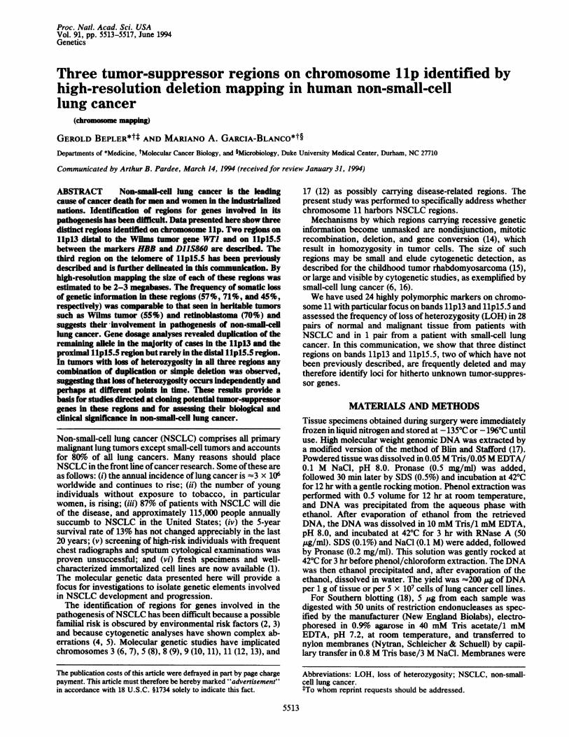
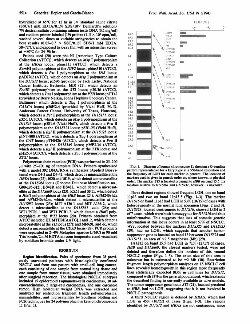
FIG. 1. Diagram of human chromosome 11 showing a G-bandingpattern representative for a karyotype at a 550-band resolution andthe frequency of LOH for each marker in percent. The location ofmarkers used is given in genetic order or, where known, in physicalorder. The marker ST5 is located proximal to HBB on band 15.5. Itslocation relative to DIIS861 and D11S932, however, is unknown.
Three distinct regions showed frequent LOH, one on band11p13 and two on band llplS.5 (Figs. 1-3). The markerDIS16 on band 11p13 had LOH in 53% (10/19) ofcases withheterozygosity in the normal lung specimen (Figs. 2 and 3).DIS323, located centromeric to DIIS16, showed LOH in 2of7 cases, which were both homozygous forDi IS16 and thusuninformative. This suggests that loss of somatic geneticinformation at this locus occurs in at least 57% of NSCLC.WTI, located between the markers DIIS325 and DIIS323(29), had no LOH, which suggests that another tumor-suppressor gene is located on band 13 between DI IS323 andDIIS151, an area of =2.5 megabases (Mb) (29).DIS12 on band 15.5 had LOH in 71% (12/17) of cases.
HBB and D1IS860, the closest markers tested, were notdeleted and therefore define the borders of this secondNSCLC region (Figs. 1-3). The exact size of this area isunknown but is estimated to be -2 Mb (30). Restrictionfragment length polymorphism analyses on 18 NSCLC celllines revealed homozygosity in this region more frequentlythan statistically expected (83% in cell lines for D1IS12,compared with 33% in the general population), which extendsthe described finding to currently available in vitro models.The tumor-suppressor gene locus ST5 (21), located proximalto HBB, had no LOH, suggesting that it is not involved inNSCLC pathogenesis.A third NSCLC region is defined by HRAS, which had
LOH in 45% (10/22) of cases (Figs. 1-3). The regionsidentified by DIIS12 and HRAS are not contiguous, since
.VICE PI PI 9 PI PI 9 PI, PI
Proc. Natl. Acad Sci. USA 91 (1994)
m

Proc. Natl. Acad. Sci. USA 91 (1994) 5515
Epidernioid (arcinomat N\denImcarcinFloiII: i(..i.I f l'c i -a 'r l, s IIak, 1 lli; t,.~Il
2 X X 2 o - XCX2Ct 2 x 2 -
009?00000*0000000OO®®0029900000
&eo&&o~eoooooeeoooooDoooo@o0000oc3oosoo®®oooocoRoooooaooooo000®o~aow®~oosoo0
CALCA ® 000@0-000000®PFH 0e0 00000i06000®DJIISI.5 OOOOQo®&OOOO)0flOD11S16 0000S 0S00@00000DI)1323 0@O*0000O00°0O000Ill' 0®0O000OO000000®Do 1S.32.5 0 0 *F O 0 Of 0 g ~O O OOC ODIIWS 0O000000000®0
DIiSI (09 0 0 0 000 0 00000®
T)YR oo006eo4e&000000oao(D3D 1h (®®00C'0*0ol A00 o
ESI (0090000 . 70
0000000000000000000OOOOOOOOOC.
OOOQOOO@OOIO®00000009§
90@G00Q@000&
0 tO 0 03 6& 0 0 e; 0\Q200000@?00C
otooo0ooo'0 09 0 t90
0 0 0 0 {a O w
E000 0®0 '09h0 0 0~0 0 * @o O 0-O5 :0ChO00 ..
E 0.1 O 0 CO O v _D
0 R(D a 6 &
OQ0 0)10 Gt-
FIG. 2. Detailed allelotyping for 24 polymorphic markers in tissue pairs from 29 patients with lung cancer. DNA from tissue pairs is labeledwith the prefix D followed by a pair of laboratory numbers given to tissue samples in consecutive order of receipt in the laboratory. e,
Heterozygosity in normal and tumor tissue; e, LOH in the tumor tissue; 0, homozygosity in normal and tumor tissue; o, no available data.
five markers, DIIS860, DIIS988, TH, INS, and IGF2, lo-cated between these loci had LOH in only one tumor (D214/15).The tumor/normal tissue pairs tested included only one
case of small-cell lung cancer (D220/21), which had no LOHin any of the tested markers on chromosome 11. The totalnumber ofNSCLC cases examined in this study is too smallto allow meaningful conclusions on the frequency and dis-tribution of the described NSCLC regions among the variouspathological subtypes and clinical-outcome groups.Gene Dosage Analysis. To assess whether LOH at the
individual loci resulted in hemizygosity or homozygosity,densitometric scanning of autoradiographic hybridizationsignals for corresponding alleles from D11S16, D11512, andHRAS was performed. An approximate doubling ofthe signalintensity for the remaining allele was observed in four ofseven interpretable cases for D11516, six of nine for D11512,and one of five for HRAS, which is consistent with geneticduplication. In all other cases LOH appeared to be the resultof simple genetic deletion.
DISCUSSIONThe analyses described provide evidence for the existence ofthree independent regions on chromosome lip with frequentLOH in NSCLC and implicate gene conversion or doublemitotic recombination and simple deletion as the mechanismsleading to genetic loss.Two previous studies have suggested the existence of
NSCLC regions on chromosome 11. Weston et a (12)studied 54 NSCLC pairs with six markers located on chro-mosome 11 (CAT, CALCA, HBB, INS, andHRAS on lip andINT2 on 11q). Results suggested frequent LOH at the HRASlocus (13 of 30 cases, 43%) which could stretch from INS to
the telomere on chromosome band 15.5. A second area ofLOH was bordered by IN72 on 11q13 and CATon 11p13, andthe frequency of LOH in this area appeared to be in the 25%range. Skinner et al. (13) studied 45 NSCLC pairs for LOHon llpl5.5 by using the markers HBB, INS, and HRAS. Nineof 40 cancers had LOH at HRAS (23%), 7 of these 9 also hadLOH at the INS locus, and 8 of these were heterozygous forHBB. Both groups ofauthors concluded that LOH forHRASis a common finding in primary NSCLC and that the spec-trum of LOH is similar in the different NSCLC subtypes.These reports favor a telomeric (INS-pter) location of aNSCLC tumor-suppressor gene, withLOH being observed in23-43% of cases, and a possible second location for atumor-suppressor gene between 11q13 and 11p13, albeit withlow-frequency LOH (2325%). We studied three markerswithin or adjacent to the latter region (CAT, D1IS149, andTYR) and found LOH in only one tumor (D188/89; see Fig.2), which accounts for 5-10%o of informative specimens. Thislow number suggests that this region is not of generalimportance for NSCLC pathogenesis. The differences inLOH frequencies among these studies are most likely causedby contamination of tumor specimens with normal diploidcells, resulting in a falsely positive heterozygosity of thetumor. Our data concur with the authors of refs. 12 and 13 inthe location of a tumor-suppressor gene in the vicinity of theHRAS marker (LOH was found in 45% of tumors). Howeverthe extent of this region (Fig. 4, HRAS locus) was unclear,and the current work shows it not to extend beyond IGF2 andDRD4. AlthoughDRD4 has been mapped telomeric to HRASby genetic linkage analysis (39), its exact physical location isunknown and may perhaps be centromeric to HRAS. Thus itremains unclear whether this LOH region includes the entiretelomere. The same region has also been implicated in thepathogenesis of breast cancer (31), rhabdomyosarcoma (15),
DRO4HJRASIGF2
DI S988DI IS860DIlS12HSBBDI I S.932D1)IS861ST5
00
*o0
t-~
0"
0OSo
occo
i-ff0
A.i
,.;-F
tr
I.
.i
4iO.;.0.
Ii
5'
@0000000
^-"I. . ..
"3 A-:~~~~~~~~~~~~~~~~~~~~~~~~~~~~~
Genetics: Bepler and Garcia-Blanco
;&ONla.,Ifr.4:.i'
,:i,. c4

5516 Genetics: Bepler and Garcia-Blanco
HRASNC
INSN C
DA -S12N C
-Al
-AA *-CoA2
: .A2 By*A - 2/Con
CALCAN C
-Al
..f}4 *& -A2
-Con
-Con
- Con
Dl 1S16N C
- A2
- A2/3
HBBN C
-Al
- Con-A2
- Con-A2
CATN C
_ O -Al
i -A2
- A2/3
-A3
* " -Con
FiG. 3. Representative Southern blots or electropherograms ofdigested PCR products of DNA from tissue pairs in Fig. 2, high-lighting the critical regions of LOH. Lanes: N, normal tissue; C, thecorresponding tumor tissue. Bands Al, A2, and A3 represent anallelic polymorphism with Al as the largest allele. Bi and B2represent a different allelic polymorphism for the same locus. Conrepresents a constant band. For HRAS, LOH for allele Al in thetumor tissue ofpair D226/27 is shown; forINS, heterozygosity in thetumor and normal tissue ofpair D228/29 is shown; for D11S12, LOHfor allele Al in the tumor tissue of pair D182/83 is shown; for HBBand CALCA, heterozygosity in the tumor and normal tissue of pairD218/19 is shown; for D1JS16, LOH for allele A2 in the tumor tissueof pair D184/85 is shown; and for CAT, heterozygosity in the tumorand normal tissue of pair D220/21 is shown.
Wilms tumor (35, 36), bladder cancer (37), and testicularcancer (38). These data are schematically summarized in Fig.4.We have found two NSCLC regions, one identified by
D11S16 on band llpi3 (Fig. 4, DJJS16) and the otheridentified by DJJS12 on band llpl5.5 (Fig. 4, DJJS12). Thefirst region, on band llpl3, has not been previously de-scribed and is clearly distal to the WTI gene (29, 40) andoutside ofthe region described by Weston et al. (12). This andthe frequency ofLOH (57%), which is comparable to the onein Wilms tumor on band llpl3 (41), strongly suggest thepresence of a tumor-suppressor gene within this region.Radice et aL (38) have described LOH in 4 of 12 testicularcancers (Fig. 4). Because many of the markers used by thoseauthors were uninformative, the minimal region of LOHcould not be determined but extended from the telomere(HRAS locus) to perhaps as far as band llql3 (INT2 locus).The second region, identified by the DJ1S12 locus, is of
particular interest because of its high frequency (71%) ofLOH, which is comparable to that found in retinoblastoma(14). Ali polymorphic markers evaluated adjacent to thislocus did not reveal LOH, which suggests that the area ofinterest is -2 Mb in size. Interestingly, data by Reeve et al.(35) and Wadey et a!. (36) suggest the presence of a secondWilms tumor locus (W12) in this region (Fig. 4). The recentdescription of loss of genetic imprinting for IGF2 in Wilmstumor (42, 43), however, suggests that loss ofgenetic controlelements adjacent to the IGF2/HI9 genes could be theimportant event within this Wilms tumorLOH domain. Therealso appears to be some overlap ofthe second NSCLC region
I)RI)4
HlkAS
16-1
I'.
1) IIs 988
D)I 15860
1)1I1IS1I-
111111'9R
1)1/ IS9 /
%IT 86
11-k-s Iin
)."'
.)! I/S I 5
fI" 1)I/s I
1.)/I.S3fVIT
K
rv T.*k_ -4
i'
F.,.>i-
0} sF' -s.F..-..
I,.
F:' :-h. ,4
[.,_.-1,.. ...
e
i
K."...K."...r....
A..,
I. 1' i ic
FIG. 4. Schematic representation of regions on chromosomellpl3-15.5 harboring potential tumor-suppressor genes. m,NSCLC regions described in this communication; c , -breast cancerregions (31-34); U, rhabdomyosarcoma regions (15, 30); C, Wilmstumor 2 regions (35, 36); A, bladder cancer region (37); Es,testicular cancer region (38).
with known areas of frequent LOH in breast cancer (32-34)and rhabdomyosarcoma (15) (Fig. 4). In addition, the cen-tromeric part of this region overlaps with a chromosomalfragment which was recently shown to encode a tumor-suppressor activity when transferred to a rhabdomyosar-coma cell line (30) (Fig. 4).
Several mechanisms leading to loss of genetic informationin two or more noncontiguous regions in NSCLC are possi-ble. Our gene dosage studies make it unlikely that the LOHevents would have occurred concurrently through a complexrearrangement of a single chromosome, since LOH at differ-ent loci in the same tumor resulted from any combination ofdeletion and duplication, suggesting that LOH at various locioccurs independently and perhaps at different points in time.This can be addressed by studies assessing the pattern ofLOH in premaliant bronchoepithelial tissue compared withcarcinoma in situ and invasive cancer. Whether LOH atdifferent loci within one tumor occurs on the same chromo-some lip cannot be determined from our studies. Studiesaddressing the parental origin of the remaining alleles couldhelp answer this question. Previous studies have demon-strated a paternal origin of the remaining alleles in rhabdo-myosarcoma (44) and Wilms tumor (45), which suggests thatLOH occurs on the same chromosome lip.
Proc. NatL Acad. Sci. USA 91 (1994)

Proc. NatL. Acad. Sci. USA 91 (1994) 5517
The data presented provide a focus for investigations toidentify and characterize tumor-suppressor genes on chromo-some lip involved in NSCLC development and progression.
This work would not have been possible without the continuedencouragement and support of Dr. Joseph C. Greenfield. Drs. DirkIglehart, Gudrun Huper, Michael Seldin, and Steven Kingsmore areacknowledged for providing specimens and methodological adviceand Drs. Russell Kaufman and John Minna for reviewing themanuscript. Funding was provided by the Department of Medicine(to G.B.) and the Duke Comprehensive Cancer Center (to M.A.G.-B.).
1. Boring, C. C., Squires, T. S. & Tong, T. (1993) Calif. CancerJ. Clin. 43, 7-26.
2. Ooi, W. L., Elston, R. C., Chen, V. W., Bailey-Wilson, J. E.& Rothschild, H. (1986) J. Nat!. Cancer Inst. 76, 217-222.
3. Sellers, T. A., Bailey-Wilson, J. E., Elston, R. C., Wilson,A. F., Elston, G. Z., Ooi, W. L. & Rothschild, H. (1990) J.Nat!. Cancer Inst. 82, 1272-1279.
4. Lukeis, R., Irving, L., Garson, M. & Hasthorpe, S. (1990)Genes Chromosomes Cancer 2, 116-124.
5. Whang-Peng, J., Knutsen, T., Gazdar, A., Steinberg, S. M.,Oie, H., Linnoila, I., Mulshine, J., Nau, M. & Minna, J. D.(1991) Genes Chromosomes Cancer 3, 168-188.
6. Brauch, H., Johnson, B., Hovis, J., Yano, T., Gazdar, A.,Pettengill, 0. S., Graziano, S., Sorenson, G. D., Poiesz, B. J.,Minna, J., Linehan, M. & Zbar, B. (1987) N. Engl. J. Med. 317,1109-1113.
7. Yokoyama, S., Yamakawa, K., Tsuchiya, E., Murata, M.,Sakiyama, S. & Nakamura, Y. (1992) CancerRes. 52, 873-877.
8. D'Amico, D., Carbone, D. P., Johnson, B. E., Meltzer, S. J. &Minna, J. D. (1992) Cancer Res. 52, 1996-1999.
9. Emi, M., Fujiwara, Y., Nakajima, T., Tsuchiya, E., Tsuda, H.,Hirohashi, S., Maeda, Y., Tsuruta, K., Miyaki, M. & Naka-mura, Y. (1992) Cancer Res. 52, 5368-5372.
10. Center, R., Lukeis, R., Dietzsch, E., Gillespie, M. & Garson,0. M. (1993) Genes Chromosomes Cancer 7, 47-53.
11. Olopade, 0. I., Buchhagen, D. L., Malik, K., Sherman, J.,Nobori, T., Bader, S., Nau, M. M., Gazdar, A. F., Minna,J. D. & Diaz, 0. (1993) Cancer Res. 53, 2410-2415.
12. Weston, A., Willey, J. C., Modali, R., Sugimura, H., McDow-ell, E. M., Resau, J., Light, B., Haugen, A., Mann, D. L.,Trump, B. F. & Harris, C. C. (1989) Proc. Natl. Acad. Sci.USA 86, 5099-5103.
13. Skinner, M. A., Vollmer, R., Huper, G., Abbott, P. & Iglehart,J. D. (1990) Cancer Res. 50, 2303-2306.
14. Cavenee, W. K., Dryja, T. P., Phillips, R. A., Benedict,W. F., Godbout, R., Gallie, B. L., Murphree, A. L., Strong,L. C. & White, R. L. (1983) Nature (London) 305, 779-784.
15. Scrable, H. J., Witte, D. P., Lampkin, B. C. & Cavenee,W. K. (1987) Nature (London) 329, 645-647.
16. Naylor, S. L., Johnson, B. E., Minna, J. D. & Sakaguchi,A. Y. (1987) Nature (London) 329, 451-454.
17. Blin, N. & Stafford, D. W. (1976) Nucleic Acids Res. 3,2303-2308.
18. Southern, E. M. (1975) J. Mol. Biol. 98, 503-517.19. Feinberg, A. P. & Vogelstein, B. (1984) Anal. Biochem. 137,
266-267.20. Williamson, R., Bowcock, A., Kidd, K., Pearson, P.,
Schmidtke, J., Ceverha, P., Chipperfield, M., Cooper, D. N.,
Coutelle, C., Hewitt, J., Klinger, K., Langley, K., Beckmann,J., Tolley, M. & Maidak, B. (1991) Cytogenet. Cell Genet. 58,1190-1832.
21. Lichy, J. H., Modi, W. S., Seuanez, H. N. & Howley, P. M.(1992) Cell Growth Difer. 3, 541-548.
22. Lichter, J. B., Barr, C. L., Kennedy, J. L., Van Tol,H. H. M., Kidd, K. K. & Livak, K. J. (1993) Hum. Mol.Genet. 2, 767-773.
23. McNoe, L. A., Eccles, M. R. & Reeve, A. E. (1992) NucleicAcids Res. 20, 1161.
24. Semenza, G. L., Dowling, C. E. & Kazazian, H. H. (1989)Nucleic Acids Res. 17, 2376.
25. Weissenbach, J., Gyapay, G., Dib, C., Vignal, A., Morissette,J., Millasseau, P., Vaysseix, G. & Lathrop, M. (1992) Nature(London) 359, 794-801.
26. Hudson, T. J., Engelstein, M., Lee, M. K., Ho, E. C., Ruben-field, M. J., Adams, C. P., Housman, D. E. & Dracopoli,N. C. (1992) Genomics 13, 622-629.
27. Edwards, A., Civitello, A., Hammond, H. A. & Caskey, C. T.(1991) Am. J. Hum. Genet. 49, 746-756.
28. Weber, J. L., Kwitek, A. E.&May, P. E. (1990) NucleicAcidsRes. 18, 4036.
29. Huff, V., Compton, D. A., Strong, L. C. & Saunders, G. F.(1990) Hum. Genet. 84, 253-257.
30. Koi, M., Johnson, L. A., Kalikin, L. M., Little, P. F. R.,Nakamura, Y. & Feinberg, A. P. (1993) Science 260, 361-364.
31. Theillet, C., Lidereau, R., Escot, C., Hutzell, P., Brunet, M.,Gest, J., Schlom, J. & Callahan, R. (1986) Cancer Res. 46,4776-4781.
32. Ali, I. U., Lidereau, R., Theillet, C. & Callahan, R. (1987)Science 238, 185-188.
33. Miyagi, M., Inazawa, J., Takita, K.-I. & Nakamura, Y. (1992)Hum. Mol. Genet. 9, 705-708.
34. Winqvist, R., Mannermaa, A., Alavaikko, M., Blanco, G.,Taskinen, P. J., Kiviniemi, H., Newsham, I. & Cavenee, W.(1993) Cancer Res. 53, 44488.
35. Reeve, A. E., Sih, S. A., Raizis, A. M. & Feinberg, A. P.(1989) Mol. Cell. Biol. 9, 1799-1803.
36. Wadey, R. B., Pal, N., Buckle, B., Yeomans, E., Pritchard, J.& Cowell, J. K. (1990) Oncogene 5, 901-07.
37. Fearon, E. R., Feinberg, A. P., Hamilton, S. H. & Vogelstein,B. (1985) Nature (London) 318, 377-380.
38. Radice, P., Pierotti, M. A., Lacerenza, S., Mondini, P., Ra-dice, M. T., Pilotti, S. & Della Porta, G. (1989) Cytogenet. CellGenet. 52, 72-76.
39. Junien, C. & van Heyningen, V. (1991) Cytogenet. Cell Genet.58, 459-554.
40. Call, K. M., Glaser, T., Ito, C. Y., Buckler, A. J., Pelletier, J.,Haber, D. A., Rose, E. A., Krai, A., Yeger, H., Lewis, W. H.,Jones, C. & Housman, D. E. (1990) Cell 60, 509-520.
41. Fearon, E. R., Vogelstein, B. & Feinberg, A. P. (1984) Nature(London) 309, 176-178.
42. Rainier, S., Johnson, L. A., Dobry, C. J., Ping, A. J., Gmndy,P. E. & Feinberg, A. P. (1993) Nature (London) 362, 747-749.
43. Ogawa, O., Eccles, M. R., Szeto, J., McNoe, L. A., Yun, K.,Maw, M. A., Smith, P. J. & Reeve, A. E. (1993) Nature(London) 362, 749-751.
44. Scrable, H., Cavenee, W., Ghavimi, F., Lovell, M., Morgan,K. & Sapienza, C. (1989) Proc. Nat!. Acad. Sci. USA 86,7480-7484.
45. Williams, J. C., Brown, K. W., Mott, M. G. & Maitland, W. J.(1989) Lancet 1, 283-284.
Genetics: Bepler and Garcia-Blanco