Calcium pump activity of the renal plasma membrane and renal microsomes
14
Biochimica et Biophysica Acta, 345 (1974) 405-418 ~tz2 Elsevier Scientific Publishing Company, Amsterdam - Printed in The Netherlands BBA 76626 CALCIUM PUMP ACTIVITY OF THE RENAL PLASMA MEMBRANE AND RENAL MICROSOMES LEON MOORE, DAVID F. FITZPATRICK*, TERESA S. CHEN** and ERWIN J. LANDON Department of Pharmacology, Vanderbilt Unicersity School of ,~Iedicine, NashL,ille, Tenn. 37232 (U.S.A.) (Received December 10th, 1973) SUMMARY ATP-dependent Ca 2 + uptake distinct from that of the mitochondria is found in both plasma membrane and microsomal membranes of rat kidney. Activity at- tributed to these fractions is enhanced by ammonium oxalate and is apparently in- sensitive to NAN3. In contrast, rat kidney mitochondrial Ca / + uptake is blocked by NaN a. The pH of optimal activity is significantly higher for the mitochondrial frac- tion. Microsomal membrane Ca z + uptake differs from that of the plasma membrane. Microsomal membranes are four times as active as the plasma membrane at high (5 mM) ATP levels. Apparent Kn, values for MgZ+-ATP differ in the two prepara- tions with a higher affinity for MgZ+-ATP found in the plasma membrane. Ca 2+ uptake activity of the plasma membrane preparation is readily inhibited by Na +. Sucrose gradient density fractionation indicates that the observed microsomal mem- brane Ca z+ pump activity is associated with membrane vesicles derived frcm the endoplasmic reticulum. Ca 2 + pump activity of both plasma membrane and micro- somal fraction is depressed in the adrenalectomized rat. This activity is not restored by a single natriuretic dose of aldosterone. INTRODUCTION ATP-dependent Ca 2 + sequestration has been extensively characterized in mi- crosomal membrane vesicles derived from skeletal muscle sarcoplasmic reticulum [1-3 ]. Lower levels of this activity have been found in microsomal membranes isolated from mammalian smooth muscle [4, 5], brain [6-8], salivary gland [9] and platelets [10]. In addition this activity is found in ghost membranes of erythrocytes [11, 12] and in skeletal muscle plasma membrane [13, 14]. A plasma membrane Ca 2+- * Present address: Department of Pharmacology, University of South Florida, Tampa, Fla., U.S.A. ** Present address: Department of Pharmacology, University of Louisville School of Medicine Louisville, Ky., U.S.A. Abbreviation: SITS, 4-acetamido-4'-isothiocyanostilbene-2,2"-disulphonic acid.
-
Upload
leon-moore -
Category
Documents
-
view
214 -
download
1
Transcript of Calcium pump activity of the renal plasma membrane and renal microsomes

Biochimica et Biophysica Acta, 345 (1974) 405-418 ~tz2 Elsevier Scientific Publishing Company, Amsterdam - Printed in The Netherlands
BBA 76626
CALCIUM PUMP ACTIVITY OF THE RENAL PLASMA MEMBRANE AND
RENAL MICROSOMES
LEON MOORE, DAVID F. FITZPATRICK*, TERESA S. CHEN** and ERWIN J. LANDON
Department o f Pharmacology, Vanderbilt Unicersity School o f ,~Iedicine, NashL, ille, Tenn. 37232 (U.S.A.)
(Received December 10th, 1973)
SUMMARY
ATP-dependent Ca 2 + uptake distinct from that of the mitochondria is found in both plasma membrane and microsomal membranes of rat kidney. Activity at- tributed to these fractions is enhanced by ammonium oxalate and is apparently in- sensitive to NAN3. In contrast, rat kidney mitochondrial Ca / + uptake is blocked by NaN a. The pH of optimal activity is significantly higher for the mitochondrial frac- tion. Microsomal membrane Ca z + uptake differs from that of the plasma membrane. Microsomal membranes are four times as active as the plasma membrane at high (5 mM) ATP levels. Apparent Kn, values for MgZ+-ATP differ in the two prepara- tions with a higher affinity for MgZ+-ATP found in the plasma membrane. Ca 2+ uptake activity of the plasma membrane preparation is readily inhibited by Na +. Sucrose gradient density fractionation indicates that the observed microsomal mem- brane Ca z+ pump activity is associated with membrane vesicles derived frcm the endoplasmic reticulum. Ca 2 + pump activity of both plasma membrane and micro- somal fraction is depressed in the adrenalectomized rat. This activity is not restored by a single natriuretic dose of aldosterone.
INTRODUCTION
ATP-dependent Ca 2 + sequestration has been extensively characterized in mi- crosomal membrane vesicles derived from skeletal muscle sarcoplasmic reticulum [1-3 ]. Lower levels of this activity have been found in microsomal membranes isolated from mammalian smooth muscle [4, 5], brain [6-8], salivary gland [9] and platelets [10]. In addition this activity is found in ghost membranes of erythrocytes [11, 12] and in skeletal muscle plasma membrane [13, 14]. A plasma membrane Ca 2+-
* Present address: Department of Pharmacology, University of South Florida, Tampa, Fla., U.S.A.
** Present address: Department of Pharmacology, University of Louisville School of Medicine Louisville, Ky., U.S.A.
Abbreviation: SITS, 4-acetamido-4'-isothiocyanostilbene-2,2"-disulphonic acid.

406
pumping mechanism is postulated to play a role in maintaining the electrochemical gradient for calcium between the external medium and cell interior [15].
Ca 2 + has been shown to play a role in modifying permeability of renal tubule cells [16]. Palmer and Posey [17] have reported evidence for ATP-dependent Ca 2+ binding in microsomal membranes prepared from rabbit kidney cortex [17]. We have recently isolated from rat kidney a subcellular fraction consisting of plasma mem- brane vesicles [18]. In the present study the existence of an ATP-dependent Ca 2+- pump activity in this plasma membrane preparation is demonstrated. This activity is also demonstrated in microsomal membrane vesicles isolated from the rat kidney. The Mg 2 +-ATP-dependent Ca z+ uptake of the renal plasma membrane and micro- somal membranes is characterized and compared with Mg 2+-ATP-dependent Ca 2 + sequestering activity of renal mitochondria.
MATERIALS AND METHODS
The animals employed in this study were male rats of the Sprague-Dawley strain weighing approximately 250 g. Nucleotides employed in this study were ob- tained from Sigma chemical company 45CAC12 (8.0 mCi/mg calcium) was obtained from New England Nuclear Corp.
Rat kidneys were homogenized in an isotonic sucrose medium employing a Potter homogenizer with a Teflon pestle. Preparation of the subcellular fractions was carried out at 0-4 °C. For purposes of the present study EDTA was omitted from the sucrose media employed in homogenization and isolation of subcellular fractions. The plasma membrane fraction was prepared by a procedure described by Fitz- patrick et al. [18]. A "light" microsomal fraction was prepared according to the procedure of Landon and Norris [19]. Kidney mitochondria were isolated as pre- viously described [20]. A succinate dehydrogenase assay according to the procedure of Pennington [21] was employed to monitor for mitochondrial contamination of the plasma membrane vesicles.
Calcium uptake was measured in the following medium: 100 mM KCI, 30 mM imidazole-histidine buffer, 5 mM ammonium oxalate, 5 mM NAN3, 5 mM MgC1 z, 5 mM ATP (pH adjusted with Tris), 20/aM CaCl 2 and 0.07 #Ci/ml 45CaClz in a total volume of 3 ml. Plasma membranes were assayed at pH 6.8, microsomes at pH 6.6 and mitochondria at pH 7.4. These pH values were chosen for optimal activity. The assay was initiated at 37 °C by the addition of 300/al of the subcellular fraction. The final protein concentrations were 0.2-0.4 mg/ml for the plasma membrane and microsomal fractions and 0.075-0.1 mg/ml for the mitochondrial fraction. Aliquots of 500/al were removed for filtration through 0.45/am membrane filters (Millipore Corporation) at 2, 10, 20 and 30 rain. The filters were prepared with a wash of 0.25 M KCI (2 ml) followed by water (10 ml). Samples were filtered with the aid of a vacuum apparatus and were washed with 0.25 M sucrose (2 ml). The filtration in general followed the procedure described by Palmer and Posey [17]. The filters were dried and 4SCa determined by liquid scintillation spectrophotometry in 2,5-diphenyloxa- zole (6 g/l) in toluene.
(Mg 2++Cai+)-stimulated ATPase activity of renal plasma membrane and renal microsomes was assayed with an incubation medium similar to that employed for measuring ATP dependent calcium uptake activity. However, no radioactive

407
calcium was necessary and the azide and oxalate were omitted. Calcium levels were varied from 0 to 100 ~M. The reaction was terminated with 5 ~o trichloroacetic acid and the liberated Pi was measured as previously described [19].
Protein content of the fractions was determined by the procedure of Suther- land et al. [22]. Nucleotides were identified on cellulose thin-layer chromatographic plates (Eastman Organic Chemicals) developed in n -bu tano l -ace tone- (NH4)OH- acetic acid-water (3.5 : 3.5 : 1.5 : 1.5 : 1, by vol.) according to the procedure de- scribed by Randerath [23]. The nucleotides were identified after incubation with the membrane fractions by spotting 5 t~1 of the 30-min incubation sample along with standard solution in the chromatographic system described above.
Density gradient studies were carried out in a continuous 20-50 percent su- crose gradient (w/v) containing 3 mM imidazole-histidine, pH 6.6. Approximately 2 mg of microsomal membrane protein were layered onto a 5 ml gradient that was then centrifuged at 130 000 ×g for 16 h at 0 °C in a Beckman SW50L rotor. Gradient fractions were obtained by puncturing the bot tom of the tube and collecting 0.7-ml samples. The following enzyme assays were carried out on the sucrose gradient frac- tions: (Na+ -- K+ )-dependent ATPase [19] as a marker for plasma membrane and N A D H oxidase [24] as a marker for endoplasmic reticulum.
RESULTS
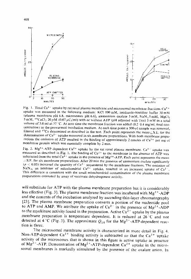
In Fig. l, an MgZ+-ATP dependent Ca z+ uptake by the plasma membrane preparation is demonstrated. In the absence of ATP approximately 2 nmoles Ca 2+ per mg protein is bound to the plasma membrane fraction within the first 2 rain of incubation. In the presence of 5 mM MgZ+-ATP a continuous C a 2+ uptake is seen over a 30-rain period. Microsomal activity is shown in the same figure and is seen to be about four times more active than that found in the plasma membrane.
Plasma membrane activity is characterized in more detail in Fig. 2. In this figure, non-ATP-dependent Ca z+ binding is subtracted. The Ca z+ uptake activity of the plasma membrane shown represents active uptake in presence of MgZ+-ATP. In the absence of oxalate the Ca/+ uptake levels off between 10 and 20 rain. In the presence of oxalate the uptake continues over the entire 30-rain period. Stimulation o f Ca 2 + uptake by oxalate has previously been interpreted as evidence for transloca- tion of calcium across the vesicle membrane in contrast to binding to external mem- brane sites [1]. Complexing of the oxalate anion with C a 2+ accumulated by the ves- icles is thought to reduce the passive outflow so that energy dependent calcium accu- mulation is measured. It is possible to conceive the observed uptake of C a 2 + as a Ca 2 + exchange between membrane and medium enhanced by the presence of Mg 2 +- ATP. However, enhanced C a 2 + uptake activity observed in presence of oxalate makes this very unlikely.
When azide is omitted f rom the plasma membrane incubation there is some increase of Ca z + uptake activity. This increase in approximately that to be expected from a two percent mitochondrial content of the plasma membrane preparation and is close to the three percent estimate of mitochondrial contamination based upon the succinate dehydrogenase assay.
Nucleotide triphosphates other than ATP do not support Ca z + uptake (Fig. 3). Mg 2+ is required for the demonstration of an ATP-dependent Ca 2 + uptake. ADP
408
50
,_c
2~
d o
I5
PLASMA MEMBRANE ~ 5
c
8 8 I~ B
I0 20 30 MINUTES
AZIDE OMITTED
C O NT RO~-L
OX AL AT~E
,; 2'0 3'o MINUTES
Fig. 1. Total Ca 2+ uptake by rat renal plasma membrane and microsomal membrane fractions. Ca z + uptake was measured in the following medium: KC1 100 mM, imidazole-histidine buffer 30 mM (plasma membrane pH 6.8, microsomes pH 6.6), ammonium oxalate 5 mM, NAN35 raM, MgCI2 5 raM, 45CAC12 20 pM (0.07/iC1/m[) with or without ATP (pH adjusted with Tris) 5 mM in a total volume of 3.0 ml at 37 ~C. At zero time the membrane fraction was added (0.2 0.4 mg/ml, final con- centration) to the prewarmed incubation medium. At each time point a 500-/4 sample was removed, filtered and 45Ca determined as described in the text. Each point represents the mean±S.E, for the determination of Ca 2 + uptake measured in six membrane preparations. With both membrane prepa- rations the omission of ATP resulted in the binding of approximately 2 nmoles of Ca z+ per mg of
membrane protein which was essentially complete by 2 min.
Fig. 2. Mg 2+-ATP dependent Ca 2 + uptake by the rat renal plasma membrane. Ca 2. uptake was measured as described in Fig. 1. The binding of Ca 2 + to the membrane in the absence of ATP was subtracted from the total Ca 2 + uptake in the presence of Mg 2 +-ATP. Each point represents the mean =LS.E. for six membrane preparations. After 20 min the presence of ammonium oxalate significantly (p < 0.05) increased the quantity of Ca z+ sequestered by the membrane fractions. The omission of NAN3, an inhibitor of mitocbondrial Ca 2+ uptake, resulted in an increased uptake of Ca 2+. This difference is consistent with the small mitochondrial contamination of the plasma membrane preparations estimated by assay of succinate dehydrogenase activity.
wil l s u b s t i t u t e f o r A T P w i t h t he p l a s m a m e m b r a n e p r e p a r a t i o n b u t it is c o n s i d e r a b l y
less e f fec t ive (Fig . 3). T h e p l a s m a m e m b r a n e f r a c t i o n was i n c u b a t e d w i t h M g 2 + - A D P
a n d t h e c o n t e n t s o f t h e i n c u b a t i o n a n a l y z e d b y a s c e n d i n g t h i n - l a y e r c h r o m a t o g r a p h y
[23]. T h e p l a s m a m e m b r a n e p r e p a r a t i o n c o n v e r t s a p o r t i o n o f t he n u c l e o t i d e p o o l to A T P a n d A M P . W e a t t r i b u t e t h e u p t a k e o f C a 2+ in t he p r e s e n c e o f M g 2 + - A D P
to t h e m y o k i n a s e ac t iv i ty f o u n d in t he p r e p a r a t i o n . A c t i v e C a 2 + u p t a k e b y t he p l a s m a
m e m b r a n e p r e p a r a t i o n is t e m p e r a t u r e d e p e n d e n t . I t is r e d u c e d a t 26 °C a n d n o t
d e t e c t e d a t 4 °C (Fig . 3). T h e a p p r o x i m a t e Q10 fo r t h e M g Z + - A T P - d e p e n d e n t r eac - t i o n is t h r ee .
T h e m i c r o s o m a l m e m b r a n e ac t i v i t y is c h a r a c t e r i z e d in m o r e de ta i l in Fig. 4. N o n - A T P - d e p e n d e n t C a 2+ b i n d i n g ac t i v i t y is s u b t r a c t e d so t h a t t he Ca 2+ u p t a k e
ac t i v i t y o f t h e m i c r o s o m e s t h a t is s h o w n in t h i s f igure is ac t ive u p t a k e in p r e s e n c e o f M g 2 + - A T P . D e m o n s t r a t i o n o f M g 2 + - A T P - d e p e n d e n t C a 2+ u p t a k e in t he m i c r o -
s o m a l m e m b r a n e s is m a r k e d l y s t i m u l a t e d b y t he p r e s e n c e o f t he o x a l a t e a n i o n . In

409
E
\
5 2 o
tS !
c
I
!
10
5
E C,
~4
3 7 °
2o i
C
,,I.-
0
E
£..)
CO LI_I -_1
o C
40
30
20
5 0 - CONTROL
/ / ~ OM ITTED
I I I
0 I0 20 30 MINUTES
Fig. 3. Characteristics of total Ca 2+ uptake by the rat renal plasma membrane preparation, lncu- bation medium for the control experiment is described in legend of Fig. 1. After 20 rain of incubation at 37 :C a 500-/d sample was removed and 4SCa was determined as described in the text. The substi- tution of other nucleotides for Mg 2+-ATP or the alteration of temper ature of incubation is indicated in the chart. Results are mean--S.E, for six membrane preparations.
Fig. 4. Mg2+-ATP de!cendent Ca 2+ uptake by the rat renal microsomal membrane preparation. Ca 2 + uptake was measured as described in Fig. 1. The binding of Ca 2 + to the membrane in the absence of ATP was subtracted from the total Ca 2 + uptake in the presence of Mg 2 +-ATP. Each point repre- sents the mean--S.E, for six membrane preparations. At 10 rain the presence of ammonium oxalate significantly increases quantities of Ca 2 + sequestered by the membrane fraction.
contrast to findings with the plasma membrane , the omission of azide does no t en- hance the Ca 2 ÷ uptake. Other nucleotide tr iphosphates do not suppor t active C a 2 +
uptake (Fig. 5) and Mg z+ is required for the A T P dependent uptake. Limited activity is seen with Mg 2 +-ADP and is a t t r ibuted to the presence of some myokinase activity in the preparat ion. As in the plasma membrane prepara t ion the C a 2+ uptake is markedly temperature dependent (Fig. 5). The approximate Q1 o for the microsomal membrane Mg 2+-ATP dependent Ca 2+ uptake is four.
Mg 2 +-ATP dependent Ca 2 + uptake activity versus the concent ra t ion of Mg 2 +- ATP and Ca 2+ is plotted for both microsomes (Fig. 6) and plasma membrane (Fig. 7). The calculated V is more than four times greater for the microsomal membranes . The calculated Km for Mg2+-ATP is 0.55 m M with the plasma membrane prepara- t ion and 3.12 with the renal microsomes. This apparent Km for Mg z +-ATP is signif- icantly lower for the plasma membrane preparat ion. Estimates of the apparent Km f o r C a 2+ gives values close to 20/~M in both preparat ions. The apparent Km for

410
. 50 -
3 7 °
c
E o
_= ¢,
,3
d o :s:
4 0 -
3 0 -
2 0 -
I 0 -
2 6 °
_ ~ a - ) u o
a_ a~ a_
o o e ~ . . . . .
Fig. 5. Characteristics o f total Ca 2+ uptake by the rat microsomal membrane preparation. Incuba- tion med ium for the control experiment is described in legend o f Fig. 1. After 20 rain o f incubation at 37 ~C a 500-pl sample was removed and ~ C a was determined as described in the text. The substi- tution o f other nucleotides for Mg 2 +-ATP or the alteration o f temperature o f incubation is indicated in the chart. Results are mean ~ S . E . for six membrane preparations.
c
E _0
c
"t o_
c
5 0 - A 2~
V Km
4 0
2C
I0
54 6 +_ I0 2 nMOLES Ca/rag protein/lOmin 2 3 5 ± 4 25.uM Ca
I00 25
~ M Ca
B V 2 8 7 -* 4 2 5 nMOLES Ca/mg protein/5 min Km 3 f2 +- 0 .298mM ATP
E e 2c
~5
I I I Jo 25 5 0 7 5 I
m M M g - A T P
Fig. 6. Mg 2+-ATP-dependent Ca 2 + uptake by the renal microsomal membrane preparation varying calcium and Mg 2 +-ATP concentrations. Incubat ion med iu m essentially that described in legend o f Fig. l , After 10 rain in (A) and 5 min in (B) a 500-ffl sample was removed, filtered and ~SCa deter- mined as described in the text. In (A) the calculated V for Ca 2 + uptake is 54.6-- 10 nmoles Ca 2 + per mg protein in 10 rain. The m a x i m u m observed velocity was 28.7 nmoles Ca 2+ per mg protein in 10 min. The apparent Km for Ca 2+ is 23 .5±4 .25 pM. Each point in A represents mean-Z-S.E, o f five membrane preparations. In (B) the calculated V for Mg2+-ATP is 28 .7~2 .15 nmoles C a z + per mg protein in 5 rain. The apparent Km for Mg2+-ATP is 3.1 ~ 0 . 3 raM. Each point in B represents the m e a n ~ S . E , for eight experiments.

411
c c
t ~ ~ F g ~ c c
C rn r n / IOmin ~) I Km 0550 -~ 0 0 7 9 mM ATD V I t t ~ I V 6,15 ± 0 5Q5 nMOLES Co/mg protein/SmiP K ± OI OLES O / g p o t e i
• ~o 25 50 75 I00 25 5 0 75 I0
~uM Co m M M g - A T P
Fig. 7. MgZ+-ATP dependent Ca =+ uptake by renal plasma membrane preparations varying Ca 2+ and Mg2+-ATP concentrations. The incubation medium is essentially that described in legend of Fig. 1. After 10 rain in (A) and 5 min in (B) a 500-pl sample was removed, filtered and 4SCa deter- mined as described in the text. In (A) the calculated V for Ca 2 + uptake is 11.8 ± 1 nmoles Ca 2 + per mg protein in 10 min. The apparent Km for calcium is 19.4:[_0.4/~M. Each point represents the mean 4- S.E. of six experiments. In (B) the velocity of calcium uptake increased with increasing Mg 2+-ATP concentrations to 2.5 raM. At higher Mg2+-ATP concentrations the Ca 2+ uptake rapidly decreased. The apparent Km for Mg2+-ATP is 0.554-0.08 mM which is significantly lower than that obtained for the microsomal membrane preparation.
Ca 2+ in renal mitochondria is found in a similar set of measurements to be 126:k 5 pM. Although in Fig. 1 with 5 mM Mg2+-ATP the Ca 2+ pump activity of micro- somes is four times greater than that of the plasma membrane, this difference is much reduced at lower and perhaps more physiological levels of ATP.
An ATPase activity dependent on Mg 2+ and stimulated by Ca 2 + is associated with skeletal muscle microsomes and is large enough to potentially relate to the Ca 2 +- sequestration system [25]. Ca 2+-stimulated Mg 2+-ATPase activity was measured in renal microsomes and plasma membrane with addition of 5, 20, 50 and 100 pM Ca 2 +.
Maximal ATPase was observed at 20/~M Ca 2 + with renal plasma membrane and 100/~M Ca 2+ with renal microsomes. The extra ATPase for 20 min averaged 600 nmoles phosphate per mg protein for the microsomes and 700 nmoles phosphate per mg protein for the plasma membrane.
The effect of replacing KCI in the medium with NaC1 or sucrose is demon- strated in Fig. 8. Substitution by Na + virtually suppresses the Mg 2 +-ATP dependent Ca 2+ uptake of the plasma membrane preparation. Sucrose in iso-osmotic concen- trations partially depresses the Mg 2+-ATP dependent uptake of the plasma membrane preparations. Sodium and sucrose have no demonstrable effect on Ca 2+ uptake in the microsomal membrane preparation. When 1 mM EDTA is included in the su- crose medium employed in the initial homogenization of the renal tissue, the Mg 2 +- A T P - d e p e n d e n t Ca 2+ uptake activity of the microsomal and plasma membrane preparations is diminished about 40°/ . For this reason no EDTA was utilized in preparat ion of cell fractions.
Ca 2+ uptake activity of kidney mitochondria has been previously studied [26, 27] but less commonly with the low levels of Ca 2+ employed in the present

412
PLASMA MEMBRANE
5 0
E I0 k~ 4C E g N
E 2 :5O 2 c~
5 z o
o
0
z ~
MICROSOMES
't
120 [- AZIDE J OMITTEb
z
E CU I I AZlUC AND \ ~) j / U X/', LATE
; ~°A 2~ <
2 G
¢ u N T R O L
20 5~ ,i ' : , , I L :
Fig. 8. The effect o f the ionic compos i t ion o f the assay medium on Mg2+-ATP-dependent Ca 2+ uptake by the p lasma membrane and microsomal membrane preparations. Incubation medium for the control experiment (KCI) is described in legend o f Fig. 1. After 20 min a 500-/d sample was withdrawn, filtered and aSCa determined as described in the text. Replacement o f KCI 100 m M by NaCI 100 m M , sucrose 200 m M or by NaCI 50 m M and KC1 50 m M are indicated on the chart. Results are m e a n - S.E. for six preparations.
Fig. 9. Mg2+-ATP dependent Ca 2+ uptake by rat renal mitochondria. Ca z+ uptake was measured in the fo l lowing medium: KCI 100 mM, imidazole-bist idine buffer (pH 7.4) 30 mM, a m m o n i u m oxalate 5 m M , N a N 3 5 mM, MgCl2 5 mM, ATP 5 m M , 45CAC12 20f fM (0.07ffCi/ml) in a total vo lume o f 3 ml at 37 °C. At zero time the mitochondria l suspension was added (0.075-0.1 mg/ml , final concentrat ion) to the prewarmed incubation medium. At each time point a 500-ffl sample was withdrawn, filtered and 45Ca determined as described in the text. Binding o f Ca 2 + to the mitochon- drial preparation in the absence o f ATP was substracted from the total Ca 2+ uptake in the presence o f 5 m M Mg 2 + - A T E Each point represents the mean ± S.E. for Ca 2 + uptake in six mitochondrial prepa- rations. There is near total inhibition o f active calcium uptake by 5 m M NAN3. When oxalate is omitted the loss o f Ca z + after 10 min may represent a substantive increase in Ca 2 + loss when oxalate is not available to remove free calcium from solution within the mitochondria.
experiments. Mg 2 +-ATP-dependent Ca 2 + uptake activity o f kidney mitochondria is shown in Fig. 9. Mitochondrial Ca 2+ uptake is completely inhibited by NaNa, but in the absence of azide a potent Ca 2 +-sequestration activity is demonstrated. Little or no oxalate effect is detected during the initial 10min of mitochondrial Ca 2+ uptake and there is no significant change when Na + replaces K +. MgZ+-ADP will not support Ca 2 + uptake unless substrate and inorganic phosphate essential for mito- chondrial systhesis o f ATP are also present (Fig. 10).
Mg 2+-ATP dependent Ca 2+ uptake in all three fractions was examined be- tween pH 6.6 and 7.4. Maximum Ca 2+ uptake by mitochondria is seen at pH 7.4. Maximum microsomal calcium uptake is seen at pH 6.6 and plasma membrane calcium uptake is optimal at pH 6.8 (Fig. l l ) .
The microsomal preparation is heterogeneous in its membrane composition. It consists of plasma membrane fragments and endoplasmic reticulum membrane fragments. Centrifugation of the microsomal fraction of a sucrose density gradient was carried out to partially separate plasma membrane from endoplasmic reticulum

413
¢,:
E O o,I
E
b_ o E
8 O9 W J 0 : S -
o
12U
1 0 0
8 0
6 0
4 0
2 0
0,_ i i ,
¢b
& S
~ o
c
E
E
o o.
F g
.J o
/ 5 - M ! T © C H O N D R I A
2
0 J ~ L I J I J ,
0 5 F P L A S M A M E M B R A N E
6 6 6 8 7 0 7 2 7 4
~ H
Fig. 10. Characteristics of total Ca 2+ uptake by rat renal mitochondria, Incubation medium for these experiments omits azide and is described in the legend of Fig. 9. After 20 min of incubation at 37 C a 500-pl sample was removed and 45Ca was determined as described in the text. The effects of omitting Mg2+-ADP or substituting other nucleotides are indicated on the chart. Each point repre- sents the mean of three mitochondrial preparations.
Fig. 1 I. The effect of pH on Mg2+-ATP stimulated Ca 2+ uptake in rat renal mitochondria, plasma membranes and microsomal membranes. Determination of calcium uptake is described in the text with pH of the imidazole-histidine buffer noted in the figure. A sample was removed from the plasma membrane and microsomal membrane incubations at 10 rain. The mitochondrial suspension was incubated for two minutes. Each point represents the meanAS.E, for six preparations of each mem- brane fraction.
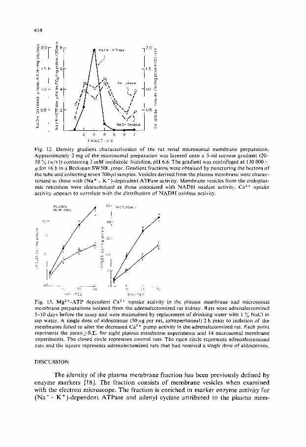
membranes. Representative findings are depicted in Fig. 12. N A D H oxidase is em- ployed as a markei for endoplasmic reticulum and ( N a + + K + ) - d e p e n d e n t ATPase for the plasma membrane. Ca 2 + uptake appears to be associated with the endoplas- mic reticulum marker enzyme in this preparation. Plasma membrane represents a minor component o f this microsomal preparation and the weaker plasma membrane Ca 2 + uptake activity would represent a very small part of the total activity observed in the microsomal preparation.
MgZ+-ATP dependent Ca 2+ pump activity has been examined in the plasma membrane preparation of rats adrenalectomized for 5-10 days. This activity is de- creased by approximately 50~o (Fig. 13). A single dose o f aldosterone (50 pg per rat, 2 h before sacrifice o f the animals) sufficient to restore N a + balance as measured by urinary electrolyte excretion [30] failed to increase Ca 2+ pump activity in the test animals (Fig. 13). Findings were similar when the microsomal membranes were examined (Fig. 13).
414
~ 2.0- !8 I £ _
:~ E ~ a o ~
A } / K - ATPase
tq B l ~% Ca uptake '
" / / ,' i fP o ~ t ~ / \ . ,d ,~
\ o ' } / J I % NA? Oli:ose
I 2 3 4 5 6 7
F R A C T I O N
2.0 E E 0 =_
1.5
LO ~
E c 0.5
3
Fig. 12. Densi ty gradient characterisation of the rat renal microsomal membrane preparation. Approximate ly 2 mg of the microsomal preparation was layered onto a 5-ml sucrose gradient (20- 50 % (w/v)) containing 3 m M imidazole-hist idine, p H 6.6. The gradient was centrifuged at 130 000 i g for 16 h in a Beckman SW50L rotor. Gradient fractions were obtained by puncturing the bottom o f the tube and collecting seven 700-ffl samples. Vesicles derived from the plasma membrane were charac- terized as those with (Na + - -K+) -dependent ATPase activity. Membrane vesicles from the endoplas- mic reticulum were characterized as those associated with N A D H oxidase activity. Ca z+ uptake activity appears to correlate with the distribution of NADI- [ oxidase activity.
0 [ _ _ _ 1 I
< 20 50 MIrlJTE3
50 I MIC ROSOM".%
40 L
u m 2O ul
o
I0
0 - - 1 1 ~ 3
I0 20 ~0
MINUTES Fig, 13. MgZ+-ATP dependent Ca 2+ uptake activity in the plasma membrane and microsomal membrane preparations isolated from the adrenalectomized rat kidney. Rats were adrena[ectomized 5 -10 days before the assay and were maintained by replacement o f drinking water with 1% NaCI in tap water. A single dose o f aldosterone (50 pg per rat, intraperitoneal) 2 h prior to isolation of the membranes failed to alter the decreased Ca 2+ pump activity in the adrenalectomized rat. Each point represents the mean~-S .E, for eight plasma membrane experiments and 14 microsomal membrane experiments. The closed circle represents control rats. The open circle represents adrenalectomized rats and the square represents adrenalectomized rats that had received a single dose o f aldosterone.
D I S C U S S I O N
The identity of the plasma membrane fraction has been previously defined by enzyme markers [18]. The fraction consists o f membrane vesicles when examined with the electron microscope. The fraction is enriched in marker enzyme activity for (Na+- -K+) -dependent ATPase and adenyl cyclase attributed to the plasma mem-

415
brane. The nonpenetrating fluorescent compound 4-acetamido-4'-isothiocyanostil- bene-2,2'-disulphonic acid (SITS) is found in this fraction when administered in vivo. The microsomal preparation contains glucose-6-phosphatase activity and a cyto- chrome b 5 spectrum in the Soret region when measured in the presence and absence of dithionite. These were absent in the plasma membrane vesicles indicating no sig- nificant microsomal endoplasmic reticulum contamination. Assay of succinate dehy- drogenase activity in current plasma membrane preparations (EDTA omitted) indicates approximately 3 ~ of the protein content of the fraction is ot mitochondrial origin.
We have been able to demonstrate Mg 2 +-ATP-dependent Ca 2 +-binding activ- ity in the plasma membrane and the microsomal membranes of rat kidney. This activity is clearly distinguished from that of mitochondria. Similar studies carried out in skeletal muscle demonstrate ATP-dependent C a 2 + binding in both the sar- colemma [13, 14] and membrane fragments of the sarcoplasmic reticulum [1-3]. The Ca 2 + uptake activity of renal plasma membrane and microsomes is substantially different. Microsomal calcium uptake activity is greater than that of the plasma membrane. The apparent Km for Mg 2 +-ATP is higher in the microsomal membranes. At Mg 2 +-ATP levels greater than 2.5 mM, the Ca z + uptake by the plasma membrane fraction sharply decreases. This is in agreement with studies of C a 2 + uptake by frag- ments of erythrocyte membranes which are presumably derived from the plasma membrane of the cell [34]. Inhibition of Ca 2+ uptake by high MgZ+-ATP levels is not significant in the microsomal membranes.
T h e C a 2 + uptake activity of renal plasma membrane but not of the microsomal membranes is sensitive to the ionic composition of the assay system. C a 2 + uptake was significantly higher in 100 mM KC1 than in iso-osmotic concentrations of NaCl or sucrose. In other isolated membrane systems effects of the ionic composition of the assay medium have been observed. Ca / + uptake is somewhat decreased by sodium ions in heart microsomes [37] and in microsomes from crustacean peripheral nerve [38]. In contrast, either monovalent cation enhances the Ca2+-stimulated, Mg 2+- dependent ATPase in the erythrocyte membrane [39, 40]. The differing characteristics of Ca 2 + uptake by the microsomal and plasma membrane preparations outlined above along with the previous enzymatic caracterization [18] of the plasma membrane fraction, strongly suggest a Ca 2 +-sequestering system distinct from and not attributable to microsomal contamination of the plasma membrane vesicles.
Plasma membrane and microsomal membrane CaZ+-uptake activity, unlike that of the mitochondria, is insensitive to NaN 3- Furthermore, the apparent affinity for Ca 2 + is much higher in both the plasma membrane and microsomal membrane fractions (Km about 20 #M) than that observed in the mitochondrial fraction (K m about 125/~M). NaCI had little effect o n C a 2 + uptake activity of renal mitochondria in contrast to effects on the plasma membrane.
Ca 2 + uptake activity of the microsomal membranes is seen in sucrose-density gradient studies to be associated with the endoplasmic reticulum. The small fragments of renal plasma membrane found in the total microsomal fraction would have low Ca z+ uptake activity and are not likely to contribute greatly to the Ca 2+ pump activity observed in this fraction.
Nucleotide triphosphates other than ATP do not support active sequestration of calcium by either plasma membrane or microsomal membranes. This is in agreement with studies of isolated erythrocyte membrane fragments [34] and isolated

416
platelet membranes [10]. Although ATP appears to be the ultimate nucleotide tri- phosphate energy source for Ca 2 + transport, resealed erythrocyte ghosts [35, 36] or erythrocyte membrane fragments with added cytosol [34] are able to transport Ca 2 + with a variety of nucleotide tfiphosphates as the energy source.
Q u a n t i t a t i v e l y Ca 2 + uptake activity in kidney membranes is much less than that found in similar membranes obtained from skeletal muscle. For optimal activity one must work with fresh subcellular preparations. Oxalate is believed in skeletal muscle studies to enhance the C a 2+ transport by precipitating accumulated C a 2+
of the vesicle and thereby maintain a low concentration of free Ca 2+ in the vesicle [31 ]. The locus of the calcium precipitate is not defined by the oxalate effect and may be either at the inner membrane or in the internal space of the vesicle. The demon- stration of a stimulation of Ca 2 + uptake by the oxalate anion in both the microsomal and plasma membrane vesicles prepared by our techniques would appear to be at variance with the observations of Palmer and Posey [17]. This, however, may not be the case. These authors studied Ca 2 + uptake in fresh preparations of the microsomal fraction of rabbit renal cortex; a system which appears to have a substantially lower velocity of uptake than the rat kidney microsomes characterized in this report. When examined after I0 min incubation, the rabbit kidney microsomes had accumulated approximately 5 nmoles of Ca 2 + per mg protein and no oxalate effect was appalent. In both the plasma membrane and microsomal membrane fractions f rom the rat kidney, we are unable to demonstrate a significant oxalate effect until greater than 10 nmoles of total C a 2 + per mg protein have been accumulated. This may suggest that the solubility product of calcium oxalate has not been exceeded in the rabbit kidney microsomal system.
An extra ATP splitting ((Mg2++CaZ+)-dependent ATPase) activity is ex- pected with the demonstration of a Mg2+-ATP dependent Ca 2+ uptake activity. This ATPase activity has been demonstrated for skeletal muscle microsomes [25], erythrocyte ghosts [32] and aorta microsomes [4]. This ATPase activity was found in our study of the renal plasma membrane and the renal microsomal membranes.
There is need to demonstrate sufficient ATP hydrolysis to account for the effect of ATP on the Ca-' + uptake. The observed ATP hydrolysis is far in excess of Ca 2 + uptake and the optimal concentration of Ca 2 + differs for the uptake and ATP hydrolysis. This is not a unique characteristic of the renal Ca2+-uptake systems. Similar results have been demonstrated in sacroplasmic reticulum vesicles from car- diac muscle (ref. 37, Fig. 2) and in vesicles from red blood cell membranes (ref. 41, Table III). Furthermore, C a 2 + uptake and Ca 2 +-stimulated M g 2 +-ATPase activities in skeletal muscle sarcolemma have differing optimal pH and C a 2+ concentration [14, 42]. There are several possible explanations for the anomalous ATPase activity. The Ca 2 + uptake may be only one of several activities coupled to Mg 2 +-ATP hydro- lysis by proteins of the cell membrane. Contractile proteins associated with the plasma membrane [43, 44] may well be expressed enzymatically as a C a 2 + - s t i m u l a t e d M g 2 +-
ATPase. Another possibility is nonspecific uncoupling of the ATPase and membrane Ca 2 + uptake resulting from vesicle damage associated with the preparative procedure. A third possibility is a mixed orientation of the isolated membrane vesicles. Steck et al. [45] have demonstrated that homogenization of human red blood ceils results in a mixture of inside-out and right side-out plasma membrane fragments. Weiner and Lee [41] observed that inside-out properties have a much higher capacity for

417
Ca 2+ sequestration. Ca 2+-stimulated Mg2+-ATPase activity is not affected by the membrane vesicle orientation.
It has previously been shown that there is a reduced ( N a + + K + ) - d e p e n d e n t ATPase activity in the rat kidney following adrenalectomy [28, 29] or following treatment with the aldosterone antagonist aldactone [29]. Dialysis o f membrane particles after addition o f 10 m M E D T A partially restores this activity in the adre- nalectomized rat kidney membranes and completely restores this acdvity in the aldac- tone treated rat kidney membrane particles [29]. It was suggested that Ca 2+ may play a role in the events described [29]. Evidence obtained in the present study would suggest that the inhibitory effect on the (Na + ÷ K +)-dependent ATPase activity seen with chronic adrenalectomy may be secondary to a depressed Ca 2+ pump activity of the cellular membranes. This Ca 2+ pump activity is decreased approximately 50 ~o in the adrenalectomized rat.
The function o f the Ca 2 + pump is not known. Homeostasis of cytoplasmic calcium levels and regulation of membrane permeability are two possibilities. Borle [3! ] in a study of cultured renal cells, analyzed the desaturation curves of cellular calcium with a three compar tment , open system model. Efflux o f Ca z+ f rom extra- cellular space was rapid. Efflux f rom a cytoplasmic compar tment was moderate. A considerably slower efflux was identified with energy dependent stores in subcel- lular compartments , presumably in mitochondria. The cytoplasmic compar tment should be regulated in part by Ca 2+-pump activity of the plasma membrane and endoplasmic reticulum. The amount o f this activity in the presence of low levels o f calcium associated with plasma membrane and endoplasmic reticulum suggests that this cytoplasmic p u m p system plays an impor tant role in the regulation o f intracellu- lar Ca 2 +.
ACKNOWLEDGEMENTS
This investigation was supported by N.1.H. research grant number A M 04703 f rom the Nat ional Institute of Arthritis, Metabolism and Digestive Diseases, N.I .H. training grant number G M 00058 f rom the Nat ional Institute o f General Medical Sciences, P.H.S. Biomedical Sciences Support grant number R R 07089, P.H.S. General Research Support Gran t number F R 05424 and a grant-in-aid f rom the Middle Tennessee Heart Association.
REFERENCES
1 Martonosi, A. and Feretos, R. (1964) J. Biol. Chem. 239, 648-658 2 Hasselbach, W. and Makinose, M. (1961) Biochem. Z. 333, 518-528 3 Meissner, G. (1973) Biochim. Biophys. Acta 297, 906-926 4 Fitzpatrick, D. F., Landon, E..l., Debbas, G. and Hurwitz, L. (1972) Science 176, 305-306 5 Hurwitz, L., Fitzpatrick, D. F., Debbas, G. and Landon, E. J. (1973) Science 179, 384-386 6 deMeis, L., Rubin-Altschul, B. M. and Machado, R. D. (1970) J. Biol. Chem. 245, 1883-1889 70tuska, M., Obtuski, I. and Ebashi, S. (1965) J'. Biochem. Tokyo 58, 188-190 8 Robinson, J. D. and Lust, W. D. (1968) Arch. Biochem. Biophys. 125, 286-294 9 Alonso, G. L., Bazerque, P. M., Arrigo, D. M. and Tumilasci, O. R. (1971) J. Gen. Physiol. 59,
340-350 10 Robblee, L. S., Shepro, D. and Belamarich, F. A. (1973) J. Gen. Physiol. 61,462-481 11 Schatzman, H. J. and Vincenzi, F. F. (1969) J. Physiol. 201, 369-395

418
12 Lee, K. S. and Shin, B. C. (1969) J. Gen. Physiol. 54, 713-729 13 Crankshaw, D. J., Kidwai, A. M. and Daniel, E. E. (1972) Proc. Int. Congr. Pbarmacol. 5th
Abstracts, p. 47, Am. Soc. Pharmacol. Exp. Then, Bethesda, Md. 14 Sulakhe, P. V., Drummond, G. I. and Ng, D. C. (1973) J. Biol. Chem. 248, 4150-4157 15 Borle, A. B. (1967) Clin. Orthop. Relat. Res. 52, 267-291 16 Kleinzeller, A., Knotkova, A. and Nedvidkova, J. (1968) J. Gen. Physiol. 51,326-333 (s) 17 Palmer, R. F. and Posey, V. A. (1970) .L Gem Physiol. 55, 89-103 18 Fitzpatrick, D. F., Davenport, G. R., Forte, L. R. and kandon, E. J. (1969) J. Biol. Chem. 244,
3561-3569 19 Landon, E. J. and Norris, J. L. (1963) Biochim. Biophys. Acta 71,266-276 20 Landon, E. J. (1967) Biochim. Biophys. Acta 143, 518-521 21 Pennington, R. J. (1961) Biochem. J. 80, 649-654 22 Sutherland, E., Cori, C. F., Haynes, R. and Olsen, N. (1949) J. Biol. Chem. 180, 825-837 23 Randerath, K. (1966) Thin-Layer Chromatography, pp. 223-225, Academic Press, New York 24 Avruch, .[. and Wallacb, D. F. H. (1971) Biochim. Biophys. Acta 233, 334-347 25 Martonosi, A. and Feretos, R. (1964) J. Biol. Chem. 239, 659-668 26 Engstrom, G. W. and DeLuca, H. F. (1964) Biochemistry 3, 379-383 27 Engstrom, G. W. and DeLuca, H. F. (1964) Biochemistry 3, 203 209 28 Chignell, C. F. and Titus, E. (1966) J. Biol. Chem. 241, 5083-5089 29 Landon, E. J., Jazab, N. and Forte, L. (1966) An:. J. Physiol. 211, 1050-1056 30 Forte, L. and Landon, E. J. (1968) Biochim. Biophys. Acta 157, 303-309 31 Weber, A. (1966) in Current Topics in Bioenergetics (Sanadi, D. R., ed.) p. 203, Academic
Press, New York 32 Schatzman, H. J. (1970) in Calcium and Cellular Function (Cuthbert, A. W., ed.) pp. 85-95 33 Borle, A. (1972) J. Membrane Biol. 10, 45-66 34 Cba, Y. N., Shin, B. C. and Lee, K. S. (1971) .I. Gem Physiol. 57, 202-215 35 OIson, E. J. and Cazort, R. J. (1969) .[. Gen. Physiol. 53, 311-322 36 Lee, K. S. and Shin, B. C. (1969) J. Gen. Physiol. 54, 713-729 37 Palmer, R. F. and Posey, V. A. (1967) J. Gen. Physiol. 50, 2085-2095 38 Lieberman, E. M., Palmer, R. F. and Collins, G. H. (1967) Exp. Cell Res. 46, 412-418 39 Schatzman, H. J. and Rossi, G. L. (1971) Biochim. Biophys. Acta 241,379-392 40 Bond, G. H. and Green, J. W. (1971) Biochim. Biophys. Acta 241, 393-398 41 Weiner, M. k. and Lee, K. S. (1972) J. Gen. Physiol. 59, 462-475 42 Sulakhe, P. V., Drummond, G. I. and Ng, D. C. (1973) J. Biol. Chem. 248, 4150-4157 43 Yang, Y-Z. and Perdue, J. F. (1972) J. Biol. Chem. 247, 4503-4509 44 Berl, S., Puszkin, S. and Nicklas, W. J. (1973) Science 179, 441-446 45 Steck, T. k., Weinstein, R. S., Straus, J. H. and Wallacb, D. F. H. (1970) Science 168, 255-257