c3bm00006k 719..727 - chem.pku.edu.cn
9
Biomaterials Science PAPER Cite this: Biomater. Sci., 2013, 1, 719 Received 4th January 2013, Accepted 19th March 2013 DOI: 10.1039/c3bm00006k www.rsc.org/biomaterialsscience Cationic, helical polypeptide-based gene delivery for IMR-90 fibroblasts and human embryonic stem cells Jonathan Yen, a Yanfeng Zhang, b Nathan P. Gabrielson, b Lichen Yin, b Linna Guan, a Isthier Chaudhury, b Hua Lu, c Fei Wang* a,d,e and Jianjun Cheng* a,b Diblock copolymers consisting of poly(ethylene glycol)-block-poly(γ-4-(((2-( piperidin-1-yl)ethyl)amino)- methyl)benzyl-L-glutamate) (PEG-b-PVBLG-8) were synthesized and evaluated for their ability to mediate gene delivery in hard-to-transfect cells like IMR-90 human fetal lung fibroblasts and human embryonic stem cells (hESCs). The PEG-b-PVBLG-8 contained a membrane-disruptive, cationic, helical polypeptide block (PVBLG-8) for complexing with DNA and a hydrophilic PEG block to improve the biocompatibility of the gene delivery vehicle. The incorporation of PEG effectively reduced the toxicity of the helical PVBLG-8 block without dramatically compromising the polymer’s ability to destabilize membranes or form complexes with DNA. PEG-b-PVBLG-8 copolymers with low (n = 76) and high (n = 287) degrees of polymerization (n) of the PVBLG-8 block were synthesized and evaluated for gene delivery. PEG-b- PVBLG-8 diblock polymers with a high degree of polymerization have a greater transfection efficiency and lower toxicity in IMR-90 cells than the commercial reagent Lipofectamine 2000. The usefulness of PEG-b-PVBLG-8 was further demonstrated via the successful transfection of hESCs without a measured loss in cell pluripotency markers. 1. Introduction Both viral and non-viral gene delivery have been explored extensively for basic research as well as therapeutic appli- cations. Viral gene delivery is typically more efficient than its non-viral counterpart but poses increased risks of immuno- genicity, insertional mutagenesis and viral integration into the host genome. 1 While the integration event results in perma- nent transgene expression—which may not be desirable for all applications—it also provides a route for the prolonged expression of undesired viral components in host cells. As non-viral gene delivery relies on synthetic polymers or lipids and DNA that is explicitly free from viral components, it is gen- erally considered a safer alternative than viral gene therapy. Therefore, there is a push to develop non-viral gene delivery materials to match the efficiency of viral vectors. 2–11 In the pursuit of regenerative medicine, non-viral gene delivery is an important tool to manipulate and control cell fate. One of the latest applications of gene delivery is the repro- gramming of terminally differentiated cells into induced pluri- potent stem cells (iPSCs) or, more recently, induced neurons, blood progenitors and cardiomyocytes. 12–14 In the pioneering work of Yu et al., a recombinant lentivirus carrying the genes for OCT4, SOX2, NANOG and LIN28 was used to reprogram IMR-90 fetal lung fibroblasts into iPSCs. 15 While the resulting iPSCs were characteristically and functionally pluripotent, they nonetheless retained remnants of viral DNA as a result of lenti- viral integration. This could have serious implications if tissue derived from such iPSCs was used therapeutically. Non-viral approaches to achieve safe, virus-free reprogramming of IMR-90 cells using synthetic polymers and lipids, however, have been fruitless as existing gene delivery materials have proven to be inefficient at mediating effective gene delivery in these cells. The genetic manipulation of human embryonic stem cells is an important tool in regenerative medicine. The control and over-expression of specific genes afforded by gene delivery are valuable not only in efforts to control stem cell fate, but also to study cell behavior in differentiation and gene targeting studies. 16 Unfortunately, recent efforts to develop new poly- meric materials for the non-viral transfection of human embryonic stem cell colonies has resulted in only slightly a Department of Bioengineering, University of Illinois at Urbana–Champaign, Urbana, IL 61801, USA b Department of Materials Science and Engineering, University of Illinois at Urbana–Champaign, Urbana, IL 61801, USA. E-mail: [email protected] c Department of Chemistry, The Scripps Research Institute, 10550 North Torrey Pines Road, La Jolla, CA 92037, USA d Department of Cell and Developmental Biology, University of Illinois at Urbana–Champaign, Urbana, IL 61801, USA. E-mail: [email protected] e Institute of Genomic Biology, University of Illinois at Urbana–Champaign, Urbana, IL 61801, USA This journal is © The Royal Society of Chemistry 2013 Biomater. Sci., 2013, 1, 719–727 | 719 Published on 08 April 2013. Downloaded on 05/06/2013 20:51:30. View Article Online View Journal | View Issue
Transcript of c3bm00006k 719..727 - chem.pku.edu.cn

BiomaterialsScience
PAPER
Cite this: Biomater. Sci., 2013, 1, 719
Received 4th January 2013,Accepted 19th March 2013
DOI: 10.1039/c3bm00006k
www.rsc.org/biomaterialsscience
Cationic, helical polypeptide-based gene delivery forIMR-90 fibroblasts and human embryonic stem cells
Jonathan Yen,a Yanfeng Zhang,b Nathan P. Gabrielson,b Lichen Yin,b Linna Guan,a
Isthier Chaudhury,b Hua Lu,c Fei Wang*a,d,e and Jianjun Cheng*a,b
Diblock copolymers consisting of poly(ethylene glycol)-block-poly(γ-4-(((2-(piperidin-1-yl)ethyl)amino)-
methyl)benzyl-L-glutamate) (PEG-b-PVBLG-8) were synthesized and evaluated for their ability to mediate
gene delivery in hard-to-transfect cells like IMR-90 human fetal lung fibroblasts and human embryonic
stem cells (hESCs). The PEG-b-PVBLG-8 contained a membrane-disruptive, cationic, helical polypeptide
block (PVBLG-8) for complexing with DNA and a hydrophilic PEG block to improve the biocompatibility
of the gene delivery vehicle. The incorporation of PEG effectively reduced the toxicity of the helical
PVBLG-8 block without dramatically compromising the polymer’s ability to destabilize membranes or
form complexes with DNA. PEG-b-PVBLG-8 copolymers with low (n = 76) and high (n = 287) degrees of
polymerization (n) of the PVBLG-8 block were synthesized and evaluated for gene delivery. PEG-b-
PVBLG-8 diblock polymers with a high degree of polymerization have a greater transfection efficiency
and lower toxicity in IMR-90 cells than the commercial reagent Lipofectamine 2000. The usefulness of
PEG-b-PVBLG-8 was further demonstrated via the successful transfection of hESCs without a measured
loss in cell pluripotency markers.
1. Introduction
Both viral and non-viral gene delivery have been exploredextensively for basic research as well as therapeutic appli-cations. Viral gene delivery is typically more efficient than itsnon-viral counterpart but poses increased risks of immuno-genicity, insertional mutagenesis and viral integration into thehost genome.1 While the integration event results in perma-nent transgene expression—which may not be desirable for allapplications—it also provides a route for the prolongedexpression of undesired viral components in host cells. Asnon-viral gene delivery relies on synthetic polymers or lipidsand DNA that is explicitly free from viral components, it is gen-erally considered a safer alternative than viral gene therapy.Therefore, there is a push to develop non-viral gene deliverymaterials to match the efficiency of viral vectors.2–11
In the pursuit of regenerative medicine, non-viral genedelivery is an important tool to manipulate and control cellfate. One of the latest applications of gene delivery is the repro-gramming of terminally differentiated cells into induced pluri-potent stem cells (iPSCs) or, more recently, induced neurons,blood progenitors and cardiomyocytes.12–14 In the pioneeringwork of Yu et al., a recombinant lentivirus carrying the genesfor OCT4, SOX2, NANOG and LIN28 was used to reprogramIMR-90 fetal lung fibroblasts into iPSCs.15 While the resultingiPSCs were characteristically and functionally pluripotent, theynonetheless retained remnants of viral DNA as a result of lenti-viral integration. This could have serious implications if tissuederived from such iPSCs was used therapeutically. Non-viralapproaches to achieve safe, virus-free reprogramming ofIMR-90 cells using synthetic polymers and lipids, however,have been fruitless as existing gene delivery materials haveproven to be inefficient at mediating effective gene delivery inthese cells.
The genetic manipulation of human embryonic stem cellsis an important tool in regenerative medicine. The control andover-expression of specific genes afforded by gene delivery arevaluable not only in efforts to control stem cell fate, but also tostudy cell behavior in differentiation and gene targetingstudies.16 Unfortunately, recent efforts to develop new poly-meric materials for the non-viral transfection of humanembryonic stem cell colonies has resulted in only slightly
aDepartment of Bioengineering, University of Illinois at Urbana–Champaign,
Urbana, IL 61801, USAbDepartment of Materials Science and Engineering, University of Illinois at
Urbana–Champaign, Urbana, IL 61801, USA. E-mail: [email protected] of Chemistry, The Scripps Research Institute, 10550 North Torrey Pines
Road, La Jolla, CA 92037, USAdDepartment of Cell and Developmental Biology, University of Illinois at
Urbana–Champaign, Urbana, IL 61801, USA. E-mail: [email protected] of Genomic Biology, University of Illinois at Urbana–Champaign, Urbana,
IL 61801, USA
This journal is © The Royal Society of Chemistry 2013 Biomater. Sci., 2013, 1, 719–727 | 719
Publ
ishe
d on
08
Apr
il 20
13. D
ownl
oade
d on
05/
06/2
013
20:5
1:30
.
View Article OnlineView Journal | View Issue
increased efficiency compared to existing commercialproducts.17
Polypeptides were among the first set of materials examinedas non-viral gene delivery agents.18–20 Given the simplicity insynthesis and formulation with anionic DNA, cationic poly-(L-lysine) (PLL) was one of the most intensively studied genedelivery polypeptides. However, as a DNA delivery vector,unmodified PLL suffered from low transfection efficiency.Even following modification with functional moieties like sac-charide,21,22 imidazole23 and guanidinium groups,24 PLL hasproven to be a largely ineffective gene delivery vector. Nonethe-less, there have also been numerous attempts to create novelgene delivery vehicles with modified polypeptides, like poly-(glycoamidoamine)s25 and HPMA-oligolysines.26
Many biologically active peptides share facially amphipathichelical domains as a common structural motif.27,28 Peptideswhich possess this structure are often able to interact with anddestabilize the lipid bilayers of cell membranes. In terms ofgene delivery, cell membrane destabilization can facilitate cellinternalization and escape from endocytic vesicles.29,30 PLLand modified PLL, however, adopt random coil structuresbecause strong intramolecular side-chain charge repulsionprohibits α-helix formation. As such, PLL functions as a con-ventional polyelectrolyte in gene delivery studies and exhibitslimited membrane activity.
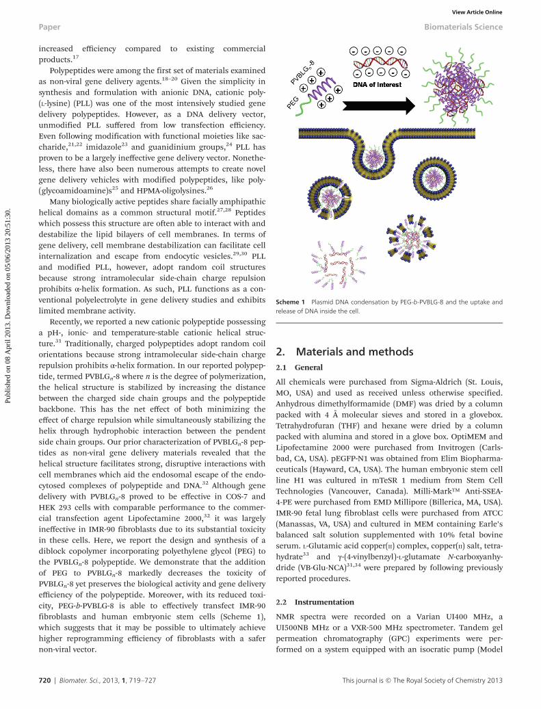
Recently, we reported a new cationic polypeptide possessinga pH-, ionic- and temperature-stable cationic helical struc-ture.31 Traditionally, charged polypeptides adopt random coilorientations because strong intramolecular side-chain chargerepulsion prohibits α-helix formation. In our reported polypep-tide, termed PVBLGn-8 where n is the degree of polymerization,the helical structure is stabilized by increasing the distancebetween the charged side chain groups and the polypeptidebackbone. This has the net effect of both minimizing theeffect of charge repulsion while simultaneously stabilizing thehelix through hydrophobic interaction between the pendentside chain groups. Our prior characterization of PVBLGn-8 pep-tides as non-viral gene delivery materials revealed that thehelical structure facilitates strong, disruptive interactions withcell membranes which aid the endosomal escape of the endo-cytosed complexes of polypeptide and DNA.32 Although genedelivery with PVBLGn-8 proved to be effective in COS-7 andHEK 293 cells with comparable performance to the commer-cial transfection agent Lipofectamine 2000,32 it was largelyineffective in IMR-90 fibroblasts due to its substantial toxicityin these cells. Here, we report the design and synthesis of adiblock copolymer incorporating polyethylene glycol (PEG) tothe PVBLGn-8 polypeptide. We demonstrate that the additionof PEG to PVBLGn-8 markedly decreases the toxicity ofPVBLGn-8 yet preserves the biological activity and gene deliveryefficiency of the polypeptide. Moreover, with its reduced toxi-city, PEG-b-PVBLG-8 is able to effectively transfect IMR-90fibroblasts and human embryonic stem cells (Scheme 1),which suggests that it may be possible to ultimately achievehigher reprogramming efficiency of fibroblasts with a safernon-viral vector.
2. Materials and methods2.1 General
All chemicals were purchased from Sigma-Aldrich (St. Louis,MO, USA) and used as received unless otherwise specified.Anhydrous dimethylformamide (DMF) was dried by a columnpacked with 4 Å molecular sieves and stored in a glovebox.Tetrahydrofuran (THF) and hexane were dried by a columnpacked with alumina and stored in a glove box. OptiMEM andLipofectamine 2000 were purchased from Invitrogen (Carls-bad, CA, USA). pEGFP-N1 was obtained from Elim Biopharma-ceuticals (Hayward, CA, USA). The human embryonic stem cellline H1 was cultured in mTeSR 1 medium from Stem CellTechnologies (Vancouver, Canada). Milli-Mark™ Anti-SSEA-4-PE were purchased from EMD Millipore (Billerica, MA, USA).IMR-90 fetal lung fibroblast cells were purchased from ATCC(Manassas, VA, USA) and cultured in MEM containing Earle’sbalanced salt solution supplemented with 10% fetal bovineserum. L-Glutamic acid copper(II) complex, copper(II) salt, tetra-hydrate33 and γ-(4-vinylbenzyl)-L-glutamate N-carboxyanhy-dride (VB-Glu-NCA)31,34 were prepared by following previouslyreported procedures.
2.2 Instrumentation
NMR spectra were recorded on a Varian UI400 MHz, aUI500NB MHz or a VXR-500 MHz spectrometer. Tandem gelpermeation chromatography (GPC) experiments were per-formed on a system equipped with an isocratic pump (Model
Scheme 1 Plasmid DNA condensation by PEG-b-PVBLG-8 and the uptake andrelease of DNA inside the cell.
Paper Biomaterials Science
720 | Biomater. Sci., 2013, 1, 719–727 This journal is © The Royal Society of Chemistry 2013
Publ
ishe
d on
08
Apr
il 20
13. D
ownl
oade
d on
05/
06/2
013
20:5
1:30
. View Article Online

1100, Agilent Technology, Santa Clara, CA, USA), a DAWNHELEOS 18-angle laser light scattering detector (also known asa multi-angle laser light scattering (MALLS) detector, WyattTechnology, Santa Barbara, CA, USA) and an Optilab rEXrefractive index detector (Wyatt Technology, Santa Barbara, CA,USA). The detection wavelength of HELEOS was set at 658 nm.Separations were performed using serially connected sizeexclusion columns (100, 500, 103 and 104 Å Phenogel columns,5 μm, 300 × 7.8 mm, Phenomenex, Torrance, CA, USA) at 60 °Cusing DMF containing 0.1 M LiBr as the mobile phase. TheMALLS detector was calibrated using pure toluene with noneed for external polymer standards and was used for thedetermination of the absolute molecular weights. The molecu-lar weights (MWs) of all polymers were determined based onthe dn/dc value of each sample calculated offline by using theinternal calibration processed by ASTRA V software (version5.1.7.3, Wyatt Technology, Santa Barbara, CA, USA). Infraredspectra were recorded on a PerkinElmer 100 serial FTIR spec-trophotometer equipped with universal attenuated total reflec-tance (ATR), which enabled the analysis of polymer samples inpowder form. Circular dichroism (CD) measurements werecarried out on a JASCO J-700 or a JASCO J-720 CD Spectro-meter. Ozone was produced by an OZV-8S ozone generatormanufactured by Ozone Solutions Inc. (Hull, IA, USA). Lyophi-lization was performed on a FreeZone lyophilizer (Labconco,Kansas City, MO, USA). Flow cytometry analysis was conductedon a BD FACSCanto 6 color flow cytometry analyzer (BectonDickinson, Franklin Lakes, NJ, USA). Cells were visualized witha Zeiss Axiovert 40 CFL fluorescence microscope equippedwith a 20× objective (Thornwood, NY, USA). Zeta potential andparticle size were analyzed with a Malvern Zetasizer (Worces-tershire, UK).
2.3 General procedure for the polymerization of VB-Glu-NCA
We followed a previously established procedure to synthesizeand polymerize NCAs to prepare polypeptides.35–38 In a glove-box, VB-Glu-NCA (56 mg, 0.2 mmol) was dissolved in DMF(1 mL) followed by the addition of PEG-amine and 1,5,7-triaza-bicyclo[4.4.0]dec-5-ene (TBD) at various monomer : amine :TBD ratios (Table 1). The polymerization solutions were stirredat room temperature for 24–60 h untill VB-Glu-NCA was con-sumed. Aliquots of the polymerization solutions were dilutedto 10 mg polymer mL−1 using DMF containing 0.1 M LiBr andanalyzed by GPC. The real-time concentration of NCA was
quantified by measuring the intensity of the anhydride bandat 1784 cm−1 by FTIR. The conversion of VB-Glu-NCA wasdetermined by comparing the VB-Glu-NCA concentration inthe polymerization solution versus the initial VB-Glu-NCA con-centration. When the polymerization was complete, themajority of the DMF was removed under vacuum and thepolymer was precipitated with ether (15 mL). The resultingPEG-b-PVBLG polymer was sonicated in ether for 5 min andcentrifuged to remove the remaining solvent. After the soni-cation–centrifugation steps were repeated two more times,PEG-b-PVBLG was collected and dried under vacuum (44 mgand 75% yield, and 35 mg and 68% yield for PEG113-b-PVBLG76 and PEG113-b-PVBLG287, respectively).
1H NMR (TFA-d, 500 MHz): δ 7.53 (d, 2H, J = 7.0 Hz, ArH), 7.39 (d, 2H, J =7.0 Hz, ArH), 6.84 (dd, 1H, J1 = 11.0 Hz, J2 = 18.0 HzC6H4CHvCH2), 5.91 (d, 1H, J = 18.0 Hz, C6H4CHvCH2), 5.43(d, 1H, J = 11.0 Hz, C6H4CHvCH2), 5.26 (m, 2H, ArCH2), 4.80(m, 1H, CHCH2CH2COOCH2), 4.13 (m, –OCH2CH2– in PEG),2.68 (m, 2H, CHCH2CH2COO), 2.30 (m, 1H, CHCH2CH2COO),2.12 (m, 1H, CHCH2CH2COO).
2.4 General procedure for the synthesis of poly(ethyleneglycol)-block-poly(γ-(4-aldehydebenzyl)-L-glutamate) (PEG-b-PABLG)
PEG-b-PVBLG (40 mg) was dissolved in chloroform (30 mL) at−78 °C. Oxygen was then bubbled into the solution for 1 minfollowed by the bubbling of ozone until the solution becameblue. The ozone was then replaced by oxygen, which wasbubbled into the solution for 2 min until the solution becamecolorless. The solution was degassed and back filled withnitrogen. Dimethyl sulfide (1 mL) was then added and thesolution was stirred at room temperature overnight. After-wards, the solvent was removed under vacuum and the result-ing PEG113-b-PABLG76 was purified by sonicating the polymerin methanol (3 × 15 mL) and collected by centrifugation.PEG113-b-PABLG76 was dried under vacuum (33 mg, 82%yield). PEG113-b-PABLG287 was synthesized from PEG113-b-PVBLG287 by following a similar procedure for the synthesis ofPEG113-b-PABLG76 with 86% yield. 1H NMR (TFA-d, 500 MHz):δ 10.31 (1H, CHOC6H4), 8.40 (d, 2H, J = 7.0 Hz, ArH), 7.96 (d,2H, J = 7.0 Hz, ArH), 5.71 (2H, CHOC6H4CH2), 5.21 (1H,CHCH2CH2CO2CH2), 4.10 (m, –OCH2CH2– in PEG), 3.12 (2H,CHCH2CH2), 2.75 (1H, CHCH2CH2), 2.56 (1H, CHCH2CH2).
2.5 General procedure for the preparation of PEG-b-PVBLG-8
PEG-b-PABLG (20 mg), N-(2-aminoethyl)piperidine (5 molarequiv. relative to the Glu repeating unit of PEG-b-PABLG) andborane–pyridine complex (5 molar equiv.) were mixed in DMF(3 mL) and stirred at 50 °C for 48 h (Table 2). The mixture waspoured into 3 M HCl (3 mL) and dialyzed against water for48 h. The resulting PEG-b-PVBLG-8 was lyophilized. The yieldsof PEG-b-PVBLG-8 copolymers were between 60 and 70%, withgrafting efficiencies greater than 95%, which was determinedas previously reported.32
Table 1 PEG113-NH2 initiated polymerization of VB-Glu-NCA
Product M : Amine : TBDaTime(h)
Conv.(%)
Mn (M*n)
b
(×103 gmol−1) MWD
PEG113-b-PVBLG76 100 : 1 : 0.1 24 80 20.7 (24.6) 1.21PEG113-b-PVBLG287 400 : 1 : 0.1 60 78 75.3 (81.4) 1.29
a TBD: 1,5,7-triazabicyclo[4.4.0]dec-5-ene. b The MW obtained(theoretical MW = Mn, PEG + 245.27 × conv. × [M]/[I]).
Biomaterials Science Paper
This journal is © The Royal Society of Chemistry 2013 Biomater. Sci., 2013, 1, 719–727 | 721
Publ
ishe
d on
08
Apr
il 20
13. D
ownl
oade
d on
05/
06/2
013
20:5
1:30
. View Article Online

2.6 General procedure for the analysis of polypeptideconformations by circular dichroism (CD)
Circular dichroism studies were performed on JASCO J-700and J-720 CD spectrometers. Samples were prepared atpolymer concentrations of 0.01–0.1 mg mL−1 unless otherwisespecified. In a representative experiment, the sample solutionwas placed in a quartz cell with a path length of 0.5 cm andthe mean residue molar ellipticity of the polymer was calcu-lated based on the measured apparent ellipticity according tothe equation: Ellipticity ([θ] in degree cm2 dmol−1) = (millide-grees × mean residue weight)/(path length in millimeters ×concentration of polypeptide in mg mL−1).39 For helix-temp-erature dependency studies, the temperature of the samplechamber containing the quartz cell was varied from 4 to 70 °Cusing a water bath. A minimum of 10 min was allowed forsample temperature equilibration prior to collecting CDmeasurements. The α-helix contents of the polypeptides werecalculated using the following equation: % α-helix = (−[θ]222 +3000)/39 000.40
2.7 Agarose gel retardation
A solution of DNA (0.5 μg) was prepared in OptiMEM (50 μL).Separately, a solution of polypeptide in OptiMEM (50 μL) wasprepared to achieve the desired polypeptide : DNA weight ratio.Following mixing of the two solutions, complexes were incu-bated at room temperature for 20 min, after which an aliquot(20 μL) was withdrawn and a loading dye (4 μL) was added.The mixture was then run on a 2% agarose gel (100 V, 60 min).DNA was stained with ethidium bromide and visualized on aGel Doc imaging system (Biorad, Herclues, CA, USA).
2.8 Characterization of polymer–DNA complex with zetapotential and dynamic light scattering
Solutions of DNA (25 μg) were prepared in OptiMEM (400 μL).Separately, a solution of polypeptide (1 mg) was prepared inOptiMEM (400 μL). A solution of Lipofectamine 2000 (50 μL,1 mg mL−1) in OptiMEM (400 μL) was also prepared as acontrol. The DNA solution was then mixed with either the
polypeptide or Lipofectamine 2000 solution and allowed toincubate at rt for 20 min. The size and surface charge of theresulting polyplexes were analyzed by dynamic light scattering(DLS) and zeta potential.
2.9 Transfection of IMR-90 with Lipofectamine 2000 andPVBLG-8 polymers
IMR-90 cells were seeded at 50 000 cells per well in 24-wellplates one day prior to transfection. On the day of transfection,plasmid pEGFP-N1 DNA (1 μL, 1 mg mL−1) was diluted withOptiMEM (50 μL). Separately, Lipofectamine 2000 (2 μL, 1 mgμL−1) or the polymer solution (10–80 μL, 1 mg mL−1) wasdiluted with OptiMEM (50 μL). The individual solutions werethen mixed gently and allowed to incubate for 5 min at rt, afterwhich they were combined and allowed to incubate at rt foranother 20 min. The cell medium was then aspirated andreplaced with pre-warmed (37 °C) OptiMEM (500 μL). Thecomplex solution was added dropwise to the cells. The cellswere then incubated at 37 °C with 5% CO2 for 4 h, after whichthe cell medium was replaced with normal culture medium(500 μL). After incubation for a total of 48 h at 37 °C with 5%CO2, the cells were imaged with a fluorescent microscopy. TheEGFP transfection efficiency was quantified by flow cytometry.
2.10 Sample preparation and flow cytometry analysis
Prior to analysis by flow cytometry, transfected cells on the 24-well plate were washed with 1× PBS (500 μL for each well) toremove any residual serum, dead cells and debris. Next,trypsin (100 μL) was added and incubated for 5–10 min todetach the cells from the plate. PBS (100 μL) was then addedand pipetted up and down to break up cell clumps. A solutionof 4% paraformaldehyde (100 μL) was added to fix the cells.Samples were kept in covered flow cytometry tubes until analy-sis (BD FACSCanto, Franklin Lakes, NJ, USA).
2.11 MTT assay of polymers
For MTT assays, 10 000 cells were seeded in each well of a96-well plate one day before transfection. The cells were thentransfected as described above, save for an 80% reduction involume and reagent quantity to accommodate the reduced wellvolume. The cells were incubated for 4 h at 37 °C in the trans-fection mix before being returned to a fresh growth medium.After 48 h, the cells were washed with PBS and an MTT solu-tion was added. Following a 4 h incubation at 37 °C, an MTTsolubilization solution (10% Triton X-100 in acidic (0.1 M HCl)isopropanol) was added to the cells and the absorbance of570 nm light was quantified on a PerkinElmer plate reader(Waltham, MA, USA).
2.12 hESC transfection
hESCs were seeded in Matrigel-coated 24-well plates. PlasmidDNA (1 μL, 1 mg mL−1) was diluted in OptiMEM (50 μL). Thepolymer solution (10–40 μL, 1 mg mL−1) was diluted withOptiMEM (50 μL). The two solutions were then vortexed gentlyand allowed to incubate for 5 min at rt, after which they werecombined and allowed to incubate for another 20 min at rt.
Table 2 Synthesis and conformation analysis of PEG-b-PVBLG-8a
Startingpolymer Product
Graftingeff.b (%)
−[θ]222 c
(10−3 degreecm2/dmol)
Helicalcontentd
(%)
PEG113-b-PABLG76
PEG113-b-PVBLG76-8(PEV-L)
>95 32.8 91.8
PEG113-b-PABLG287
PEG113-b-PVBLG287-8(PEV-H)
>95 34.6 96.4
a Reducing reagent (5 molar equiv.) was used. Reaction was carried outfor 48 h at 50 °C. b The grafting efficiency was determined by 1H NMRanalysis. c The mean residue molar ellipticity was calculated byfollowing literature-reported formulas: ellipticity ([θ]222 nm in cm2
degree dmol−1) = (millidegrees × mean residue weight)/(path length inmillimeters × concentration of polypeptide in mg mL−1). d The α-helixcontents of the polypeptides were calculated using the followingequation: % α-helix = (−[θ]222 + 3000)/39 000.40
Paper Biomaterials Science
722 | Biomater. Sci., 2013, 1, 719–727 This journal is © The Royal Society of Chemistry 2013
Publ
ishe
d on
08
Apr
il 20
13. D
ownl
oade
d on
05/
06/2
013
20:5
1:30
. View Article Online

Next, the mixtures were added to the cells dropwise andallowed to incubate at 37 °C for 4 h. The medium was thenaspirated and fresh medium was added. After 48 h, the cellswere stained with DAPI (250 μL, 3 nM) and SSEA-4-PE (250 μL,0.02 mg mL−1), a pluripotency cell marker, for 30 min at37 °C.
3. Results3.1 Synthesis and characterization of PEG-b-PVBLG-8 (PEV)
γ-(4-Vinylbenzyl)-L-glutamate N-carboxyanhydride (VB-Glu-NCA)was prepared by following previously reported methods.31,32,34
The ring-opening polymerization of VB-Glu-NCA with PEG-amine as the macroinitiator yielded PEG-block-poly(γ-(4-vinyl-benzyl)-L-glutamate) (PEG-b-PVBLG) with controlled molecularweights (MWs) and narrow molecular-weight distributions(Scheme 2). At the VB-Glu-NCA–PEG-amine ratio of 100, theobtained Mn of 20.7 × 10³ g mol−1 agreed well with the theoreti-cal Mn of 24.6 × 10³ g mol−1 and had a narrow molecularweight distribution of 1.21 (entry 1, Table 1). Two PEG-b-PVBLGcopolymers were prepared with degrees of polymerization (DP)of 76 (PEG-b-PVBLG76) and 287 (PEG-b-PVBLG287) of the PVBLGblock (Table 1). The ozonation of PEG-b-PVBLG yielded PEG-b-poly(γ-(4-aldehydebenzyl-L-glutamate) (PEG-b-PABLG), whichserved as the reactive intermediate that, through subsequenthydroamination and reduction with N-(2-aminoethyl)piperi-dine, yielded the desired PEG-b-PVBLG76-8 (PEV-L) and PEG-b-PVBLG287-8 (PEV-H). Grafting efficiencies greater than 95%were achieved for both PEV-L and PEV-H (Table 2).
Both PEV-L and PEV-H are highly soluble in water at pH1–10 (>50 mg mL−1), which is drastically different from the
corresponding parental (PEG-b-PVBLG) and intermediate poly-mers (PEG-b-PABLG) that are insoluble in water. The excellentwater solubility of PEV-L and PEV-H is clearly related to theircharged side groups, which make it possible for the appli-cation of PEV at physiological pH. Both PEV-L and PEV-Hshowed the characteristic CD spectra of an α-helix with twominima at 208 and 222 nm (Fig. 1a), consistent with our pre-viously reported α-helical conformation of PVBLG-8.32,41
Helical contents of greater than 90% were observed for bothPEV-L and PEV-H at pH 3 when the side chain amine groupsare protonated (Table 2). As expected, the charge repulsion ofthe side groups had minimal effect on helix stability becausethe charged amine groups were placed far away from the poly-peptide backbone. Furthermore, the helicity—as measured bythe value of −[θ]222—was shown to be stable against pH andsalt changes in the surrounding environment. For example,the −[θ]222 values of PEV-L and PEV-H remained unchangedwhen the solution pH was increased from 1 to 9 (Fig. 1b). Thehelices of PEV-L and PEV-H were also fairly stable in concen-trated denaturing conditions, such as in 1 M NaCl (Fig. 1d)and 2 M urea (Fig. 1d) aqueous solutions. These observationssuggested that PEVs would maintain their helical confor-mation in various extracellular and intracellular environmentswith well-preserved properties throughout the gene transfec-tion processes.
Complex formation with PEG-b-PVBLG-8 with DNA. Theability of PEG-b-PVBLG-8 to bind and complex with DNA wasexamined using a gel retardation assay. Polymer was mixedwith plasmid DNA at DNA : polymer weight ratios between 1 : 1and 1 : 60 and run on an agarose gel. The results for PEV-L canbe seen in Fig. 2a. The addition of polymer in excess of 1 : 2(DNA : polymer weight ratio) resulted in the formation ofstable complexes which prohibited the migration of DNA
Scheme 2 The chemical route for the preparation of PEG-b-PVBLG-8 fromVB-Glu-NCA.
Fig. 1 (a) CD spectra in water of PEV-L, PEV-H, DNA–PEV-L, and DNA–PEV-H at1 : 40 weight ratio at pH 3. (b) The pH dependence of the residue molar ellipti-city at 222 nm for PEV-L and PEV-H at 0.05 mg mL−1. (c) Salt dependence ofresidue ellipticity at 222 nm for PEV-L and PEV-H at pH 3 and 0.05 mg mL−1. (d)The helical stabilities of PEV-L and PEV-H at pH 3 and 0.05 mg mL−1 in the pres-ence of urea.
Biomaterials Science Paper
This journal is © The Royal Society of Chemistry 2013 Biomater. Sci., 2013, 1, 719–727 | 723
Publ
ishe
d on
08
Apr
il 20
13. D
ownl
oade
d on
05/
06/2
013
20:5
1:30
. View Article Online

under an electrophoretic force. Interestingly, at a 1 : 2 DNA :polymer weight ratio, the DNA could still be seen in theloading well, indicating incomplete condensation. However,the DNA was no longer visible when sufficient polymer wasadded to achieve a 1 : 10 DNA : polymer weight ratio, indicatingcomplete condensation. As can be seen in Fig. 1a, DNAbinding does not affect the α-helicity of the peptide.
3.2 Dynamic light scattering
Dynamic light scattering revealed complexes formed betweenDNA and Lipofectamine 2000 at a 1 : 2 DNA : Lipofectamine2000 weight ratio to be approximately 574 nm in diameter.Meanwhile, the hydrodynamic diameters of complexes ofPEV-L and PEV-H at a 1 : 40 DNA : polymer weight ratio weresubstantially smaller—about 107 nm and 246 nm for PEV-Land PEV-H, respectively (Fig. 2b). Testing of DNA : polymerweight ratios less than and greater than 1 : 40 did not dramati-cally change the measured diameter—provided that aminimum amount of polymer was added to achieve complexa-tion. For example, the diameters of complexes made withDNA : PEV-L weight ratios of 1 : 10, 1 : 20, 1 : 40 and 1 : 80 were117 nm, 115 nm, 107 nm, and 84 nm, respectively. Thissuggests that once a minimum amount of polypeptide ispresent—presumably enough to achieve a 1 : 10 DNA : polymerweight ratio based on Fig. 2a—the excess polymer is not incor-porated into the complexes and exists freely in the solution.Furthermore, despite the rod-like structure of the helical poly-peptide, complexes formed between DNA and PEG-PVBLG-8possessed a globular structure under TEM (Fig. 2c). Zetapotential measurements were performed to ensure that thecomplex formed between PEV-L (or PEV-H) and DNA wereoverall positively charged. PEV–DNA complexes formed pos-sessed zeta potentials between 2 and 10 mV, while PVBLG-8/DNA complexes possessed zeta potentials between 20 and30 mV (data not shown). The addition of the PEG block to thepolypeptide shielded the cationic charge of the PVBLG-8block.
3.3 Toxicity
The toxicity of the PEV-H was compared with PVBLG-8homopolymer (P0) and Lipofectamine 2000 via MTT assays inIMR-90 cells. PVBLG-8 (P0) was found to be notably toxic to
IMR-90 cells. At 1 : 40 DNA : polymer weight ratio, a viabilityof 6% was observed. The new diblock polymers of both low(PEV-L) and high (PEV-H) molecular weight showed sub-stantially reduced toxicity to IMR-90 cells (Fig. 3a). Increasingthe DNA : polymer ratio resulted in increased toxicity for bothPEV-L and PEV-H polymers. PEV-H–DNA complexes showedslightly reduced toxicity compared to the PEV-L–DNA complex,but both PEV-L- and PEV-H polymers were less toxic than Lipo-fectamine 2000 under similar conditions.
3.4 Transfection
Transfection experiments were performed with the diblock co-polymers to determine whether their reduced toxicity impactedtheir ability to effectively deliver genes to IMR-90 cells. Asshown in Fig. 3b, the commercial reagent Lipofectamine 2000resulted in approximately 19% of treated cells expressing thedelivered GFP transgene. Unmodified PVBLG-8 (P0), on theother hand, had a transfection efficiency of only 1.4% with lowcell viability. While PVBLG-8 performed comparable to Lipo-fectamine 2000 in COS-7 and HeLa cells in previous studies,its poor performance as shown in Fig. 3b is likely due to theexcessive toxicity of the polypeptide in IMR-90 cells. Reducingthe toxicity of PVBLG-8 through the addition of PEG blocksresulted in an increase in IMR-90 transfection efficiency of9.4% and 21.4% for PEV-L and PEV-H, respectively. Because ofthe reduced toxicity of PEV-L and PEV-H, the DNA dosagecould be increased from 1 μg to 2 μg, which resulted in slightlyhigher transfection efficiencies, 13% and 27%, for PEV-L andPEV-H formulations, respectively (data not shown). Althoughthis increase in efficiency was modest, it should be highlighted
Fig. 2 DNA–polymer complex analysis: (a) Gel retardation of the PEV-L atdifferent DNA to polymer weight ratios. (b) Particle size analysis of the PEV-Lcopolymer and the DNA complex with different PVBLG-8 chain lengths andDNA to polymer weight ratios. (c) TEM image of PEV-L with the DNA complex.Scale bar: 200 nm.
Fig. 3 In vitro analysis in IMR-90 cells. (a) MTT cell viability assay of the DNA–polymer complex with different polymers and at different DNA to polymerweight ratios in IMR-90. The amount of DNA was fixed at 1 μg. (b) Initial testingfor PEV-L along with Lipofectamine 2000 (Lip) and PVBLG-8 (P0) with varyingpEGFP-N1 plasmid and polymer amounts. Transfection efficiency was analysed48 h post-transfection with flow cytometry. (c) Fluorescent images of the trans-fection using Lip and PEV-H. Scale bars = 0.25 mm.
Paper Biomaterials Science
724 | Biomater. Sci., 2013, 1, 719–727 This journal is © The Royal Society of Chemistry 2013
Publ
ishe
d on
08
Apr
il 20
13. D
ownl
oade
d on
05/
06/2
013
20:5
1:30
. View Article Online

that the diblock polymers appeared to have reduced toxicity.For example, even though both Lipofectamine 2000- andPEV-H-transfected cells expressed similar amounts of GFP inFig. 3c, the cells transfected with PEV-H possessed an overallhealthier phenotype with flat and elongated shapes asopposed to cells transfected with Lipofectamine 2000 thatappeared to be sparse and rounded.
To further demonstrate the application of PEG-b-PVBLG-8,we next evaluated the gene delivery efficiency to H1 hESC.PEV-H transfection in separated H1 cells resulted in highertransfection efficiency than in cells plated as colonies (Fig. 4aand b). Moreover, the polymer was shown to have no impacton hESC pluripotency 48 h post-transfection. This was evi-denced by the similar expression of stage-specific embryonicantigen-4 (SSEA-4) both before and after transfection in colo-nies and single cells (Fig. 4c). Further evidence of pluripotencywas shown through a Western blot of the OCT4 pluripotencytranscription factor before and after transfection with PEV-H(Fig. 4d). Combined with its efficiency, the mild cell impact ofthe PEV-H made it a promising reagent to manipulate thegene expression of human stem cells.
4. Discussion
Transfection efficiency is often limited due to the toxicity ofvectors. Generally, the more efficient the transfection material,the greater the impact it may have on cell health. This islargely due to the requirements of effective gene delivery—namely, a high polycationic charge to condense DNA and
enable cell surface binding and membrane-lytic properties tofacilitate intracellular escape from endocytic vesicles. Whilegood for the delivery of nucleic acids, highly cationic materialscan also bind and interfere with the function of necessary pro-teins within the cell. Moreover, the membrane lytic effect ofmaterials can be unspecific and may act in a desirable as wellas an undesirable manner. Effective gene delivery materialsare able to balance their positive and negative tendencies sothat they are efficient enough to allow macromolecules likeDNA to enter the cells but safe enough that the cells are notirreparably damaged during the process. In this paper, wefocus on the previously described PVBLG-8 materials and tryto reduce their overall toxicity while maintaining their effectivegene delivery performance.
Previous characterizations of the helical polypeptidePVBLG-8 revealed that it can operate as an effective deliveryvector for both DNA and siRNA.41 In both cases, its perform-ance was demonstrated to be tied to its ability to form stablehelices that cause pore formation within membranes. In thecase of DNA delivery, the membrane lytic potential was essen-tial to the escape of DNA–polypeptide complexes from endo-cytic vesicles. In the case of siRNA delivery, the helicalPVBLG-8 caused pore formation within cell membranes toallow the non-endocytic diffusion of siRNA into the cellcytosol. Therefore, in our desire to reduce the toxicity of thePVBLG-8 materials, it was also essential to retain their helicalstructure. Unfortunately, just as helicity makes the materialseffective delivery agents, it is also a contributing factor tooverall toxicity. As such, we sought a strategy to append chargeshielding materials to the helical PVBLG-8. By shielding thepositive charge, we hoped to minimize the toxicity of thematerial by reducing its electrostatic attraction with negativelycharged cell membranes. At the same time, since the helicityof the PVBLG-8 block was maintained (Fig. 1a), we hoped thatthe material would retain its ability to effectively escape theendosome and mediate effective gene delivery (Scheme 1).
To test the feasibility of incorporation of charge shieldinggroups, PEG was covalently conjugated with PVBLG-8 to yieldthe diblock polymer PEG-b-PVBLG-8. The PEG used had a MWof 5000 Da (DP of 113) and was conjugated to helical PVBLG-8with DPs of 76 and 287 to yield the diblock materials PEV-Land PEV-H, respectively (Scheme 2). Circular dichroism exper-iments revealed that the incorporation of PEG did not alter thepresence and stability of the helices even after the DNA isbound and condensed by the peptide (Fig. 1a). Moreover, thematerials were also able to bind and condense plasmid DNAinto spherical particles with diameters of the order of 100 nm(Fig. 2a–c). Despite the inclusion of PEG, these particles weredemonstrated to retain an overall positive surface chargesimilar to commercial materials like Lipofectamine 2000 byanalyzing their surface zeta potential (data not shown).
Toxicity measurements with the diblock materials revealedthat the inclusion of a PEG block substantially increased thebiocompatibility of the materials in IMR-90 cells. For example,treatment with the unmodified PVBLG-8 left only approxi-mately 5% of IMR-90 cells viable. However, with the addition
Fig. 4 EGFP plasmid transfection efficiency using Lip and PEV-H of H1 hESC as(a) small colonies and (b) single cells as analysed by flow cytometry. (c) Brightfield and fluorescence imaging of PEV-H transfection of EGFP plasmid into hESCas colonies and single cells. Colonies were stained with Hoechst and pluripo-tency marker SSEA-4 antibody conjugated with PE. Scale bar: 250 μm. (d)Western blot of cells isolated 72 h post-transfection demonstrating the proteinexpression of OCT4 in hESC H1 cells.
Biomaterials Science Paper
This journal is © The Royal Society of Chemistry 2013 Biomater. Sci., 2013, 1, 719–727 | 725
Publ
ishe
d on
08
Apr
il 20
13. D
ownl
oade
d on
05/
06/2
013
20:5
1:30
. View Article Online

of a PEG block, cell viability increased dramatically and wasless toxic than Lipofectamine 2000 (Fig. 3a). With its improvedtoxicity profile, PEG-b-PVBLG-8 was able to mediate effectivetransfection in IMR-90 cells. Previously, the toxicity of thematerials was so extreme that only 1.4% of cells were success-fully transfected. With the reduced toxicity, that number wasincreased to approximately 20%—an increase of approximately14-fold. With its improved safety profile, PEG-b-PVBLG-8 wasmild enough to transfect H1 human embryonic stem cellswithout affecting the expression of cell pluripotency markers(Fig. 4).
5. Conclusions
We prepared diblock copolymers consisting of poly(ethyleneglycol)-block-poly(γ-4-(((2-(piperidin-1-yl)ethyl)amino)methyl)-benzyl-L-glutamate) (PEG-b-PVBLG-8) and evaluated their capa-bility to mediate gene delivery in IMR-90 human fetal lungfibroblasts and human embryonic stem cells (hESCs). ThePEG-b-PVBLG-8 contained a membrane-disruptive, cationic,helical polypeptide block (PVBLG-8) for complexing with DNAand a hydrophilic PEG block to improve the biocompatibilityof the gene delivery vehicle. PEG-b-PVBLG-8 copolymers withlow (n = 76) and high (n = 287) degrees of polymerization (n) ofthe PVBLG-8 block were synthesized. We found that the incor-poration of PEG effectively reduced the toxicity of the helicalPVBLG-8 block without dramatically compromising the com-plexation of copolymers with DNA and their transfectionefficiencies. PEG-b-PVBLG-8 diblock polymer with a highdegree of polymerization had a greater transfection efficiencyand lower toxicity in IMR-90 cells than the commercial reagentLipofectamine 2000. The usefulness of PEG-b-PVBLG-8 wasfurther demonstrated via the successful transfection of hESCswithout a measured loss in cell pluripotency markers. In con-trast to many other polymer- and lipid-based transfectionsystems which utilize the proton sponge mechanism (e.g. poly-ethylenimine) or lipid mixing (e.g. DOTAP, DOPE, etc.) to facili-tate endosomal escape, the peptides described here make useof a novel pore formation mechanism. As endocytic escape isgenerally considered one of the most challenging aspects ofgene delivery, this alternative endosomolytic mechanism mayprove useful when working with cell lines not readily amenableto transfection by current methods (i.e. IMR-90 and hES cells).As both IMR-90 and hESCs are important cell lines that arecommonly used in the field of regenerative medicine, effectivenon-viral gene delivery to these cells is an important step inbringing regenerative medicine closer to clinical applications.
Acknowledgements
This work was supported by NIH (Director’s New InnovatorAward 1DP2OD007246 and 1R21EB013379 for J.C) and NSF(CHE 1153122). J.Y. was funded at UIUC by National ScienceFoundation (NSF) Grant 0965918 IGERT: Training the Next
Generation of Researchers in Cellular and Molecular Mech-anics and BioNanotechnology.
References
1 U. P. Davé, N. A. Jenkins and N. G. Copeland, Gene therapyinsertional mutagenesis insights, Science, 2004, 303, 333.
2 K. Leong, H. Mao, K. Roy, S. Walsh and J. August, DNA-polycation nanospheres as non-viral gene delivery vehicles,J. Controlled Release, 1998, 53, 183.
3 K. Kizjakina, J. M. Bryson, G. Grandinetti andT. M. Reineke, Cationic glycopolymers for the delivery ofpDNA to human dermal fibroblasts and rat mesenchymalstem cells, Biomaterials, 2011, 33, 1851–1862.
4 P. M. McLendon, K. M. Fichter and T. M. Reineke, Poly (glyco-amidoamine) vehicles promote pDNA uptake throughmultiple routes and efficient gene expression via caveolae-mediated endocytosis, Mol. Pharm., 2010, 7, 738–750.
5 S. Srinivasachari and T. M. Reineke, Versatile supramolecu-lar pDNA vehicles via “click polymerization” of β-cyclodex-trin with oligoethyleneamines, Biomaterials, 2009, 30,928–938.
6 F. Yang, S. W. Cho, S. M. Son, S. R. Bogatyrev, D. Singh,J. J. Green, Y. Mei, S. Park, S. H. Bhang and B. S. Kim,Genetic engineering of human stem cells for enhancedangiogenesis using biodegradable polymeric nanoparticles,Proc. Natl. Acad. Sci. U. S. A., 2010, 107, 3317–3322.
7 F. Yang, J. Green, T. Dinio, L. Keung, S. Cho, H. Park,R. Langer and D. Anderson, Gene delivery to human adultand embryonic cell-derived stem cells using biodegradablenanoparticulate polymeric vectors, Gene Ther., 2009, 16,533–546.
8 M. S. Shim and Y. J. Kwon, Controlled delivery of plasmidDNA and siRNA to intracellular targets using ketalized poly-ethylenimine, Biomacromolecules, 2008, 9, 444–455.
9 M. S. Shim and Y. J. Kwon, Acid-transforming polypeptidemicelles for targeted nonviral gene delivery, Biomaterials,2010, 31, 3404–3413.
10 M. S. Shim and Y. J. Kwon, Dual mode polyspermine withtunable degradability for plasmid DNA and siRNA delivery,Biomaterials, 2011, 32, 4009–4020.
11 X. Jiang, Y. Zheng, H. H. Chen, K. W. Leong, T. H. Wangand H. Q. Mao, Dual-sensitive micellar nanoparticles regu-late DNA unpacking and enhance gene-delivery efficiency,Adv. Mater., 2010, 22, 2556–2560.
12 U. Pfisterer, A. Kirkeby, O. Torper, J. Wood, J. Nelander,A. Dufour, A. Björklund, O. Lindvall, J. Jakobsson andM. Parmar, Direct conversion of human fibroblasts todopaminergic neurons, Proc. Natl. Acad. Sci. U. S. A., 2011,108, 10343–10348.
13 R. Ambasudhan, M. Talantova, R. Coleman, X. Yuan,S. Zhu, S. A. Lipton and S. Ding, Direct reprogramming ofadult human fibroblasts to functional neurons underdefined conditions, Cell Stem Cell, 2011, 113–118.
Paper Biomaterials Science
726 | Biomater. Sci., 2013, 1, 719–727 This journal is © The Royal Society of Chemistry 2013
Publ
ishe
d on
08
Apr
il 20
13. D
ownl
oade
d on
05/
06/2
013
20:5
1:30
. View Article Online

14 E. Szabo, S. Rampalli, R. M. Risueño, A. Schnerch,R. Mitchell, A. Fiebig-Comyn, M. Levadoux-Martin andM. Bhatia, Direct conversion of human fibroblasts to multi-lineage blood progenitors, Nature, 2010, 468, 521–526.
15 J. Yu, M. A. Vodyanik, K. Smuga-Otto, J. Antosiewicz-Bourget, J. L. Frane, S. Tian, J. Nie, G. A. Jonsdottir,V. Ruotti and R. Stewart, Induced pluripotent stem celllines derived from human somatic cells, Science, 2007, 318,1917–1920.
16 J. Zou, M. L. Maeder, P. Mali, S. M. Pruett-Miller,S. Thibodeau-Beganny, B. K. Chou, G. Chen, Z. Ye,I. H. Park and G. Q. Daley, Gene targeting of a disease-related gene in human induced pluripotent stem andembryonic stem cells, Cell Stem Cell, 2009, 5, 97–110.
17 J. J. Green, B. Y. Zhou, M. M. Mitalipova, C. Beard,R. Langer, R. Jaenisch and D. G. Anderson, Nanoparticlesfor gene transfer to human embryonic stem cell colonies,Nano Lett., 2008, 8, 3126–3130.
18 M. Monsigny, A. C. Roche, P. Midoux and R. Mayer, Glyco-conjugates as carriers for specific delivery of therapeuticdrugs and genes, Adv. Drug Delivery Rev., 1994, 14, 1–24.
19 E. Wagner, D. Curiel and M. Cotten, Delivery of drugs, pro-teins and genes into cells using transferrin as a ligand forreceptor-mediated endocytosis, Adv. Drug Delivery Rev.,1994, 14, 113–135.
20 S. K. Cho and Y. J. Kwon, Polyamine/DNA polyplexes withacid-degradable polymeric shell as structurally and func-tionally virus-mimicking nonviral vectors, J. ControlledRelease, 2011, 150, 287–297.
21 T. Ferkol, J. C. Perales, F. Mularo and R. W. Hanson, Recep-tor-mediated gene transfer into macrophages, Proc. Natl.Acad. Sci. U. S. A., 1996, 93, 101–105.
22 P. Erbacher, M. T. Bousser, J. Raimond, M. Monsigny,P. Midoux and A. C. Roche, Gene transfer by DNA/glycosy-lated polylysine complexes into human blood monocyte-derived macrophages, Hum. Gene Ther., 1996, 7, 721–729.
23 D. Putnam, C. A. Gentry, D. W. Pack and R. Langer,Polymer-based gene delivery with low cytotoxicity by aunique balance of side-chain termini, Proc. Natl. Acad.Sci. U. S. A., 2001, 98, 1200–1205.
24 T. Okuda, A. Sugiyama, T. Niidome and H. Aoyagi, Charac-ters of dendritic poly (-lysine) analogues with the terminallysines replaced with arginines and histidines as gene car-riers in vitro, Biomaterials, 2004, 25, 537–544.
25 N. P. Ingle, B. Malone and T. M. Reineke, Poly (glycoami-doamine)s: a broad class of carbohydrate-containing poly-cations for nucleic acid delivery, Trends Biotechnol., 2011,443–453.
26 R. N. Johnson, D. S. H. Chu, J. Shi, J. G. Schellinger,P. M. Carlson and S. H. Pun, HPMA-oligolysine copolymersfor gene delivery: optimization of peptide length and polymermolecular weight, J. Controlled Release, 2011, 303–311.
27 D. E. Robertson, R. S. Farid, C. C. Moser, J. L. Urbauer,S. E. Mulholland, R. Pidikiti, J. D. Lear, A. J. Wand,
W. F. DeGrado and P. L. Dutton, Design and synthesis ofmulti-haem proteins, Nature, 1994, 425–432.
28 V. Muñoz and L. Serrano, Elucidating the folding problemof helical peptides using empirical parameters, Nat. Struct.Mol. Biol., 1994, 1, 399–409.
29 K. M. Stewart, K. L. Horton and S. O. Kelley, Cell-penetrat-ing peptides as delivery vehicles for biology and medicine,Org. Biomol. Chem., 2008, 6, 2242–2255.
30 S. Deshayes, M. Morris, G. Divita and F. Heitz, Cell-pene-trating peptides: tools for intracellular delivery of thera-peutics, Cell. Mol. Life Sci., 2005, 62, 1839–1849.
31 H. Lu, J. Wang, Y. G. Bai, J. W. Lang, S. Y. Liu, Y. Lin andJ. Cheng, Ionic polypeptides with unusual helical stability,Nat. Commun., 2011, 2, 206.
32 N. P. Gabrielson, H. Lu, L. C. Yin, D. Li, F. Wang andJ. Cheng, Reactive and bioactive cationic α-helical polypep-tide template for nonviral gene delivery, Angew. Chem., Int.Ed., 2012, 51, 1143–1147.
33 W. A. R. Vanheeswijk, M. J. D. Eenink and J. Feijen, Animproved method for the preparation of gamma-esters ofglutamic-acid and beta-esters of aspartic-acid, Synthesis-Stuttgart, 1982, 744–747.
34 H. Lu, Y. G. Bai, J. Wang, N. P. Gabrielson, F. Wang, Y. Linand J. Cheng, Ring-opening polymerization of gamma-(4-vinylbenzyl)-L-glutamate N-carboxyanhydride for the syn-thesis of functional polypeptides, Macromolecules, 2011, 44,6237–6240.
35 H. Lu, J. Wang, Y. Lin and J. Cheng, One-pot synthesisof brush-like polymers via integrated ring-opening meta-thesis polymerization and polymerization of amino acidN-carboxyanhydrides, J. Am. Chem. Soc., 2009, 131, 13582–13583.
36 H. Lu and J. Cheng, N-Trimethylsilyl amines for controlledring-opening polymerization of amino acid N-carboxyanhy-drides and facile end group functionalization of polypep-tides, J. Am. Chem. Soc., 2008, 130, 12562–12563.
37 H. Lu and J. Cheng, Hexamethyldisilazane-mediated con-trolled polymerization of alpha-amino acid N-carboxyanhy-drides, J. Am. Chem. Soc., 2007, 129, 14114–14115.
38 Y. G. Bai, H. Lu, E. Ponnusamy and J. Cheng, Synthesis ofhybrid block copolymers via integrated ring-openingmetathesis polymerization and polymerization of NCA,Chem. Commun., 2011, 47, 10830–10832.
39 N. J. Greenfield, Using circular dichroism spectra to esti-mate protein secondary structure, Nat. Protoc., 2006, 1,2876–2890.
40 J. A. Morrow, M. L. Segall, S. Lund-Katz, M. C. Phillips,M. Knapp, B. Rupp and K. H. Weisgraber, Differences instability among the human apolipoprotein E isoformsdetermined by the amino-terminal domain, Biochemistry,2000, 39, 11657–11666.
41 N. P. Gabrielson, H. Lu, L. Yin, K. H. Kim and J. Cheng, Acell-penetrating helical polymer for siRNA delivery to mam-malian cells, Mol. Ther., 2012, 20, 1599–1609.
Biomaterials Science Paper
This journal is © The Royal Society of Chemistry 2013 Biomater. Sci., 2013, 1, 719–727 | 727
Publ
ishe
d on
08
Apr
il 20
13. D
ownl
oade
d on
05/
06/2
013
20:5
1:30
. View Article Online


![PDF [727 KB]](https://static.fdocuments.in/doc/165x107/586f55111a28ab3f228bbd63/pdf-727-kb.jpg)