C300$-2 T0$-#1 (- P0$1130$ U+$0...
23
Margaret Ann Azzaro, RN, MSN, BSN Sue Hill, RN, BSN, CWOCN Linda Lankenau, MSN, RN, CWOC Sharon Lepper, BSN, RN, WOCN Charlotte Lisco, RN, BSN, CWON NURSE ALERT Current Trends in Pressure Ulcer Prevention
Transcript of C300$-2 T0$-#1 (- P0$1130$ U+$0...

Margaret Ann Azzaro, RN, MSN, BSN
Sue Hill, RN, BSN, CWOCN
Linda Lankenau, MSN, RN, CWOC
Sharon Lepper, BSN, RN, WOCN
Charlotte Lisco, RN, BSN, CWON
NURSE ALERT
Current Trends in Pressure Ulcer Prevention

TABLE OF CONTENTS
Heel Pressure Ulcers: What You Need to Know
Device Related Pressure Ulcers
Moisture Associated Skin Damage (MASD)
1 – 4
5 – 8
9 – 14
15 – 19Caring for the Bariatric Patient

Heel Pressure Ulcers: What You Need to Know
How big is the problem?
With or without underlying diabetes, there is a reported 19% – 32% occurrence in
acute care. People are living longer, entering hospitals with multiple comorbities and
complex care needs, and undergoing more surgical procedures at advanced age that put
them at high risk, particularly when hip and lower extremity orthopedic procedures are re-
quired. In October, 2008 the Center for Medicare and Medicaid Services (CMS) announced they would no
longer provide reimbursement for significant hospital acquired pressure ulcers.
Heel ulcers are the second most common site for pressure ulcers, and are on the rise !!
41% of all deep tissue injuries are found in the heels
Why are heels so vulnerable?
The calcaneus bone is the largest bone of the foot and is wide for its skin surface area. Little subcutaneous
fat surrounds the pointed end of the bone. The shock absorptive capacity of the
heel decreases with age leaving it more susceptible to forces of pressure, friction,
and shear. The sole of the foot has no sebaceous glands; lack of lubrication leaves
the skin more vulnerable to drying and cracking. Circulatory impairment secondary
to age, ASD, vascular, ischemic, and obstructive insufficiencies place heels at high
risk. Peripheral vascular changes that occur with diabetes cause narrowing and
hardening of the blood vessels, particularly those of the legs and feet. Decreased
blood flow results in damage to nerves, and reduced tissue tolerance to pressure. Loss of sensation
secondary to diabetic neuropathy can prevent patients from feeling ischemic pain that causes a normally
sensate patient to move his/her leg to relieve the pressure and stop the pain.
Other contributing factors...
Heels are often ignored during doctor and nursing skin assessments, both on admission and during
the hospital stay.
Most commonly used risk assessment tools do not have a subscale for non-movement of lower
extremities. Risk assessments in general don’t address the specific risk factors for the development
of heel pressure ulcers.
Heels are under the bedcovers and are not incontinent therefore, they do not require the frequent
assessment, cleansing and lubrication that the incontinent patient does
Patients with diabetes are 4x more likely to develop a heel ulcer
1.
The Result
Too often, prevention and treatment interventions are inappropriate which in turn increases health
care expenditures. Quality of care and comfort for the patient decreases, while the potential for
regulatory penalties and lawsuits climb.
Potential personal costs to the patient can include
• Depression
• Increased length of stay in the acute care facility
• The need to GO to a facility for additional treatment(s) upon hospital discharge
• Possible surgical intervention
• Pain
• Danger of local or systemic infection; osteomyelitis
• Risk of amputation
Litigation Costs: $250,000 median cost per judgment
Potential facility costs
• Increased length of stay and medical costs for the patient who develops a hospital acquired
pressure ulcer
• Potential for litigation, reduced reimbursement and fines from overseeing regulatory commissions.
• Institution reputation for quality of care.
Said to cost from $2,000 to $30,000 to treat a heel pressure ulcer,
up to $70,000 to treat a complex, full-thickness ulcer
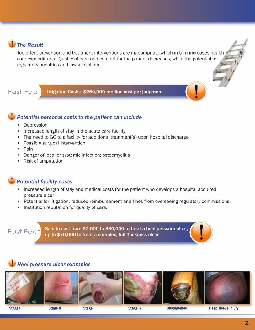
Heel pressure ulcer examples
Stage I Stage II Stage III Stage IV Unstageable Deep Tissue Injury
2.

What are the particular risks for a heel pressure ulcer?
• History of previous heel ulcer
• Immobility
• Multiple co-morbidities (emphasis on diabetes mellitus)
• Devices that place pressure on heels (TEDS, traction, CPMs, compression hose)
• Lower extremity vascular disease
• Vasoconstrictive drugs and sedation used in critical care
• Epidural and general anesthsia
• Lower extremity contractures that lead to constant unrelieved pressure
• Lower extremity orthopedic surgeries
• Lower extremity edema
• Ventilator dependency
• Agitation that results in friction and tissue distortion to heel skin
Simple education is important in pressure ulcer prevention
What can I do to prevent heel pressure ulcers in my patient?
• Be aware of the specific risk factors for heel pressure ulcer development.
(i.e. Braden mobility score 3 or less, unable to lift foot off bed unassisted or reposition independently)
• Assess and document heel skin integrity on admission and each shift.
• Treat dry skin with a moisturizer twice daily to decrease friction and shear.
• Institute regular, frequent repositioning of the extremity.
• Float heels of at risk patients; position pillows lengthwise from the knee to just above the heel,
suspending heel off the support surface.
• Consider protective heel boot if prolonged inactivity (i.e. greater than 6 hours) occurs.
• Initiate prevention protocols on at-risk patients early and aggressively.
• Provide range of motion exercises to ankles every 12 hours.
• Remove TED stockings, CPMs, compression hose, ace wraps, per facility protocol for skin assessments.
• Mobilize patients as soon as possible.
• Consult WOC nurse if patient develops a heel ulcer or deep tissue injury.
• Consult PT if patient has foot drop or is at risk for developing a plantar flexion contracture at the ankle.
• Protect patients at risk during OR and long stays in ED.
3.

Barczak CA, Barnett RI, Childs EJ, et al. Fourth national pressure ulcer prevalence survey. Adv Wound Care 1997: 10:18-26.
Salcido, R, Lee, A, Ahn, C. Heel pressure ulcers: purple heel and deep tissue injury.
Adv Wound Care 2011: 24(8) 374-382.
VanGilder C, MacFarlane GD, Harrison P, Lachenbruch C, Meyer S. The demographics of suspected deep tissue injury in the
United States: an analysis of the International Pressure Ulcer Prevalence Survey 2006-2009. Adv Skin Wound Care. 2010:
23(6)254-61.
(2) Vangilder C, Macfarlane, GD, a Meyer S. Results of nine international pressure ulcer prevalence surveys. 1989 to 2005.
Ostomy Wound Management 2008; 54(2)40-54.
Slowikowski G, Funk M. Factors associated with pressure ulcers in patients in a surgical intensive care setting. Journal of
Wound, Ostomy and Continence Nursing. 2010;37(6);619-626
Meyers T. Preventing heel pressure ulcers and plantar flexion contractures in high risk sedated patients. Journal of Wound,
Ostomy and Continence Nursing 2010;37(4)372-378.
Bennet RG, O'Sullivan JO, DeVito EM, Remsburg R.J AM Geriatric Soc. 2000; 48(1): 73-81
Ayello EA, Levine JM, Roberrtson S. CMS Updates on MDS 3.0 Section M: Skin Conditions-Change in Coding of Blister Heel
Ulcers. Adv Skin and Wound Care 2010: 23:9.
Fowler Yvonne. 21092 Head over Heels: Best Practices for Preventng Heel Ulcers. WOCN poster presentation2007.
Frain R. Decreasing the Incidence of Heel Pressure Ulcers in Long Term Care by Increasing Awareness: Results of a One Year
Program. OWM 2008; 54:2.
Langemo D, and Thompson P. Heel Pressure Ulcers: Stand Guard. Adv Skin and Wound Care 2008; 21:6.
NPUAP Guidelines on Pressure Ulcers 2007, npuap.org.
Wilson M. Heel Pressure Ulcers: An Overview of Pressure-Relieving Equipment. Wound Essentials 2007.
WOCN Society Guideline for Prevention and Management of Pressure Ulcers. June 2010, wocn.org.
References

Moisture Associated Skin Damage (MASD)
What is MASD?
Moisture Associated Skin Damage (MASD) is an inflammation of the skin which may or may not be
seen as skin erosion (defined as loss of some or all of the epidermis leaving a denuded surface) or
cutaneous secondary skin infection. It includes incontinence-associated dermatitis (IAD) second-
ary to chronic exposure to urine , stool, or both, and intertriginous (area where two skin surfaces
may touch or rub together) dermatitis (ITD) secondary to moisture trapped in skin folds. It can ex-
tend from the buttocks throughout the perineal area to the posterior and inner thighs.
Fecal Incontinence is more skin damaging than urine
Effects of Chronic Moisture
Chronic moisture compromises the barrier function of the skin. The damage from urine comes from the al-
kaline moisture environment produced by urine coming into contact with fecal bacteria present in the per-
ineal skin area. Skin damage from liquid stool is secondary to the intestinal enzymes lipase and protease
that erode the keratinocyte protective first layer of the skin. Chronic liquid stool incontinence can also lead
to compromised nutrition which in turn can negatively impact overall skin integrity.
Other Contributing Factors...
Factors affecting a patient’s tissue tolerance to incontinence include: • Age
• Health status
• Poor nutritional status
• Skin exposure to friction and shear
• Type of incontinence
• Skin integrity (if edematous or inflamed the skin is at higher
risk for damage)
• Fever
• Functional ability to reach a toilet
• Cognitive ability to recognize the need to reach a toilet
• Severity of illness
• Health comorbidities
5.

Why is MASD so Important?
First, because of the patient impact. Issues include pain, which can be severe and constant depending
on the level of skin damage and cleansing techniques, and the possible development of secondary skin
infections such as candidiasis (yeast) and/ or bacterial related skin infections. Misidentification of the
cause of the skin damage can result in ineffective treatment plans. And MASD increases patient risk for
pressure ulcer development. Onset of incontinence associated dermatitis can occur quickly in critically ill
acute care patients. Frequent liquid stool and diminished cognition are key risk factors.
Legal Implications
Legal implications relate to the financial consequences when skin breakdown from moisture is
misidentified as a hospital acquired pressure ulcer. This can lead to the possibility of fines from
accrediting institutions or The Center for Medicare and Medicaid Services (CMS), and denial of
reimbursement to hospitals from insurance carriers for costs associated with pressure ulcer
treatments if the MASD related skin breakdown progresses to a Stage III or IV pressure ulcer.
Medicare/Medicaid may not cover cases of MASD
Nursing Impact
Nursing is impacted by the time and effort it takes to care for skin damaged by incontinence, and having to
answer family questions about how and why did this happen ? and what is being done about it ?....
Pride in providing quality care to all patients is a goal of every nurse.
How to differentiate IAD (incontinence-associated dermatitis) from Stage I and II Pressure Ulcers
Main Cause
Location
Skin Characteristics
Pain
Odor
Note
Exposure to urine/ stool/ or both
In the perineal area and skin folds where stool and urine can accumulate
Shiny, red, glistening. Usually no slough or devitalized tissue in the area
Can be burning, itching, tingling, severe
Urine or fecal
Candidiasis (fungal rash) often seen
IAD
6.

How to differentiate IAD (incontinence-associated dermatitis) from Stage I and II Pressure Ulcers
Main Cause
Location
Skin Characteristics
Pain
Odor
Note
Skin exposure to pressure, shear, heat and humidity secondary to inability to change
positions, inactivity, and/or overall immobility
Stage I and II Pressure Ulcers
Usually over bony prominences, or secondary to external pressures from devices such ascasts, athrombic stockings, ace wraps, or other medical devices
Stage I- non-blanchable erythema (redness) of intact skin in light-skinned person; slightbruising, pain to touch, and/or increased warmth in dark skinned person. Stage II- partialthickness abrasion with pink wound bed, superficial skin ulcerations tend to have distinctwound margins
May be sharp and intensified with movement, usually not itchy
None, unless infected
Erythema/ bruising/ and/or discomfort of Stage I ulcer may resolve with off-loading andfrequent repositioning. Stage II shallow ulcer generally heals in by re-epithelialization overa 2 week period with treatment and off-loading.
7 Steps in Preventing MASD
1. Visual inspection and patient history are key to accurately diagnosing and treating. On hospital admission
and through-out the hospital stay, thorough exam of perineal , buttock, sacral crease skin and skin folds
should be done in patients with suspected or known moisture problems (especially those with existing i
continence, anasarca, obesity, significant sweating problems, etc.)
2. Prompt removal of urine and stool, timely diaper or underpad changes, and gentle but thorough cleansing
with a pH neutral cleanser are essential.
3. Application of skin protectants/barrier ointments that are petroleum or dimethicone based protect the skin
from continuing insult.
4. Assisting patients to the toilet or commode at regular intervals, particularly early in the morning and
before bed can be very helpful.
5. Aggressive repositioning that off-loads the patient’s sacrum and ischial bony areas helps prevent
progression to pressure related skin breakdown.
6. Use of pressure redistributing overlays and mattresses may be very effectively used for this high risk
population.
7. Diversion products such as indwelling urinary catheters and/or fecal management products should be
considered and initiated only per institution protocol.
Stage I
Stage II
7.

Document Daily
Documentation of incontinence episodes, the patient’s skin condition, and treatment efforts
should be done daily. If there is lack of improvement in eliminating or minimizing the cause(s) of
incontinence, there should be a record of MD notification and actions taken.
Clinical evidence supports having a defined skin care
protocol and availability of quality incontinence care
products to achieve best outcomes of patient protection
and decreased incidence of IAD complications.
References
Black, J, Gray, M, et al. MASD Part 2: Incontinence-Associated Dermatitis and Intertriginous Dermatitis. J Wound
Ostomy Continence Nurs. 2011; 38 359-370.
Bliss D, Savik, K, et al. Incontinence-Associated Dermatitis in Critically Ill Adults. J Wound Ostomy Continence Nurs.
2011;38: 433-445.
National Pressure Ulcer Advisory Panel: Pressure Ulcer Stages Revised by NPUAP 2007, NPUAP.org
WEB MD : definitions of intertriginous areas and skin erosion
8.

Device Related Pressure Ulcers
Definitions and Device Risks
The National Pressure Ulcer Advisory Panel (NPUAP) defines pressure ulcers as lo-
calized injury to the skin and/or underlying tissue usually over a bony prominence,
as a result of pressure, or pressure in combination with shear. The panel also rec-
ognizes that medical devices used to monitor or treat patients are also likely sources
of pressure ulcers. And since most hospitalized patients are placed on some kind
of medical device, the potential risks are huge.
Patients with devices are 2-4 times more at risk for PU
Side Effects of Medical Devices
Medical Devices and the Affected Areas
Typical medical devices in the hospital setting include: • Sequential compression devices/Stockings - Lower Extremities/Achilles/Knees
• Cervical collars - Neck/Occiput/Clavicle
• Endotracheal tubes - Lips/Tongue
• BiPap - Nasal Bridge
• Fecal containment devices - Perianal/Buttocks
• Nasal cannulas - Ears/Nares
• Pulse oximetry probes (When used incorrectly)
• Nasogastric tubes - Nares/Bridges/Nose
• Splints, braces and wraps - (When in contact with the skin) Heels
• Urinary catheters (Misplacement of tubing) - Upper Thighs
9.

Challenges of Medical Devices
Medical devices are common and often crucial in proper patient care. However these same devices, by
design, can also create pressure in unexpected places. Since most devices require a secure fit to work
properly, pressure ulcers can result in areas other than the common bony prominences, such as the ears,
cheeks and abdomen. The materials used in tandem with the devices, like tape or straps, can also make
skin inspection difficult. The pressure ulcer often takes the shape of the device obstructing any sign of
the problem at hand. The microclimate of the patient’s skin can also be altered from heat and humidity
generated from the devices. These moist environments are ideal for rapid skin deterioration.
Patients who develop edema AFTER medical devices have been placed and secured are at hight risk for
the development of medical device related pressure ulcers. Frequent assessment is critical!
Side Effects of Medical Devices
When Attaching Medical Devices
• Assess skin that is in contact with devices per policy
• Pay close attention to areas with minimal adipose tissue
• Fasten and position tubes and lines with minimal tension
10.
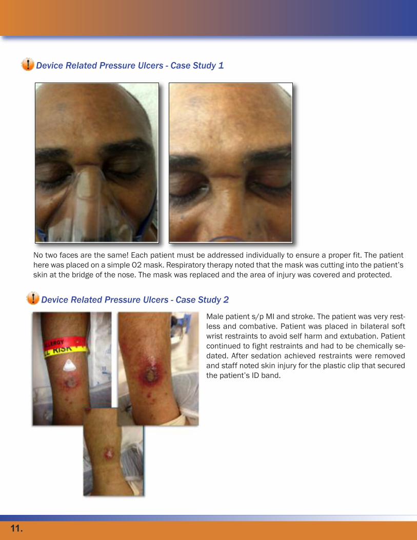
Device Related Pressure Ulcers - Case Study 1
No two faces are the same! Each patient must be addressed individually to ensure a proper fit. The patient
here was placed on a simple O2 mask. Respiratory therapy noted that the mask was cutting into the patient’s
skin at the bridge of the nose. The mask was replaced and the area of injury was covered and protected.
Male patient s/p MI and stroke. The patient was very rest-
less and combative. Patient was placed in bilateral soft
wrist restraints to avoid self harm and extubation. Patient
continued to fight restraints and had to be chemically se-
dated. After sedation achieved restraints were removed
and staff noted skin injury for the plastic clip that secured
the patient’s ID band.
Device Related Pressure Ulcers - Case Study 2
11.

When caring for Bariatric Patients there are several things to be considered
when protecting your patient from Device Related Injury.
Skin folds should be inspected routinely with each position adjustment, not
only for moisture and subsequent damage from the perspiration, wound
drainage, urine or fecal contact that we see in all patients but also for items
inadvertently left in the bed. Skin folds can obscure items such as:
• Cleansing items
• Caps from medical devices
• Alcohol prep wipes
• Dropped food
• Syringes
Use of a wicking product may be used in skin folds to assist in healing the skin of a patient with wound leakage
from open wound or a weeping fungal infection. Care must be used in the choice of this product so that the
product does not in itself increase pressure on the vulnerable damaged skin. You should never use wash clothes,
pillowcases or similar items that can increase the level of moisture in the skin folds.
Weeping wound sites in skin folds offer a distinct challenge to the care of bariatric patients. Use of cream or
ointment that will adhere to the skin in the face of moisture is paramount. Most successful is a product that will
adhere to the moist weeping tissue but wash off easily with soap and water. It must be soft, flexible and easy to
apply. It should be inert, non-toxic and contain no alcohol or latex. It should be non-sensitizing, cause no friction
and not be absorbed systemically.
Device Related Injury to Bariatric Patients
Less is Best – recommendation by NPUAP as a best practice for all
patients, including the bariatric population
Preventing and Managing Device-Related Pressure Ulcers
Because the medical devices are necessary in the hospital setting eliminating their usage is out of the ques-
tion. But, preventing and treating pressure ulcers caused by these devices remains a requirement. In order to
find that balance, devices should be repositioned often. Assessments, including loosening and removing de-
vices on every shift, are critical. And manufacturers instructions for removal and application should be closely
followed. If straps securing the devices can use less pressure and still maintain their effectiveness this might
be a viable option for controlling device-related pressure ulcers. Adding a thin hydrocolloid dressing to the
bridge of the nose when using BIPAP to stabilize skin can help reduce friction and shear. Also ensure the
patient receives a proper sized and fitted masked to minimize unnecessary pressure to the bony areas of the
face. When securing NG Tubes try not to chevron tubes to the nose, instead secure tube with transparent
12.

Incidence of Device Related Pressure Ulcers
Cervical Collars Users
• 33-44% of users (Davis et al, 1995)
• 45% of all reportable device related PU (Nix et al, 2009)
Pulse Oximetry Users in ICU
• 5% in ICU (Wille et al, 2000)
Nasoenteric (NG) Tube
• 12% of all device related PU (Penney et al, 2010)
Respiratory Devices
• 33% of all reportable device related PU (Nix et al, 2010)
•Tracheostomies 14% of all device related (Penney et al, 2010)
Pediatric Pressure Ulcers
• 50% (Baharestani and Ratliff, 2007)
Nebraska Medical Center P&I
• 34.5% (Black et al , 2010)
Duke University Hospital
• 42% (Penny et al, 2010)
PUs on the lips, cheeks, ear lobes, nasal bridge, occiput, fingertips and
webbing of the thumbs all have their origin with medical devices.
Preventing and Managing Device-Related Pressure Ulcers
dressing so the tube is "free-floating" in the nare and not riding up on the nostril. Also make sure that the NG
tube is away from patient’s cheeks and ears when in a side lying position. ET tubes should be re-positioned
q 8 hours and avoid using tape to secure devices. Commercially marketed securement devices that allow
movement of the tube should be utilized and is a best practice.
When turning patients, be sure to secure all lines, tubes and connectors away from patient’s skin. Be sure
that sequential tubing is not lying under the patient’s heels. Telemetry and SPO2 connects should not be left
under the patient, the connectors are hard and can lead to a device related injury very quickly.
Finally, another effective resource is the development of a team consisting of nursing, respiratory and wound
care to address the device related pressure ulcers from respiratory equipment. Assigning clear responsibilities
with real accountability can be a tremendous motivator in keeping device related pressures in check.
Check out the NPUAPs position paper on mucosal pressure ulcers and their
recommendation not to stage.
http://www.npuap.org/Mucosal_Pressure_Ulcer_Position_Statement_final.pdf
13.

References
Black J, Edsberg L, Baharestani M, Langemo D, Goldberg M, McNichol, L, Cuddigan, J, National Pressure Ulcer Advi-
sory Panel. Pressure Ulcers: Avoidable or Unavoidable? Results of the National Pressure Advisory Panel Consensus
Conference. Ostomy Wound Management. Feb, 2011; 24-37.
Nix D. Overview of Device Related Pressure Ulcers.
Black J, Cuddigan J, Walko M, Didier LA, Lander M, Kelpe M. Medical device related pressure ulcers in hospitalized
patients. The International Wound Journal. 2010; Vol 7, No. 5, 358 - 365.
14.

Caring for the Bariatric Patient
Bariatrics is branch of medicine that deals with the causes, prevention, and treatment of obesity. And, according
to the CDC Behavioral Risk Factor Surveillance System, since 1991 obesity rates in the Midwest have risen to
nearly 29% of the population. The Body Mass Index (BMI) is a number calculated from a person’s weight and
height and is an indicator in categorizing obesity.
According to the federal guidelines on identification, evaluation and treatment of the overweight and obese,
persons with a BMI greater than 30 may be considered obese. About 97 million adults in the United States are
overweight or obese per the National Heart, Lung, and Blood Institute Expert Panel June 1998. 66% of adults
in U.S. are overweight and about 33% are obese. Excess fat at the waist has also been linked to several other
health disorders, including high blood pressure, regardless of total body fat.
Visceral fat has been linked to metabolic disturbances and increased risk for cardiovascular disease and type
2 diabetes. In women, it is also associated with breast cancer and the need for gallbladder surgery. One reason
excess visceral fat is so harmful could be its location near the portal vein, which carries blood from the intestinal
area to the liver. Substances released by visceral fat, including free fatty acids, enter the portal vein and travel
to the liver, where they can influence the production of blood lipids. Visceral fat is directly linked with higher
total cholesterol and LDL (bad) cholesterol, lower HDL (good) cholesterol, and insulin resistance.
In all patients, the skin is critical part of care. Obese patients are no different. Pres-
sure redistribution must be started in early admission, however obese patients often
have difficulty breathing and as a result are fearful of position changes.
One way to provide safe lifting and repositioning for the bariatric patient is to es-
tablish a lift team. A lift team not only provides safety for the patient, but also for
the staff. One such finding was when a lift team was established at Indiana Uni-
versity Hospital. The team began as a research study to determine the best trans-
ferring and repositioning devices for bariatric patients. The resulting decrease in
patient handing injury associated costs were unexpected. A physical therapist
trained all the members of the new lift team in proper ergonomic positioning to pre-
vent staff and patient injuries during lifting and repositioning. A variety of devices
were used during the study but overwhelmingly the outcome was by using such a
team the facility decreased their patient injury expenses by almost $250,000 per
year.
Proper equipment is also important when caring for the obese patient. Since most hospital beds have weight
limits in the 300 range, it is important to find a bed that will accommodate the patient’s weight.
Bariatrics Defined
97 million adults in the US are overweight or obese
“Less is best” when placing linens, pads, etc in the
patient’s bed. - NAPUP
15.

Treating obesity, however, can be expensive. Medicare will not reimburse for bed purchases and the spe-
cialty beds for bariatric patients is on a CAPPED basis – so the most expensive beds are often not fully re-
imbursable.
Monitoring outcomes is very important with the bariatric patient. Your facility should monitor and allow for
changes whenever the satisfaction of both patient and staff is decreased. Education should also be of-
fered to ensure basic bariatric skills are accomplished safely and effectively.
How to Assess the Bariatric Patient
Assessing the bariatric patient can be achieved by following these steps:
• Skin: look for pressure ulcers in unusual places such as skin folds or sides of feet.
• Deep skin folds may harbor fungal growth.
• Circulation: when patient is lying flat, the weight of the pannus might impede circulation to
lower extremities. It is common to find effects of venous stasis on lower extremities.
• Wound care: may have impaired healing and increased risk of infection.
Steinmann, RN Magazine 2000
Common complications in the obese population include:
• Malnutrition
Poor diet choices often made by patient in choosing only empty high calorie foods, nutrition
consult along with definitive lab studies such as pre-albumin or transferrin
• Sleep Apnea
Excess fatty tissue in neck causes narrowing, cutting off breathing. May require CPAP
• Obesity Hypoventilation Syndrome
Weight of fatty tissue on rib cage & chest prevents full expansion leading to ventilatory insufficiency
• Delayed Wound Healing
Blood supply to fatty tissue may be insufficient to provide adequate oxygen and nutrition needed for
healing, often leading to dehiscence
• Immobility
Physical therapy start within first 24 hours to offer passive and active exercises to reduce possible
deconditioning
Bryant and Nix 2007
Skin irritations are also a common problem among the bariatric population. Tears in the skin often occur
between folds due to friction or if skin is held too taut. Treatment typically involved methods to allow free
air movement between skin folds by using products like folded towels, static air seating cushions, or wick-
ing agents. Education of staff about proper turning without stretching the skin is imperative.
16.

The three most common skin irritations faced by obese patients are: • Pressure Ulcers
Can be at risk for atypical ulcers from pressure within skin folds, tubes or catheters, or from ill-fitting chairs
and beds.
• Candidiasis
Yeast like fungus that lives in moist dark environments like skin folds. Moisture from perspiration, urine or
wound drainage exacerbates this condition.
• Contact Dermatitis
Moisture barriers that adhere to weeping or moist skin may need a coating of skin barrier to enhance ad-
herence. This is often confused with stage I pressure wounds.
The obese population is also susceptible to other health conditions. Lym-
phedema, also known as lymphatic obstruction, is a condition of localized fluid
retention caused by a compromised lymphatic system. The lymphatic system
(often referred to as the body's "second" circulatory system) collects and filters
the interstitial fluid of the body. Massive localized lymphedema (MLL) is an
extreme example of the subcutaneous fat deposits pathognomonic of lym-
phedema. Patients with this condition seek treatment when the size of the tis-
sue mass starts to interfere with their daily activities or when excoriation or
skin breakdown occurs. Treatment for Lymphedema includes Complete De-
congestive Therapy [CDT], or wrapping. Medicare does not cover garments
for patients who do not have wounds so any type of garment or device for lym-
phedema management is non-reimbursable for Medicare beneficiaries.
Elephantiasis nostras verrucosa (ENV)
This is an unusual progressive cutaneous hypertrophy due to chronic lymphedema, and repeated inflam-
matory episodes. It usually manifests over the lower extremities as non-pitting edema with lichenification,
hyperkeratotic papules, nodules, and verrucous, cobblestone-like placques.
The pannus is a medical term for a hanging flap of tissue. It can come in many different sizes and shapes
and can become very large, even hanging down below the knees. The extra tissue of a hanging pannus
can make personal hygiene difficult. Skin conditions such as yeast infections under the pannus are common
problems. A massive hanging pannus can get in the way of walking.
And, even a smaller pannus can be an annoyance with clothing as the individual sits or stands Panniculus
size has their own term to designate the size and amount of restriction it can bring to the patient. Patients
are rated on a 1-5 scale with 5 being the most severe.
Again, assessment and monitoring is key when caring for the obese patient. A focused physical exam must
include any factors that could impair healing process: impaired perfusion, sensation, or systemic infection.
Vascular assessment, lab tests and x-rays as needed are also part of the focused physical exam.
Patient with Lymphedema
17.

And caregivers should also take into account the patient’s overall nutrition. Without full patient function ca-
pacity the chances for healing are also limited and should be determined with the assessment. Reassess-
ment must be done whenever the ulcer does not show signs of healing within 2 weeks or if the patient’s
physical/physiological status changes.
Finally, it is important to address the patient’s pain level and to manage accordingly. Always use a validated
pain scale appropriate for your patient. Use a lift or transfer device to minimize friction and/or shear when
repositioning. Avoid postures that increase pressure and always minimize pressure ulcer pain by handling all
wounds gently.
SPECIAL FEATUREDevice Related Injury to Bariatric Patients
When caring for Bariatric Patients there are several things to be considered when protecting your patient
from Device Related Injury.
Skin folds should be inspected routinely with each position adjustment, not only for moisture and subse-
quent damage from the perspiration, wound drainage, urine or fecal contact that we see in all patients but
also for items inadvertently left in the bed. Skin folds can obscure items such as:
• Cleansing items
• Caps from medical devices
• Alcohol prep wipes
• Dropped food
Placing a hydrocolloid on the top of the thigh can give the bariatric patient a safe position to attach a Foley
catheter holder to prevent the tubing from causing pressure on the inner thigh when patient is positioned on
their side. The hydrocolloid may be left in place for up to 10 days.
Use of a wicking product may be used in skin folds to assist in healing the skin of a patient with wound leak-
age from open wound or a weeping fungal infection. Care must be used in the choice of this product so that
the product does not in itself increase pressure on the vulnerable dam-
aged skin. You should never use wash clothes, pillowcases or similar
items that can increase the level of moisture in the skin folds.
Weeping wound sites in skin folds offer a distinct challenge to the care of
bariatric patients. Use of cream or ointment that will adhere to the skin in
the face of moisture is paramount. Most successful is a product that will
adhere to the moist weeping tissue but wash off easily with soap and
water. It must be soft, flexible and easy to apply. It should be inert, non-
toxic and contain no alcohol or latex. It should be non-sensitizing, cause
no friction and not be absorbed systemically.
18.

CDC Behavioral Risk Factor Surveillance System, 2007
National Heart, Lung and Blood Institute Expert Panel, June 1998, 2001-
2004, 2007
Steinmann, RN Magazine 2000
Bryant and Nix, 2007
Lymphedema in the Morbidly Obese Patient: Unique Challenges in a Unique
Population. Volume: 54 Issue Number 1: C. Fife, MD; and M. Carter, PhD
Elephantiasis nostras verrucosa on the legs and abdomen with morbid obe-
sity in an Indian lady - Podila S Sarma MD1, Ashok Ghorpade MD MNAMS2
Dermatology Online Journal 14 (12): 20
NPUAP Quick Reference 2009
References

Charlotte Lisco, RN, BSN, CWON
Sue Hill, RN, BSN, CWOCN
Linda Lankenau, MSN, RN, CWOC
Sharon Lepper, BSN, RN, WOCN
Margaret-Ann Azzaro, RN, MSN, BSN
Charlotte Lisco is a Certified Wound, Ostomy Nurse currently on staff at Kaiser Permanente Medical Center
in Fontana, California. Charlotte has over 10 years of nursing experience ranging from Registered Nurse in
the Oncology Unit to Clinical Nurse Educator to her current position as CWON.
Sue Hill is a Certified Wound, Ostomy, Continence Nurse in the Phoenix, Arizona area. She currently works
PRN in the Wound, Ostomy, Continence department at the Mayo Clinic Hospital in Phoenix, Arizona.
Linda Lankenau is a Certified Wound, Ostomy, Continence Nurse in the greater New Jersey area. Most re-
cently Linda held the position of CWON at the Pennsylvania Hospital, in Philadelphia, where she specialized
in the management of clients with ostomies, wounds, tubes, fistulas, pressure ulcers, skin integrity and in-
continence problems.
Margaret-Ann is a Clinical Nurse Specialist ins Springfield, MA. She offers her clinical expertise to other
nurses to advance their practices and improve outcomes. Her specialized training, assists her in diagnosing
and treating illnesses through the delivery of evidence-based nursing interventions.
Sharon Lepper is a Certified Wound, Ostomy, Continence Nurse in the Indianapolis area. Most recently
Sharon held the position of WOCN at Indiana University/Methodist Hospitals, Clarian Health Partners. Along
with her nursing responsibilities, Sharon spent a great deal of her career as an educator. Among her tenure,
she taught wound care to staff on all in-patient units and educated new ostomy patients and their families
on proper ostomy pre and post-op care.

250 N. Belmont Ave. • Indianapolis, IN 46222
800.966.3462 • www.ehob.com