C 1+ 2 SURGICL PATHOLOGY OF OESOPHAGUS
91
SURGICAL PATHOLOGY OF ESOPHAGUS 4 hours
description
med
Transcript of C 1+ 2 SURGICL PATHOLOGY OF OESOPHAGUS

SURGICAL PATHOLOGY OF ESOPHAGUS
4 hours
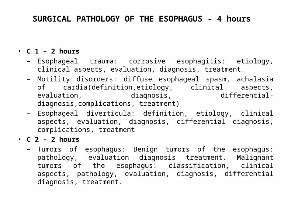
SURGICAL PATHOLOGY OF THE ESOPHAGUS - 4 hours
• C 1 – 2 hours– Esophageal trauma: corrosive esophagitis: etiology, clinical aspects,
evaluation, diagnosis, treatment.– Motility disorders: diffuse esophageal spasm, achalasia of
cardia(definition,etiology, clinical aspects, evaluation, diagnosis, differential-diagnosis,complications, treatment)
– Esophageal diverticula: definition, etiology, clinical aspects, evaluation, diagnosis, differential diagnosis, complications, treatment
• C 2 – 2 hours – Tumors of esophagus: Benign tumors of the esophagus: pathology,
evaluation diagnosis treatment. Malignant tumors of the esophagus: classification, clinical aspects, pathology, evaluation, diagnosis, differential diagnosis, treatment.
2
SURGICAL PATHOLOGY OF ESOPHAGUS
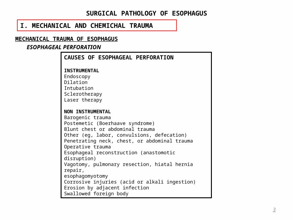
MECHANICAL TRAUMA OF ESOPHAGUSESOPHAGEAL PERFORATION
3
I. MECHANICAL AND CHEMICHAL TRAUMA
CAUSES OF ESOPHAGEAL PERFORATION INSTRUMENTALEndoscopyDilationIntubationSclerotherapyLaser therapy NON INSTRUMENTALBarogenic traumaPostemetic (Boerhaave syndrome)Blunt chest or abdominal traumaOther (eg, labor, convulsions, defecation)Penetrating neck, chest, or abdominal traumaOperative traumaEsophageal reconstruction (anastomotic disruption)Vagotomy, pulmonary resection, hiatal hernia repair,esophagomyotomyCorrosive injuries (acid or alkali ingestion)Erosion by adjacent infectionSwallowed foreign body

SURGICAL PATHOLOGY OF ESOPHAGUS
Pathophysiology • Regardless of the specific cause, the resulting mediastinitis the resulting mediastinitis and his severe consequences demand prompt demand prompt
recognition and treatment of the esophageal disruptionrecognition and treatment of the esophageal disruption. • Esophageal and gastric contents are suckedsucked into the mediastinum by respiratory movements and negative
intrathoracic pressure. • Salivary enzymes, gastric acid, bile, and food Salivary enzymes, gastric acid, bile, and food enter the mediastinum, the presence of oral bacteria the presence of oral bacteria in these
fluids initiates a fulminant infection a fulminant infection and an inflammatory response progresses.• This mediastinal “burn” mediastinal “burn” produces massive fluid accumulation, which can displace the trachea, heart, or lungs• The entire process is aggravated if there is preexisting esophageal disease preexisting esophageal disease causing obstruction distal to the
perforation.Clinical Features
• Patients with esophageal perforation characteristically present with: – cervical or thoracic pain, – difficulty swallowing, – respiratory distress, – fever.
• Pain features depends with esophageal perforation location– Cervical or upper thoracic esophagus generally cause cervical or high retrosternal pain– Middle or distal esophagus produce anterior thoracic, posterior thoracic, interscapular, or epigastric pain. – Upper thoracic esophagealUpper thoracic esophageal perforations perforations may produce signs of right pleural effusionright pleural effusion, while– Distal esophagealDistal esophageal perforation perforation is associated with left pleural effusionleft pleural effusion.
4
I. MECHANICAL AND CHEMICHAL TRAUMA ESOPHAGEAL PERFORATION
SURGICAL PATHOLOGY OF ESOPHAGUS
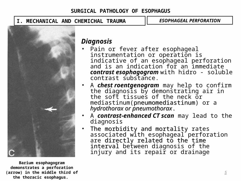
Diagnosis• Pain or fever after esophageal instrumentation or
operation is indicative of an esophageal perforation and is an indication for an immediate contrast esophagogram with hidro - soluble contrast substance.
• A chest roentgenogram may help to confirm the diagnosis by demonstrating air in the soft tissues of the neck or mediastinum(pneumomediastinumpneumomediastinum) or a hydrothorax or hydrothorax or pneumothoraxpneumothorax. .
• A contrast-enhanced CT scan may lead to the diagnosis• The morbidity and mortality The morbidity and mortality rates associated with
esophageal perforation are directly related to the time directly related to the time intervalinterval between diagnosis of the injury and its repair or drainage
5
I. MECHANICAL AND CHEMICHAL TRAUMA
Barium esophagogram demonstrates a perforation (arrow) in the middle third of the thoracic esophagus.
ESOPHAGEAL PERFORATION

SURGICAL PATHOLOGY OF ESOPHAGUS
Management(Principles of Surgical Treatment)• The initial treatment of an acute esophageal perforation focuses on:
– decreasing bacterial and chemical contamination of the mediastinum– restoring intravascular volume losses.
• Oral intake is withheld, the patient is instructed not to swallow saliva. A disposable oral dental suction is often helpful for evacuating oral secretions.
• Broad-spectrum intravenous antibiotics with activity against oral flora are administered using a combination of a cephalosporin (cefazolin or cefamandole), 1 g/4 h, and an aminoglycoside (gentamicin or tobramycin), 1 to 1.5 mg/kg/8 h, and metronidasole 2g/24h.
• Nasogastric tube decompression of the stomach is instituted to minimize possible gastroesophageal reflux and further soiling of the mediastinum.
• Therapy of esophageal perforation is influenced by: – The location of the tear– The size of the tear – The cause of the tear, – The length of delay in diagnosis, – The extent of mediastinal and pleural contamination– The presence of intrinsic esophageal disease.
• The treatment of an acute esophageal perforation must be individualized.
6
I. MECHANICAL AND CHEMICHAL TRAUMA ESOPHAGEAL PERFORATION

SURGICAL PATHOLOGY OF ESOPHAGUS
Nonoperative Therapy • Although most esophageal perforations require operative intervention; only selected patients may be
managed nonoperatively with: – Cessation of oral intake, – Administration of antibiotics, – Intravenous hydration until the disruption heals or the small contained cavity begins to decrease in size.
• Criteria for nonoperative therapy of an esophageal perforation include the following:– A local, contained disruption without evidence of pleural contamination (hydrothorax or pneumothorax), – A walled-off extravasation in which contrast material drains back into the esophagus, – Minimal or no symptoms, – Minimal or no evidence of systemic infection (fever or leukocytosis).
• The usual clinical settings in which such perforations are encountered are: – cervical esophageal tears caused by esophagoscopy; – intramural dissections that have occurred during dilation of a stricture or pneumatic dilation for
achalasia; – asymptomatic esophageal anastomotic disruption discovered on a routine postoperative contrast study.
7
I. MECHANICAL AND CHEMICHAL TRAUMA ESOPHAGEAL PERFORATION
SURGICAL PATHOLOGY OF ESOPHAGUS
• When treating such perforations conservatively, – oral hygiene should be optimized to minimize further contamination by oral bacteria– A nasogastric tube is seldom helpful. – Nutrition may be maintained by a nasogastric feeding tube, gastrostomy, or jejunostomy or by intravenous
hyperalimentation until oral intake can be resumed, usually 1 to 3 weeks after the injury. • nonoperative therapy is best suited for patients presenting no more than 24 hours after the injury with no
systemic evidence of sepsis and clearly demonstrable, contained, internally drained leaks on barium esophagogram.
• Infants with iatrogenic perforation can often be successfully managed without operation. • Perforations complicating pneumatic dilation for achalasia occur in 4% to 6% of patients, and most are small and
well-managed medically with antibiotics and intravenous hyperalimentation.• For the remainder of patients with perforations, operative therapy is generally indicated.
OPERATIVE THERAPY OF ESOPHAGEAL PERFORATIONSCervical and Upper Thoracic Esophageal Perforations lead to:
• Progressive contamination of the mediastinum as infection descends dependently along the fascial planes from the neck.
• Unless adequate drainage is accomplished, death from mediastinitis follows.• Most cervical and upper thoracic perforations may be adequately drained through a cervical approach, placing
drains in the retroesophageal space.• An incision is made parallel to the anterior border of the sternocleidomastoid muscle, which is retracted laterally along with the carotid sheath and its contents.
The trachea, thyroid gland, and strap muscles are retracted medially. It may be necessary to divide the omohyoid muscle, middle thyroid vein, and occasionally the inferior thyroid artery to reach the prevertebral fascia. Once this is identified, blunt finger dissection into the prevertebral space gives access to the abscess cavity, and appropriate drains are placed and brought out through the skin incision.
• When a cervical esophageal perforation extends into either pleural cavity or the lower mediastinum, the cervical approach is inadequate, and transthoracic drainage is required.
8
I. MECHANICAL AND CHEMICHAL TRAUMA ESOPHAGEAL PERFORATION

SURGICAL PATHOLOGY OF ESOPHAGUS
Operative Therapy Of Esophageal Perforations(suite)
9
I. MECHANICAL AND CHEMICHAL TRAUMA
Approach for drainage of a cervical esophageal perforation.
ESOPHAGEAL PERFORATION

SURGICAL PATHOLOGY OF ESOPHAGUS
Operative Therapy Of Esophageal Perforations(suite)Thoracoesophageal Perforations
Normal esophagus• The earlier an esophageal perforation is recognized and treated, the better is the chance for successful
primary repair.• Most agree that such perforations that are not associated with intrinsic esophageal disease are best treated
with primary repair of the tear combined with wide mediastinal drainage.• A change in philosophy has occurred regarding the application of primary repair to perforations occurring in
an normal esophagus regardless of the duration of the injury.• Perforations of the lower third of the esophagus are approached through a left thoracotomy in the sixth or
seventh interspace, while more proximal thoracic esophageal tears are approached through a right thoracotomy.
• Mediastinal drainage is achieved by opening the mediastinal pleura from the level of the tear to the thoracic inlet superiorly and the diaphragm inferiorly, irrigating the mediastinum, and placing a large-bore chest tube that allows transpleural drainage.
• Perforations of the intraabdominal esophagus unassociated with pleural contamination are approached through the abdomen.
Esophagus With Intrinsic Disease• Perforations associated with distal obstruction from intrinsic esophageal disease constitute a problem
because breakdown of an attempted repair is common in the presence of distal obstruction. • The associated obstruction must be relieved at the same time of repair and drainage.
10
I. MECHANICAL AND CHEMICHAL TRAUMA ESOPHAGEAL PERFORATION

SURGICAL PATHOLOGY OF ESOPHAGUS
Operative Therapy Of Esophageal Perforations(suite)• Patients with intrinsic esophageal disease that cannot be treated effectively by more conservative means are
best treated by esophageal resection. • Immediate esophageal substitution with colon(retrosternal) or stomach (in the posterior mediastinum) in the
native esophageal bed.
11
I. MECHANICAL AND CHEMICHAL TRAUMA
Final position of the mobilized stomach in the posterior mediastinum after transhiatal esophagectomy and cervical esophagogastric anastomosis.
ESOPHAGEAL PERFORATION

SURGICAL PATHOLOGY OF ESOPHAGUS
Operative Therapy Of Esophageal Perforations(suite)• In situations in which immediate esophageal reconstruction is not possible, the stomach is divided from the
esophagus, the cardia is oversewn, The intrathoracic esophagus is then mobilized through the diaphragmatic hiatus and a cervical incision, delivering the entire thoracic esophagus through the neck wound and placing it on the anterior chest wall.
• The mediastinum can be copiously irrigated through the cervical incision and the diaphragmatic hiatus at the time of esophagectomy
• A feeding jejunostomy is used for enteral alimentation until reconstruction is performed several weeks later.
12
I. MECHANICAL AND CHEMICHAL TRAUMA
Irrigation of the posterior mediastinum after transhiatal esophagectomy for irreparable esophageal disruption.
ESOPHAGEAL PERFORATION
SURGICAL PATHOLOGY OF ESOPHAGUS
Late Esophageal Perforation • The longer the time interval between the occurrence of the perforation and operative treatment, the more
inflamed are the tissues adjacent to the tear and, at least theoretically, the greater is the risk of failure of primary suture repair.
• Patients with late-recognized esophageal perforations have been treated in a variety of ways, with wide drainage alone, drainage and closure, drainage over a T-tube, esophageal resection, exclusion and diversion, and even nonoperative management.
13
I. MECHANICAL AND CHEMICHAL TRAUMA ESOPHAGEAL PERFORATION
SURGICAL PATHOLOGY OF ESOPHAGUS
CAUSTIC INJURY OF ESOPHAGUS• Caustic ingestion occurs in two categories of patients:
– Children younger than 5 years of age who accidentally swallow these agents – Adults who are attempting suicide.
• The most common agents responsible for caustic esophageal injuries are alkalis(sodiun hydroxide), acids, bleach, and detergents containing sodium tripolyphosphatesodium tripolyphosphate.
• Ingestion of detergents and bleach causes only mild esophageal irritation, which heals without adverse sequelae.
• Acids and alkalis, may have devastating effects that range from acute multiorgan necrosis and perforation to chronic esophageal and gastric strictures.
• Alkalis are more destructive, producing liquefaction necrosis, which almost ensures deep penetration• Acids usually cause coagulation necrosis that in part limits the depth of the injury. • In response to either ingested acid or alkali, reflex pyloric spasm occurs, with resultant pooling of these agents
in the gastric antrum. • Laboratory studies using the canine model have shown that both cricopharyngeal and pyloric sphincter spasm
occur when concentrated lye enters the esophagus and stomach. The esophagus contracts vigorously, propelling the caustic into the stomach. Pyloric and gastric contraction follows and propels the caustic agent back up into the esophagus. This seesaw movement of the caustic agent between the esophagus and stomach occurs for several minutes until both gastric and esophageal atony occur as the result of extensive damage to both organs.
14
I. MECHANICAL AND CHEMICHAL TRAUMA
SURGICAL PATHOLOGY OF ESOPHAGUS
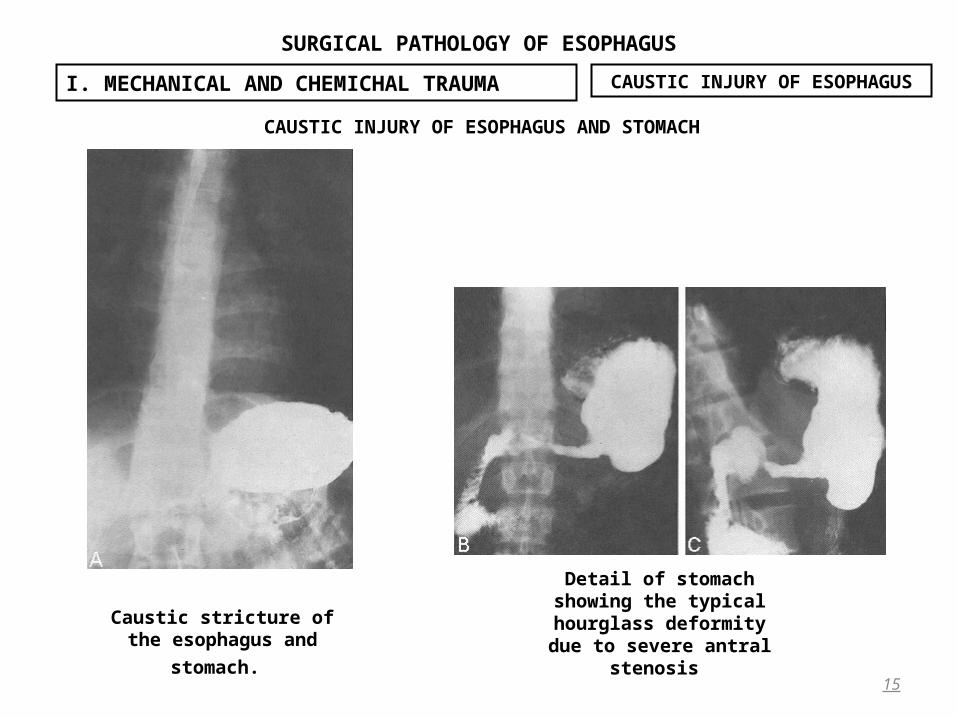
CAUSTIC INJURY OF ESOPHAGUS AND STOMACH
15
I. MECHANICAL AND CHEMICHAL TRAUMA
Detail of stomach showing the typical hourglass
deformity due to severe antral stenosis
Caustic stricture of the esophagus and stomach.
CAUSTIC INJURY OF ESOPHAGUS
SURGICAL PATHOLOGY OF ESOPHAGUS
Clinical Features• The clinical manifestations of caustic ingestion are directly related to the amount and character of the agent
ingested. • Solid alkali typically causes burns of the mouth, pharynx, and upper esophagus. The resulting severe pain
usually causes immediate expectoration so that relatively little of the caustic agent is swallowed. On examination, the mucosa of the mouth and oropharynx shows patchy areas of white to gray-black pseudomembranes.
• Patients may present with– Excessive salivation– Hoarseness, – Stridor, – Aphonia, and – Dyspnea from laryngotracheal edema or destruction.
• Liquid alkali ingestion. This form of alkali is usually swallowed quickly, producing less injury to the mouth and pharynx but more damage to the esophagus, stomach, or both.
• Patients may present with – Dysphagia, – Odynophagia, and – Aspiration,– Severe retrosternal, back, or abdominal pain and – signs of peritoneal irritation suggest that mediastinitis or peritonitis resulting from esophageal or gastric
perforation has occurred.
16
I. MECHANICAL AND CHEMICHAL TRAUMA CAUSTIC INJURY OF ESOPHAGUS

SURGICAL PATHOLOGY OF ESOPHAGUS
• Acid ingestion, gastric injury is more common; therefore, signs and symptoms are frequently localized to the abdomen.
• When esophageal or gastric perforation results from caustic ingestion, – progressively severe sepsis and – hypovolemic shock until appropriate resuscitative measures are instituted.
• In the absence of gastric or esophageal perforation, the acute clinical manifestations typically resolve within several days, with clinical improvement lasting for several weeks.
• After this, symptoms due either to esophageal or gastric stricture formation begin. Although only 10% to 25% of adult patients who ingest solid alkali develop strictures, most patients who ingest liquid alkali have severe esophageal and usually gastric injury that often results in stricture formation. Children with limited exposure from accidental ingestions are less likely to have severe injuries. Acid ingestion most often results in stricture or contracture of the antrum or pylorus.
Immediate Diagnosis And Treatment• Acute caustic ingestion is an indication for hospitalization. • Initial management centers on stabilizing the patient and assessing the severity of the injury. • Vomiting should not be induced. • Because caustic injuries produce almost instantaneous tissue damage, • Attempts to dilute the agent by having the patient drink water are futile and dangerous. In fact, this may only
aggravate the problem by producing increased gastric distention and vomiting. • Oral intake should be withheld and hypovolemia corrected with intravenous fluids.
17
I. MECHANICAL AND CHEMICHAL TRAUMA CAUSTIC INJURY OF ESOPHAGUS
SURGICAL PATHOLOGY OF ESOPHAGUS
Immediate Diagnosis And Treatment (suite)• Careful observation for evidence of airway obstruction is mandatory. Endotracheal intubation or
tracheostomy may be required if there is significant laryngeal edema or actual laryngeal destruction. • Broad-spectrum antibiotics are indicated once the diagnosis of substantial esophageal injury has been
established to diminish the risk of pulmonary infection from aspiration as well as bacterial invasion through the damaged esophageal wall.
• Although corticosteroids have been advocated in the acute phase of caustic ingestion to minimize subsequent stricture formation, their efficacy has not been established.
• Because corticosteroids may mask signs of sepsis and visceral perforation and impair healing, their use in caustic esophageal injury is potentially deleterious and is therefore not recommended.
• A relatively urgent contrast examination of the esophagus may provide important information in the patient with a caustic injury.
• Radiographically, acute mucosal esophageal injuries are seen as blurred irregular margins with linear streaking of contrast in deeper ulcers. Submucosal edema may be manifest by scalloped or straightened esophagogastric junction margins.
• Dilation of the esophagus and stomach, gastric ulcerations, air in the gastric wall, and frank extravasation of contrast material from the esophagus or stomach are common.
• A contrast esophagogram is the best way to make the diagnosis of esophageal perforation and should be performed if the diagnosis is suspected either at the time of admission or in subsequent follow-up.
• Identification of the site of perforation is vitally important vitally important in the planning of subsequent intervention. The initial esophagogram in these patients can be performed with a water-soluble agent (eg, Gastrografin), but dilute barium provides much better mucosal detail and should be used if the diagnosis of perforation is suspected.
18
I. MECHANICAL AND CHEMICHAL TRAUMA CAUSTIC INJURY OF ESOPHAGUS
SURGICAL PATHOLOGY OF ESOPHAGUS
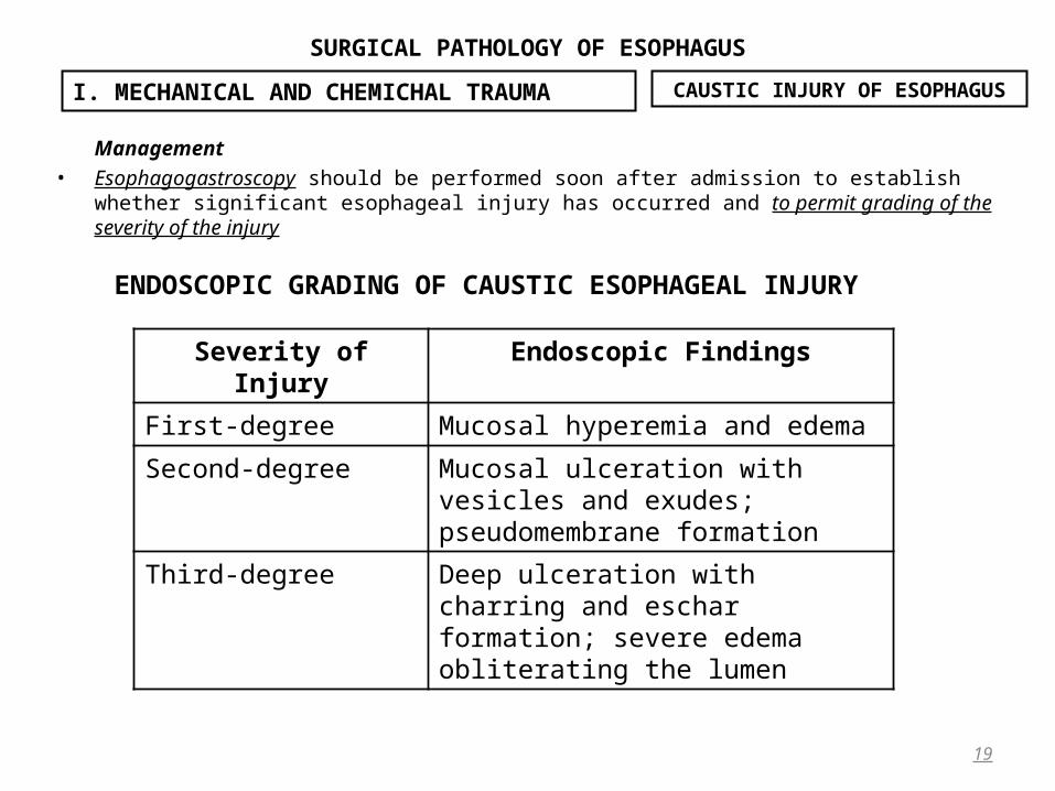
Management• Esophagogastroscopy should be performed soon after admission to establish whether significant esophageal
injury has occurred and to permit grading of the severity of the injury
19
I. MECHANICAL AND CHEMICHAL TRAUMA
ENDOSCOPIC GRADING OF CAUSTIC ESOPHAGEAL INJURY
CAUSTIC INJURY OF ESOPHAGUS
Severity of Injury Endoscopic Findings
First-degree Mucosal hyperemia and edema
Second-degree Mucosal ulceration with vesicles and exudes; pseudomembrane formation
Third-degree Deep ulceration with charring and escharformation; severe edema obliterating the lumen

SURGICAL PATHOLOGY OF ESOPHAGUS
Management (suite)• After the initial resuscitative and diagnostic measures are performed, patients with caustic injuries must be
observed carefully. • Those with first-degree burns require no other specific therapy for 24 to 48 hours. The incidence of subsequent
esophageal stricture is low in patients with such injuries. • Those who have second- or third-degree burns require careful and more prolonged observation for evidence of
esophageal or gastric necrosis during the acute phase of the injury. • Full-thickness necrosis of the esophagus, stomach, or other organs requires emergent resection. • Patients with free intraperitoneal air, mediastinal air, extravasation of contrast material from the stomach or
esophagus, peritonitis, or abdominal or mediastinal sepsis require immediate surgical exploration. Similarly, exploration is indicated in patients with severe persistent back or retrosternal pain suggesting mediastinitis and in those with metabolic acidosis suggesting visceral necrosis.
• When esophageal or gastric resection for acute caustic injury is required, restoration of alimentary continuity should be deferred until the patient has recovered from the acute insult and the development of chronic stricture formation in retained organs can be evaluated.
• Esophageal stricture formation after second- and third-degree burns is the rule, and dilation therapy has been the traditional therapy for chronic caustic esophageal strictures.
• Dilation therapy should not be instituted until at least 6 to 8 weeks after the injury, when reepithelialization is complete, to minimize the risk of esophageal perforation
• If a caustic esophageal stricture is perforated during dilation, esophagectomy and visceral esophageal substitution is the best approach because repair of a perforation proximal to a stricture is rarely successful.
20
I. MECHANICAL AND CHEMICHAL TRAUMA CAUSTIC INJURY OF ESOPHAGUS
SURGICAL PATHOLOGY OF ESOPHAGUS
Management (suite)• Strictures that cannot be adequately dilated (to a 46F dilator or larger for adults) and those that remain
refractory to dilation after 6 to 12 months require esophageal substitution, usually with colon or stomach which is the preferred esophageal substitute, but its use in these patients may be precluded by gastric scarring and contracture secondary to the original injury.
• Severe esophageal strictures resulting from caustic ingestion have been managed in the past by retrosternal colonic interposition, leaving the native, destroyed esophagus in situ in the posterior mediastinum.
• Recent data, favor resection of the damaged esophagus in virtually every case, for several reasons:• First, the obstructed esophagus can develop into a posterior mediastinal retention cyst or abscess. • Second, caustic injuries may result in destruction of the lower esophageal sphincter, resulting reflux
esophagitis in the retained esophagus • Finally, the risk of esophageal carcinoma developing after a caustic injury is about 1000 times the usual risk,
with an incidence of 0.8% to 4%, typically after a latent period of 20 to 40 years. • Therefore, a young patient whose caustic esophageal stricture is simply bypassed must be followed
indefinitely for the development of carcinoma in the native esophagus, • Resection of the strictured esophagus also permits placement of the esophageal substitute in the posterior
mediastinum in the original bed. This is the shortest and most direct route between the neck and abdominal cavity and does not require resection of the clavicle and adjacent sternum to enlarge the superior opening into the anterior mediastinum, as is required when carrying out a retrosternal esophageal substitution.
21
I. MECHANICAL AND CHEMICHAL TRAUMA CAUSTIC INJURY OF ESOPHAGUS

SURGICAL PATHOLOGY OF ESOPHAGUS
22
II. DIVERTICULA OF ESOPHAGUS

SURGICAL PATHOLOGY OF ESOPHAGUS
• An esophageal diverticulum is an epithelial-lined mucosal pouch that protrudes from the esophageal lumen. • Most esophageal diverticula are acquired, and they occur predominantly in adults. • Esophageal diverticula may be classified according to: • Their location:
– Pharyngoesophageal (Zenker) diverticula occur at the junction of the pharynx and esophagus; – Parabronchial (mid-esophageal) diverticula occur in proximity to the tracheal bifurcation; and – Epiphrenic (supradiaphragmatic) diverticula occur in the distal 10 cm of the esophagus.
• Their structure:– Diverticula containing all layers of the normal esophageal wall (mucosa, submucosa, and muscle) are termed
true diverticula, – Diverticula containing only mucosa and submucosa are termed false diverticula. • Ethiopathogenical aspects: ‒ Pressure diverticula - Most esophageal diverticula arise because elevated intraluminal pressureelevated intraluminal pressure forces forces the
mucosa and submucosa to herniate through the esophageal musculature; these are false diverticulafalse diverticula. . – Traction diverticula result from external inflammatory reaction in adjacent mediastinal lymph nodes that
adhere to the esophagus and pull the wall toward them as healing and contraction occurs, and these are true true diverticuladiverticula.
• Pharyngoesophageal and epiphrenic Pharyngoesophageal and epiphrenic diverticula are pulsion pulsion diverticula that are generally associated with abnormal esophageal motilityabnormal esophageal motility.
• Parabronchial diverticula Parabronchial diverticula are usually but not always of the traction varietytraction variety and include all layers of the esophageal wall.
23
II. DIVERTICULA OF ESOPHAGUS
SURGICAL PATHOLOGY OF ESOPHAGUS
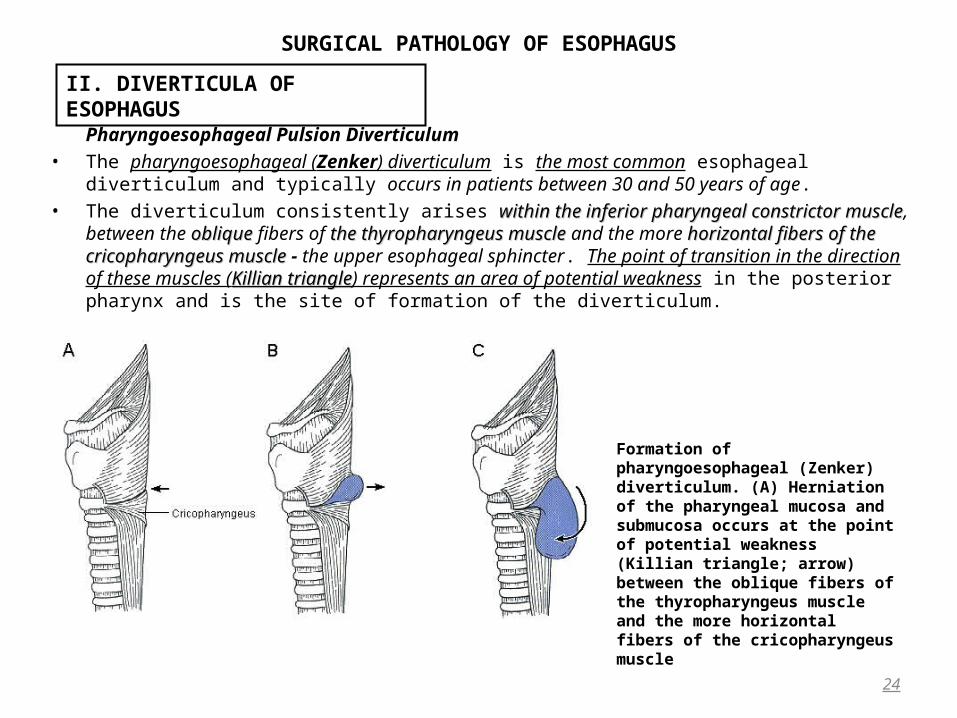
Pharyngoesophageal Pulsion Diverticulum• The pharyngoesophageal (Zenker) diverticulum is the most common esophageal diverticulum and typically
occurs in patients between 30 and 50 years of age. • The diverticulum consistently arises within the inferior pharyngeal constrictor musclewithin the inferior pharyngeal constrictor muscle, between the obliqueoblique
fibers of the thyropharyngeus muscle the thyropharyngeus muscle and the more horizontal fibers of the cricopharyngeus musclehorizontal fibers of the cricopharyngeus muscle - the upper esophageal sphincter. The point of transition in the direction of these muscles (Killian triangleKillian triangle) represents an area of potential weakness in the posterior pharynx and is the site of formation of the diverticulum.
24
II. DIVERTICULA OF ESOPHAGUS
Formation of pharyngoesophageal (Zenker) diverticulum. (A) Herniation of the pharyngeal mucosa and submucosa occurs at the point of potential weakness (Killian triangle; arrow) between the oblique fibers of the thyropharyngeus muscle and the more horizontal fibers of the cricopharyngeus muscle
SURGICAL PATHOLOGY OF ESOPHAGUS
Pharyngoesophageal Pulsion Diverticulum(suite 1)• Manometric measurementManometric measurement of upper esophageal sphincter function is difficult with existing standard recording
equipment.• Some degree of incoordination in the swallowing mechanismincoordination in the swallowing mechanism, is thought to be the basis for formation of the
Zenker diverticulum. • Pharyngeal contraction Pharyngeal contraction that occurs inappropriatelyinappropriately after cricopharyngeal closureafter cricopharyngeal closure has been demonstrated in
these patients. • Regardless of the precise motor dysfunction, a pulsion diverticulum would not occur in these patients unless
there were some reason for unusually elevated esophageal pressures.• As the swallowed bolus exerts pressure within the pharynx, mucosa and submucosa herniate through the
anatomically weak area above the cricopharyngeus muscle. The diverticulum gradually enlarges with time, extending over the cricopharyngeus muscle, and dissects downward in the prevertebral space posterior to the esophagus and occasionally into the superior mediastinum. Clinical Features
• Patients with pharyngoesophageal diverticula characteristically present with complaints of :– Cervical dysphagia, – Effortless regurgitation of undigested food or pills, – A gurgling sensation in the neck on swallowing, – Periodic choking, and – Aspiration
• Marked weight loss and dysphagia in an elderly patient may be misdiagnosed as an esophageal malignancy
25
II. DIVERTICULA OF ESOPHAGUS

SURGICAL PATHOLOGY OF ESOPHAGUS
Pharyngoesophageal Pulsion Diverticulum(suite 2)Diagnosis
• The diagnosis of a Zenker diverticulum is established with a barium esophagogram. • In evaluating the patient with a Zenker diverticulum, it must be realized that it is the degree of upper
esophageal sphincter muscle dysfunction, not the absolute size of the pouch, that determines the severity of symptoms experienced by these patients.
26
II. DIVERTICULA OF ESOPHAGUS
Posteroanterior (A) and oblique (B) views from barium esophagogram in an 15-cm pharyngoesophageal diverticulum
Small Zenker diverticulum. (A) The 2.5-cm pouch and the esophageal narrowing distal to it representing the tight cricopharyngeus sphincter. (B) Detail of pouch showing retained barium.

SURGICAL PATHOLOGY OF ESOPHAGUS
Pharyngoesophageal Pulsion Diverticulum(suite 3)• The first surgical approaches to Zenker diverticula involved simply excising the pouch and suturing the
pharyngeal defect. • The underlying upper esophageal sphincter dysfunction was not appreciated, and there was a high incidence
of suture line disruption with resulting cervical and mediastinal infection. • Cricopharyngeal myotomy, which relieves the relative obstruction distal to the pouch, is regarded as the
most important aspect of surgical treatment in these patients.
27
II. DIVERTICULA OF ESOPHAGUS
Cervical esophagomyotomy and concomitant pharyngoesophageal diverticulum resection.
SURGICAL PATHOLOGY OF ESOPHAGUS
Pharyngoesophageal Pulsion Diverticulum(suite 4)• The results of treatment(diverticulectomy) are excellent, and recurrence is rare if the relative obstruction distal to
the pouch has been relieved by complete division of the upper esophageal sphincter. • An alternative approach is diverticulopexy, which involves mobilizing the pouch, inverting it, and suspending it
from adjacent tissues so that the mouth is dependent. This operation is successful only if combined with a cervical esophagomyotomy.
• Endoscopic division of the common wall between the diverticulum (internal pharyngoesophago-myotomy, or the Dohlman procedure) has been used with success by a small number of surgeons for treatment of Zenker diverticulum.
Mid-esophageal Traction Diverticulum • Mediastinal granulomatous disease (eg, tuberculosis or histoplasmosis) is the common cause • This type of diverticulum is much smaller than the pulsion diverticulum and has a characteristic blunt tapered tip
that points toward the adjacent subcarina and parabronchial lymph nodes to which it adheres • It is typically diagnosed as an incidental finding on a barium esophagogram and almost always is asymptomatic.• No specific treatment is indicated. • At times, however, inflammatory necrosis of the granulomatous reaction may produce a fistula between the
esophagus and the tracheobronchial tree, requiring division of the fistula and interposition of normal tissues.• Mid-esophageal traction diverticula must be differentiated from pulsion diverticula, which may also occur in this
location and are associated with neuromotor esophageal dysfunction, as is the case with epiphrenic diverticula.
28
II. DIVERTICULA OF ESOPHAGUS

SURGICAL PATHOLOGY OF ESOPHAGUS
Epiphrenic diverticulum• An epiphrenic or supradiaphragmatic diverticulum occurs within the distal 10 cm of the thoracic esophagus.• It is a pulsion diverticulum that arises because of abnormally elevated intraluminal esophageal pressure
29
II. DIVERTICULA OF ESOPHAGUS
Barium esophagogram showing an epiphrenic diverticulum as well as a small traction diverticulum (arrow) of the middle esophagus.

SURGICAL PATHOLOGY OF ESOPHAGUS
Epiphrenic diverticulum(suite 1)• Although many patients do not have symptoms at the time of diagnosis on barium esophagogram, others have
symptoms from the frequently associated esophageal conditions: hiatal hernia, diffuse esophageal spasm, achalasia, reflux esophagitis, and carcinoma.
• Dysphagia and regurgitation are the common symptoms of an epiphrenic diverticulum, and retrosternal pain may occur from associated diffuse esophageal spasm.
• Esophageal manometry and acid reflux testing in these patients should be performed to define the associated motor abnormality and to assess competence of the lower esophageal sphincter mechanism
• Pouches smaller than 3 cm and causing little or no symptoms require no treatment. • Severe dysphagia, chest pain, or an anatomically dependent or enlarging pouch are indications for repair. Unless
there is an associated distal esophageal stricture or tumor, it must be inferred that the patient with an epiphrenic diverticulum has abnormally elevated intraesophageal pressure that has produced the pouch and is the result of neuromotor dysfunction. This can be documented manometrically.
• The surgical approach to epiphrenic diverticula is through a left sixth or seventh interspace posterolateral thoracotomy. This is the case even for diverticula that present to the right of the esophagus.
• A long extramucosal thoracic esophagomyotomy is performed from the level of the aortic arch to the esophagogastric junction.
• If there is an associated hiatal hernia or incompetent lower esophageal sphincter, an antireflux operation should be carried out at the same operation. If an adequate esophagomyotomy is performed, and the abnormally elevated intraesophageal pressure is thus relieved, suture line disruption and recurrence of the diverticulum are rare.
30
II. DIVERTICULA OF ESOPHAGUS
SURGICAL PATHOLOGY OF ESOPHAGUS
Classification: Primary and Secondary
• Primary motor disorderPrimary motor disorder implies that the cause of the muscular defect is not knownmuscular defect is not known.• There are four identifiable categories of primary motor disordersprimary motor disorders:
– Achalasia,– DES, – Nutcracker esophagus, and– Hypertensive LES. – Nonspecific motor disorder Includes those patients whose motor function is clearly abnormal but who do
not fall into one of the four major categories. • These five categories are derived from the manometric features on stationary motility. • In clinical activity these categories may not be as distinct as the classification implies. For example, intermediate
forms exist. • Secondary motor disordersSecondary motor disorders are the result of some systemic disease affecting the esophagussystemic disease affecting the esophagus. • The most common secondary motor disorder is the hypoperistalsis associated with complicated GERD, but the
term usually refers to a systemic connective tissue or neuromuscular disease, such as scleroderma or polymyositis.
Pharyngoesophageal Disorders• Disorders of the pharyngoesophageal phase of swallowing result from a discoordination of the neuromuscular
events involved in : – chewing, – initiation of swallowing, – propulsion of the material from the oropharynx to the cervical esophagus.
31
III. FUNCTIONAL MOTOR DISORDERS OF ESOPHAGUS
SURGICAL PATHOLOGY OF ESOPHAGUS
• The causes of pharyngoesophageal dysphagia are: – Neuromuscular diseases. The most important are
• cerebrovascular disease, • myasthenia gravis, • Parkinson disease, • motor neuron disease, • multiple sclerosis, and
– Muscular diseases such as • myotonic dystrophy and • polymyositis.
– Structural lesions of pharynx, including • tumors, • Zenker diverticula, and • scarring of the tongue or pharynx from caustic injury, • previous surgery, or • radiotherapy.
– Extrinsic compression (rarely) from goiter or cervical spine osteophytes.
Pathophysiology • All the diseases mentioned earlier produce their effects by disrupting one or more of the components of the
pharyngeal function: ‒ Weakness or immobility of the tongue Weakness or immobility of the tongue produces difficulty in oropharyngeal transfer.‒ Paralysis of the soft palateParalysis of the soft palate this accounts for the frequent occurrence of nasal regurgitation and the nasal quality to the
voice in these patients. ‒ If the larynx cannot be elevatedIf the larynx cannot be elevated, there is loss of airway protection, and patients are prone to aspiration.
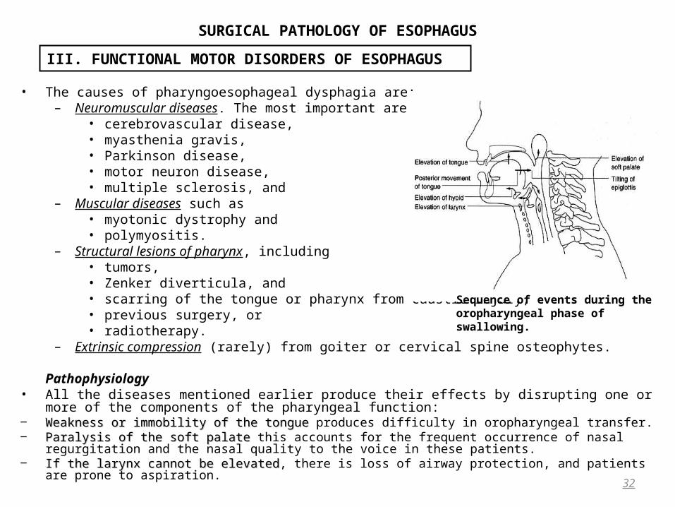
32
III. FUNCTIONAL MOTOR DISORDERS OF ESOPHAGUS
Sequence of events during the oropharyngeal phase of swallowing.
SURGICAL PATHOLOGY OF ESOPHAGUS
• In neuromuscular diseases, dysphagia is often worse for liquids than for solids. Clinical features
– Choking, – Repetitive pneumonia, – Nasal regurgitation, and hoarseness are also prominent features. – A prominent cough on assuming the recumbent position suggests the presence of Zenker diverticulum
because retained food then flows back into the pharynx. • In neuromuscular disorders examination may reveal a characteristic pattern of signs depending on the underlying
cause easily recognizable. • The voice may sound weak from vocal cord paralysis, or “wet” because of uncleared laryngeal secretions, or it
may exhibit a nasal quality from palatal paresis. It is worthwhile watching the patient swallow during the examination.
33
III. FUNCTIONAL MOTOR DISORDERS OF ESOPHAGUS

SURGICAL PATHOLOGY OF ESOPHAGUS
Investigation • Until recently, objective assessment of these conditions has been difficult, because of the rapidity of the events
during the oropharyngeal phase of swallowing. Careful analysis of – videoroentgenographic studies, – esophagoscopy, – manometry with specially designed catheters, and – 24-hour esophageal pH monitoring can identify the cause of a pharyngoesophageal dysfunction in most of
the conditions described. • Videoroentgenography is the most objective test to evaluate oropharyngeal bolus transport, pharyngeal
contraction, relaxation of the pharyngoesophageal segment, and the dynamics of airway protection during swallowing.
• Carefully performed motility studies may demonstrate insufficient relaxation or premature contraction of the cricopharyngeus, high sphincter pressure, inadequate pharyngeal pressurization, or an elevated intrabolus pressure suggesting decreased compliance of the pharyngoesophageal segment. Treatment
• Therapeutic options in all these diseases are limited by the nature of the pathology. • Medical treatment is confined to:
– Drug treatment for a specific neurologic condition (eg, myasthenia gravis or Parkinson disease), and – Therapy from a speech pathologist designed to train the patient to maximize residual function.
34
III. FUNCTIONAL MOTOR DISORDERS OF ESOPHAGUS

SURGICAL PATHOLOGY OF ESOPHAGUS
• The surgeon’s role is to reduce outflow resistance by performing cricomyotomy. Initially, this was recommended only for patients with demonstrable failure of UES relaxation. More recently, a number of reports indicate that a wide variety of neuromuscular diseases may be improved by cricomyotomy.This is because a weak or uncoordinated pharyngeal contraction may be sufficient to permit improved swallowing if outflow resistance is reduced.
• The outcome of cricomyotomy is also affected by the presence of more distal esophageal disease; when gross GERD and an associated motility defect of the esophageal body coexist, the risk of aspiration of gastric juice is increased.
• Thus, all surgical procedures for this condition should include a myotomy of the cricopharyngeus and proximal esophagus.
35
III. FUNCTIONAL MOTOR DISORDERS OF ESOPHAGUS
Completed cricomyotomy
SURGICAL PATHOLOGY OF ESOPHAGUS
• All operations on the cervical esophagus carry the risk of hematoma formation and laringeal recurrent nerve paralysis. If the diverticulum is opened during operation, there is a significantly increased risk of salivary fistula and wound infection.
• Finally, in patients who fail to benefit from reduction of outflow resistance and swallowing therapy, the only option is tube feeding. (A percutaneous endoscopic gastrostomy but a jejunostomy is the most trouble-free solution but requires a general anesthetic.
Primary Motor Disorder Of The EsophagusAchalasia
• Achalasia is the best known primary motility disorder of the esophagus. • It is characterized by failure of esophageal body peristalsis and incomplete relaxation of the LES. • It is generally thought to be caused by:
– neuronal degeneration neuronal degeneration in the myenteric plexus of the esophageal wall, causing aperistalsis, and by – loss of activity of inhibitory neurons loss of activity of inhibitory neurons in the LES, leading to incomplete relaxation.
• The cause of the neuronal degeneration is obscure; there is some evidence that previous infection with varicella-zoster virus may be responsible.
• There is also some experimental evidence, however, that obstruction at the gastroesophageal junction may produce a condition with the radiologic and manometric features of achalasia.
• This evidence suggests that outflow resistance is a primary phenomenon and that degeneration of the esophageal body is secondary.
36
III. FUNCTIONAL MOTOR DISORDERS OF ESOPHAGUS
SURGICAL PATHOLOGY OF ESOPHAGUS
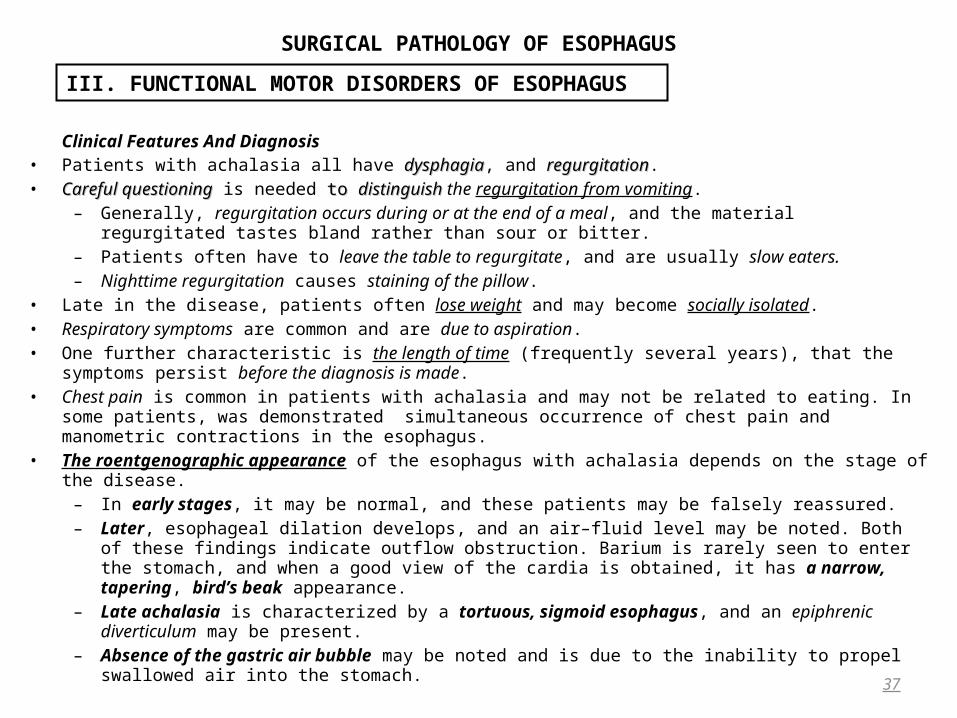
Clinical Features And Diagnosis• Patients with achalasia all have dysphagiadysphagia, and regurgitationregurgitation. • Careful questioningCareful questioning is needed to to distinguish distinguish the regurgitation from vomiting.
– Generally, regurgitation occurs during or at the end of a meal, and the material regurgitated tastes bland rather than sour or bitter.
– Patients often have to leave the table to regurgitate, and are usually slow eaters. – Nighttime regurgitation causes staining of the pillow.
• Late in the disease, patients often lose weight and may become socially isolated. • Respiratory symptoms are common and are due to aspiration. • One further characteristic is the length of time (frequently several years), that the symptoms persist before the
diagnosis is made. • Chest pain is common in patients with achalasia and may not be related to eating. In some patients, was
demonstrated simultaneous occurrence of chest pain and manometric contractions in the esophagus.• The roentgenographic appearance of the esophagus with achalasia depends on the stage of the disease.
– In early stages, it may be normal, and these patients may be falsely reassured. – Later, esophageal dilation develops, and an air–fluid level may be noted. Both of these findings indicate
outflow obstruction. Barium is rarely seen to enter the stomach, and when a good view of the cardia is obtained, it has a narrow, tapering, bird’s beak appearance.
– Late achalasia is characterized by a tortuous, sigmoid esophagus, and an epiphrenic diverticulum may be present.
– Absence of the gastric air bubble may be noted and is due to the inability to propel swallowed air into the stomach.
37
III. FUNCTIONAL MOTOR DISORDERS OF ESOPHAGUS
SURGICAL PATHOLOGY OF ESOPHAGUS
38
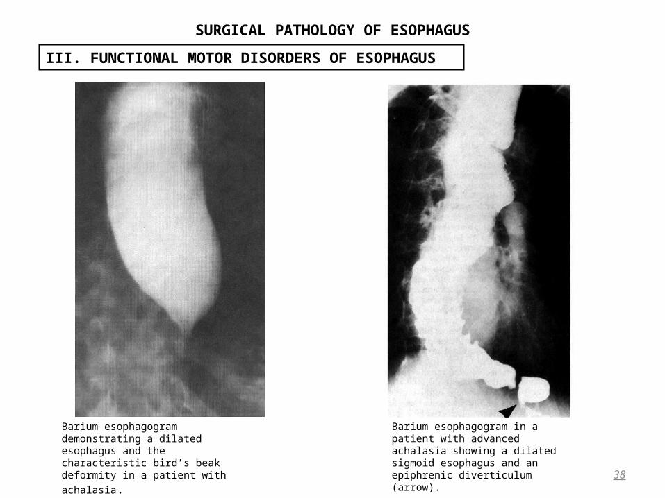
III. FUNCTIONAL MOTOR DISORDERS OF ESOPHAGUS
Barium esophagogram in a patient with advanced achalasia showing a dilated sigmoid esophagus and an epiphrenic diverticulum (arrow).
Barium esophagogram demonstrating a dilated esophagus and the characteristic bird’s beak deformity in a patient with achalasia.
SURGICAL PATHOLOGY OF ESOPHAGUS
• ENDOSCOPY frequently reveals: – Residual liquid or food in the esophagus. – The narrowing at the lower end which permits the passage of the endoscope, usually with a characteristic popping
sensation – Mild esophagitis may be observed, sometimes attributable to fermentation or stagnation of esophageal contents,
in untreated cases. – When the patient has had previous treatment for achalasia(pneumatic dilation), such inflammation is likely to be
caused by gastroesophageal reflux, – In every patient with presumed achalasia it is important to view the cardia from below with the endoscope
retroflexed because a small infiltrating gastroesophageal tumor may otherwise be missed.
• MANOMETRY is required to establish the diagnosis of achalasia. • The following are classic features of achalasia seen at stationary manometry:
– Elevated LES pressure – Incomplete LES relaxation – Absence of esophageal body peristalsis – Positive intraesophageal body pressure
• Not every patient has all four features. • Data are limited with regard to 24-hour pH and motility studies in patients with achalasia. • Generally, excessive acid exposure is rare before dilation or operation. • A characteristic pattern is the gradual fall in pH over a period of hours, and this may represent fermentation of
residual food material because it clears after swallowing water. • When true reflux episodes occur, they are prolonged because of the absence of peristalsis.
39
III. FUNCTIONAL MOTOR DISORDERS OF ESOPHAGUS

SURGICAL PATHOLOGY OF ESOPHAGUS
Treatment • Although some patients show a short-lived symptomatic and manometric response to calcium-channel blocking
agents, • The mainstay of treatment for achalasia is either balloon dilation or surgery. • The description of botulinum toxin injection has created much interest, but the reduction in LES pressure obtained
by the investigators is small and the follow-up short. Its role is therefore unproven. • Balloon dilation has the advantages that it can be done on an outpatient basis and has minimal recovery time. It is
less likely to be effective than surgical treatment and frequently needs to be repeated.• The risk of gastroesophageal reflux after dilation is not known because large studies of 24-hour pH monitoring
after dilation are lacking, but the risk of clinically significant symptoms appears to be low. • All surgical procedures employ a variant of Heller myotomy, in which the circular muscle of the lower esophagus is
divided. In the United States, most myotomies are carried out through the chest, but the abdominal route is favored in Europe.
• Regardless of the route chosen, the four important principles are • (1) adequate myotomy, • (2) minimal hiatal disturbance, • (3) antireflux protection without the creation of obstruction, and • (4) prevention of closure of the myotomy with healing. • The advent of minimally invasive surgery has led to the development of thoracoscopic and laparoscopic
myotomies, and these are widely performed with comparable results to open surgery. • There is broad agreement that if the myotomy is performed through the abdomen, an antireflux procedure should
be added, Either a posterior (Toupet) or anterior (Dor) hemifundoplication should be used.
40
III. FUNCTIONAL MOTOR DISORDERS OF ESOPHAGUS
SURGICAL PATHOLOGY OF ESOPHAGUS
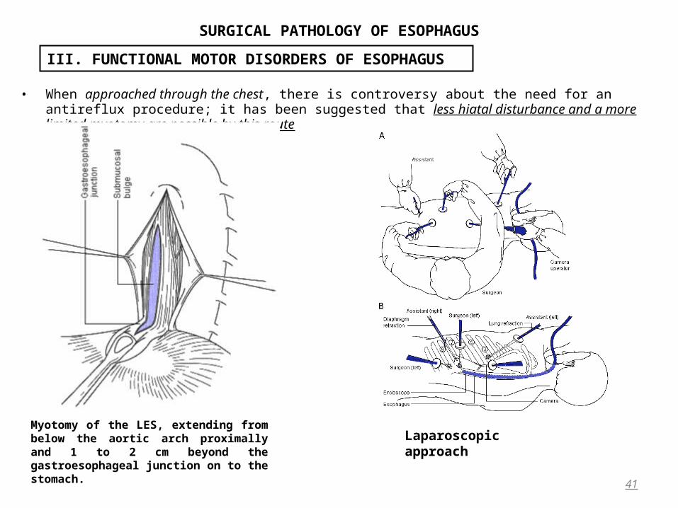
• When approached through the chest, there is controversy about the need for an antireflux procedure; it has been suggested that less hiatal disturbance and a more limited myotomy are possible by this route
41
III. FUNCTIONAL MOTOR DISORDERS OF ESOPHAGUS
Myotomy of the LES, extending from below the aortic arch proximally and 1 to 2 cm beyond the gastroesophageal junction on to the stomach.
Laparoscopic approach
SURGICAL PATHOLOGY OF ESOPHAGUS
Diffuse Esophageal Spasm • Diffuse esophageal spasm is an esophageal motor disorder characterized clinically by:
– substernal chest pain or – dysphagia.
• DES differs from classic achalasia in that it represents a primary disease of the esophageal body and produces:– Less dysphagia, – Causes more chest pain, and has – Less effect on the patient’s general condition. – True symptomatic DES is more rare than achalasia.
Roentgenographic abnormalities, such as segmental spasm segmental spasm with compartmentalization of the esophagus compartmentalization of the esophagus or formation of a diverticulumformation of a diverticulum, are the anatomic correlates of the disordered motility function.
Manometric abnormalities in DES can be present over the total length of the smooth muscle portion of the esophageal body. In segmental esophageal spasm, the manometric abnormalities are confined to a short segment of the esophagus. The classic manometric finding in these patients is the frequent occurrence of simultaneous and repetitive esophageal contractions, which may be of abnormally high amplitude or long duration.
• Key to the diagnosis of DES is that the esophagus retains a degree of peristaltic ability, in contrast to that of Key to the diagnosis of DES is that the esophagus retains a degree of peristaltic ability, in contrast to that of achalasia. achalasia. A criterion of 20% or more simultaneous contractions in 10 wet swallows has been used to define DES.
• The LES in patients with DES usually shows normal resting pressure and relaxation on deglutition. • A hypertensive sphincter with poor relaxation may also be present and may represent early achalasia.• In patients with advanced disease, the radiographic appearance of tertiary contractions appears helical and has
been termed corkscrew esophagus or pseudodiverticulosis.• DES is a benign disease that rarely causes nutritional problems and does not lead to life-threatening
complications. For this reason, symptom control is the only significant goal of treatment.
42
III. FUNCTIONAL MOTOR DISORDERS OF ESOPHAGUS

SURGICAL PATHOLOGY OF ESOPHAGUS
• Medical treatment for DES is designed to abolish strong simultaneous contractions and generally employs calcium-channel blockers or long-acting nitrates.
• The surgical option is to perform myotomy of the esophageal body. • Surgery for DES is not as successful as for achalasia and is considered only when medical treatment is ineffective. • Esophageal body myotomy should always be accompanied by myotomy of the LESEsophageal body myotomy should always be accompanied by myotomy of the LES (with partial fundoplication if
performed by open surgery) because even a normal LES can impose an outflow resistance too great for the myotomized body to overcome.
43
III. FUNCTIONAL MOTOR DISORDERS OF ESOPHAGUS
Barium esophagograms of two patients with diffuse esophageal spasm, showing corkscrew esophagi with multiple contractions
SURGICAL PATHOLOGY OF ESOPHAGUS
Nutcracker Esophagus • This term nutcracker esophagus is used to describe a manometric abnormality in which the amplitude of
esophageal body peristalsis is greater than 2 standard deviations above normal. • It was first recognized when increasing numbers of patients with noncardiac chest pain were investigated by
esophageal manometry, and is the most common primary motility disorder of the esophagus. • The dominant symptom of this condition is central crushing chest pain. • It may have no relation to food ingestion but differs from angina in that it more frequently comes on at rest. • Dysphagia or classic heartburn may be present, but this tends to be overshadowed by the chest pain. • Patients with nutcracker esophagus are usually referred from cardiologists with normal coronary angiograms and
a request for esophageal motility testing. • Barium radiography and endoscopy are not usually helpful. • The pathognomonic feature on manometry is the presence of prolonged high-amplitude waves, with a peak of
more than 180 mmHg. • The waves are normally peristaltic. Many patients with noncardiac chest pain are found to have increased
esophageal acid exposure, and this subgroup is important to identify because they respond well to fundoplication. • Myotomy for isolated nutcracker esophagus with symptoms of chest pain has a low success rate, and the
mainstay of treatment for these patients is muscle relaxants, such as nitrates and calcium-channel blockers. • If features of DES are discovered on ambulatory manometry, myotomy is more likely to be successful.
44
III. FUNCTIONAL MOTOR DISORDERS OF ESOPHAGUS

SURGICAL PATHOLOGY OF ESOPHAGUS
Secondary motor disorders of esophagus• Many connective tissue and neuromuscular diseases affect the esophageal body, but the most significant is
scleroderma. Most patients with this condition develop dysphagia. • The loss of esophageal function is caused by replacement of the muscle of the lower esophagus and LES by
fibrous tissue. • The manometric hallmark of the condition is absence of LES pressure and severely impaired contraction amplitude
in the smooth muscle portion of the esophagus. • The grossly defective LES allows superimposed reflux-induced injury to occur, accelerating the loss of body
function. • Many patients experience esophageal strictures. Antireflux surgery in this situation must involve a partial
fundoplication, but some patients eventually require esophageal replacement. Sometimes, the situation is compounded by a severe delay in gastric emptying, and patients are greatly improved by performing total gastrectomy and reconstruction with a Hunt-Lawrence jejunal pouch in a Roux-en-Y fashion.
• Surgical treatment of motor disorders by myotomy cannot normally reverse the disease process; rather, it creates a defect to overcome an existing defect. In advanced disease when residual esophageal function has been destroyed, myotomy is ineffective. Further, the superimposition of an esophageal stricture on top of a primary motor disorder makes any procedure aimed at preserving the esophagus unlikely to succeed. If more than one myotomy has been attempted in the past, it is highly unlikely that any procedure short of esophagectomy will provide symptomatic relief. The indications and choice of esophageal substitute are considered in the section on esophageal replacement in benign disease.
45
III. FUNCTIONAL MOTOR DISORDERS OF ESOPHAGUS

SURGICAL PATHOLOGY OF ESOPHAGUS
• Gastroesophageal reflux is a normal phenomenon. • This can be measured only by 24-hour pH monitoring. • Most normal people experience short episodes of reflux, usually after meals.• Gastroesophageal reflux disease occurs when esophageal acid exposure exceeds that of a normal
population. • Other definitions used in the past were either nonspecific (eg, symptoms of heartburn or regurgitation) or indirect (eg, the presence of a hiatal hernia), or they
detected the disease only when complications such as esophagitis were present.
• The ready availability of 24-hour esophageal pH monitoring allows the physician The ready availability of 24-hour esophageal pH monitoring allows the physician to quantitate the to quantitate the abnormality, to assess objectively the response to treatment, and to formulate a logical approach to therapy. abnormality, to assess objectively the response to treatment, and to formulate a logical approach to therapy.
• It is estimated that 7% of Americans suffer from daily heartburn, and up to 30% use antacids at least once a month.
• Most people whose symptoms are controlled by such means do not consult a physician, and of those who do, few are referred to surgeons. Pathophisiology
• Pathologic gastroesophageal reflux may result from the following causes:– A mechanical defect in the LESA mechanical defect in the LES. . This accounts for about 50% to 60% of patients with increased
esophageal acid exposure. It is important to identify these patients because they generally have a good outcome after antireflux surgery but a poor response to medical treatment.
– Inefficient esophageal clearanceInefficient esophageal clearance of refluxed gastric juice and – Abnormalities of the gastric reservoir that augment physiologic refluxAbnormalities of the gastric reservoir that augment physiologic reflux. Clinical features
• Symptoms of GERD can be classified as either: – Typical (ie, heartburn and regurgitation) or – Atypical (ie, noncardiac chest pain, pulmonary problems such as asthma, recurrent pneumonia or
progressive fibrosis, laryngeal symptoms such as hoarseness and aspiration, and loss of dental enamel). 46
IV. Gastroesophageal Reflux Disease 1
SURGICAL PATHOLOGY OF ESOPHAGUS
• Heartburn is the most common symptom associated with GERD, usually occurring 30 to 60 minutes after meals. Heartburn exacerbated by lyinglying flatflat or bending overbending over suggests a profound weakness of the LES. It may be associated with
• Belching and regurgitation of acid into the throat. If the regurgitated material comes from the esophagus, it tastes bland and suggests a motor disorder, and if it regurgitates from the stomach and tastes bitter, it suggests GRD.
• Variable respiratory symptoms may result if the regurgitation is associated with aspiration:– Sometimes, the picture resembles asthma, and GERD should always be considered in managing this
condition. – A history of isolated episodes of pneumonia or – Frequent bouts of wheezing and coughing at night is also suggestive of GERD. – Hoarseness may be present from laryngeal irritation.
• Dysphagia resulting from GERD is not a typical clinical sign and results from a motility disorder secondary to esophagitis, loss of esophageal compliance, or stricture formation. Patients usually localize dysphagia to the level of the lower sternum, but we have found that cervical dysphagia is common in GERD. Patients’ localization of the site of obstruction is not always reliable; generally, an obstructing lesion does not cause symptoms to be perceived distal to the lesion. It is common to find that heartburn ceases to be a prominent symptom when a stricture has developed. By contrast, the sudden development or rapid progression of dysphagia suggests a tumor. In the absence of a history of heartburn, a squamous cancer of the esophagus is likely, but if heartburn was prominent, the most common cause is adenocarcinoma arising in Barrett’s esophagus.
• Angina-like chest pain, sometimes called noncardiac chest pain, is frequently caused by GERD. These patients often describe other classic symptoms of GERD, which tend to be mild and overshadowed by the chest pain. Of patients with angiographically negative chest pain, 20% to 50% have an esophageal cause, and of these, 50% have increased esophageal acid exposure.
• Epigastric pain and nausea may be associated with other symptoms of GERD and usually result from pathologic DGR or delayed gastric emptying. It is important to recognize these symptoms before offering a patient antireflux surgery because they may persist after operation, and the patient should be warned of their presence and the possibility of future medical or surgical therapy.
47
IV. Gastroesophageal Reflux Disease 2

SURGICAL PATHOLOGY OF ESOPHAGUS
• Bloating is mainly a gastric symptom suggesting gastric dilation secondary to aerophagia or delayed gastric emptying. It may be accompanied by adaptive relaxation of the abdominal muscles causing visible distention. Investigations
• the initial investigations include a barium esophagogram and upper gastrointestinal endoscopy. In patients with GERD, these only uncover a pathologic lesion if a complication of the disease, such as esophagitis, stricture, or Barrett’s esophagus, or a potentially related condition, such as hiatus hernia, is present.
• The next step in investigation is physiologic testing of the esophagus and stomach using esophageal manometry and pH monitoring. Additional tests depend on the abnormalities revealed by these basic assessments.
• Combined pH monitoring and :– chest roentgenography is helpful if there are respiratory symptoms; – gastric emptying tests, – gastric acid analysis for hypersecretion, and – esophageal and gastric bile probe monitoring may be required to elucidate gastric symptoms.
• Ambulatory esophageal motility may help define an esophageal motility disorder if stationary manometry is equivocal.
• As a result of this process of investigation, a comprehensive understanding of esophageal function will be reached, enabling the physician to identify the etiologic factor responsible and predict the outcome of alternative treatments.
Complications of the GERD• Complications of GERD are defined by the presence of tissue injury and include:
– Esophagitis, – Stricture, and – Barrett’s esophagus.
48
IV. Gastroesophageal Reflux Disease 3

SURGICAL PATHOLOGY OF ESOPHAGUS
• Why some patients experience complications and others do not is not known, • Several factors appear to be associated: 1. The status of the LES has emerged as a significant factor in several long-term studies, and LES dysfunction
predicts a poor response to medical treatment. The table below shows the relation of a defective sphincter to complications in 150 consecutive adult patients with proven gastroesophageal reflux.
• Note that Barrett’s esophagus is almost always associated with a mechanically defective sphincter.
• LES failure is an early event in the pathogenesis of GERD and that patients with tissue injury have more profound impairment of LES function.
49
IV. Gastroesophageal Reflux Disease 4
COMPLICATIONS OF GASTROESOPHAGEAL REFLUX DISEASE IN 150 CONSECUTIVE ADULT PATIENTS
Complication Patients Normal LES (%) Defective LES (%)None 59 58 42Esophagitis 47 23* 77Stricture 19 11 89Barrett’s esophagus 25 0 100 LES, lower esophageal sphincter.* Grade of esophagitis more severe with defective LES.

SURGICAL PATHOLOGY OF ESOPHAGUS
2. A defect of esophageal clearance that prolongs the contact time between the refluxate and the mucosa is likely to lead to increased esophageal injury. This may be due to failure of esophageal propulsion, as in primary motor disorders. More commonly, the defect in clearance is secondary to reflux-induced damage, creating a vicious cycle of increasing esophageal injury.Patients with Patients with strictures and Barrett’s esophagus may thus have a profound defect in esophageal strictures and Barrett’s esophagus may thus have a profound defect in esophageal contractility. contractility. When the injury extends beyond the mucosa, the consequent interference with esophageal function may not revert to normal when the mucosa has healed : the mucosa may heal by intensive acid-suppression therapy, the abnormalities in the LES and esophageal body generally do not. This is This is because the mucosa is repeatedly being because the mucosa is repeatedly being renewed, whereas muscle cells once damaged are unlikely to recover. renewed, whereas muscle cells once damaged are unlikely to recover.
3. The presence of a hiatal hernia is also associated with more complications of GERD. The cause-and-effect relation between hiatal herniation and GERD is controversial : – Early workers used the terms hiatal hernia and reflux esophagitis as near synonyms, whereas – Later studies showed that the feature that distinguished pathologic from physiologic reflux was not the
presence of a hiatal hernia but rather the LES pressure. – As the diagnosis of hiatal herniation has become more standardized, it has become clear that the presence of a
hiatal hernia interferes with the emptying of the distal esophagus and causes a defect in acid clearance. Thus, patients with GERD associated with a hiatal hernia have more complications of the disease than those without. Conversely, the prevalence of hiatal herniation in patients with GERD increases as the complications become more severe. Most patients with Barrett’s esophagus or stricture have a hiatal hernia.
4. The composition of the refluxed material also has an effect on the development of complications. The injurious effect of refluxed gastric juice depends on a number of factors:
50
IV. Gastroesophageal Reflux Disease 5
SURGICAL PATHOLOGY OF ESOPHAGUS
– Pepsin-induced mucosal damage is likely only at a pH of 1.0 to 2.5, but in the presence of bile salts and a higher pH, trypsin may be more important.
– Not only is trypsin activated at a pH higher than 5.0, but the solubility of potentially injurious bile– salts is greatest at neutral pH. In the clinical situation, complications of GERD are more common when there
is an alkaline component to the refluxate. – In Barrett’s esophagus, the development of complications such as stricture and ulceration is strongly
associated with increased alkaline exposure. – The presence of acid or alkaline reflux and the presence of a mechanically defective sphincter are
independent determinants of mucosal damage, and when combined, the effects are additive. A patient with both features has a 95% incidence of complications.
Esophagitis • Esophagitis is usually diagnosed by the presence of macroscopic mucosal erosions at endoscopy.
– Mere erythema of the mucosa is subjective, especially on a video screen, and is consequently of little significance.
– Erosions first appear on the apex of distal mucosal folds and progress to affect multiple folds, eventually becoming confluent.
• Histologically, erosions are characterized by loss of surface epithelium and neutrophil infiltration. Histologic abnormalities of esophagitis when the epithelium is visually normal are of uncertain relevance. Esophageal Ulceration
• Historically, esophageal ulcers were the first clinical manifestation of GERD to be described. • They resemble peptic ulcers in the stomach or duodenum in that they have a tendency to penetrate deeply and
lead to bleeding or perforation. • They are found most commonly in association with Barrett’s esophagus, often near the squamocolumnar junction
and, when healed, may lead to the high mid-esophageal stricture characteristic of that condition.
51
IV. Gastroesophageal Reflux Disease 6

SURGICAL PATHOLOGY OF ESOPHAGUS
Esophageal Ulceration • Historically, esophageal ulcers were the first clinical manifestation of GERD to be described.
– They resemble peptic ulcers in the stomach or duodenum in that they have a tendency to penetrate deeply and lead to bleeding or perforation.
– They are found most commonly in association with Barrett’s esophagus, often near the squamocolumnar junction and,
– When healed, may lead to the high mid-esophageal stricture characteristic of that condition.Esophageal Stricture
• More severe esophagitis causes circumferential changes that can cause fibrosis in the deeper layers, leading to stricture and esophageal shortening.
• Strictures have an inflammatory component as well as fibrous replacement of muscle. Improvement in the former is partly responsible for diminished dysphagia after corrective antireflux surgery or intensive medical treatment. Most reflux strictures occur in the distal esophagus unless Barrett’s esophagus is present, in which case the stricture is often more proximal. The development of a reflux stricture causes slowly progressive dysphagia for solids, usually after a long history of heartburn and regurgitation. Rapidly progressive dysphagia or severe weight loss are uncommon and suggest malignancy.
52
IV. Gastroesophageal Reflux Disease 7
SURGICAL PATHOLOGY OF ESOPHAGUS
Barrett’s Esophagus • The condition in which the esophagus is lined with columnar epithelium was first described by Norman Barrett in
1950, although he incorrectly believed it to be congenital in origin.• It is now realized that Barrett’s esophagus represents advanced GERD and is found in 7% to 10% of patients with
GERD.• It is characterized :
– endoscopically by the presence of velvety orange-red mucosa that lines the esophagus, and – histologically by the presence of columnar epithelium.
• The visual appearance at endoscopy can be confused with herniation of normal gastric mucosa above the crura, and in the past, Barrett’s esophagus was only diagnosed if the columnar mucosa extended 2 cm or more above the esophagogastric junction.
• The histologic hallmark of Barrett’s esophagus is the presence of specialized columnar epithelium, which shows the presence of specialized columnar epithelium, which shows features of features of intestinal metaplasiaintestinal metaplasia,, easily recognized by the presence of goblet cellsthe presence of goblet cells. These features may be seen in biopsies of segments less than 2 cm above the esophagogastric junction, sometimes called short-segment Barrett’s esophagus. Short-segment Barrett’s esophagus often appears as a small tongue of columnar epithelium extending above the Z-line into the lower esophagus. The presence of specialized epithelium is now regarded as the pathognomonic feature of Barrett’s esophagus regardless of how far it extends into the esophagus. Barrett’s esophagus can exist alone or can be complicated by ulceration, stricture, and malignant change.
• Once Barrett’s epithelium is present, medical therapy or antireflux surgery rarely causes it to regress.Once Barrett’s epithelium is present, medical therapy or antireflux surgery rarely causes it to regress. Unless it (Barrett’s epithelium) is actually ablated (eg, with laser therapy), it persists.
• The most significant feature of Barrett’s esophagus is ITS MALIGNANT POTENTIAL. • The metaplastic epithelium usually undergoes dysplastic change before becoming frankly neoplastic, but the
changes may be focal and thus missed on biopsy. • Most pathologists distinguish only two grades of dysplasia:
– Low grade dysplasia and – High-grade dysplasia which is synonymous with carcinoma in situ, and up to half of esophagi removed for up to half of esophagi removed for
such a condition demonstrate foci of invasive carcinoma. such a condition demonstrate foci of invasive carcinoma. 53
IV. Gastroesophageal Reflux Disease 8

SURGICAL PATHOLOGY OF ESOPHAGUS
• The exact magnitude of the risk of malignancy is debated and ranges from 1 per 50 to 1 per 150 patient-years. Even the most conservative estimates indicate a risk 40 times that seen in the general population.
• Adenocarcinoma of the esophagus is rapidly increasing in most Western countries, and Barrett’s esophagus is the only known risk factor. In the United States, adenocarcinoma accounted for about 3% of esophageal cancers In the United States, adenocarcinoma accounted for about 3% of esophageal cancers between about 1930 and 1970; between about 1930 and 1970; since the mid-1970s, its incidence has risen by 10% per yearsince the mid-1970s, its incidence has risen by 10% per year. . It now accounts for It now accounts for almost 50% of all esophageal cancersalmost 50% of all esophageal cancers. .
• The male/female ratio is 5:1. • Physiologic dysfunction in Barrett’s esophagus is characteristic of advanced reflux disease: a defective LESa defective LES, poor poor
distal esophageal body peristalsisdistal esophageal body peristalsis, and fixed hiatal herniation fixed hiatal herniation are all common. • Mucosal insensitivity to acid-induced pain is present and may explain why many patients present lateMucosal insensitivity to acid-induced pain is present and may explain why many patients present late. • Abnormal composition of gastric juice may be found, specifically the presence of duodenal juice. In the past, this was
inferred by the presence of so-called alkaline reflux (increased percentage of time at a pH of more than 7.0) on esophageal pH monitoring, but reports monitoring bilirubin confirm that Barrett’s esophagus is frequently associated with excessive bile in the esophagus.
• Repetitive injury from noxious gastric juice can lead to mutations during the repair process in can lead to mutations during the repair process in the p53 genethe p53 gene,, a gene that controls programmed cell death. Patients with adenocarcinoma arising in Barrett’s esophagus have a high Patients with adenocarcinoma arising in Barrett’s esophagus have a high incidence of p53 mutations. incidence of p53 mutations.
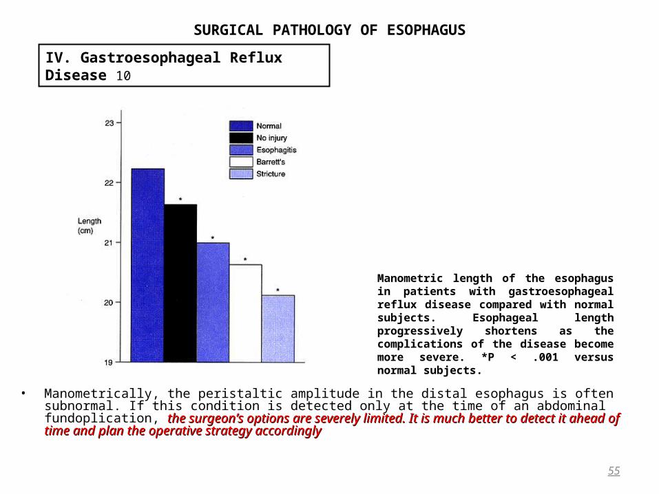
Short Esophagus • The term short esophagusThe term short esophagus is used by surgeons to describe the situation in the operating room when the when the
gastroesophageal junction cannot be brought down into the abdominal cavity without tensiongastroesophageal junction cannot be brought down into the abdominal cavity without tension. • Esophageal shortening begins to occur early in the development of GERD • Manometric studies demonstrate that shortening of the esophageal body increases as complications become
more severe. • Shortening of the longitudinal muscle, is associated with hiatal herniation, and periesophageal inflammation.
Radiologically, it is associated with fixation of the hiatal hernia; that is, the hernia does not reduce in the upright position after a swallow. Any hernia greater than 5 cm in length is likely to be associated with esophageal shortening.
54
IV. Gastroesophageal Reflux Disease 9
SURGICAL PATHOLOGY OF ESOPHAGUS
• Manometrically, the peristaltic amplitude in the distal esophagus is often subnormal. If this condition is detected only at the time of an abdominal fundoplication, the surgeon’s options are severely limited. It is much better to the surgeon’s options are severely limited. It is much better to detect it ahead of time and plan the operative strategy accordinglydetect it ahead of time and plan the operative strategy accordingly
55
IV. Gastroesophageal Reflux Disease 10
Manometric length of the esophagus in patients with gastroesophageal reflux disease compared with normal subjects. Esophageal length progressively shortens as the complications of the disease become more severe. *P < .001 versus normal subjects.

SURGICAL PATHOLOGY OF ESOPHAGUS
Surgical Treatment• The aim of surgery is to restore the patient to a life free of symptoms, without the need to take regular
medications, and without undue social, dietary, or other lifestyle restrictions. • The status of a patient whose reflux symptoms must be controlled by
– taking regular acid suppression therapy, – taking prokinetic agents, – avoiding late meals and – rich or spicy food, – eschewing: tea, coffee, alcohol, tobacco, chocolate and peppermint, – wearing only loose clothes, and – sleeping with the head of the bed elevated cannot be considered ideal.
• Only two randomized trials have compared the relative merits of medical versus surgical treatment.Only two randomized trials have compared the relative merits of medical versus surgical treatment. • Both showed a clear advantage for surgical treatment, but some are reluctant to accept this conclusion, arguing
that the medical treatment in both did not include omeprazole. • An ongoing trial comparing laparoscopic Nissen fundoplication with proton pump inhibitors may provide a
conclusion more relevant to current practice. • There is no doubt that proton pump inhibitors represent a great advance in the medical treatment of GERD, but
until recently, long-term use was discouraged by the US Food and Drug Administration.long-term use was discouraged by the US Food and Drug Administration.
56
IV. Gastroesophageal Reflux Disease 11

SURGICAL PATHOLOGY OF ESOPHAGUS
• Serum gastrin levels are usually raised in patients on long-term omeprazoleSerum gastrin levels are usually raised in patients on long-term omeprazole, and there are theoretic and experimental reasons to believe that the trophic effect of long-term gastrin elevations may predispose to the trophic effect of long-term gastrin elevations may predispose to neoplasianeoplasia. In rats, gastric carcinoid tumors have been reportedIn rats, gastric carcinoid tumors have been reported.
• Long-term omeprazole Long-term omeprazole use in patients with severe esophagitis generally heals the esophagitisgenerally heals the esophagitis if a high dose is given but is associated with atrophic gastritisbut is associated with atrophic gastritis.
• No reports of cancer in humans attributable to omeprazole have been madeNo reports of cancer in humans attributable to omeprazole have been made. • A limiting factor in the medical treatment of GERDA limiting factor in the medical treatment of GERD is that treatment addresses only acid suppressiontreatment addresses only acid suppression, ignoring the
other potentially injurious components of the refluxate, which continue to cause damage despite symptomatic continue to cause damage despite symptomatic relief. relief.
• The traditional reasons for an internist to refer a patient with GERD for surgery areThe traditional reasons for an internist to refer a patient with GERD for surgery are: – an unsatisfactory response to medical treatment and – the development of uncontrollable complications.
Operative Indications • The first requirement in the consideration of antireflux surgery are:• Objective demonstration of the presence of GERD by 24-hour pH monitoring. Second, Objective demonstration of the presence of GERD by 24-hour pH monitoring. Second, • The patient must have either symptoms or complications of the disease. The patient must have either symptoms or complications of the disease. • The disease should be caused by a defect remediable by surgical therapyThe disease should be caused by a defect remediable by surgical therapy, such as a mechanically defective LES.
Studies have indicated that a Nissen fundoplication has beneficial effects in addition to restoring the characteristics of the LES. It may accelerate gastric emptying and reduce the frequency of transient LES relaxations. Consequently, even in patients without defective sphincters, there are situations in which a Nissen fundoplication can correct the underlying abnormality.
57
IV. Gastroesophageal Reflux Disease 12
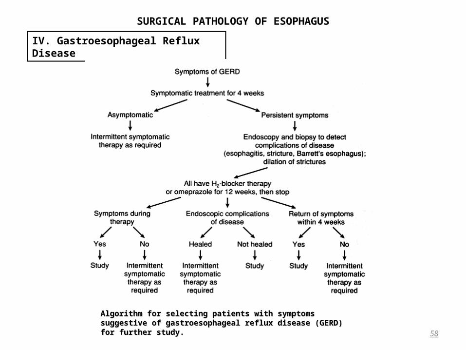
SURGICAL PATHOLOGY OF ESOPHAGUS
58
IV. Gastroesophageal Reflux Disease
Algorithm for selecting patients with symptoms suggestive of Algorithm for selecting patients with symptoms suggestive of gastroesophageal reflux disease (GERD) for further study. gastroesophageal reflux disease (GERD) for further study.

SURGICAL PATHOLOGY OF ESOPHAGUS
59
IV. Gastroesophageal Reflux Disease
CONCEPTUAL SCHEME OF THE APPROPRIATE TREATMENT AT EACH STAGE OF THE SPECTRUM OF GASTROESOPHAGEAL REFLUX DISEASE
(GERD).

SURGICAL PATHOLOGY OF ESOPHAGUS
Nissen Fundoplication
60
IV. Gastroesophageal Reflux Disease
Dissection of the left crus and the angle of His.
A. Exposure for crural closure.
B. The completed closure.
Creation of the fundoplication.
Fixation of the fundoplication.

SURGICAL PATHOLOGY OF ESOPHAGUS
Partial Fundoplications • The most widely used is the transabdominal Toupet proceduretransabdominal Toupet procedure, in which the anterior and posterior lips of the the anterior and posterior lips of the
fundoplication are sutured, not to each other, but to the right and left crura and to the esophageal wall, to fundoplication are sutured, not to each other, but to the right and left crura and to the esophageal wall, to produce a 270-degree fundoplicationproduce a 270-degree fundoplication. It can be performed without having to divide the short gastric arteries.
61
IV. Gastroesophageal Reflux Disease
SURGICAL PATHOLOGY OF ESOPHAGUS
General considerations• Most esophageal tumors are malignant; less than 1% are benign.• A knowledge of the anatomic relations between the esophagus and adjacent structures is important both in
understanding the presentation of esophageal tumors at various levelsthe presentation of esophageal tumors at various levels and in planning therapy. planning therapy. • For example, tumors involving the cervicothoracic esophagus (the segment from the cricopharyngeal
sphincter to the thoracic inlet at the level of the suprasternal notch) often involve the larynx and therefore require a laryngopharyngectomy for complete resection.
• The upper thoracic esophagus is contiguous with the posterior membranous trachea anteriorly and the aortic arch and great vessels. Thus, patients with cancer involving the upper thoracic esophagus should routinely undergo preoperative bronchoscopy to rule out invasion of the posterior membranous trachea, which would preclude resection. When resecting an upper thoracic esophageal tumor through a thoracotomy, the approach is a right fourth or fifth interspace incision because the aortic arch interferes with mobilization of the upper thoracic esophagus through the left chest.
• Mid-thoracic esophageal tumors can involve the carina or proximal main-stem bronchus, particularly where the esophagus passes behind the left main-stem bronchus, the common site for presentation of a malignant tracheoesophageal fistula. Once again, because of its anatomic proximity to the tracheobronchial tree, a mid-thoracic esophageal tumor may require a right thoracotomy, which provides optimal exposure to the carina and proximal bronchi.
• Distal esophagus Distal esophageal tumors are approached transthoracically through a left-sided approach because the most distal esophagus and esophagogastric junction cannot be adequately visualized through the right chest.
62
V. Esophageal Tumours
SURGICAL PATHOLOGY OF ESOPHAGUS
• Another important anatomic consideration when performing esophageal resection is the tunique submucosa of the esophagus, the unusual fat content of which allows a great deal of mobility of the overlying mucosa.
• Unless great care is taken to ensure that every anastomotic stitch transfixes the submucosa, an anastomotic leak may occur as a result of the mucosa retracting proximally and accurate apposition of the mucosa not being achieved.
• The esophagus is a mucosa-lined muscular tube that lacks a serosa. It is surrounded by adventitia, or mediastinal connective tissue, which is a loose fibroareolar layer.
• Transmural invasion by esophageal carcinomas is exceedingly common, the tumor not being limited by overlying pleura, in contrast to intestinal cancers, which often extend to, but not through, the adjacent peritoneum.
Benign tumors and Cysts of the esophagus
• Benign tumors of the esophagus are rare, constituting only 0.5% to 0.8% of esophageal neoplasms. • They are classified into two major groups:
– epithelial (mucosal) and epithelial (mucosal) and – intramural (extramucosal) . intramural (extramucosal) .
• Even more rare are – heterotopic collections of tissue withinheterotopic collections of tissue within the esophageal wall.
63
V. Esophageal Tumours

SURGICAL PATHOLOGY OF ESOPHAGUS
Leiomyomas
• Leiomyomas are the most common benign intramural esophageal tumor which occur in patients between 20 and 50 years.
• The tumors are multiple in 3% to 10% of patients, they have no established gender preponderance, and can occur at any level within the esophagus but rarely occur in the cervical segment.
• More than 80% of esophageal leiomyomas occur in the middle and lower thirds of the esophagusMore than 80% of esophageal leiomyomas occur in the middle and lower thirds of the esophagus. • Because calcification can occur within a leiomyoma, this must be considered in the differential diagnosis of a
calcified mediastinal mass.• Histologically, leiomyomas are composed of interlacing bundles of smooth muscle cellsinterlacing bundles of smooth muscle cells.• Tumors less than 5 cm in diameter rarely cause symptoms. • When larger than this, dysphagia, retrosternal pressure, and pain are the common complaints.• When a leiomyoma virtually encircles the esophageal lumen, obstruction and regurgitation can occur. • Bleeding more often occurs with the malignant form of the tumor, leiomyosarcoma. • Malignant degeneration of leiomyomas is exceedingly rareexceedingly rare, with fewer than 10 reported cases. • Most leiomyomas, however, are solitary and vary from 2 to 5 cm in diameter. • Another interesting variation of this tumor is diffuse leiomyomatosis of the esophagus, in which there is
extensive infiltration of the entire esophagus.
64
V. Esophageal Tumours

SURGICAL PATHOLOGY OF ESOPHAGUS
65
V. Esophageal Tumours
Esophagogram showing a leiomyoma with the typical acute angle at its junction with the esophageal wall.
•Esophageal leiomyomas produce a characteristic smooth, concave submucosal defect with sharp borders and abrupt sharp angles where the tumor meets the normal esophageal wall on barium swallow examination.
•The tumor often appears to lie half within and half outside the esophagus
•EsophagoscopyEsophagoscopy is indicated to exclude the presence of carcinoma. If the radiologic impression of a leiomyoma is confirmed endoscopically, a biopsy of the mass should not be performedshould not be performed so that subsequent extramural resection is not complicated by scarring at the biopsy site. At esophagoscopy, these tumors are characteristically mobile, have an intact overlying mucosa, and can be displaced by the advancing esophagoscope.
• Endoscopic ultrasonography has provided a new means for evaluating the esophageal leiomyoma, which is seen as a distinct intramural mass of characteristic low echodensity.
•An asymptomatic leiomyoma or one discovered incidentally on a barium swallow examination can be safely observed and followed with periodic barium esophagograms and endoscopic ultrasonography.
•Tumors that are symptomatic or larger than 5 cm in diameter should be excised.
Benign tumors and Cysts of the esophagus(suite)

SURGICAL PATHOLOGY OF ESOPHAGUS
• Once the esophagus is encircled and the tumor located, the overlying longitudinal muscle is split in the direction of its fibers. The tumor is then gently dissected away from the contiguous underlying submucosa and adjacent muscle. When enucleation of the tumor is complete, the longitudinal esophageal muscle is reapproximated, although a large extramucosal defect may be left without complication.
• When resection is complete, leiomyomas virtually never recurleiomyomas virtually never recur.Polyps
• Benign polyps of the esophagus are rare and typically arise in the cervical esophagus. • Traction on these polyps caused by repeated peristaltic contractions results in progressive lengthening of their
pedicles. • This may be responsible for their dramatic presentations, extruding into and even out of the mouth or producing extruding into and even out of the mouth or producing
asphyxia as the upper airway becomes obstructedasphyxia as the upper airway becomes obstructed. • Most benign polyps occur in older men, and these frequently are attached to the cricoid cartilage. • The tumors typically produce dysphagia, but hematemesis or melena may occur if the overlying mucosa becomes
ulcerated. • These polyps tend to be solitary with a long, cylindric configuration that may produce marked esophageal dilation.
Histologically, they are composed of fibrovascular tissue with varying amounts of associated fat. • Barium swallow findings may be nondiagnostic or inaccurately interpreted in these patients. The polyp may be
overlooked as an air bubble or may be misdiagnosed as a carcinoma, or even as a foreign body or achalasia if it has caused marked esophageal dilation .
• Similarly, esophagoscopy may fail to define the polypmay fail to define the polyp, particularly if the pedicle is not demonstrated and the mucosa overlying the polyp is normal. The endoscopist simply passes the lesion, which is soft and easily displaced with the esophagus.
• Although esophageal polyps have been removed endoscopically by electrocoagulation of the pedicle, the the recommended approach is resection through a lateral cervical esophagotomy, delivering the polyp from the recommended approach is resection through a lateral cervical esophagotomy, delivering the polyp from the esophagus, resecting its mucosal base of origin, and repairing the defect under direct vision esophagus, resecting its mucosal base of origin, and repairing the defect under direct vision
66
V. Esophageal Tumours Benign tumors and Cysts of the esophagus(suite)
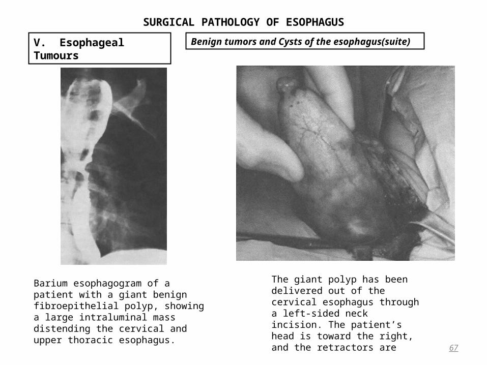
SURGICAL PATHOLOGY OF ESOPHAGUS
67
V. Esophageal Tumours Benign tumors and Cysts of the esophagus(suite)
The giant polyp has been delivered out of the cervical esophagus through a left-sided neck incision. The patient’s head is toward the right, and the retractors are
Barium esophagogram of a patient with a giant benign fibroepithelial polyp, showing a large intraluminal mass distending the cervical and upper thoracic esophagus.

SURGICAL PATHOLOGY OF ESOPHAGUS
Hemangiomas • Esophageal hemangiomas are rare, constituting 2% to 3% of benign tumors.• They are generally asymptomatic, they can be responsible for periodic gastrointestinal bleeding or even massive
and fatal hematemesis. • Asymptomatic lesions discovered incidentally during performance of an esophagoscopy should be followed with
periodic endoscopy. • Those that have bled require treatment, and although resection has been the standard approach, laser endoscopylaser endoscopy
provides an effective alternative for control of the small bleeding sites visualized through the esophagoscope. Miscellaneous Benign Tumors
• Benign esophageal tumors other than leiomyomas and polyps are extremely rare. – Granular cell myoblastomasGranular cell myoblastomas actually arise from Schwann cells, not muscle as their name implies. They
produce dysphagia, retrosternal pain, nausea, and vomitingdysphagia, retrosternal pain, nausea, and vomiting. They are difficult to diagnose endoscopically because of their submucosal location and have a characteristic grayish yellow appearance. The overlying mucosa typically shows pseudoepitheliomatous hyperplasia, which may be misdiagnosed histologically as squamous cell carcinoma. Local excision is sufficient treatment of symptomatic tumors.
– PapillomasPapillomas, sessile lobulated tumors that have a fibrous core and are covered by squamous mucosa, have been reported. Most occur in association with some degree of esophageal obstruction, most often in the distal esophagus. Papillomas have been postulated to represent localized epithelial hyperplasia or even to be represent localized epithelial hyperplasia or even to be premalignant lesionspremalignant lesions, but their true significance is unknown. On the basis of their size and radiographic configuration, papillomas at times warrant esophageal exploration to exclude malignancy, but a major resection should be avoided because local excision is adequate therapy.
– Esophageal adenomas, Esophageal adenomas, – Carcinoid tumorsCarcinoid tumors, and – Inflammatory pseudotumorsInflammatory pseudotumors also have been reported but are so rare that they are mentioned only for the
sake of completeness.
68
V. Esophageal Tumours Benign tumors and Cysts of the esophagus(suite)

SURGICAL PATHOLOGY OF ESOPHAGUS
Cysts • Esophageal cysts arise as outpouchings of the embryonic foregutoutpouchings of the embryonic foregut. • The esophageal duplication cyst is a variation of the foregut cyst; it extends along the length of the thoracic
esophagus and is lined by squamous epithelium. It has submucosal and muscle layers, the latter of which interdigitate with the outer longitudinal muscle layer of the normal esophagus.
• Three quarters of esophageal duplication cysts present in childhood, and more than 60% are located along the right side of the esophagus.
• Esophageal duplication cysts are frequently associated with vertebral anomalies (Klippel-Feil deformity or spina bifida) and spinal cord abnormalities.
• More than 60% of esophageal cysts cause either respiratory or esophageal symptoms in the first year of lifein the first year of life. Those located in the upper third of the esophagus tend to present in infancy, while lower-third cysts may be asymptomatic initially and present later in childhood.
• Adults present with: dysphagia, choking, or retrosternal pain when previously asymptomatic cysts enlarge as a result of bleeding or infection. In the rare cyst that contains ectopic gastric mucosa, a perforation of the cyst may In the rare cyst that contains ectopic gastric mucosa, a perforation of the cyst may occur. occur.
• The diagnosis of an esophageal cyst can usually be made on the basis of its typical radiographic appearance. On the standard posteroanterior chest roentgenogram, the cyst may cause:
– displacement of the trachea; on – lateral chest roentgenogram, it may appear as a retrocardiac posterior mediastinal mass.
• The barium esophagogram demonstrates a smooth extramucosal esophageal mass that rarely communicates with the esophageal lumen.
• With computed tomography (CT), may be identified The cystic nature of the lesion and its relation with adjacent mediastinal structures
• When a duplication cyst is suspected, spinal radiographs should be obtained preoperatively to identify an origin of the cyst in the notochord.
• Because esophageal cysts have a predilection for bleeding, ulceration, perforation, and infection, excision is Because esophageal cysts have a predilection for bleeding, ulceration, perforation, and infection, excision is generally recommended.generally recommended. This can generally be achieved with low morbidity by an extramucosal resection.
69
V. Esophageal Tumours Benign tumors and Cysts of the esophagus(suite)
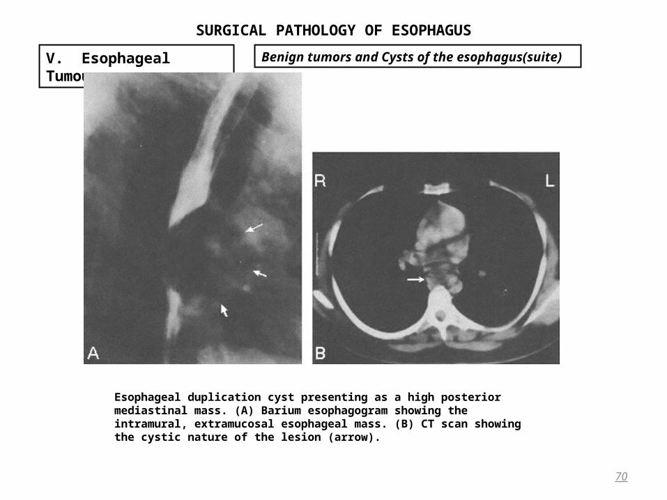
SURGICAL PATHOLOGY OF ESOPHAGUS
70
V. Esophageal Tumours Benign tumors and Cysts of the esophagus(suite)
Esophageal duplication cyst presenting as a high posterior mediastinal mass. (A) Barium esophagogram showing the intramural, extramucosal esophageal mass. (B) CT scan showing the cystic nature of the lesion (arrow).
SURGICAL PATHOLOGY OF ESOPHAGUS
Heterotopic Tumors • Islets of columnar mucosa may be found lining the pharynx and esophagus. Given the embryologic replacement of
the initial columnar ciliated epithelium by stratified squamous epithelium, the occurrence of preserved inlet patches of columnar epithelium is readily explained
• These islets are much more common near the upper end than the lower end of the esophagus. • Endoscopically, they are described as an inlet patch of columnar mucosa. • This tissue is not to be confused with Barrett’s mucosanot to be confused with Barrett’s mucosa and has little, if any, premalignant disposition. • There have also been isolated reports of
– sebaceous gland tumors as well as – ectopic pancreatic and thyroid tissue within the esophagus. These are primarily autopsy reports that have
little clinical significance.
71
V. Esophageal Tumours Benign tumors and Cysts of the esophagus(suite)

SURGICAL PATHOLOGY OF ESOPHAGUS
Squamous Cell Carcinoma • World-wide, 95% of all esophageal cancers are squamous cell carcinomas. • In the United States and Europe, however, the incidence of adenocarcinoma arising in Barrett’s mucosathe incidence of adenocarcinoma arising in Barrett’s mucosa is is
increasing at an alarming rateincreasing at an alarming rate and in many areas surpasses that of squamous cell tumors.surpasses that of squamous cell tumors.• The incidenceThe incidence of squamous cell carcinoma of the esophagus throughout the world is relatively low 3 or 4 per
100,000 population.• This disease occurs most commonly in the seventh decade of life and generally is 1.5 to 3 times more common in
men than in women.• The cause of esophageal carcinoma is unknown. It is thought to occur most often as a result of prolonged
exposure of the esophageal mucosa to noxious stimuli in patients who have a genetic predisposition to the disease:
– Carcinogenic nitrosamines in the soilnitrosamines in the soil, – Contamination of foods by mutagenic fungi, Geotrichum candidummutagenic fungi, Geotrichum candidum, – The ingestion of very hot teaingestion of very hot tea – Chewing tobacco with or without betel nut, betel leafChewing tobacco with or without betel nut, betel leaf,
– Alcohol consumption and cigarette smokingAlcohol consumption and cigarette smoking– malnutrition, malnutrition, – vitamin deficiencies, vitamin deficiencies, – anemia, anemia, – poor oral hygiene, dental caries, poor oral hygiene, dental caries, – previous gastric surgeryprevious gastric surgery– certain premalignant esophageal conditions certain premalignant esophageal conditions
72
V. Esophageal Tumours Malignant Esophageal Tumors
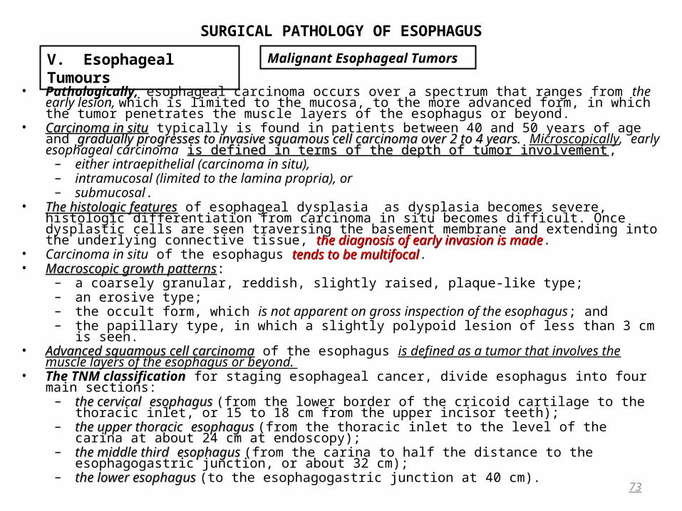
SURGICAL PATHOLOGY OF ESOPHAGUS
• Pathologically, esophageal carcinoma occurs over a spectrum that ranges from the early lesion, which is limited to the mucosa, to the more advanced form, in which the tumor penetrates the muscle layers of the esophagus or beyond.
• Carcinoma in situCarcinoma in situ typically is found in patients between 40 and 50 years of age and gradually progresses to gradually progresses to invasive squamous cell carcinoma over 2 to 4 years.invasive squamous cell carcinoma over 2 to 4 years. Microscopically, early esophageal carcinoma is defined in is defined in terms of the depth of tumor involvementterms of the depth of tumor involvement,
– either intraepithelial (carcinoma in situ), – intramucosal (limited to the lamina propria), or – submucosal.
• The histologic featuresThe histologic features of esophageal dysplasia as dysplasia becomes severe, histologic differentiation from carcinoma in situ becomes difficult. Once dysplastic cells are seen traversing the basement membrane and extending into the underlying connective tissue, the diagnosis of early invasion is madethe diagnosis of early invasion is made.
• Carcinoma in situ of the esophagus tends to be multifocaltends to be multifocal. • Macroscopic growth patternsMacroscopic growth patterns: :
– a coarsely granular, reddish, slightly raised, plaque-like type; – an erosive type; – the occult form, which is not apparent on gross inspection of the esophagus; and – the papillary type, in which a slightly polypoid lesion of less than 3 cm is seen.
• Advanced squamous cell carcinomaAdvanced squamous cell carcinoma of the esophagus is defined as a tumor that involves the muscle layers of the esophagus or beyond.
• The TNM classification for staging esophageal cancer, divide esophagus into four main sections:– the cervicalthe cervical esophagus esophagus (from the lower border of the cricoid cartilage to the thoracic inlet, or 15 to 18 cm
from the upper incisor teeth); – the upper thoracicthe upper thoracic esophagus esophagus (from the thoracic inlet to the level of the carina at about 24 cm at
endoscopy); – the middle thirdthe middle third esophagus esophagus (from the carina to half the distance to the esophagogastric junction, or about 32
cm); – the lower esophagus the lower esophagus (to the esophagogastric junction at 40 cm).
73
V. Esophageal Tumours Malignant Esophageal Tumors

SURGICAL PATHOLOGY OF ESOPHAGUS
• Using this arbitrary division of the esophagus, – 8% of squamous cell carcinomas occur in the cervical esophagus, – 55% in the upper and mid-thoracic segments, and – 37% in the lower thoracic segment.
• Macroscopically, 60% of these lesions are fungating intraluminal growths, – 25% are ulcerative lesionsulcerative lesions associated with extensive infiltration of the adjacent esophageal wall, – 15% are infiltratinginfiltrating.
• Microscopically, most squamous cell carcinomas of the esophagus are moderately differentiatedmoderately differentiated and contain islands of atypical squamous cells that infiltrate the underlying adjacent normal tissuesatypical squamous cells that infiltrate the underlying adjacent normal tissues and contain keratin pearl formation and intercellular bridges between the tumor cells.
• Esophageal carcinoma tends to be multifocal,multifocal, and a patient who survives treatment of one carcinoma has at least twice the risk of developing a second primary esophageal neoplasm.
• Esophageal carcinoma is notorious for its aggressive biologic behavioris notorious for its aggressive biologic behavior.• It tends to infiltrate locally, involving adjacent lymph nodes and spreading along the extensive submucosal
esophageal lymphatic channels. • Lack of an esophageal serosa favors tumor extension into adjacent structures such as the pericardium, aorta, the pericardium, aorta,
tracheobronchial tree, diaphragm, stomach, and left recurrent laryngeal nervetracheobronchial tree, diaphragm, stomach, and left recurrent laryngeal nerve. • Mediastinal, supraclavicular, or celiac lymph node metastases are present in at least 75% of patients with
esophageal cancer at the time of initial diagnosis. • Cervical esophageal cancers tend to drain to the deep cervical, paraesophageal, posterior mediastinal, and
tracheobronchial lymph nodes, while the lower esophageal tumors spread to paraesophageal, celiac, and splenic hilar lymph nodes.
• Distant spread to the liver and lungs is seen in 90% of cases at autopsy. • The prognosis of invasive SCC is dismal, with 5% to 12% of patients surviving 5 years. • Unfortunately, extraesophageal tumor extension is present in 70% of cases at the time of diagnosisextraesophageal tumor extension is present in 70% of cases at the time of diagnosis, and when when
lymph node metastases are present, 5-year survival is only 3%, compared with 42% when there is no lymph node lymph node metastases are present, 5-year survival is only 3%, compared with 42% when there is no lymph node spread. spread.
74
V. Esophageal Tumours Malignant Esophageal Tumors
SURGICAL PATHOLOGY OF ESOPHAGUS
Adenocarcinoma • Adenocarcinomas account for 2.5% to 8% of primary esophageal cancers, but the frequency of this tumor is
increasing at a rate surpassing that of any other cancer.• This increase is the result of the growing prevalence of adenocarcinoma arising in Barrett’s mucosagrowing prevalence of adenocarcinoma arising in Barrett’s mucosa. • Adenocarcinomas most often involve the distal third of the esophagusAdenocarcinomas most often involve the distal third of the esophagus, have a peak incidence in the sixth decade
of life, and are three times more common in men than in woman. • Esophageal adenocarcinoma has three potential originsEsophageal adenocarcinoma has three potential origins:
– The malignant degeneration of metaplastic columnar epithelium (Barrett’s mucosa),The malignant degeneration of metaplastic columnar epithelium (Barrett’s mucosa), – Heterotopic islands of columnar epitheliumHeterotopic islands of columnar epithelium, or – The esophageal submucosal glandsThe esophageal submucosal glands. !!! In addition, the esophagus may be involvedesophagus may be involved secondarily by a gastric carcinoma growing upwardsecondarily by a gastric carcinoma growing upward.
• Severe gastroesophageal reflux is a major factor in the development of a columnar epithelium–lined (Barrett’s) esophagus.
• Refluxed gastric acid, proteases, and bile erode the normal squamous epitheliumerode the normal squamous epithelium, and the residual pluripotential the residual pluripotential basal cells may differentiate along multiple cell lines, producing a variety of columnar epithelial cell typesbasal cells may differentiate along multiple cell lines, producing a variety of columnar epithelial cell types.
• The diagnosis of Barrett’s esophagus is established at endoscopy by histologic documentation of columnar mucosa extending into the tubular esophagus at least 2 cm above the anatomic esophagogastric junction. This metaplasia may extend up to the thoracic inlet
• It has been estimated that patients with Barrett’s esophagus are 40 times more likely to develop adenocarcinoma than the general population. Barrett’s mucosa occurs in three characteristic histologic patternsBarrett’s mucosa occurs in three characteristic histologic patterns:
– Gastric fundus-type epithelium,Gastric fundus-type epithelium, which has a foveolar surface pattern (no villi), but contains glands with parietal cells, chief cells, and mucous cells
– Junctional-type epitheliumJunctional-type epithelium, in which there are no villi present and in which cardiac-type mucous glands without parietal or chief cells are seen. The mucosa has a foveolar pattern that is flat and typically is seen in normal colon, gastric cardia, and villous atrophy of the small bowel.
75
V. Esophageal Tumours Malignant Esophageal Tumors
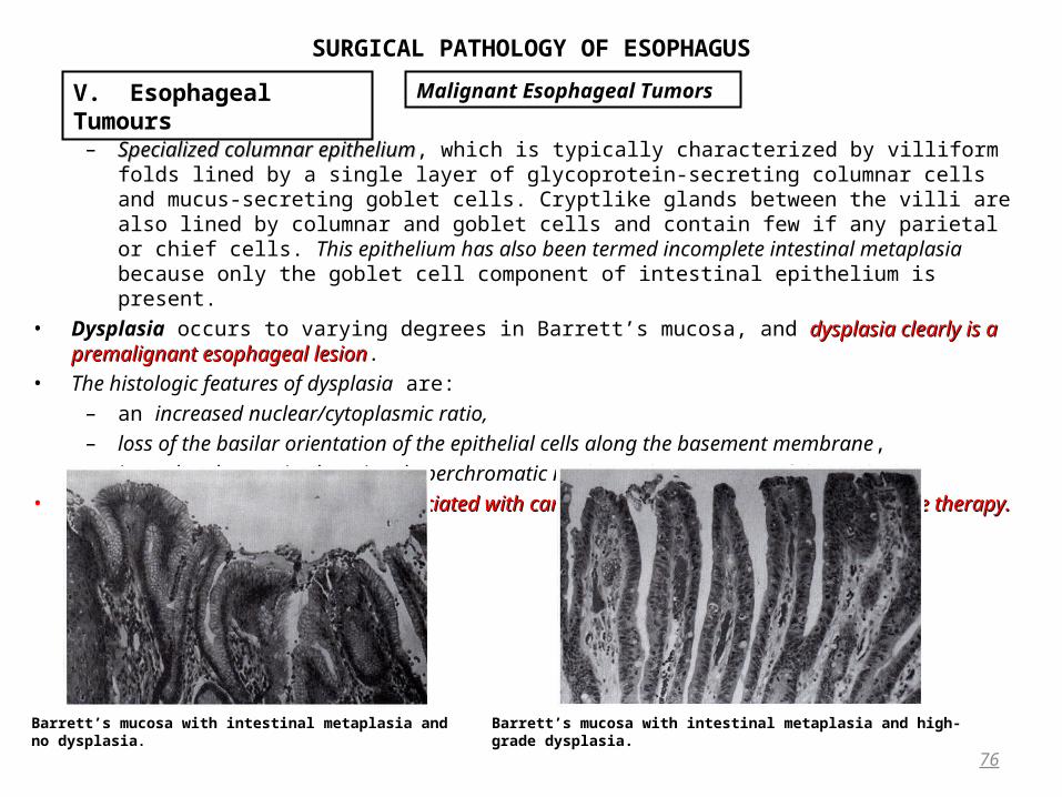
SURGICAL PATHOLOGY OF ESOPHAGUS
– Specialized columnar epitheliumSpecialized columnar epithelium, which is typically characterized by villiform folds lined by a single layer of glycoprotein-secreting columnar cells and mucus-secreting goblet cells. Cryptlike glands between the villi are also lined by columnar and goblet cells and contain few if any parietal or chief cells. This epithelium has also been termed incomplete intestinal metaplasia because only the goblet cell component of intestinal epithelium is present.
• Dysplasia occurs to varying degrees in Barrett’s mucosa, and dysplasia clearly is a premalignant esophageal dysplasia clearly is a premalignant esophageal lesionlesion.
• The histologic features of dysplasia are: – an increased nuclear/cytoplasmic ratio, – loss of the basilar orientation of the epithelial cells along the basement membrane, – irregular chromatin clumping, hyperchromatic nuclei, and prominence of the nucleoli
• Severe dysplasia is almost always associated with carcinoma in situ and mandates aggressive therapy. Severe dysplasia is almost always associated with carcinoma in situ and mandates aggressive therapy.
76
V. Esophageal Tumours Malignant Esophageal Tumors
Barrett’s mucosa with intestinal metaplasia and no dysplasia. Barrett’s mucosa with intestinal metaplasia and high-grade dysplasia.
SURGICAL PATHOLOGY OF ESOPHAGUS
• Esophageal adenocarcinoma has an aggressive biologic behavioraggressive biologic behavior that is characterized by frequent transmural invasion and lymphatic spread.
• Because these tumors arise in the lower third of the esophagus, paraesophageal, celiac axis, and splenic hilum paraesophageal, celiac axis, and splenic hilum lymph node metastases are commonlymph node metastases are common. .
• The lung and liver are the visceral organs most frequently involved by metastases. • Esophageal adenocarcinoma is associated with a 5-year survival rate of 0% to 7%.5-year survival rate of 0% to 7%. • Without lymph node involvement, survival of 5 years is possible, compared with an average survival of only 9
months in patients with lymph node involvement. Other Malignancies
• Anaplastic small cellAnaplastic small cell (oat cell) carcinoma arises in the esophagus from the same argyrophilic cells that give rise to this tumor in the lung. As is the case with their pulmonary counterparts, these tumors contain neurosecretory granules on electron microscopy. They are extremely aggressive tumors, they are commonly associated with distant spread at the time of diagnosis, and survival beyond 1 year is rare.
• Adenoid cystic esophageal carcinomaAdenoid cystic esophageal carcinoma is another rare lesion, and fewer than 50 cases have been reported. These tumors typically occur in the middle third of the esophagus, are discovered late in their course, metastasize widely, and are associated with a median survival of only 9 months.
• About 100 cases of malignant melanoma of the esophagusAbout 100 cases of malignant melanoma of the esophagus have been reported, and these rare lesions constitute less than 0.1% of esophageal malignancies. Malignant melanoma may involve the esophagus either as a primary tumor or as a secondary metastasis. In the former situation, it is thought to arise from melanocytes that occur in the esophagus. These tumors typically present as large (7 cm or more) polypoid masses, which may or may not be pigmented. The average survival is only 13.4 months, and less than 5% survive 5 years. Metastasis to liver, lymph nodes, lung, and brain is common.
• Carcinosarcoma Carcinosarcoma describes a lesion of the esophagus that has histologic features of both squamous cell carcinoma and malignant spindle cell sarcoma. These typically polypoid tumors generally occur in the distal two thirds of the esophagus, grow to large size (10 to 15 cm), and have a poor prognosis, with 2% to 6% of patients surviving 5 years.
77
V. Esophageal Tumours Malignant Esophageal Tumors
SURGICAL PATHOLOGY OF ESOPHAGUS
Local Effects • SYMPTOMS FROM ESOPHAGEAL CARCINOMA MAY BE INSIDIOUS IN ONSET:
– nonspecific retrosternal discomfort, – indigestion, or – transient dysphagia.
• Early esophageal carcinomaEarly esophageal carcinoma that is limited to the mucosa or submucosa may be completely asymptomaticmay be completely asymptomatic or may produce localized spasm that is manifested as periodic esophageal obstructionmay produce localized spasm that is manifested as periodic esophageal obstruction.
• Because the esophagus is a distensible tube, a major portion of the circumference must be involved before Because the esophagus is a distensible tube, a major portion of the circumference must be involved before obstructive symptoms developobstructive symptoms develop.
• Many patients subconsciously alter their eating habits by eliminating some foods, chewing their food more thoroughly, and using more liquids to wash down food.
• By the time of presentation to a physician with a complaint of dysphagia, symptoms have often been present for 6 to 8 months.
• DYSPHAGIADYSPHAGIA is the most common presenting symptom of esophageal carcinoma. It develops in 90% of patients and is the primary manifestation of the disease in more than 80%. It may be:
– a subtle retrosternal discomfortsubtle retrosternal discomfort as a bolus of food is swallowed, – a transient feeling of retrosternal discomfort with swallowingtransient feeling of retrosternal discomfort with swallowing that may not recur for several weeks or
months, – painful swallowing (odynophagia),painful swallowing (odynophagia),– complete esophageal obstructioncomplete esophageal obstruction.
• WEIGHT LOSSWEIGHT LOSS is the next most common symptom and is present in about 40% of patients with esophageal carcinoma.
• PAINPAIN is the initial symptom in 10% of patients. It may be:– precordial, retrosternal, epigastric, or intrascapular. – Transient retrosternal pain radiating to the back or neck as the solid bolus of food passes through the tumor
and causes local distention or muscle contraction has a much different implication than constant, boring retrosternal or epigastric pain, which more often represents local invasion by the tumor.
78
V. Esophageal Tumours Malignant Esophageal Tumors

SURGICAL PATHOLOGY OF ESOPHAGUS
• REGURGITATION OF UNDIGESTED FOODREGURGITATION OF UNDIGESTED FOOD that has not passed through the esophagus should not be confused with the vomiting of gastric contents.
• RESPIRATORY SYMPTOMSRESPIRATORY SYMPTOMS may be due either to aspiration or to direct invasion of the tracheobronchial tree by the tumor. These symptoms include cough, dyspnea, pleuritic pain, or hemoptysiscough, dyspnea, pleuritic pain, or hemoptysis.
• HEMATEMESISHEMATEMESIS is a rare, early symptom of esophageal carcinoma, but bleeding from an esophageal malignancy is seldom of sufficient quantity to cause melena.
• HOARSENESSHOARSENESS from recurrent laryngeal nerve involvement is an ominous sign of unresectability. • The course of the left main-stem bronchusThe course of the left main-stem bronchus anterior to the esophagus at the level of the carina is significant in the
patient with a mid-esophageal tumor because the common wall between the esophagus and left main-stem bronchus may become involved with tumor and lead to the development of a malignant tracheoesophageal a malignant tracheoesophageal fistulafistula. Systemic Effects
• Although the systemic effects of esophageal carcinoma are less well recognized than the local effects, they may have important clinical significance.
– Weight lossWeight loss and– Negative nitrogen balanceNegative nitrogen balance due to starvation have direct implications on the morbidity and mortality of esophageal resection
in these patients. Virtually every patient with advanced esophageal obstruction is – Dehydrated Dehydrated and total body volume depleted from impaired oral intake. The patient with esophageal obstruction is prone to
the development of – Severe hypokalemiaSevere hypokalemia with secondary muscle weakness. One to 2 L of saliva is produced each day, and the concentration of
potassium within saliva (20 mEq/mL) is higher than that in any other gastrointestinal secretions. Patients who are unable to swallow their saliva, therefore, may present with marked hypokalemia.
– Fever and systemic toxicityFever and systemic toxicity may be due to aspiration from the obstructed esophagus. • The production of parathormone by some squamous cell esophageal carcinomasThe production of parathormone by some squamous cell esophageal carcinomas has been documented and may
result in hypercalcemia, even in the absence of bone metastaseshypercalcemia, even in the absence of bone metastases.• Preoperative hypercalcemia in the patient with esophageal carcinoma and no demonstrable bone metastases has
been suggested to be a poor prognostic sign.• Apparently a vagal nerve–mediated response, the occurrence of “swallow syncope“swallow syncope” has been reported in a few
patients with esophageal obstruction due to carcinoma.
79
V. Esophageal Tumours Malignant Esophageal Tumors

SURGICAL PATHOLOGY OF ESOPHAGUS
History and Physical Examination • 90% of patients with esophageal carcinoma experience dysphagia as their primary presenting symptom, thus a
complaint of dysphagia in any adult 50 years of age or older cannot be taken lightly. • A complaint of dysphagia warrants both a barium swallowbarium swallow examination and an endoscopic evaluationendoscopic evaluation to rule out
the presence of carcinoma. The combination of esophageal biopsy and brushings for cytologic evaluation establishes a diagnosis of carcinoma in 95% of patients with malignant strictures.
• Patients with long-standing reflux symptoms that have been well controlled with medical therapy who then who then develop an increase in retrosternal discomfortdevelop an increase in retrosternal discomfort should not be presumed to have esophagitisshould not be presumed to have esophagitis. Rather, they should they should undergo appropriate radiographic and endoscopic evaluationundergo appropriate radiographic and endoscopic evaluation.
• Aside from evidence of weight loss, most patients with esophageal carcinoma have few objective findings on physical examination to aid in the diagnosis.
• Nonetheless, careful examination for cervical or supraclavicular lymph node metastases, abdominal masses, and liver nodularity is warranted. The finding of a hard, supraclavicular lymph node in the patient with an intrathoracic esophageal carcinoma warrants fine-needle aspiration biopsy. If metastatic disease is documented, the presence of a stage IV tumor has been established.
• Resectional therapy of the esophageal tumor in this situation is seldom justified because the patient’s expected survival is so poor.
• Laboratory studies should include a complete blood countcomplete blood count, blood urea nitrogenblood urea nitrogen, and serum creatinineserum creatinine to assess the state of hydration, as well as liver function testsliver function tests, including total protein and albumin levelstotal protein and albumin levels to assess nutrition. Serum electrolytes, particularly potassium and calcium levels, should also be obtainedSerum electrolytes, particularly potassium and calcium levels, should also be obtained.
• In obtaining a history from the patient who complains of dysphagia, the physician should ask the patient to the physician should ask the patient to localize with one finger on the anterior chest or neck the point at which food lodges when swallowinglocalize with one finger on the anterior chest or neck the point at which food lodges when swallowing. The patient with a mechanical esophageal obstruction such as a carcinoma is able to localize the consistent point of is able to localize the consistent point of obstruction without difficultyobstruction without difficulty. This is in contrast to the patient with neuromotor obstruction, who may only sense slow esophageal emptying diffusely in the retrosternal area.
80
V. Esophageal Tumours Malignant Esophageal Tumors Diagnostic Investigations

SURGICAL PATHOLOGY OF ESOPHAGUS
Radiographic Studies • A barium swallow examinationA barium swallow examination is the first study that should be obtained in a patient who complains of dysphagia.
Tumors of the cervical esophagus are most difficult to identify by barium swallow examination, and carcinoma of the cardia may be confused with achalasia, a benign stricture, or esophageal spasm.
• The typical esophageal carcinoma presents radiographically as an irregular, rigid narrowing of the esophageal wall.
• The normal mucosal pattern is frequently destroyed. Polypoid fungating tumors present as irregular filling defects with ulcerated borders within the esophagus.
• An old dictum relates that an esophagus that is dilated proximal to a stenosis is most indicative of a benign chronic obstruction, whereas an esophagus proximal to a carcinoma has “not had enough time” to dilate. (This observation has proved to be incorrect on numerous occasions).
• Similarly, although a smooth, tapered radiographic esophageal stricture supposedly reflects benign disease, any any stenosis merits esophageal biopsy and brushings for cytologic evaluation to rule out carcinomastenosis merits esophageal biopsy and brushings for cytologic evaluation to rule out carcinoma.
• The barium swallow examination may also show a soft tissue mass adjacent to the esophageal tumora soft tissue mass adjacent to the esophageal tumor indicative of indicative of extraesophageal local invasionextraesophageal local invasion.
• In half of patientshalf of patients with esophageal carcinoma is the plain chest radiograph abnormalis the plain chest radiograph abnormal, the most common findings being:
– an air–fluid level in the obstructed esophagus, – a dilated esophagus, – abnormal mediastinal soft tissue representing adenopathy, – pleural effusions, or – pulmonary metastases.
81
V. Esophageal Tumours Malignant Esophageal Tumors Diagnostic Investigations
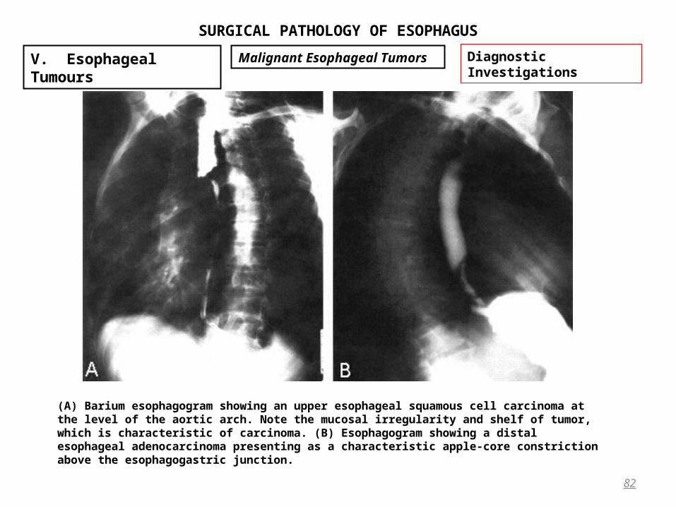
SURGICAL PATHOLOGY OF ESOPHAGUS
82
V. Esophageal Tumours Malignant Esophageal Tumors Diagnostic Investigations
(A) Barium esophagogram showing an upper esophageal squamous cell carcinoma at the level of the aortic arch. Note the mucosal irregularity and shelf of tumor, which is characteristic of carcinoma. (B) Esophagogram showing a distal esophageal adenocarcinoma presenting as a characteristic apple-core constriction above the esophagogastric junction.

SURGICAL PATHOLOGY OF ESOPHAGUS
• The chest and upper abdominal CT scanThe chest and upper abdominal CT scan is the standard radiographic technique for stagingstandard radiographic technique for staging esophageal carcinomaesophageal carcinoma. – esophageal wall thickness should not exceed 5 mm on CT scanning, – regional adenopathy or – pulmonary, liver, adrenal, or distant nodal metastasis. – CT scan, guided tissue diagnosis with fine-needle aspiration biopsy is warranted. A positive histologic
diagnosis of stage IV carcinoma translates to an average survival of only 6 to 12 months, and therefore an operation of the magnitude of esophagectomy is contraindicated.
• Several investigators have reported the value of CT in evaluating resectability of esophageal carcinomathe value of CT in evaluating resectability of esophageal carcinoma. Bronchoscopy
• BronchoscopyBronchoscopy should be performed in patients with carcinoma of the upper and middle thirds of the esophagus to exclude invasion of the posterior membranous trachea or main-stem bronchi, which precludes a safe esophagectomy. Other Studies.
– Magnetic resonance imaging to evaluate mediastinal invasion has not gained widespread popularity. – Bone scan is not warranted unless the patient has specific complaints suggesting that bone metastases exist.– routine brain scans are not indicated because brain metastases from carcinoma of the esophagus are
uncommon (less than 4% in patients being evaluated for esophagectomy).20
83
V. Esophageal Tumours Malignant Esophageal Tumors Diagnostic Investigations
SURGICAL PATHOLOGY OF ESOPHAGUS
Esophagoscopy • Esophagoscopy is one of the most important diagnostic tools in the assessment of the patient with esophageal
symptoms from any cause. The flexible fiberoptic esophagoscope permits endoscopic assessment with greater ease gf obstructing lesions of esophagusEndoscopic Vital Staining, Endoscopic Ultrasound and Endoscopic Abrasive cytologyEndoscopic Vital Staining, Endoscopic Ultrasound and Endoscopic Abrasive cytology
• Vital stainingVital staining of the esophageal mucosa is a technique that is useful in detecting dysplastic esophageal lesions that are not obvious on direct endoscopic assessment. Carcinoma in situ (intraepithelial carcinoma) or microinvasive carcinoma may appear endoscopically as flat, nondescript lesions (leukoplakia or erythroplakia) and therefore can be difficult to diagnose. Lugol (3% iodide) solution or 2% toluidine blue may be applied through the esophagoscope to the esophageal mucosa.
– Lugol Lugol solution stains normal glycogenic esophageal mucosa brown, while abnormal mucosa (early carcinoma, esophagitis, Barrett’s mucosa) remains unstained.
– Toluidine blueToluidine blue is a metachromatic stain with affinity for cell nuclei. Therefore, tissues with a high cellular density and high nucleus/cytoplasm ratio take up the stain quickly and retain it for about 1 hour.
• Endoscopic ultrasoundEndoscopic ultrasound is being used with increasing frequency as an adjunct to the standard radiologic and endoscopic assessment of esophageal disease. It offers the potential for more sensitive staging of esophageal It offers the potential for more sensitive staging of esophageal carcinoma by detecting the depth of invasion and the presence of abnormal mediastinal adenopathycarcinoma by detecting the depth of invasion and the presence of abnormal mediastinal adenopathy. Ultrasound permits the endoscopic delineation of the mucosa, submucosa, and muscular layers of the esophagus as well as endoscopic delineation of the mucosa, submucosa, and muscular layers of the esophagus as well as adjacent tissues. Lymph nodes as small as 5 mm can be recognized with this instrument. adjacent tissues. Lymph nodes as small as 5 mm can be recognized with this instrument.
• Endoscopic Abrasive cytologyEndoscopic Abrasive cytology using a swallowed balloon catheter (balloon cytology) has been extremely effective in screening for carcinoma
• Combining abrasive cytology with vital staining of the esophageal mucosa may prove to yield the best sensitivity Combining abrasive cytology with vital staining of the esophageal mucosa may prove to yield the best sensitivity and specificity for screening populations. and specificity for screening populations.
84
V. Esophageal Tumours Malignant Esophageal Tumors Diagnostic Investigations
SURGICAL PATHOLOGY OF ESOPHAGUS
Premalignant Esophageal Lesions Chronic irritation of esophageal mucosaChronic irritation of esophageal mucosa by a variety of noxious stimulinoxious stimuli (alcohol, tobacco, hot foods and liquids) eventually may lead to the development of esophageal carcinoma. A variety of esophageal lesions have a recognized premalignant nature:
• The premalignant nature of Barrett’s esophagitisBarrett’s esophagitis was discussed earlier. • Reflux EsophagitisReflux Esophagitis constitutes a chronic chemical injury of the esophageal mucosa, it is regarded as a potentially
premalignant abnormality of the esophagus that requires aggressive medical therapy or surgical control. • About 10% to 12% of patients with achalasia of cardiaachalasia of cardia who are observed for 15 years or longer develop
esophageal carcinoma. The cause is thought to be related to the irritating effects of the fermenting intraesophageal contents on the adjacent esophageal mucosa.
• Plummer-Vinson syndromePlummer-Vinson syndrome (Paterson-Kelly syndrome or sideropenic dysphagiasideropenic dysphagia) is a premalignant esophageal condition. The term sideropenic dysphagia refers to the development of cervical dysphagia in patients who have iron-deficiency anemia.
• Familial keratosis palmaris et plantaris (tylosis)Familial keratosis palmaris et plantaris (tylosis) is associated with an increased incidence of esophageal carcinoma • Radiation EsophagitisRadiation Esophagitis during the course of treatment for lymphoma, lung, breast, or other mediastinal
malignancies are at increased risk for developing esophageal carcinoma years later. • Within Esophageal DiverticulaEsophageal Diverticula several isolated reports have been made of esophageal carcinomas that have been
found incidentally presumably as a result of the irritating effects on the mucosa of stagnant, putrefying food within the pouch. Esophageal diverticula are therefore also regarded as premalignant esophageal lesions, although this occurrence is extremely rare.
85
V. Esophageal Tumours Malignant Esophageal Tumors Premalignant Lesions

SURGICAL PATHOLOGY OF ESOPHAGUS
Treatment of Esophageal Cancer• Therapy of esophageal carcinoma is influenced by the knowledge that in most of these patients, local tumor
invasion or distant metastatic disease precludes cure. Neither chemotherapy, radiotherapy, nor surgery alone has achieved significant and consistent long-term survival in patients with esophageal carcinoma. Radiation
• Although squamous cell carcinoma is generally regarded as a radiosensitive and therefore potentially curable radiosensitive and therefore potentially curable tumortumor, radiotherapy alone alone has not achieved cure in most of these patients.has not achieved cure in most of these patients. Radiotherapy is usedRadiotherapy is used in the treatment of esophageal carcinoma to provideto provide either palliation or curepalliation or cure as an adjunct to esophagectomy.
• Palliative radiotherapyPalliative radiotherapy in the range of 4000 to 5000 cGy over 3 to 4 weeks relieves dysphagia sufficiently in nearly half of patients with advanced metastatic carcinoma and severe dysphagia to allow them to swallow liquids and diet supplements.
• ““Curative” supervoltage radiotherapyCurative” supervoltage radiotherapy is delivered in doses of 5000 to 7000 cGy over 5 to 7 weeks, using rotational and oblique ports to avoid spinal cord injuryto avoid spinal cord injury. Unfortunately, the average 5-year survival after such treatment is between 6% and 10% in most series because radiation alone fails to control either the primary tumor or distant metastatic disease.
• Thus, reported 5-year survival rates after esophageal resection for carcinoma usually average between 10% and 15%, with more than 80% of patients dying within 1 year of diagnosis. Several Japanese reports indicate 5-year Several Japanese reports indicate 5-year survival rates of 25% to 38%, with combined preoperative radiotherapy followed by resection. survival rates of 25% to 38%, with combined preoperative radiotherapy followed by resection. Intubation
• A variety of endoesophageal tubes (Celestin, Fell, Mackler, Mousseau-Barbin, Souttar, Wilson-Cook) have been used to provide palliation in patients with esophageal carcinoma.
• A variety of expandable intraesophageal metallic stentsexpandable intraesophageal metallic stents have been used to achieve palliation in patients with unresectable esophageal carcinoma.26,27 These stents are easier to insert than the older plastic tubes, have a larger lumen, and theoretically, carry less risk of perforation. They are inserted under fluoroscopy using a flexible esophagoscope.
86
V. Esophageal Tumours Malignant Esophageal Tumors Principles of Treatment
SURGICAL PATHOLOGY OF ESOPHAGUS
Laser • Endoscopic laser fulguratingEndoscopic laser fulgurating of esophageal carcinoma, particularly with the Nd:YAG laser, has been used to
achieve temporary relief of the esophageal obstruction in patients with unresectable tumors. Generally, multiple Generally, multiple sessions are requiredsessions are required to resect sufficient tumor to achieve an adequate lumen, and functional success with restoration of comfortable swallowing is achieved in 75% to 80% of patientscomfortable swallowing is achieved in 75% to 80% of patients. Bypass
• A variety of surgical procedures, such as substernal gastric or colon bypass, have been developed as palliative substernal gastric or colon bypass, have been developed as palliative internal bypasses of unresectable esophageal carcinomas.internal bypasses of unresectable esophageal carcinomas. This is simply too large an operation for a patient with so advanced a malignancy. Surgical Resection
• Transthoracic Resection.Transthoracic Resection. For most patients with localized esophageal carcinoma, resection provides the most effective and reliable palliation of dysphagia.
– The traditional surgical approach to distal esophageal carcinoma is a left thoracoabdominal incision (Fig. 19-11). After resecting the distal esophagus, proximal stomach, and adjacent lymph nodes, an intrathoracic esophagogastric anastomosis is performed.
– Tumors involving the mid-esophagus are resected through either a thoracoabdominal or separate thoracic and abdominal incisions, and a high intrathoracic esophagogastric anastomosis is performed (Fig. 19-12).
• Transhiatal ResectionTranshiatal Resection. During the past two decades, the technique of transhiatal esophagectomy without thoracotomy has been popularized as an operation that minimizes the factors responsible for most poor results from traditional transthoracic esophageal resection and reconstruction. In this operation, irrespective of the level of the tumor, the entire intrathoracic esophagus is resected, the stomach is repositioned in the posterior mediastinum in the original esophageal bed, and the gastric fundus is anastomosed to the cervical esophagus above the level of the clavicles.
87
V. Esophageal Tumours Malignant Esophageal Tumors Principles of Treatment

SURGICAL PATHOLOGY OF ESOPHAGUS
88
V. Esophageal Tumours Malignant Esophageal Tumors Principles of Treatment
Standard thoracoabdominal esophagogastrectomy for carcinomas of the distal esophagus and cardia. (A) Thoracoabdominal incision. (B) Tissue to be resected (colored area). (C) Completed reconstruction after intrathoracic esophagogastric anastomosis
Standard thoracoabdominal esophagogastrectomy for tumors of the upper and middle thirds of the thoracic esophagus. (A) Either the continuous thoracoabdominal incision or separate thoracic and abdominal incisions are used. (B) Portion of esophagus to be resected (colored area). (C) Completed reconstruction with high intrathoracic esophagogastric anastomosis and gastric drainage procedure.

SURGICAL PATHOLOGY OF ESOPHAGUS
89
V. Esophageal Tumours Malignant Esophageal Tumors Principles of Treatment
Transhiatal EsophagectomyTranshiatal EsophagectomyMobilization of the thoracic esophagus from the posterior mediastinum
SURGICAL PATHOLOGY OF ESOPHAGUS
• This operation has been criticized because of its limited exposure of the intrathoracic esophagus through the diaphragmatic hiatus and, therefore, the risk of intraoperative bleeding from the divided aortic esophageal branches. In addition, one cannot carry out a complete mediastinal lymph node dissection through the diaphragmatic hiatus for purposes of staging or potential cure. Radical Resection.
• The radical transthoracic esophagectomyradical transthoracic esophagectomy with en bloc dissection of contiguous lymph node–bearing tissues for esophageal carcinoma. This is a much more formidable operation, the results of which, when compared with those of transhiatal esophagectomy without thoracotomy and no formal lymph node dissection. As a general rule, the stomach is the preferred visceral esophageal substitute, being far more resilient than intestine and readily reaching to the neck for replacement of the entire esophagus. Colonic interposition is a major operative undertaking in patients with esophageal carcinoma and should be used only in selected cases in which the stomach is not available for esophageal replacement. Multimodality Therapy
• Efforts have been made to improve survival in patients with esophageal carcinoma by using multimodality therapy in combination with surgery.
• Combined preoperative chemotherapychemotherapy and radiotherapyradiotherapy before transhiatal esophagectomy for carcinoma, for example, has provided encouraging survival statistics. They received 3 weeks of chemotherapy with cisplatin, vinblastine, and 5-fluorouracil, concurrent with 3750 to 4500 cGy of radiotherapy. After a 3-week recovery period, transhiatal esophagectomy was accomplished.
90
V. Esophageal Tumours Malignant Esophageal Tumors Principles of Treatment
SURGICAL PATHOLOGY OF ESOPHAGUS
91
V. Esophageal Tumours Malignant Esophageal Tumors Principles of Treatment