Tumours of the Oesophagus
22
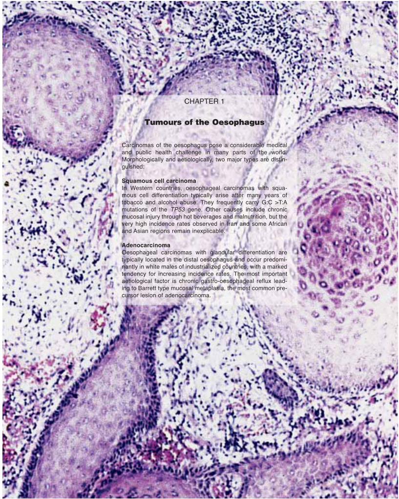
CHAPTER 1 Tumours of the Oesophagus Carcinomas of the oesophagus pose a considerable medical and public health challenge in many parts of the world. Morphologically and aetiologically, two major types are distin- guished: Squamous cell carcinoma In Western countries, oesophageal carcinomas with squa- mous cell differentiation typically arise after many years of tobacco and alcohol abuse. They frequently carry G:C >T:A mutations of the TP53 gene. Other causes include chronic mucosal injury through hot beverages and malnutrition, but the very high incidence rates observed in Iran and some African and Asian regions remain inexplicable. Adenocarcinoma Oesophageal carcinomas with glandular differentiation are typically located in the distal oesophagus and occur predomi- nantly in white males of industrialized countries, with a marked tendency for increasing incidence rates. The most important aetiological factor is chronic gastro-oesophageal reflux lead- ing to Barrett type mucosal metaplasia, the most common pre- cursor lesion of adenocarcinoma.
Transcript of Tumours of the Oesophagus
CHAPTER 1
Tumours of the Oesophagus
Carcinomas of the oesophagus pose a considerable medicaland public health challenge in many parts of the world.Morphologically and aetiologically, two major types are distin-guished:
Squamous cell carcinomaIn Western countries, oesophageal carcinomas with squa-mous cell differentiation typically arise after many years oftobacco and alcohol abuse. They frequently carry G:C >T:Amutations of the TP53 gene. Other causes include chronicmucosal injury through hot beverages and malnutrition, but thevery high incidence rates observed in Iran and some Africanand Asian regions remain inexplicable.
AdenocarcinomaOesophageal carcinomas with glandular differentiation aretypically located in the distal oesophagus and occur predomi-nantly in white males of industrialized countries, with a markedtendency for increasing incidence rates. The most importantaetiological factor is chronic gastro-oesophageal reflux lead-ing to Barrett type mucosal metaplasia, the most common pre-cursor lesion of adenocarcinoma.
01 19.7.2006 7:22 Page 9

10 Tumours of the oesophagus
Epithelial tumours
Squamous cell papilloma 8052/01
Intraepithelial neoplasia2
SquamousGlandular (adenoma)
CarcinomaSquamous cell carcinoma 8070/3Verrucous (squamous) carcinoma 8051/3Basaloid squamous cell carcinoma 8083/3Spindle cell (squamous) carcinoma 8074/3Adenocarcinoma 8140/3Adenosquamous carcinoma 8560/3Mucoepidermoid carcinoma 8430/3Adenoid cystic carcinoma 8200/3Small cell carcinoma 8041/3Undifferentiated carcinoma 8020/3Others
Carcinoid tumour 8240/3
Non-epithelial tumours
Leiomyoma 8890/0Lipoma 8850/0Granular cell tumour 9580/0Gastrointestinal stromal tumour 8936/1
benign 8936/0uncertain malignant potential 8936/1malignant 8936/3
Leiomyosarcoma 8890/3Rhabdomyosarcoma 8900/3Kaposi sarcoma 9140/3Malignant melanoma 8720/3Others
Secondary tumours
WHO histological classification of oesophageal tumours
_____________1 Morphology code of the International Classification of Diseases for Oncology (ICD-O) {542} and the Systematized Nomenclature of Medicine (http://snomed.org).
Behaviour is coded /0 for benign tumours, /1 for unspecified, borderline or uncertain behaviour, /2 for in situ carcinomas and grade III intraepithelial neoplasia, and /3 formalignant tumours.
2 Intraepithelial neoplasia does not have a generic code in ICD-O. ICD-O codes are available only for lesions categorized as glandular intraepithelial neoplasia grade III(8148/2), squamous intraepithelial neoplasia, grade III (8077/2), and squamous cell carcinoma in situ (8070/2).
_____________1 {1, 66}. This classification applies only to carcinomas.2 A help desk for specific questions about the TNM classification is available at http://tnm.uicc.org.
TNM classification1
T – Primary TumourTX Primary tumour cannot be assessedT0 No evidence of primary tumourTis Carcinoma in situT1 Tumour invades lamina propria or submucosaT2 Tumour invades muscularis propriaT3 Tumour invades adventitiaT4 Tumour invades adjacent structures
N – Regional Lymph NodesNX Regional lymph nodes cannot be assessedN0 No regional lymph node metastasisN1 Regional lymph node metastasis
M – Distant MetastasisMX Distant metastasis cannot be assessedM0 No distant metastasisM1 Distant metastasis
For tumours of lower thoracic oesophagusM1a Metastasis in coeliac lymph nodesM1b Other distant metastasis
For tumours of upper thoracic oesophagusM1a Metastasis in cervical lymph nodesM1b Other distant metastasis
For tumours of mid-thoracic oesophagusM1a Not applicableM1b Non-regional lymph node
or other distant metastasis
Stage Grouping
Stage 0 Tis N0 M0Stage I T1 N0 M0Stage IIA T2 N0 M0
T3 N0 M0Stage IIB T1 N1 M0
T2 N1 M0Stage III T3 N1 M0
T4 Any N M0Stage IVA Any T Any N M1aStage IVB Any T Any N M1b
TNM classification of oesophageal tumours
01 19.7.2006 7:22 Page 10

11Squamous cell carcinoma
DefinitionSquamous cell carcinoma (SCC) of theoesophagus is a malignant epithelialtumour with squamous cell differentia-tion, microscopically characterised bykeratinocyte-like cells with intercellularbridges and/or keratinization.
ICD-O Code 8070/3
EpidemiologySquamous cell carcinoma of the oeso-phagus shows great geographical diver-sity in incidence, mortality and sex ratio.In Western countries, the age-standar-dized annual incidence in most areasdoes not exceed 5 per 100,000 popula-tion in males and 1 in females. There are,however, several well-defined high-riskareas, e.g. Normandy and Calvados inNorth-West France, and Northern Italy,where incidence may be as high as 30per 100,000 population in males and 2 infemales {1020, 1331}. This type of can-cer is much more frequent in Easterncountries and in many developing coun-tries. Regions with very high incidencerates have been identified in Iran, CentralChina, South Africa and Southern Brazil.In the city of Zhengzhou, capital ofHenan province in China, the mortalityrate exceeds 100 per 100,000 populationin males and 50 in females {1116, 2191}.
In both high-risk and low-risk regions,this cancer is exceedingly rare beforethe age of 30 and the median age isaround 65 in both males and females.Recent changes in the distribution pat-tern in France indicate that the rate ofSCC has increased steadily in low-riskareas, particularly among females,whereas there may be a slight decreasein high-risk areas. In the United States, asearch in hospitalisation records of mili-tary veterans indicates that SCC is 2-3times more frequent among blacks thanamong Asians, Whites or NativeAmericans {453}.
AetiologyTobacco and alcohol. In Western coun-tries, nearly 90% of the risk of SCC canbe attributed to tobacco and alcohol.Each of these factors influences the riskof oesophageal cancer in a different way.With regard to the consumption of tobac-co, a moderate intake during a long peri-od carries a higher risk than a high intakeduring a shorter period, whereas thereverse is true for alcohol. Both factorscombined show a multiplicative effect,even at low alcohol intake. In high-riskareas of North-West France and NorthernItaly, local drinking customs may partiallyexplain the excess incidence of SCC{523, 1020}. In Japanese alcoholics, a
polymorphism in ALDH2, the geneencoding aldehyde dehydrogenase 2,has been shown to be significantly asso-ciated with several cancers of the upperdigestive tract, including squamous cellcancer. This observation suggests a rolefor acetaldehyde, one of the main car-cinogenic metabolites of alcohol in thedevelopment of oesophageal carcinoma{2177}.Nutrition. Risk factors other than tobac-co and alcohol play significant roles inother regions of the world. In high-riskareas of China, a deficiency in certaintrace elements and the consumption ofpickled or mouldy foods (which arepotential sources of nitrosamines) havebeen suggested.Hot beverages. Worldwide, one of themost common risk factors appears to bethe consumption of burning-hot bevera-ges (such as Mate tea in South America)which cause thermal injury leading tochronic oesophagitis and then to precan-cerous lesions {1116, 2191, 387}.HPV. Conflicting reports have proposeda role for infectious agents, includinghuman papillomavirus (HPV) infection.Although HPV DNA is consistentlydetected in 20 to 40% of SCC in high-riskareas of China, it is generally absent inthe cancers arising in Western countries{954, 679}.
Squamous cell carcinoma of the oesophagus
H.E. Gabbert Y. NakamuraT. Shimoda J.K. FieldP. Hainaut H. Inoue
Fig. 1.02 Squamous cell carcinoma of the oesophagus. Age-standardized incidencerates per 100,000 and proportions (%) due to alcohol and tobacco (dark-blue).
Fig. 1.01 Worldwide annual incidence (per 100,000) of oesophageal cancer inmales. Numbers on the map indicate regional average values.
5.27.8
19.3
11.9
51.6
4.611.0
< 2.2 < 3.8 < 5.8 < 9.5 < 51.7
China, Henan
Iran, North East
South Africa
India, Bombay
Urugay
China, Hong Kong
Italy, North East
USA, New York
France, Calvados
1%
70%
50%
50%
20%
90%
90%
70%
90%
0 50 100 150 200
01 19.7.2006 7:22 Page 11

12 Tumours of the oesophagus
Associations between achalasia, Plum-mer-Vinson syndrome, coeliac diseaseand tylosis (focal nonepidermolytic pal-moplantar keratoderma) with oeso-phageal cancer have also been de-scribed.
LocalizationOesophageal SCC is located predomi-nantly in the middle and the lower third ofthe oesophagus, only 10-15% being situ-ated in the upper third {1055}.
Clinical featuresSymptoms and signsThe most common symptoms of ad-vanced oesophageal cancer are dys-phagia, weight loss, retrosternal or epi-gastric pain, and regurgitation causedby narrowing of the oesophageal lumenby tumour growth {606}. Superficial SCCusually has no specific symptoms butsometimes causes a tingling sensation,and is, therefore, often detected inciden-tally during upper gastrointestinalendoscopy {464, 1874}.
Endoscopy and vital stainingSuperficial oesophageal cancer is com-monly observed as a slight elevation orshallow depression on the mucosalsurface, which is a minor morphologicalchange compared to that of advancedcancer. Macroscopically, three types canbe distinguished: flat, polypoid and ulcer-ated. Chromoendoscopy utilizing toluidineblue or Lugol iodine spray may be of value{465, 481}. Toluidine blue, a metachromat-ic stain from the thiazine group, has a par-ticular affinity for RNA and DNA, andstains areas that are richer in nuclei thanthe normal mucosa. Lugol solution reactsspecifically with glycogen in the normalsquamous epithelium, whereas precan-cerous and cancerous lesions, but alsoinflamed areas and gastric heterotopia,are not stained. However, the superficialextension of carcinomas confined to themucosa can not be clearly recognized bysimple endoscopy.
Endoscopic ultrasonographyEndoscopic ultrasonography is used toevaluate both depth of tumour infiltrationand para-oesophageal lymph nodeinvolvement in early and advancedstages of the disease {1509, 1935}. Forthe evaluation of the depth of infiltration,high frequency endoscopic ultrasono-graphy may be used {1302}. In general,
Fig. 1.03 Macroscopic images of squamous cell carcinoma (SCC) of the oesophagus. A Flat superficial type.B Lugol iodine staining of the specimen illustrated in A. C Polypoid SCC. D Longitudinal sections of carcino-ma illustrated in C. E Deeply invasive polypoid SCC. F Longitudinal sections of carcinoma illustrated in E.
A
C
B
F
D
E
01 19.7.2006 7:22 Page 12

13Squamous cell carcinoma
oesophageal carcinoma presents onendosonography as a circumscribed ordiffuse wall thickening with a predomi-nantly echo-poor or echo-inhomoge-neous pattern. As a result of tumourpenetration through the wall and intosurrounding structures, the endosono-graphic wall layers are destroyed.
Computed tomography (CT) and magnet-ic resonance imaging (MRI)In advanced carcinomas, CT and MRIgive information on local and systemicspread of SCC. Tumour growth is char-acterized as swelling of the oesophagealwall, with or without direct invasion tosurrounding organs {1518}. Cervical,abdominal and mediastinal node enlarg-ement is recorded. Three-dimensionalCT or MRI images may be presented asvirtual endoscopy, effectively demon-strating T2-T4 lesions, but not T1 lesions.
MacroscopyThe gross appearance varies accordingto whether it is detected in an early or anadvanced stage of the disease. Amongearly SCC, polypoid, plaque-like, de-pressed and occult lesions have beendescribed {161, 2183}. For the macro-scopic classification of advanced oeso-phageal SCC, Ming {1236} has proposedthree major patterns: fungating, ulcera-tive, and infiltrating. The fungating patternis characterized by a predominantly exo-phytic growth, whereas in the ulcerativepattern, the tumour growth is predomi-nantly intramural, with a central ulcerationand elevated ulcer edges. The infiltrativepattern, which is the least common one,also shows a predominantly intramuralgrowth, but causes only a small mucosaldefect. Similar types of macroscopic
growth patterns have been defined in theclassification of the Japanese Society forEsophageal Diseases {58}.
Tumour spread and stagingFor the staging of SCC, the TNM system(tumour, node, metastasis) establishedby the International Union AgainstCancer (UICC) is the most widely usedsystem. Its usefulness in the planning oftreatment and in the prediction of prog-nosis has been validated {1104, 895, 66,1, 772}.Superficial oesophageal carcinoma.When the tumour is confined to themucosa or the submucosa, the termsuperficial oesophageal carcinoma isused irrespective of the presence ofregional lymph node metastases {58,161}. In China and in Japan, the termearly oesophageal carcinoma is oftenused defining a carcinoma that invadesno deeper than the submucosa but hasnot metastasised {609}. In several studiesfrom Japan, superficial carcinomasaccounted for 10-20% of all resected car-cinomas, whereas in Western countries
superficial carcinomas are much less fre-quently reported {543}. About 5% ofsuperficial carcinomas that have invadedthe lamina propria display lymph nodemetastases, whereas in carcinomas thatinvade the submucosa the risk of nodalmetastasis is about 35% {1055}. Fortumours that have infiltrated beyond thesubmucosa, the term advanced oeso-phageal carcinoma is applied. Intramural metastases. A special featureof oesophageal SCC is the occurrence ofintramural metastases, which have beenfound in resected oesophageal speci-mens in 11-16% of cases {896, 987}.These metastases are thought to resultfrom intramural lymphatic spread with theestablishment of secondary intramuraltumour deposits. Intramural metastasesare associated with an advanced stageof disease and with shorter survival.Second primary SCC. Additionally, theoccurrence of multiple independent SCChas been described in between 14 and31% of cases, the second cancers beingmainly carcinomas in situ and superficialSCC {1154, 989, 1507}.Treatment groups. Following the clinicalstaging, patients are usually divided intotwo treatment groups: those with locore-gional disease in whom the tumour ispotentially curable (e.g. by surgery,radiotherapy, multimodal therapy), andthose with advanced disease (meta-stases outside the regional area or inva-sion of the airway) in whom only palliativetreatment is indicated {606}. Oeso-phageal SCC limited to the mucosa maybe treated by endoscopic mucosalresection due to its low risk of nodalmetastasis. Endoscopic mucosal resec-tion is also indicated for high-gradeintraepithelial neoplasia. Tumours thathave invaded the submucosa or those inmore advanced tumour stages have
Fig. 1.06 Primary squamous cell carcinoma (CA) ofoesophagus with an intramural metastasis (M) nearthe oesophagogastric junction.
Fig. 1.05 A Endoscopic view of a superficial squa-mous cell carcinoma presenting as a large nodule(CA) in a zone of erosion. B After spraying of 2%iodine solution, the superficial extent of the tumourbecomes visible as unstained light yellow area (CA,arrows).
A
B
Fig. 1.04 Catheter probe ultrasonograph of a squa-mous cell carcinoma, presenting as hypoechoiclesion (arrow).
CA
CA
M
CA
01 19.7.2006 7:22 Page 13

more than 30% risk of lymph nodemetastasis, and endoscopic therapy isnot indicated {465}. Additionally, clinicalstaging is performed in order to deter-mine the success of treatment, e.g. fol-lowing radio- and/or chemotherapy.
Tumour spreadThe most common sites of metastasis ofoesophageal SCC are the regional lymphnodes. The risk of lymph node metasta-sis is about 5% in carcinomas confinedto the mucosa but over 30% in carcino-mas invading the submucosa and over80% in carcinomas invading adjacentorgans or tissues {772}. Lesions of theupper third of the oesophagus most fre-quently involve cervical and mediastinallymph nodes, whereas those of the mid-dle third metastasise to the mediastinal,cervical and upper gastric lymph nodes.Carcinomas of the lower third preferen-tially spread to the lower mediastinal andthe abdominal lymph nodes {28}. Themost common sites of haematogenousmetastases are the lung and the liver{1153, 1789}. Less frequently affectedsites are the bones, adrenal glands, andbrain {1551}. Recently, disseminatedtumour cells were identified by means ofimmunostaining in the bone marrow ofabout 40% of patients with oesophagealSCC {1933}. Recurrence of cancer fol-lowing oesophageal resection can belocoregional or distant, both with approx-imately equal frequency {1185, 1027}.
Histopathology Oesophageal SCC is defined as the pen-etration of neoplastic squamous epitheli-um through the epithelial basement mem-brane and extension into the lamina pro-pria or deeper tissue layers. Invasioncommonly starts from a carcinoma in situwith the proliferation of rete-like projec-tions of neoplastic epithelium that pushinto the lamina propria with subsequentdissociation into small carcinomatous cellclusters. Along with vertical tumour cellinfiltration, usually a horizontal growthundermines the adjacent normal mucosaat the tumour periphery. The carcinomamay already invade intramural lymphaticvessels and veins at an early stage of dis-ease. The frequency of lymphatic andblood vessel invasion increases withincreasing depth of invasion {1662}.Tumour cells in lymphatic vessels and inblood vessels may be found progressive-ly several centimetres beyond the gross
tumour. The carcinoma invades the mus-cular layers, enters the loose fibrousadventitia and may extend beyond theadventitia, with invasion of adjacentorgans or tissues, especially the tracheaand bronchi, eventually with the formationof oesophagotracheal or oesophago-bronchial fistulae {1789}. Oesophageal SCC displays differentmicroscopic patterns of invasion, whichare categorised as ‘expansive growth’ or‘infiltrative growth’. The former pattern ischaracterized by a broad and smoothinvasion front with little or no tumour celldissociation, whereas the infiltrative pat-tern shows an irregular invasion front anda marked tumour cell dissociation. The degree of desmoplastic or inflamma-tory stromal reaction, nuclear polymor-phism and keratinization is extremelyvariable. Additionally, otherwise typicaloesophageal SCC may contain small fociof glandular differentiation, indicated bythe formation of tubular glands or mucin-producing tumour cells {987}.
Verrucous carcinoma (ICD-O 8051/3)This rare variant of squamous cell carci-noma {19} is histologically comparable toverrucous carcinomas arising at othersites {969}. On gross examination, itsappearance is exophytic, warty, cauli-flower-like or papillary. It can be found inany part of the oesophagus. Histologi-cally, it is defined as a malignant papil-lary tumour composed of well differentia-ted and keratinized squamous epitheli-um with minimal cytological atypia, andpushing rather than infiltrating margins{2066}. Oesophageal verrucous carcino-ma grows slowly and invades locally, witha very low metastasising potential.
Spindle cell carcinoma (ICD-O 8094/3)This unusual malignancy is defined as asquamous cell carcinoma with a variable
sarcomatoid spindle cell component. It isalso known by a variety of other terms,including carcinosarcoma, pseudosarco-matous squamous cell carcinoma, poly-poid carcinoma, and squamous cell car-cinoma with a spindle cell component{1055}. Macroscopically, the tumour ischaracterized by a polypoid growth pat-tern. The spindle cells may be capable ofmaturation, forming bone, cartilage andskeletal muscle cells {662}. Alternatively,they may be more pleomorphic, resem-bling malignant fibrous histiocytoma. Inthe majority of cases a gradual transitionbetween carcinomatous and sarcomatouscomponents has been observed on thelight microscopic level. Immunohisto-chemical and electron microscopic stu-dies indicate that the sarcomatous spin-dle cells show various degrees of epithe-lial differentiation. Therefore, the sarcoma-
14 Tumours of the oesophagus
Fig. 1.07 Squamous cell carcinoma with transmuralinvasion. M, remaining intact mucosa.
Fig. 1.09 Verrucous carcinoma. A Typical exo-phytic papillary growth. B High degree of differen-tiation.
B
Fig. 1.08 Squamous cell carcinoma invading thin-walled lymphatic vessels.
M
A
01 19.7.2006 7:22 Page 14
15Squamous cell carcinoma
tous component may be metaplastic.However, a recent molecular analysis of asingle case of a spindle cell carcinomashowed divergent genetic alterations inthe carcinomatous and in the sarcoma-tous tumour component suggesting twoindependent malignant cell clones {823}.
Basaloid squamous cell carcinoma (ICD-O 8083/3) This rare but distinct variant of oeso-phageal SCC {1961} appears to be iden-tical to the basaloid squamous cell carci-nomas of the upper aerodigestive tract{109}. Histologically, it is composed ofclosely packed cells with hyperchromat-ic nuclei and scant basophilic cyto-plasm, which show a solid growth pat-tern, small gland-like spaces and foci ofcomedo-type necrosis. Basaloid squa-mous cell carcinomas are associated
with intraepithelial neoplasia, invasiveSCC, or islands of squamous differentia-tion among the basaloid cells {2036}. Theproliferative activity is higher than in typi-cal SCC. However, basaloid squamouscell carcinoma is also characterized by ahigh rate of apoptosis and its prognosisdoes not differ significantly from that ofthe ordinary oesophageal SCC {1663}.
Precursor lesions Most studies on precursor lesions ofoesophageal SCC have been carried outin high-risk populations, especially in Iranand Northern China, but there is no evi-dence that precursor lesions in low-riskregions are substantially different. Thedevelopment of oesophageal SCC isthought to be a multistage process whichprogresses from the conversion of nor-mal squamous epithelium to that withbasal cell hyperplasia, intraepithelialneoplasia (dysplasia and carcinoma insitu), and, finally, invasive SCC {354,1547, 377}.
Intraepithelial neoplasia. This lesion isabout eight times more common in highcancer-risk areas than in low-risk areas{1547}, and is frequently found adjacentto invasive SCC in oesophagectomyspecimens {1154, 988}. Morphologicalfeatures of intraepithelial neoplasiainclude both architectural and cytologicalabnormalities. The architectural abnor-mality is characterized by a disorganisa-tion of the epithelium and loss of normalcell polarity. Cytologically, the cells exhibitirregular and hyperchromatic nuclei, anincrease in nuclear/cytoplasmic ratio andincreased mitotic activity. Dysplasia isusually graded as low or high-grade. Inlow-grade dysplasia, the abnormalitiesare often confined to the lower half of theepithelium, whereas in high-grade dys-plasia the abnormal cells also occur in
the upper half and exhibit a greaterdegree of atypia. In carcinoma in situ, theatypical cells are present throughout theepithelium without evidence of maturationat the surface of the epithelium {1154}. Ina two-tier system, severe dysplasia andcarcinoma-in-situ are included under therubric of high-grade intraepithelial neo-plasia, and may have the same clinicalimplications {1055}.Epidemiological follow-up studies sug-gest an increased risk for the subse-quent development of invasive SCC forpatients with basal cell hyperplasia (rela-tive risk: 2.1), low-grade dysplasia (RR:2.2), moderate-grade dysplasia (RR:15.8), high-grade dysplasia (RR: 72.6)and carcinoma in situ (RR: 62.5) {377}.
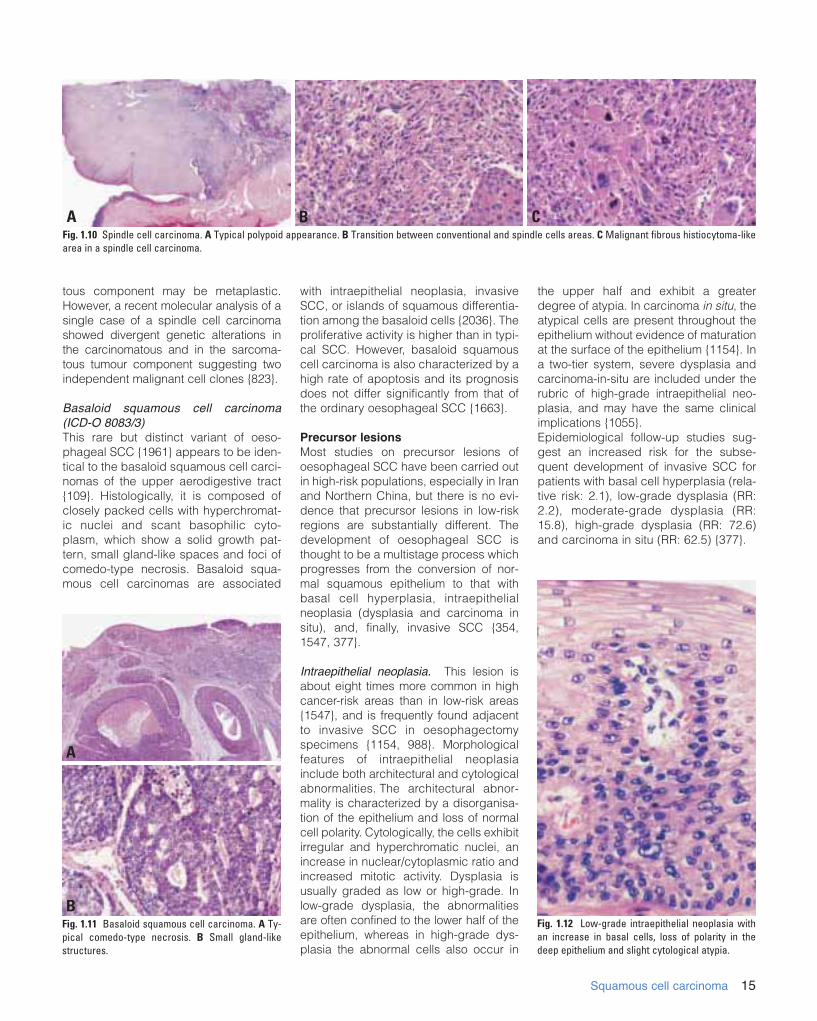
Fig. 1.10 Spindle cell carcinoma. A Typical polypoid appearance. B Transition between conventional and spindle cells areas. C Malignant fibrous histiocytoma-likearea in a spindle cell carcinoma.
B CA
Fig. 1.11 Basaloid squamous cell carcinoma. A Ty-pical comedo-type necrosis. B Small gland-likestructures.
A
BFig. 1.12 Low-grade intraepithelial neoplasia withan increase in basal cells, loss of polarity in thedeep epithelium and slight cytological atypia.
01 19.7.2006 7:22 Page 15

16 Tumours of the oesophagus
Basal cell hyperplasiaThis lesion is histologically defined as anotherwise normal squamous epitheliumwith a basal zone thickness greater than15% of total epithelial thickness, withoutelongation of lamina propria papillae{377}. In most cases, basal cell hyper-plasia is an epithelial proliferative lesionin response to oesophagitis, which is fre-quently observed in high-risk populationsfor oesophageal cancer {1547}.
Squamous cell papilloma (ICD-O 8052/0)Squamous cell papilloma is rare andusually causes no specific symptoms. Itis a benign tumour composed of hyper-plastic squamous epithelium coveringfinger-like processes with cores derivedfrom the lamina propria. The polypoidlesions are smooth, sharply demarcated,and usually 5 mm or less in maximumdiameter {249, 1428}. Rarely, giant papil-lomas have been reported, with sizes upto 5 cm {2037}. Most squamous cellpapillomas represent single isolatedlesions, typically located in the distal to
middle third of the oesophagus, but mul-tiple lesions occur. Histologically, cores of fibrovascular tis-sue are covered by mature stratifiedsquamous epithelium. The aetiologicalrole of human papillomavirus (HPV)infection has been investigated in seve-ral studies, but the results were inconclu-sive {248}. Malignant progression to SCCis extremely rare.In Japan, oesophageal squamous cellcarcinoma is diagnosed mainly based onnuclear criteria, even in cases judged tobe non-invasive intraepithelial neoplasia(dysplasia) in the West. This difference indiagnostic practice may contribute to therelatively high rate of incidence and goodprognosis of superficial squamous cellcarcinoma reported in Japan {1682}.
Grading Grading of oesophageal SCC is tradition-ally based on the parameters of mitoticactivity, anisonucleosis and degree ofdifferentiation.Well differentiated tumours have cytolo-gical and histological features similar tothose of the normal oesophageal squa-mous epithelium. In well differentiatedoesophageal SCC there is a high propor-tion of large, differentiated, keratinocyte-like squamous cells and a low proportionof small basal-type cells, which are loca-ted in the periphery of the cancer cellnests {1055}. The occurrence of kera-tinization has been interpreted as a signof differentiation, although the normaloesophageal squamous epithelium doesnot keratinize. Poorly differentiated tumours predomi-nantly consist of basal-type cells, whichusually exhibit a high mitotic rate.Moderately differentiated carcinomas,between the well and poorly differentia-ted types, are the most common type,accounting for about two-thirds of alloesophageal SCC. However, since nogenerally accepted criteria have beenidentified to score the relative contribu-tion of the different grading parameters,grading of SCC suffers from a great inter-observer variation. Undifferentiated carcinomas are definedby a lack of definite light microscopicfeatures of differentiation. However, ultra-structural or immunohistochemical inves-tigations may disclose features of squa-mous differentiation in a subset of light-microscopically undifferentiated carcino-mas {1881}.
Fig. 1.14 Squamous cell papilloma of distal oeso-phagus. This lesion was negative for human papilloma-virus by in situ hybridisation.
Fig. 1.13 High grade intraepithelial neoplasia of oesophageal squamous epithelium. Architectural disarray,loss of polarity and cellular atypia are much greater than shown in Fig. 1.12. Changes in D extend to theparakeratotic layer of the luminal surface.
A B
C D
01 19.7.2006 7:22 Page 16

17Squamous cell carcinoma
Genetic susceptibility Familial predisposition of oesophagealcancer has been only poorly studiedexcept in its association with focal non-epidermolytic palmoplantar keratoderma(NEPPK or tylosis) {1279, 1278, 752}.This autosomal, dominantly inherited dis-order of the palmar and plantar surfacesof the skin segregates together withoesophageal cancer in three pedigrees,two of which are extensive {456, 1834,693}. The causative locus has been des-ignated the tylosis oesophageal cancer(TOC) gene and maps to 17q25 betweenthe anonymous microsatellite markersD17S1839 and D17S785 {1594, 899}.The genetic defect is thought to be in amolecule involved in the physical struc-ture of stratified squamous epitheliawhereby loss of function of the gene mayalter oesophageal integrity thereby mak-ing it more susceptible to environmentalmutagens.Several structural candidate genes suchas envoplakin (EVPL), integrin β4(ITGB4) and plakoglobin have beenexcluded as the TOC gene following inte-gration of the genetic and physical mapsof this region {1595}. The importance ofthis gene in a larger population thanthose afflicted with the familial disease isindicated by the association of thegenomic region containing the TOCgene with sporadic squamous celloesophageal carcinomas {2020, 823},Barrett adenocarcinoma of the oesopha-gus {439}, and primary breast cancers{549} using loss of heterozygosity stud-ies.
GeneticsAlterations in genes that encoderegulators of the G1 to S transition of cellcycle are common in SCC. Mutation inthe TP53 gene (17p13) is thought to bean early event, sometimes already
detectable in intraepithelial neoplasia.The frequency and type of mutationvaries from one geographic area to theother, suggesting that some TP53 muta-tions may occur as the result of exposureto region-specific, exogenous risk fac-tors. However, even in SCC from WesternEurope, the TP53 mutation spectrumdoes not show the same tobacco-associ-ated mutations as in lung cancers{1266}. Amplification of cyclin D1(11q13) occurs in 20-40% of SCC and isfrequently detected in cancers that retainexpression of the Rb protein, in agree-ment with the notion that these two fac-tors cooperate within the same signallingcascade {859}. Inactivation of CDKN2Aoccurs essentially by homozygous dele-tion or de novo methylation and appearsto be associated with advanced cancer.Other potentially important genetic alter-ations include transcriptional inactivationof the FHIT gene (fragile histidine triad, a
presumptive tumour suppressor on3p14) by methylation of 5’ CpG islands,and deletion of the tylosis oesophagealcancer gene on 17q25 {2020, 1264}.Furthermore, analysis of clones on3p21.3, where frequent LOH occurs inoesophageal cancer {1274}, recently ledto identification of a novel gene termedDLC1 (deleted in lung and oesophagealcancer-1) {365}. Although the function ofthe DLC1 gene remains to be clarified,RT-PCR experiments indicated that 33%of primary cancers of lung and oesopha-gus lacked DLC1 transcripts entirely orcontained increased levels of nonfunc-tional DLC1 mRNA. Recent evidencesuggests that LOH at a new, putativetumour suppressor locus on 5p15 mayoccur in a majority of SCC {1497}.Amplification of several proto-oncogeneshas also been reported (HST-1, HST-2,EGFR, MYC) {1266}. How these variousgenetic events correlate with phenotypic
Fig. 1.16 Location of the tylosis oesophageal cancer gene on chromosome 17q.
A B CFig. 1.15 Squamous cell carcinoma. A Moderately differentiated. B Well differentiated with prominent lymphoid infiltrate. C Well differentiated areas (left) contrastwith immature basal-type cells of a poorly differentiated carcinoma (right).
Location of the tylosisoesophageal cancergene by haplotype analy-sis
1cM/ 500 Kb
01 19.7.2006 7:22 Page 17

changes and co operate in the sequenceof events leading to SCC is still specula-tive.
Prognosis and prognostic factors Overall, the prognosis of oesophagealSCC is poor and the 5-year survival ratesin registries are around 10%. Cure isforeseen only for superficial cancer. Thesurvival varies, depending upon tumourstage at diagnosis, treatment received,patient’s general health status, morpho-logical features and molecular features ofthe tumour. In the past, studies on prog-nostic factors were largely focused onpatients who were treated by surgery,whereas factors influencing survival ofpatients treated by radiotherapy or bymultimodal therapy have been investi-gated only rarely.
Morphological factors The extent of spread of the oesophagealSCC is the most important factor forprognosis, the TNM classification beingthe most widely used staging system.Staging. All studies indicate that thedepth of invasion and the presence ofnodal or distant metastases are inde-pendent predictors of survival {1104,895, 772}. In particular, lymph nodeinvolvement, regardless of the extent ofthe primary tumour, indicates a poorprognosis {1862, 912, 1873}. Morerecently, the prognostic significance ofmore sophisticated methods for thedetermination of tumour spread havebeen evaluated, including the ratio ofinvolved to resected lymph nodes{1603}, immunohistochemically deter-mined lymph node micrometastases{824, 1327} and micrometastases in thebone marrow {1933}. However, currentdata are still too limited to draw final con-clusions on the prognostic value.Differentiation. The prognostic impact oftumour differentiation is equivocal, possi-bly due to the poor standardisation of thegrading system and to the high prognos-tic power of tumour stage. Althoughsome studies have shown a significantinfluence of tumour grade on survival{709, 772}, the majority of studies havenot {443, 1858, 1601, 1660}. Otherhistopathological features associatedwith a poor prognosis include the pres-ence of vascular and/or lymphatic inva-sion {772, 1662} and an infiltrative growthpattern of the primary tumour {1660}.Lymphocytic infiltration. Intense lympho-
cytic response to the tumour has beenassociated with a better prognosis{1660, 443}.Proliferation. The cancer cell prolifera-tion index, determined immunohisto-chemically by antibodies such as PCNAor Ki- 67 / MIB-1, have been studiedextensively. However, the proliferationindex does not appear to be an inde-pendent prognostic factor {2189, 1005,1659, 779}.
DNA ploidy. Aneuploidy of cancer cells,as determined by flow cytometry or byimage analysis, has been identified in55% to 95% of oesophageal SCC {935}.Regarding the prognostic impact, patientswith diploid tumours usually survive longerthan those with aneuploid tumours.However, a prognostic impact independ-ent of tumour stage has been shown onlyin two studies {422, 1195}, whereas themajority of studies have not verified this
0 10 20 30 40 50 60 70
SCC
ADC
G:C>A:T
G:C>A:T (CpG)
G:C>C:G
G:C>T:A
G:C>C:G
Deletions, insertions, complex mutations
G:C>A:T
G:C>A:T (CpG)
G:C>T:A
Deletions, insertions, complex mutations
18 Tumours of the oesophagus
Gene Location Tumor abnormality Function
TP53 17p13 Point mutation, LOH G1 arrest, apoptosis,genetic stability
p16, p15, 9p22 Homozygous loss CDK inhibitorARF/CDKN2 Promoter methylation (cell cycle control)
Cyclin D1 11q13 Amplification Cell cycle control
EGFR 17p13 Amplification, overexpression Signal transduction(membrane Tyr kinase)
c-myc 8q24.1 Amplification Transcription factor
Rb 13q14 LOH Cell cycle controlAbsence of expression
TOC 17q25 LOH Tumour suppressor
FEZ1 8p22 Transcription shutdown Transcription factor
DLC1 3p21.3 Transcription shutdown Growth inhibition
Table 1.01Genetic alterations in squamous cell carcinoma of the oesophagus.
Fig. 1.17 Spectrum of TP53 mutations in squamous cell carcinoma (SCC) and adenocarcinoma (ADC) of theoesophagus.
01 19.7.2006 7:22 Page 18

19Squamous cell carcinoma
finding {935}. Therefore, the determinationof DNA ploidy is currently not consideredto improve the prognostic information pro-vided by the TNM system {1055}.Extent of resection. The frequency oflocoregional recurrence is negativelycorrelated with the distance of the pri-mary tumour to the proximal resectionmargin and possibly to preoperativechemotherapy {1890, 1027}.Molecular factorsThe TP53 gene is mutated in 35% to 80%
of oesophageal SCC {1266}. Whereassome studies indicated a negative prog-nostic influence of p53 protein accumula-tion in cancer cell nuclei {1743, 277}, oth-ers did not observe any prognostic valueof either immunoexpression or TP53mutation {2014, 1661, 1008, 779, 319}. Other potential prognostic factors includegrowth factors and their receptors {927},oncogenes, including c-erbB-2 and int-2{778}, cell cycle regulators {1748, 1297},tumour suppressor genes {1886}, redox
defence system components, e.g., metal-lothionein and heat shock proteins {897},and matrix proteinases {1303, 1947,2155}. Alterations of these factors inoesophageal SCC may enhance tumourcell proliferation, invasiveness, andmetastatic potential, and thus may beassociated with survival. However, noneof the factors tested so far has enteredclinical practice.
Fig. 1.19 Immunoreactivity for epidermal growthfactor receptor (EGFR) in oesophageal squamouscell carcinoma.
Fig. 1.20 Fluorescence in situ hybridisation demon-strating cyclin D1 in squamous carcinoma cells.
Fig. 1.18 TP53 immunoreactivity in squamous cellcarcinoma of the oesophagus.
Multiple LOHAmplification of CMYC, EGFR, CYCLIND1, HST1...
Overexpression of CYCLIN D1LOH at 3p21; LOH at 9p31
LOH 3p14 (FHIT); LOH 17q25 (TOC)TP53 mutations
Normal oesophagus Oesophagitis Low-grade High-grade Invasive SCCintraepithelial neoplasia intraepithelial neoplasia
Fig. 1.21 Putative sequence of genetic alterations in the development of squamous cell carcinoma of the oesophagus.
01 19.7.2006 7:22 Page 19

20 Tumours of the oesophagus
DefinitionA malignant epithelial tumour of theoesophagus with glandular differentia-tion arising predominantly from Barrettmucosa in the lower third of the oeso-phagus. Infrequently, adenocarcinomaoriginates from heterotopic gastricmucosa in the upper oesophagus, orfrom mucosal and submucosal glands.
ICD-O Code 8140/3
EpidemiologyIn industrialized countries, the incidenceand prevalence of adenocarcinoma ofthe oesophagus has risen dramatically{1827}. Population based studies in theU.S.A. and several European countriesindicate that the incidence of oeso-phageal adenocarcinoma has doubledbetween the early 1970s to the late1980s and continues to increase at a rateof about 5% to 10% per year {152, 153,370, 405, 1496}. This is paralleled by ris-ing rates of adenocarcinoma of thegastric cardia and of subcardial gastriccarcinoma. It has been estimated thatthe rate of increase of oesophageal andoesophagogastric junction adenocarci-noma in the U.S.A. during the pastdecade surpassed that of any other typeof cancer {152}. In the mid 1990s theincidence of oesophageal adenocarcino-ma has been estimated between 1 and 4per 100,000 per year in the U.S.A. andseveral European countries and thusapproaches or exceeds that of squa-mous cell oesophageal cancer in theseregions. In Asia and Africa, adenocarci-noma of the oesophagus is an uncom-mon finding, but increasing rates arealso reported from these areas. In addition to the rise in incidence, ade-nocarcinoma of the oesophagus and ofthe oesophagogastric junction sharesome epidemiological characteristicsthat clearly distinguish them from squa-mous cell oesophageal carcinoma andadenocarcinoma of the distal stomach.These include a high preponderance forthe male sex (male:female ratio 7:1), a
higher incidence among whites and anaverage age at the time of diagnosis ofaround 65 years {1756}.
Aetiology Barrett oesophagusThe epidemiological features of adeno-carcinoma of the distal oesophagus andoesophagogastric junction match thoseof patients with known intestinal metapla-sia in the distal oesophagus, i.e. Barrettoesophagus {1605, 1827}, which hasbeen identified as the single most impor-tant precursor lesion and risk factor foradenocarcinoma of the distal oesopha-gus, irrespective of the length of the seg-ment with intestinal metaplasia.Intestinal metaplasia of the oesophagusdevelops when the normal squamousoesophageal epithelium is replaced bycolumnar epithelium during the processof healing after repetitive injury to theoesophageal mucosa, typically associat-ed with gastro-oesophageal reflux dis-ease {1798, 1799}. Intestinal metaplasia can be detected inmore than 80% of patients with adenocar-cinoma of the distal oesophagus. {1756,1824}. A series of prospective endoscop-ic surveillance studies in patients withknown intestinal metaplasia of the distaloesophagus has shown an incidence ofoesophageal adenocarcinoma in theorder of 1/100 years of follow up {1799}.This translates into a life-time risk foroesophageal adenocarcinoma of about10% in these patients. The length of theoesophageal segment with intestinalmetaplasia, and the presence of ulcera-tions and strictures have been implicatedas further risk factors for the developmentof oesophageal adenocarcinoma bysome authors, but this has not been con-firmed by others {1799, 1797, 1827}.The biological significance of so-calledultrashort Barrett oesophagus or intestin-al metaplasia just beneath a normal Zline has yet to be fully clarified {1325}.Whether adenocarcinoma of the gastriccardia or subcardial gastric cancer isalso related to foci of intestinal metapla-
sia at or immediately below the gastriccardia {715, 1797, 1722} is discussed inthe chapter on adenocarcinoma of theoesophagogstric junction. Despite thebroad advocation of endoscopic surveil-lance in patients with known Barrettoesophagus, more than 50% of patientswith oesophageal adenocarcinoma stillhave locally advanced or metastatic dis-ease at the time of presentation {1826}. Chronic gastro-oesophageal reflux is theusual underlying cause of the repetitivemucosal injury and also provides anabnormal environment during the healingprocess that predisposes to intestinalmetaplasia {1799}. Data from Swedenhave shown an odds ratio of 7.7 for oeso-phageal adenocarcinoma in personswith recurrent reflux symptoms, as com-pared with persons without such symp-toms {1002, 1001}. The more frequent, more severe, andlonger-lasting the symptoms of reflux, thegreater the risk. Among persons withlong-standing and severe symptoms ofreflux, the odds ratio for oesophagealadenocarcinoma was 43.5. Based onthese data a strong and probably causalrelation between gastro-oesophagealreflux, one of the most common benigndisorders of the digestive tract, andoesophageal adenocarcinoma has beenpostulated.Factors predisposing for the developmentof Barrett oesophagus and subsequentadenocarcinoma in patients with gastro-oesophageal reflux disease include amarkedly increased oesophageal expo-sure time to refluxed gastric and duode-nal contents due to a defective barrierfunction of the lower oesophageal sphinc-ter and ineffective clearance function ofthe tubular oesophagus {1823, 1827}.Experimental and clinical data indicatethat combined oesophageal exposure togastric acid and duodenal contents (bileacids and pancreatic enzymes) appearsto be more detrimental than isolatedexposure to gastric juice or duodenalcontents alone {1241, 1825}. Combinedreflux is thought to increase cancer risk
M. Werner R. LambertJ.F. Flejou G. KellerP. Hainaut H.J. SteinH. Höfler
Adenocarcinoma of the oesophagus
01 19.7.2006 7:22 Page 20

21Adenocarcinoma
by promoting cellular proliferation, and byexposing the oesophageal epithelium topotentially genotoxic gastric and intestin-al contents, e.g. nitrosamines {1825}.
TobaccoSmoking has been identified as anothermajor risk factor for oesophageal adeno-carcinoma and may account for as muchas 40% of cases through an early stagecarcinogenic effect {562, 2204}.
ObesityIn a Swedish population-based case con-trol study, obesity was also associatedwith an increased risk for oesophagealadenocarcinoma. In this study the adjust-ed odds ratio was 7.6 among persons inthe highest body mass index (BMI) quar-tile compared with persons in the lowest.Obese persons (BMI > 30 kg/m2) had anodds ratio of 16.2 as compared with theleanest persons (persons with a BMI < 22kg/m2) {1002}. The pathogenetic basis ofthe association with obesity remains to beelucidated {310}.
AlcoholIn contrast to squamous cell oesopha-geal carcinoma, there is no strong rela-tion between alcohol consumption andadenocarcinoma of the oesophagus.
Helicobacter pyloriThis infection does not appear to be apredisposing factor for the developmentof intestinal metaplasia and adenocarci-noma in the distal oesophagus. Accor-ding to recent studies, gastric H. pyloriinfection may even exert a protectiveeffect {309}.
LocalizationAdenocarcinoma may occur anywhere ina segment lined with columnar metaplas-tic mucosa (Barrett oesophagus) butdevelops mostly in its proximal verge.Adenocarcinoma in a short segment ofBarrett oesophagus is easily mistaken foradenocarcinoma of the cardia. Sinceadenocarcinoma originating from the dis-tal oesophagus may infiltrate the gastriccardia and carcinoma of the gastric car-dia or subcardial region may grow intothe distal oesophagus these entities arefrequently difficult to discriminate (seechapter on tumours of the oesopha-gogastric junction). As an exception, ade-nocarcinoma occurs also in the middle orproximal third of the oesophagus, in the
latter usually from a congenital islet of het-erotopic columnar mucosa (that is pres-ent in up to 10% of the population).
Barrett oesophagusSymptoms and signsBarrett oesophagus as the precursor ofmost adenocarcinomas is clinically silentin up to 90% of cases. The symptomatol-ogy of Barrett oesophagus, when pres-ent, is that of gastro-oesophageal reflux{1011}. This is the condition where theearly stages of neoplasia (intraepithelialand intramucosal neoplasia) should besought.
EndoscopyThe endoscopic analysis of the squamo-columnar junction aims at the detectionof columnar metaplasia in the distaloesophagus. At endoscopy, the squamo-columnar junction (Z-line) is in the thorax,just above the narrowed passage acrossthe diaphragm. The anatomical land-marks in this area are treated in thechapter on tumours of the oesophago-gastric junction.If the length of the columnar lining in thisdistal oesophageal segment is 3 cm, itis termed a long type of Barrett metapla-sia. When the length is < 3 cm, it is ashort type. Single or multiple finger-like(1-3 cm) protrusions of columnar mucosaare classified as short type. In patientswith short segment (< 3 cm) Barrettoesophagus the risk for developing ade-nocarcinoma is reported to be lowercompared to those with long segmentBarrett oesophagus {1720}.As Barrett oesophagus is restricted tocases with histologically confirmed intes-tinal metaplasia, adequate tissue sam-pling is required.
HistopathologyBarrett epithelium is characterized by twodifferent types of cells, i.e. goblet cellsand columnar cells, and has also beentermed ‘specialized’, ‘distinctive’ orBarrett metaplasia. The goblet cells stainpositively with Alcian blue at low pH (2.5).The metaplastic epithelium has a flat orvilliform surface, and is identical to gastricintestinal metaplasia of the incompletetype (type II or III). Rarely, foci of completeintestinal metaplasia (type I) with absorp-tive cells and Paneth cells may be found.The mucous glands beneath the surfaceepithelium and pits may also containmetaplastic epithelium. Recent studiessuggest that the columnar metaplasiaoriginates from multipotential cells locatedin intrinsic oesophageal glands {1429}.
Intraepithelial neoplasia in Barrett oesophagusMacroscopyIntraepithelial neoplasia generally has nodistinctive gross features, and is detectedby systematic sampling of a flat Barrettmucosa {634, 1573}. The area involved isvariable, and the presence of multipledysplastic foci is common {226, 1197}.In some cases, intraepithelial neoplasiapresents as one or several nodularmasses resembling sessile adenomas.Rare dysplastic lesions have been con-sidered true adenomas, with an expand-ing but localised growth resulting in awell demarcated interface with the sur-rounding tissue {1459}.
MicroscopyEpithelial atypia in Barrett mucosa is usu-ally assessed according to the system
Fig. 1.22 Endoscopic ultrasonograph of Barrett T1adenocarcinoma. The hypoechoic tumour liesbetween the first and second hyperechoic layers(markers). The continuity of the second layer (sub-mucosa) is respected.
Table 1.02Pattern of endoscopic ultrasound in oesophagealcancer. There are three hyper- and two hypo-echoic layers; the tumour mass is hypoechoic.
T1 The 2nd hyperechoic layer(submucosa) is continuous
T2 The 2nd hyperechoic layer(submucosa) is interruptedThe 3rd hyperechoic layer(adventitia) is continuous
T3 The 3rd hyperechoic layer(aventitia) is interrupted
T4 The hypoechoic tumour iscontinuous with adjacent structures
01 19.7.2006 7:22 Page 21

22 Tumours of the oesophagus
devised for atypia in ulcerative colitis,namely: negative, positive or indefinitefor intraepithelial neoplasia. If intra-epithelial neoplasia is present, it shouldbe classified as low-grade (synonymouswith mild or moderate dysplasia) or high-grade (synonymous with severe dyspla-sia and carcinoma in situ) {1582, 1685}.The criteria used to grade intraepithelialneoplasia comprise cytological and archi-tectural features {75}.
Negative for intraepithelial neoplasiaUsually, the lamina propria of Barrettmucosa contains a mild accompanyinginflammatory infiltrate of mononuclearcells. There may be mild reactive
changes with enlarged, hyperchromaticnuclei, prominence of nucleoli, andoccasional mild stratification in the lowerportion of the glands. However, towardsthe surface there is maturation of theepithelium with few or no abnormalities.These changes meet the criteria of atypianegative for intraepithelial neoplasia, andcan usually be separated from low-gradeintraepithelial neoplasia.Atypia indefinite for intraepithelial neo-plasia. One of the major challenges forthe pathologist in Barrett oesophagus isthe differentiation of intraepithelial neo-plasia from reactive or regenerativeepithelial changes. This is particularlydifficult, sometimes even impossible, if
erosions or ulcerations are present{1055}. In areas adjacent to erosions andulcerations, the metaplastic epitheliummay display villiform hyperplasia of thesurface foveolae with cytological atypiaand architectural disturbances. Theseabnormalities are usually milder thanthose observed in intraepithelial neopla-sia. There is a normal expansion of thebasal replication zone in regenerativeepithelium versus intraepithelial neopla-sia, where the proliferation shifts to moresuperficial portions of the gland {738}. Ifthere is doubt as to whether reactive andregenerative changes or intraepithelialneoplasia is present in a biopsy, the cat-egory atypia indefinite for intraepithelialneoplasia is appropriate and a repeatbiopsy after reflux control by medicalacid suppression or anti-reflux therapy isindicated.Low-grade and high-grade intraepithelialneoplasia. Intraepithelial neoplasia inBarrett metaplastic mucosa is defined asa neoplastic process limited to theepithelium {1582}. Its prevalence inBarrett mucosa is approximately 10%,and it develops only in the intestinal typemetaplastic epithelium.Cytological abnormalities typically extendto the surface of the mucosa. In low-grade intraepithelial neoplasia, there isdecreased mucus secretion, nuclearpseudostratification confined to the lowerhalf of the glandular epithelium, occa-sional mitosis, mild pleomorphism, andminimal architectural changes. High-grade intraepithelial neoplasiashows marked pleomorphism anddecrease of mucus secretion, frequentmitosis, nuclear stratification extending
Fig. 1.24 Barrett oesophagus with low-grade intraepithelial neoplasia on the left and high-grade on the right.Note the numerous goblet cells showing a clear cytoplasmic mucous vacuole indenting the adjacent nucleus.
Fig. 1.23 Barrett oesophagus. A Haphazardly arranged glands (right) adjacent to hyperplastic squamous epithelium (left). B Goblet cells and columnar cells form vil-lus-like structures over chronically inflamed stroma. There is no intraepithelial neoplasia.
A B
01 19.7.2006 7:22 Page 22

23Adenocarcinoma
to the upper part of the cells and glands,and marked architectural aberrations.The most severe architectural changesconsist of a cribriform pattern that is afeature of high-grade intraepithelial neo-plasia as long as the basement mem-brane of the neoplastic glands has notbeen disrupted. The diagnostic repro-ducibility of intraepithelial neoplasia is farfrom perfect; significant interobservervariation exists {1572}.
AdenocarcinomaSymptoms and signs Dysphagia is often the first symptom ofadvanced adenocarcinoma in the oesophagus. This may be associatedwith retrosternal or epigastric pain orcachexia.
EndoscopyThe endoscopic pattern of the earlytumour stages may be that of a smallpolypoid adenomatous-like lesion, butmore often it is flat, depressed, elevatedor occult {1011, 1009}. Areas with high
grade intraepithelial neoplasia are oftenmulticentric and occult. Therefore asystematic tissue sampling has beenrecommended when no abnormality isevident macroscopically {483}. The usualpattern of advanced adenocarcinoma atendoscopy is that of an axial, and oftentight, stenosis in the distal third of theoesophagus; with a polypoid tumour,bleeding occurs at contact.
RadiologyThis approach is still proposed in the pri-mary diagnosis of oesophageal cancerwhen endoscopic access is not easilyavailable {1058}. Today, barium studiesare helpful mostly for the analysis ofstenotic segments; they are less efficientthan endoscopy for the detection of flatabnormalities. Computerised tomogra-phy will detect distant thoracic andabdominal metastases.
Endoscopic ultrasonographyAt high frequency, some specificities inthe echoic pattern of the mucosa andsubmucosa of the columnar lined oeso-phagus are displayed. However, the pro-cedure is only suitable for the staging oftumours previously detected at endo-scopy; the tumour is hypoechoic. Lymphnodes adjacent to the oesophageal wallcan also be visualised by this technique{1614}.
MacroscopyThe majority of primary adenocarcino-mas of the oesophagus arise in the lowerthird of the oesophagus within a segmentof Barrett mucosa {1055}. Adjacent to thetumour, the typical salmon-pink mucosa
of Barrett oesophagus may be evident,especially in early carcinomas. In theearly stages, the gross findings of Barrettadenocarcinoma may be subtle withirregular mucosal bumps or smallplaques. At the time of diagnosis, mosttumours are advanced with deep infiltra-tion of the oesophageal wall. Theadvanced carcinomas are predominantlyflat and ulcerated with only one thirdhaving a polypoid or fungating appear-ance. Occasionally, multifocal tumours
A BFig. 1.25 High-grade intraepithelial neoplasia in Barrett oesophagus. A Marked degree of stratification with nuclei being present throughout the thickness of the epithe-lium. Foci of cribriform, back-to-back glands. B Highly atypical cells lining tubular structures.
Fig. 1.27 Highly infiltrative adenocarcinoma inBarrett oesophagus (pT3), with extension into thecardia.
Fig. 1.26 Mucinous adenocarcinoma arising inBarrett oesophagus. Large mucinous lakes extendthroughout the oesophageal wall.
01 19.7.2006 7:22 Page 23

24 Tumours of the oesophagus
may be present {1055, 1770}. The rareadenocarcinomas arising independentlyof Barrett oesophagus from ectopic gas-tric glands and oesophageal glands dis-play predominantly ulceration and poly-poid gross features, respectively. Thesetumours are also found in the upper andmiddle third of the oesophagus {265,1204}, but are rare.
HistopathologyAdenocarcinomas arising in the settingof Barrett oesophagus are typically papil-lary and/or tubular. A few tumours are of
the diffuse type and show rare glandularformations, and sometimes signet ringcells {1458, 1770}. Differentiation mayproduce endocrine cells, Paneth cellsand squamous epithelium. Mucinousadenocarcinomas, i.e. tumours with morethan 50% of the lesion consisting ofmucin, also occur.
GradingMost adenocarcinomas arising fromBarrett mucosa are well or moderatelydifferentiated {1458}, and display wellformed tubular or papillary structures.
The well differentiated tumours may posea diagnostic problem in biopsy speci-mens because the infiltrating componentmay be difficult to recognize as invasive{1055} since Barrett mucosa often hasirregular dispersed glands. Glandularstructures are only slightly formed inpoorly differentiated adenocarcinomasand absent in undifferentiated tumours.Small cell carcinoma may show foci ofglandular differentiation. It is discussedin the chapter on endocrine neoplasmsof the oesophagus.
Tumour spread and stagingAdenocarcinomas spread first locally andinfiltrate the oesophageal wall. Distalspread to the stomach may occur.Extension through the oesophageal wallinto adventitial tissue, and then into adja-cent organs or tissues is similar to squa-mous cell carcinoma. Common sites oflocal spread comprise the mediastinum,tracheobronchial tree, lung, aorta, peri-cardium, heart and spine {1055, 1789}.Barrett associated adenocarcinomametastasizes to para-oesophageal andparacardial lymph nodes, those of thelesser curvature of the stomach and theceliac nodes. Distant metastases occurlate. The TNM classification used for SCCis applicable to Barrett adenocarcinomaand provides prognostically significantdata {1945}.
Other carcinomas Adenosquamous carcinoma(ICD-O code: 8560/3)This carcinoma has a significant squa-mous carcinomatous component that isintermingled with a tubular adenocarci-noma.Mucoepidermoid carcinoma(ICD-O code: 8430/3)This rare carcinoma shows an intimatemixture of squamous cells, mucus secret-ing cells and cells of an intermediatetype.Adenoid cystic carcinoma(ICD-O code: 8200/3) This neoplasm is also infrequent andbelieved to arise, like the mucoepider-moid variant, from oesophageal glands{265, 2066}. Both lesions tend to be ofsalivary gland type, and small tumoursmay be confined to the submucosa.However, the ordinary oesophageal ade-nocarcinoma can also arise from ectopicgastric glands, or oesophageal glands{1204, 1055}.
Fig. 1.28 Adenocarcinoma, tubular type. A Well differentiated, B moderately differentiated and C poorly dif-ferentiated.
A
C
B
01 19.7.2006 7:22 Page 24

25Adenocarcinoma
Genetic susceptibility Several lines of evidence suggest thatthere is a genetic susceptibility to oeso-phageal adenocarcinoma arising fromBarrett oesophagus. The almost exclu-sive occurrence of Barrett oesophagus inwhites and its strong male predominancehint at the involvement of genetic factors{1605}. Several reports describe familialclustering of Barrett oesophagus, adeno-carcinoma and reflux symptoms in up tothree generations, with some familiesshowing an autosomal dominant patternof inheritance with nearly complete pene-trance {470, 480, 482, 569, 861, 1537,1610, 1959}. Although shared dietary orenvironmental factors in these familiescould play a role, the earlier age of onsetof Barrett in some families suggests theinfluence of genetic factors {861}. Themolecular factors that determine thisgenetic susceptibility are largely un-known and linkage analysis in familieshas not been reported. Recently, an asso-ciation between a variant of the GSTP1(glutathione S-transferase P1) gene andBarrett oesophagus and adenocarcino-ma has been demonstrated {1994}. GSTsare responsible for the detoxification ofvarious carcinogens, and inherited dif-ferences in carcinogen detoxificationcapacity may contribute to the develop-ment of Barrett epithelium and adenocar-cinoma.
Genetics In Barrett oesophagus a variety of mole-cular genetic changes has been correlat-ed with the metaplasia-dysplasia-carci-noma sequence (Fig. 1.21) {2091}.Prospective follow-up of lesions biopsiedat endoscopy show that alterations inTP53 and CDKN2A occur at early stages{112, 1337}.TP53. In high-grade intraepithelial neo-plasia a prevalence of TP53 mutations ofapproximately 60% is found, similar toadenocarcinoma {789}. Mutation in oneallele is often accompanied by loss of theother (17p13.1). Mutations occur indiploid cells and precede aneuploidy.The pattern of mutations differs signifi-cantly from that in squamous cell carcino-mas. This is particularly evident for thehigh frequency of G:C>A:T transitionmutations, which prevail in adenocarcino-mas but are infrequent in SCC (Fig. 1.17).CDKN2A. Alterations of CDKN2A, alocus on 9p21 encoding two distincttumour suppressors, p16 and p19arf
include hypermethylation of the p16 pro-motor and, more rarely, mutations andLOH {948}.FHIT. Among other early changes in thepremalignant stages of metaplasia arealterations of the transcripts of FHIT, apresumptive tumour suppressor genespanning the common fragile site FRA3B{1222}.
LOH and gene amplification. A number ofother loci are altered relatively late duringthe development of adenocarcinoma,with no obligate sequence of events.Prevalent changes (> 50%) include LOHon chromosomes 4 (long arm) and 5(several loci including APC) and amplifi-cation of ERBB2 {1266, 1264}.Phenotypic changes in Barrett oesopha-
Fig. 1.29 Adenoid cystic carcinoma showing typical cribriform pattern resembling its salivary gland coun-terpart.
Table 1.03Genes and proteins involved in carcinogenesis in Barrett oesophagus.
Factor Comment
Tumour suppressor genesTP53 60% Mutation – high-grade intraepithelial neoplasia and carcinomaAPC Late in intraepithelial neoplasia-carcinoma sequenceFHIT Common, early abnormalitiesCDKN2A (p16) Hypermethylation common in intraepithelial neoplasia
Growth factor receptorsCD95/APO/Fas Shift to cytoplasm in carcinomaEGFR Expressed in 60% carcinomas, gene amplificationc-erbB2 Late in dysplasia-carcinoma sequence, gene amplification
Cell adhesionE-cadherin Loss of expression in intraepithelial and invasive carcinomaCatenins Similar loss of expression to E-cadherin
ProteasesUPA Prognostic factor in carcinoma
ProliferationKi-67 Abnormal distribution in high-grade intraepithelial neoplasia
Membrane traffickingrab11 High expression in low-grade intraepithelial neoplasia
01 19.7.2006 7:22 Page 25

26 Tumours of the oesophagus
C. CapellaE. SolciaL.H. SobinR. Arnold
Endocrine tumours of the oesophagus
DefinitionEndocrine tumours of the oesophagusare rare and include carcinoid (well dif-ferentiated endocrine neoplasm), smallcell carcinoma (poorly differentiatedendocrine carcinoma), and mixedendocrine-exocrine carcinoma.
ICD-O codesCarcinoid 8240/3Small cell carcinoma 8041/3Mixed endocrine-exocrine
carcinoma 8244/3
EpidemiologyIn an analysis of 8305 carcinoid tumoursof different sites, only 3 (0.04%) carci-noids of the oesophagus were reported{1251}. They represented 0.05% of allgastrointestinal carcinoids reported in thisanalysis and 0.02% of all oesophagealcancers. All cases were in males and pre-sented at a mean age of 56 years {1251}.Small cell carcinoma occurs mainly in thesixth to seventh decade and is twice ascommon in males as females {190, 421,765, 1026}. The reported frequenciesamong all oesophageal cancers werebetween 0.05% to 7.6 % {190, 421, 765,1026}.
The few mixed endocrine-exocrine carci-nomas were in males at the sixth decade{256, 301}.
Aetiological factors Patients with small cell carcinomas oftenhave a history of heavy smoking and onereported case was associated with longstanding achalasia {93, 1539}. A case ofcombined adenocarcinoma and carci-noid occurred in a patient with a Barrettoesophagus {256}. Small cell carcinomahas also been associated with Barrettoesophagus {1678, 1813}.
LocalizationCarcinoid tumours are typically locatedin the lower third of the oesophagus{1329, 1567, 1754}. Almost all small cellcarcinomas occur in the distal half of theoesophagus {190, 421}.
Clinical featuresDysphagia, severe weight loss andsometimes chest pain are the main symp-toms of endocrine tumours of the oesoph-agus. Patients with small cell carcinomasoften present at an advanced stage {765,1026}. Inappropriate antidiuretic hor-mone syndrome and hypercalcemia havebeen reported {421}. In addition, a case
of watery diarrhoea, hypokalaemia-achlor-hydria (WDHA) syndrome, due toectopic production of VIP by a mixed-cell(squamous-small cell) carcinoma of theoesophagus has been described {2070}.
Fig. 1.30 Small cell carcinoma of the oesophagus.
gus include expansion of the Ki-67 prolif-eration compartment correlating with thedegree of intraepithelial neoplasia {738}.Molecules involved in membrane traffick-ing such as rab11 have been reported tobe specific for the loss of polarity seen inlow-grade intraepithelial neoplasia{1566}. In invasive carcinoma, reducedexpression of cadherin/catenin complexand increased expression of various pro-teases are detectable. Non-neoplasticBarrett oesophagus expresses theMUC2 but not the MUC1 mucin geneproduct, whereas neither is expressed inintraepithelial neoplasia in Barrett
oesophagus {298}. Invasive lesionsexhibit variable expression of MUC1 andMUC2.
Prognostic factors The major prognostic factors in adeno-carcinoma of the oesophagus are thedepth of mural invasion and the pres-ence or absence of lymph node or dis-tant metastasis {734, 1049, 1458, 1945}.Gross features and histological differenti-ation do not influence prognosis. Theoverall 5-year survival rate after surgeryis less than 20% in most series includinga majority of advanced carcinomas. The
survival rates are better in superficial(pT1) adenocarcinoma, ranging from65% to 80% in different series {735,1219}.Since the stage at the time of diagnosisis the most important factor affecting out-come, endoscopic surveillance of Barrettpatients with early detection of their ade-nocarcinomas, results in better progno-sis in most cases {1995}.
01 19.7.2006 7:22 Page 26

27Lymphoma
MacroscopyAll reported oesophageal carcinoidswere of large size (from 4 to 7 cm in diam-eter) and infiltrated deeply the oeso-phageal wall {1329, 1567, 1754}. Smallcell carcinomas usually appear as fun-gating or ulcerated masses of large size,measuring from 4 to 14 cm in greatestdiameter.
HistopathologyCarcinoid (well differentiated endocrineneoplasm)All carcinoids so far reported in the litera-ture have been described as deeply infil-trative tumours, with high mitotic rate andmetastases {1329, 1567, 1754}.Microscopically, they are composed ofsolid nests of tumour cells that show pos-itive stain for Grimelius and neuron-spe-cific enolase {1567}, and characteristicmembrane-bound neurosecretory gran-ules at ultrastructural examination {1754}.
Small cell carcinoma (poorly differentiat-ed endocrine carcinoma)Small cell carcinoma of the oesophagusis indistinguishable from its counterpart
in the lung according to histological andimmunohistochemical features as well asclinical behaviour. The cells may besmall with dark nuclei of round or ovalshape and scanty cytoplasm, or be larg-er with more cytoplasm (intermediatecells) forming solid sheets and nests.There may be foci of squamous carcino-ma, adenocarcinoma, and/or mucoepi-dermoid carcinoma, a finding that raisesthe possibility of an origin of tumour cellsfrom pluripotent cells present in thesquamous epithelium or ducts of thesubmucosal glands {190, 1887}. Argyro-phylic granules can be demonstrated byGrimelius stain, and small dense-coregranules are always detected by elec-tron microscopy {781}. Immunohistochemical reactions for neu-ron-specific enolase, synaptophysin,chromogranin and leu7 usually are posi-tive and represent useful diagnosticmarkers {723}. Some cases have beenassociated with calcitonin and ACTHproduction {1272}.
Mixed endocrine-exocrine carcinomaIn the few reported cases {256, 301}, the
tumours combined a gastrointestinal-type adenocarcinoma with the trabecu-lar-acinar component of a carcinoid. Inone case the carcinoid component waspositive for Grimelius stain, Fontanaargentaffin reaction and formaldehydeinduced fluorescence for amines {301}.
Prognostic factorsTwo of three oesophageal carcinoidsfrom the analysis of 8305 cases of carci-noid tumours {1251} were associatedwith distant metastases and one {1567}of the three reported cases {1329, 1567,1754} died 29 months after surgery.The prognosis of small cell carcinoma ofthe oesophagus is poor, even when theprimary growth is limited {190, 421}. Thesurvival period is usually less than 6months {816 and thus similar to that ofpatients with small cell carcinoma of thecolon {765, 1026}. Multidrug chemother-apy may offer temporary remission {765,816, 1026, 1678}.
DefinitionPrimary lymphoma of the oesophagus isdefined as an extranodal lymphoma aris-ing in the oesophagus with the bulk ofthe disease localized to this site {796}.Contiguous lymph node involvement anddistant spread may be seen but the pri-mary clinical presentation is in theoesophagus with therapy directed at thissite.
Clinical featuresThe oesophagus is the least commonsite of involvement with lymphoma in thedigestive tract, accounting for less than1% of lymphoma patients {1399}. Oeso-phageal involvement is usually second-
ary either from the mediastinum, fromnodal disease or from a primary gastriclocation. Patients are frequently male andusually over 50 years old. Tumoursinvolving the distal portion of the oesoph-agus may cause dysphagia {644}.
Histopathology Primary oesophageal lymphomas maybe of the large B-cell type or may be low-grade B-cell MALT lymphomas {1794}.MALT lymphomas show morphologicaland cytological features common toMALT lymphomas found elsewhere in thedigestive tract. Lymphoid follicles aresurrounded by a diffuse infiltrate of cen-trocyte-like (CCL) cells showing a vari-
able degree of plasma cell differentia-tion. Infiltration of these cells into theoverlying epithelium is usually seen.Characteristically the CCL cells expresspan-B-cell markers CD20 and CD79aand they are negative for CD5 and CD10.They express bcl-2 protein and may bepositive with antibodies to CD43. Due tothe rarity of these lesions, moleculargenetics data are not available. In common with other sites in the diges-tive tract, secondary involvement of theoesophagus may occur in disseminationof any type of lymphoma.Primary oesophageal T-cell lymphomahas been described but is exceedinglyrare {547}.
A. WotherspoonA. ChottR.D. GascoyneH.K. Müller-Hermelink
Lymphoma of the oesophagus
01 19.7.2006 7:22 Page 27
28 Tumours of the oesophagus
DefinitionA variety of rare benign and malignantmesenchymal tumours that arise in theoesophagus. Among these, tumours ofsmooth muscle or ‘stromal’ type are mostcommon.
ICD-O codesLeiomyoma 8890/0Leiomyosarcoma 8890/3Gastrointestinal
stromal tumour (GIST) 8936/3Granular cell tumour 9580/0Rhabdomyosarcoma 8900/3Kaposi sarcoma 9190/3
ClassificationThe morphological definitions of theselesions follow the WHO histological clas-sification of soft tissue tumours {2086}.Stromal tumours are described in detailin the chapter on gastric mesenchymaltumours.
EpidemiologyLeiomyoma is the most common mes-enchymal tumour of the oesophagus. Itoccurs in males at twice the frequency
as females and has a median age distri-bution between 30 and 35 years {1712,1228}. Sarcomas of the oesophagusaccounted for 0.2% of malignant oeso-phageal tumours in SEER data from theUnited States from 1973 to 1987. Maleswere more frequently affected thanfemales by nearly 2:1 {1928}. Adultsbetween the 6th and 8th decades areprimarily affected. Oesophageal stromaltumours show demographics similar tothose of sarcomas {1228}.
LocalizationLeiomyomas and stromal tumours aremost frequent in the lower oesophagusand begin as intramural lesions. Thelarger tumours can extend to medi-astinum and form a predominantly medi-astinal mass. Leiomyomatosis formsworm-like intramural structures that mayextend into the upper portion of thestomach.
Clinical featuresDysphagia is the usual complaint, butmany leiomyomas and a small proportionof stromal tumours are asymptomaticand are incidentally detected by X-ray asmediastinal masses. Since most sarco-mas project into the lumen, they are rela-tively easy to diagnose by endoscopy orimaging studies. The endoscopic patternis that of a submucosal tumour with aswelling of a normal mucosa. Endo-scopic ultrasound helps in determiningthe actual size of the tumour, its positionin the oesophageal wall and its eventualposition in the mediastinum. A CT scanof the mediastinum is then a useful com-pliment. Most tumours less than 3 cm indiameter are benign. Endoscopic tissuesampling (large biopsy or fine needleaspiration) is difficult and not very reli-able for the assessment of malignancy.
MacroscopyLeiomyomas vary in size from a few mil-limeters up to 10 cm in diameter (aver-age 2-3 cm). They may be spherical, orwhen larger they can form sausage-likemasses with a large longitudinal dimen-
sion or dumb-bell shaped masses withcircular involvement {1712, 1228}. Largeleiomyomas (over 0.5 kg) have beendescribed {968}. Sarcomas, most ofthem representing malignant gastroin-testinal stromal tumours (GISTs), are typi-cally multinodular or less commonlyplaque-like masses resembling sarco-mas of the soft tissues. Many oeso-phageal sarcomas protrude into themediastinum.
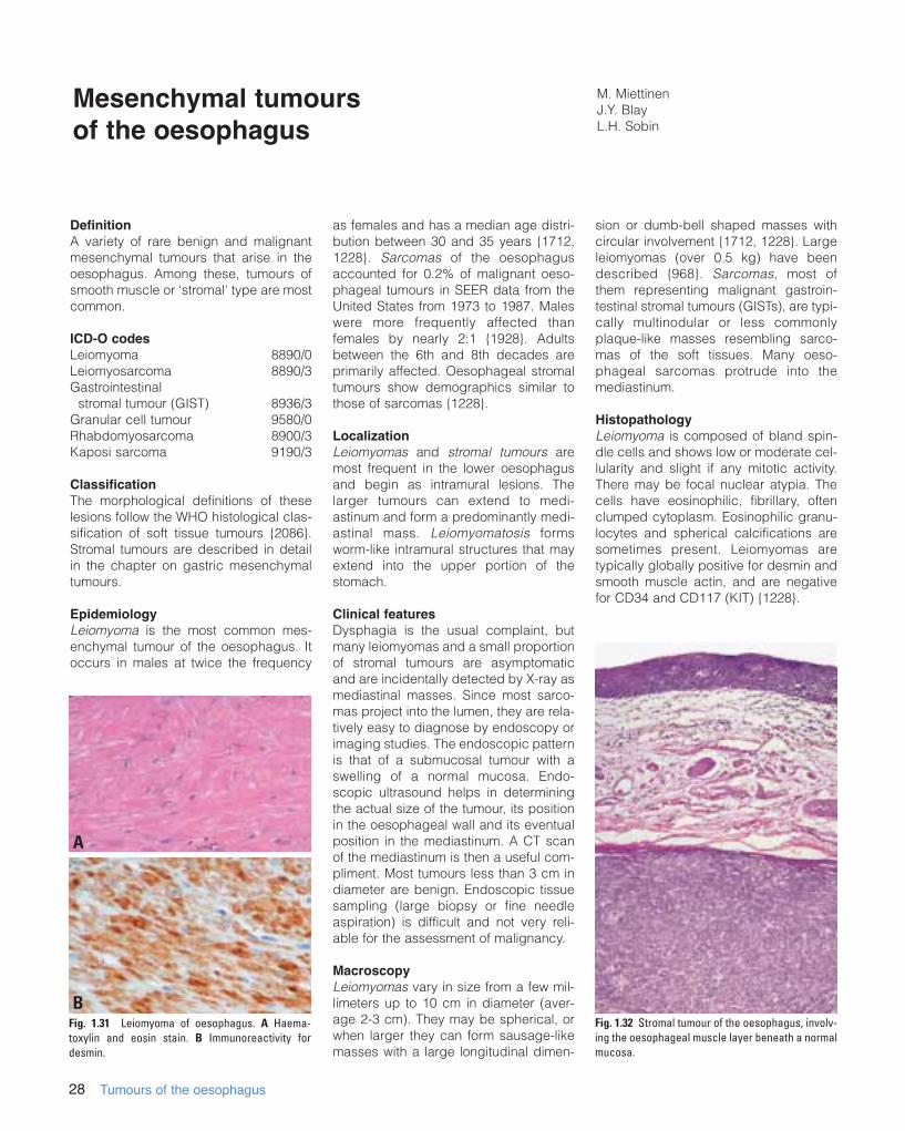
HistopathologyLeiomyoma is composed of bland spin-dle cells and shows low or moderate cel-lularity and slight if any mitotic activity.There may be focal nuclear atypia. Thecells have eosinophilic, fibrillary, oftenclumped cytoplasm. Eosinophilic granu-locytes and spherical calcifications aresometimes present. Leiomyomas aretypically globally positive for desmin andsmooth muscle actin, and are negativefor CD34 and CD117 (KIT) {1228}.
M. MiettinenJ.Y. BlayL.H. Sobin
Mesenchymal tumours of the oesophagus
Fig. 1.31 Leiomyoma of oesophagus. A Haema-toxylin and eosin stain. B Immunoreactivity fordesmin.
Fig. 1.32 Stromal tumour of the oesophagus, involv-ing the oesophageal muscle layer beneath a normalmucosa.
A
B
01 19.7.2006 7:22 Page 28

29Mesenchymal tumours
Leiomyosarcoma, a malignant tumourfeaturing differentiated smooth musclecells, is rare in the oesophagus. In arecent series, such tumours comprised4% of all combined smooth muscle andstromal tumours. They were largetumours that presented in older adults,and all patients died of disease.Diagnosis is based on demonstration ofsmooth muscle differentiation by α-smooth muscle actin, desmin or both,and lack of KIT expression {1228}.Stromal tumours (GISTs) are rare in theoesophagus, and comprise 20-30% ofthe combined cases of smooth muscleand stromal tumours. Like elsewhere inthe digestive system, they predominantlyoccur in older adults between the 6thand 8th decades; oesophageal stromaltumours may have a male predomi-nance. Most oesophageal examples arespindle cell tumours, and a minority areepithelioid. Oesophageal GISTs are iden-tical with their gastric counterparts bytheir positivity for KIT and CD34, variablereactivity for smooth muscle actin andgeneral negativity for desmin. Most areclinically malignant, and commonlydevelop liver metastases. The oeso-phageal tumours analyzed to date haveshown similar c-kit mutations (exon 11)as observed in gastric and intestinalGISTs {1228}. The pathological featuresare described with gastric GISTs.
Granular cell tumours are usually detect-ed endoscopically as nodules or smallsessile polyps predominantly in the distaloesophagus {1216, 7}. Benign behaviouris the rule, but a case of malignantoesophageal granular cell tumour hasbeen reported. The tumours are usuallysmall, up to 1-2 cm in diameter, and aregrossly yellow, firm nodules. Histologi-cally they are composed of sheets of ovalto polygonal cells with a small centralnucleus and abundant granular slightlybasophilic cytoplasm. This is due toextensive accumulation of lysosomesfilled with lamellar material. Granular celltumours are typically PAS- and S100-pro-tein positive and negative for desmin,actin, CD34 and KIT. Tumours thatencroach upon the mucosa may elicit apseudocarcinomatous squamous hyper-plasia {862, 1710}.Rhabdomyosarcoma has been reportedin older adult patients in distal oesopha-gus. A few well-documented cases haveshown features similar to embryonalrhabdomyosarcoma {2002}. Demonstra-tion of skeletal muscle differentiation bythe presence of cross-striations, electronmicroscopy, or immunohistochemistry isrequired for the diagnosis.Synovial sarcoma has been reported inchildren and in older adults. Thesetumours usually present as polypoidmasses in the proximal oesophagus{168, 149}.Kaposi sarcoma may appear as amucosal or less commonly more exten-sive mural lesion, usually in HIV-positivepatients. Histologically typical are spin-dle cells with vascular slit formations andscattered PAS-positive globules. Thetumour cells are positive for CD31 andCD34.
GradingHistological grading follows the systemscommonly used for soft tissue tumours.Mitotic activity is the main criterion forgrading stromal sarcomas andleiomyosarcomas, namely those tumourswith over 10 mitoses per 10 HPF are con-sidered high-grade.
GeneticsSomatic deletions and gene rearrange-ments involving the genes encodingalpha5 and 6 chains of collagen type IVhave been described in oesophagealleiomyomatosis associated with Alportsyndrome {1704, 1982} and in sporadic
leiomyoma {683}, whereas these tumoursdo not have c-kit gene mutations com-monly found in GISTs {1018}. Compar-ative genomic hybridization studies haveshown that oesophageal leiomyomas donot have losses of chromosome 14, asoften seen in GIST, but instead havegains in chromosome 5 {450, 1664}.Oesophageal stromal tumours show simi-lar c-kit mutations as observed in gastricand intestinal GISTs (see stomach mes-enchymal tumours) {1228}.Kaposi sarcoma is positive for humanherpesvirus 8 by PCR.
PrognosisThe prognosis of oesophageal sarcomas,like carcinomas, is largely dependent onthe size, depth of invasion, and presenceor absence of metastasis.
Fig. 1.33 Granular cell tumour of oesophagus. Fig. 1.34 Kaposi sarcoma in a patient with acquiredimmunodeficiency syndrome.
1 cm
01 19.7.2006 7:22 Page 29

30 Tumours of the oesophagus
Secondary tumours
DefinitionTumours of the oesophagus that originatefrom but are discontinuous with a primarytumour elsewhere in the oesophagus oran extra-oesophageal neoplasm.
IncidenceMetastatic spread to the oesophagus isuncommon. An unusually high frequency(6.1% of autopsy cases) was reportedfrom Japan {1249}.
Origin of metastasesThe concept of intramural metastasis inoesophageal squamous cell carcinomais discussed in the chapter on squamouscell carcinoma of the oesophagus.Neoplasms of neighbouring organs suchas pharynx or gastric cardia {714} canspread to the oesophagus via lymphat-ics. Haematogenous metastases fromany primary localization may occur.Reported primary sites include thyroid{335}, lung {1416, 1249}, breast {2143,
1249, 545}, skin {1569, 1203}, kidney{1956}, prostate {1318} and ovary {1249}.
LocalizationThe most common site of involvement isthe middle third of the oesophagus.
Clinical featuresThe leading symptom is dysphagia,whereas achalasia and upper gastroin-testinal bleeding with anemia are unusu-al {545}. Barium swallow examination,endoscopy, computed tomography andmagnetic resonance imaging demon-strate in most cases a submucosaltumour, but any aspect resembling a pri-mary oesophageal carcinoma may beobserved {545, 1318, 714}.
Histopathology and predictive factorsSubmucosal localization without invasionof the mucosa is characteristic for ametastasis. Early metastases of gastricand oesophageal tumours into theoesophagus may be local indicators ofsystemic spread {896, 714}. The pres-
ence of metastasis in the oesophagus isa sign of poor prognosis, but the outcomeis much better when the primary tumourgrowth rate is slow, and when othermetastases are excluded {1416, 1249}.
Melanoma
ICD-O Code 8720/3
Malignant melanoma in the oesophagusis much more commonly metastatic thanprimary. Primary oesophageal mela-nomas are usually polypoid and are clini-cally aggressive lesions {400, 353}. Theyare believed to arise from a zone of atyp-ical junctional proliferation of melano-cytes and such a proliferation is oftenpresent adjacent to the invasive tumour,although it may not be observed inadvanced disease. The histology of theinvasive component is indistinguishablefrom cutaneous melanoma {409}. Growthis typically expansile rather than infiltra-tive.
G. IlyésA. KádárN.J. Carr
Secondary tumours and melanoma of the oesophagus
Fig. 1.35 Primary melanoma of the oesophagus (ME). The gastro-oesophagealjunction is on the left (arrows).
Fig. 1.36 Primary malignant melanoma of the distal oesophagus. Zone of atypi-cal junctional proliferation of melanocytes located adjacent to the invasivetumour. This supports the diagnosis of a primary melanoma.
ME
01 19.7.2006 7:22 Page 30