by Smac/Diablo Author Manuscript NIH Public Access 1,3 2,3...
20
A Dimeric Smac/Diablo Peptide Directly Relieves Caspase-3 Inhibition by XIAP: Dynamic and Cooperative Regulation of XIAP by Smac/Diablo Zhonghua Gao 1,3 , Yuan Tian 2,3 , Junru Wang 1 , Qian Yin 3,4 , Hao Wu 3,4 , Yue-Ming Li 2,3 , and Xuejun Jiang 1,3 1 Cell Biology Program, Memorial Sloan-Kettering Cancer Center, New York, NY 10021 2 Molecular Pharmacology and Chemistry Program, Memorial Sloan-Kettering Cancer Center, New York, NY 10021 3 Graduate School of Biomedical Sciences, Weill Medical College of Cornell University, New York, NY 10021 4 Department of Biochemistry, Weill Medical College of Cornell University, New York, NY 10021 Abstract Caspase activation, the executing event of apoptosis, is under deliberate regulation. IAP proteins inhibit caspase activity whereas Smac/Diablo antagonizes IAP. XIAP, a ubiquitous IAP, can inhibit both caspase-9, the initiator caspase of the mitochondrial apoptotic pathway, and the downstream effector caspases, caspase-3 and caspase-7. Smac neutralizes XIAP inhibition of caspase-9 by competing for binding of the BIR3 domain of XIAP with caspase-9, whereas how Smac liberates effector caspases from XIAP inhibition is not clear. It is generally believed that binding of Smac with IAP generates a steric hindrance that prevents XIAP from inhibiting effector caspases, and therefore small molecule mimics of Smac are not able to reverse inhibition of the effector caspases. Surprisingly, we show here that binding of a dimeric Smac N-terminal peptide with the BIR2 domain of XIAP effectively antagonizes inhibition of caspase-3 by XIAP. Further, we defined the dynamic and cooperative interaction of Smac with XIAP: binding of Smac with the BIR3 domain anchors the subsequent binding of Smac with the BIR2 domain, which in turn attenuates the caspase-3-inhibitory function of XIAP. We also show that XIAP homotrimerizes via its C-terminal Ring domain, making its inhibitory activity towards caspase-3 more susceptible to Smac. INTRODUCTION Apoptosis, a cellular process that plays crucial roles in multiple physiological and pathological events, is executed by the activity of a family of cysteine proteases known as caspases (1,2). In mammals, a major caspase activation pathway is the mitochondrial cytochrome c-mediated pathway (3). This pathway mediates apoptosis triggered by a plethora of death stimuli including DNA damage, growth factor deprivation, and various other stressful conditions. Mechanistically, these stimuli converge on mitochondria and cause release of cytochrome c (4), which subsequently induces the mediator protein Apaf-1 to form an oligomeric protein complex, the apoptosome (5,6). The apoptosome recruits and activates the initiator caspase, caspase-9, and the apoptosome-caspase-9 holoenzyme Address correspondence to: Xuejun Jiang, Cell Biology Program, Memorial Sloan-Kettering Cancer Center, 1275 York Avenue, Box 522, New York, NY 10021, USA. Tel: 212-639-6814. [email protected]. NIH Public Access Author Manuscript J Biol Chem. Author manuscript; available in PMC 2011 October 26. Published in final edited form as: J Biol Chem. 2007 October 19; 282(42): 30718–30727. doi:10.1074/jbc.M705258200. NIH-PA Author Manuscript NIH-PA Author Manuscript NIH-PA Author Manuscript
Transcript of by Smac/Diablo Author Manuscript NIH Public Access 1,3 2,3...
A Dimeric Smac/Diablo Peptide Directly Relieves Caspase-3Inhibition by XIAP: Dynamic and Cooperative Regulation of XIAPby Smac/Diablo
Zhonghua Gao1,3, Yuan Tian2,3, Junru Wang1, Qian Yin3,4, Hao Wu3,4, Yue-Ming Li2,3, andXuejun Jiang1,3
1Cell Biology Program, Memorial Sloan-Kettering Cancer Center, New York, NY 100212Molecular Pharmacology and Chemistry Program, Memorial Sloan-Kettering Cancer Center,New York, NY 100213Graduate School of Biomedical Sciences, Weill Medical College of Cornell University, New York,NY 100214Department of Biochemistry, Weill Medical College of Cornell University, New York, NY 10021
AbstractCaspase activation, the executing event of apoptosis, is under deliberate regulation. IAP proteinsinhibit caspase activity whereas Smac/Diablo antagonizes IAP. XIAP, a ubiquitous IAP, caninhibit both caspase-9, the initiator caspase of the mitochondrial apoptotic pathway, and thedownstream effector caspases, caspase-3 and caspase-7. Smac neutralizes XIAP inhibition ofcaspase-9 by competing for binding of the BIR3 domain of XIAP with caspase-9, whereas howSmac liberates effector caspases from XIAP inhibition is not clear. It is generally believed thatbinding of Smac with IAP generates a steric hindrance that prevents XIAP from inhibiting effectorcaspases, and therefore small molecule mimics of Smac are not able to reverse inhibition of theeffector caspases. Surprisingly, we show here that binding of a dimeric Smac N-terminal peptidewith the BIR2 domain of XIAP effectively antagonizes inhibition of caspase-3 by XIAP. Further,we defined the dynamic and cooperative interaction of Smac with XIAP: binding of Smac with theBIR3 domain anchors the subsequent binding of Smac with the BIR2 domain, which in turnattenuates the caspase-3-inhibitory function of XIAP. We also show that XIAP homotrimerizes viaits C-terminal Ring domain, making its inhibitory activity towards caspase-3 more susceptible toSmac.
INTRODUCTIONApoptosis, a cellular process that plays crucial roles in multiple physiological andpathological events, is executed by the activity of a family of cysteine proteases known ascaspases (1,2). In mammals, a major caspase activation pathway is the mitochondrialcytochrome c-mediated pathway (3). This pathway mediates apoptosis triggered by aplethora of death stimuli including DNA damage, growth factor deprivation, and variousother stressful conditions. Mechanistically, these stimuli converge on mitochondria andcause release of cytochrome c (4), which subsequently induces the mediator protein Apaf-1to form an oligomeric protein complex, the apoptosome (5,6). The apoptosome recruits andactivates the initiator caspase, caspase-9, and the apoptosome-caspase-9 holoenzyme
Address correspondence to: Xuejun Jiang, Cell Biology Program, Memorial Sloan-Kettering Cancer Center, 1275 York Avenue, Box522, New York, NY 10021, USA. Tel: 212-639-6814. [email protected].
NIH Public AccessAuthor ManuscriptJ Biol Chem. Author manuscript; available in PMC 2011 October 26.
Published in final edited form as:J Biol Chem. 2007 October 19; 282(42): 30718–30727. doi:10.1074/jbc.M705258200.
NIH
-PA Author Manuscript
NIH
-PA Author Manuscript
NIH
-PA Author Manuscript
activates the downstream effector caspases, caspase-3 and caspase-7. This caspase cascadeleads to apoptotic cell death associated with a series of hallmark morphological features.
In cells, the mitochondrial apoptotic pathway is regulated at multiple stages. The Bcl-2family of proteins regulates release of mitochondrial apoptotic proteins such as cytochromec (7–10). The pathway can also be regulated downstream of cytochrome c release. Forexample, it has be found that oncoprotein prothymosin-α and tumor suppressor PHAPproteins modulate the assembly and activity of the apoptosome-caspase-9 holoenzyme,likely via regulating the nucleotide binding/exchanging function of Apaf-1 (11,12). Further,the enzymatic activity of caspases can be directly regulated by IAP (Inhibitor of Apoptosis)proteins (13). IAP proteins all contain one or more BIR (Baculoviral IAP Repeat) domains,which are thought to be responsible for caspase inhibition. For example, XIAP, a ubiquitousmember of IAP family, contains three BIR domains (from N terminus, BIR1, BIR2, andBIR3). During the process of apoptosis, the inhibitory function of IAP can be antagonizedby Smac/Diablo (14,15) and Omi/HtrA2 (16–20), which are also released from mitochondriaas cytochrome c.
The molecular basis underlying neutralization of IAP by Smac is intriguing and elegant.Smac is dimeric protein, and is posttranslationally processed in mitochondria (14,15,21).The N terminus of processed Smac, starting with the residues AVPI, has been shown to benecessary and sufficient for removal of inhibition of caspase-9 by XIAP (21). Structuralstudies demonstrated that the AVPI residues of Smac interact specifically and tightly with aconserved groove within XIAP BIR3 domain (22,23). Subsequently, it was revealed thatXIAP BIR3 domain interacts with caspase-9 in a strikingly similar manner. The cleaved,activated caspase-9 exposes a new N terminus similar to that of Smac, which is required forinteraction of caspase-9 with the groove region of XIAP BIR3 domain (24,25). Therefore,Smac neutralizes XIAP inhibition of caspase-9 by competing for the same specific bindingsite of XIAP with caspase-9.
In addition to the initiator caspase-9, XIAP can inhibit the effector caspases of themitochondrial pathway as well (26). Such inhibition can also be counteracted by Smac (21).However, the mechanism how Smac does so is not clear, because Smac can only interactwith the BIR2 and BIR3 domains of XIAP (21), whereas XIAP utilizes a linker regionbetween its BIR1 and BIR2 domains, instead of one of BIR domains, to inhibit the effectorcaspases. This linker region can strongly interact with the active sites of both caspase-3 andcaspase-7, as defined biochemically and structurally (26–30). Currently, it is generallyaccepted that binding of Smac with the BIR2 and BIR3 domains of XIAP creates a sterichindrance that is essential for preventing binding of XIAP linker region with effectorcaspases, thus achieving neutralization of XIAP inhibition (27–29,31).
IAP proteins are frequently overexpressed in human cancers, indicating their oncogenicfunction and the potential to be therapeutic targets (32–35). Indeed, small moleculecompounds and antisense oligonucleotides targeting IAP have been developed as anticancerproto-drugs (36–38). Interestingly, the mechanism by which the short N terminus of Smacantagonizes IAP was also explored for anticancer agent development. A successful examplefor such mechanism-based design is a synthesized chemical compound mimicking thestructure of the dimeric Smac N-terminal sequence, AVPI (33). The non-peptide nature ofthis molecule provides desirable cell-permeability and stability, and this molecule possessesstrong ability to potentiate apoptosis in cancer cells, thus a promising anticancer drug lead.However, it is predicted by the current model that the effect of such Smac N-terminal mimicmight be limited, because it is supposed to be able to only antagonize inhibition of caspase-9by IAP, but not able to antagonize inhibition of effector caspases by IAP, which shouldrequire the bulky Smac protein to create steric hindrance (32,39).
Gao et al. Page 2
J Biol Chem. Author manuscript; available in PMC 2011 October 26.
NIH
-PA Author Manuscript
NIH
-PA Author Manuscript
NIH
-PA Author Manuscript

In this study, we investigate the biochemical mechanisms by which Smac antagonizesinhibition of the effector caspase, caspase-3, by XIAP. We show that the dimeric Smac N-terminal peptide alone can effectively restore caspase-3 activity inhibited by XIAP. Ourstudy also indicates that Smac interacts with the BIR3 and then BIR2 domains of XIAPsequentially, and such dynamic interaction cooperatively neutralizes inhibition of caspase-3by the linker region of XIAP.
EXPERIMENTAL PROCEDURESReagents
Caspase-3 substrate, Ac-DEVD-AMC (Cat # 235425), and caspase-9 substrate, Ac-LEHD-AFC (Cat # 218765), were purchased from Calbiochem. Anti-Smac antibody (Cat # 05-681)was obtained from Upstate; anti-Flag antibody M2 was from Sigma.
PlasmidsFor His-tagged recombinant proteins, full-length human XIAP (residue 10-497) or XIAP-ΔRing (10-356) was generated by PCR and subcloned into pET21a (Novagen). Site directedMutagenesis was performed to generate BIR2 (D214S) and BIR3 mutant (E314A) of full-length XIAP. For GST-tagged recombinant proteins, full-length human XIAP (1-497) orBIR3 domain (238-358) was generated by PCR and subcloned into pGEX2T (GEHealthcare). For purification of heterodimer Smac, PCR product from pET15b-Smac(AVPI)-His was inserted into pOKD4 to generate pOKD4-Smac (MVPI)-Flag.
Peptide SynthesisAll peptides were prepared using standard Fmoc solid phase chemistry on a peptidesynthesizer (Protein Technologies, Inc.). The dimeric peptide (dAVPI) was synthesizedusing Universal PEG NovaTag resin (Cat # 04-12-3911, EMD Biosciences). The Fmocprotecting group of the Universal PEG NovaTag resin was first removed using 20%piperidine in DMF. Then individual Fmoc-AA-OH was coupled to the resin. The aminogroup of N-terminal amino acid was protected by Boc group, rather than Fmoc. After thefirst peptide was synthesized, the Mmt protecting group was removed with 1.0 M HOBt inTFE/DCM. The produced resin was used for the synthesis of second peptides using the sameprocedure. The final cleavage and deprotection were performed with a cocktail (TFA: water:TIS, 95:2.5:2.5, v/v). The cleaved peptides were extracted repeatedly with ethyl ether andpurified by reverse phase HPLC on a C18 column (ZORBAX 300 SB-C18, AgilentTechnologies), using a 0–30% water/acetonitrile gradient in the presence of 0.1%trifluoroacetic acid (TCA). Peptide identity was verified by LC-MS/MS (AgilentTechnologies).
Preparation of Cytosolic Fractions from HeLa CellsHeLa cells are harvested by centrifugation at 1000 × g for 5 min. After washed with PBS,cell pellets were suspended in 5 × volume of Buffer A (20 mM HEPES, pH 7.5, 10 mMKCl, 1 mM EDTA, 1 mM EGTA, 1.5 mM MgCl2, 1 mM DTT) supplemented with mixtureproteases inhibitors (Roche Diagnostics). After incubation on ice for 15 min, cells werebroken by passing through 22-gauge needles 25 times. Cell lysates were then subject tocentrifugation at 750 × g for 10 min at 4°C. Supernatants were further centrifuged at 12,000× g for 10 min at 4°C, followed by final centrifugation at 100,000 × g for 2 hr at 4°C toobtain HeLa S-100 fraction (HS100).
Gao et al. Page 3
J Biol Chem. Author manuscript; available in PMC 2011 October 26.
NIH
-PA Author Manuscript
NIH
-PA Author Manuscript
NIH
-PA Author Manuscript
Recombinant ProteinsGenerally, for His-tagged proteins, plasmids were transformed into BL21 (DE3) andcultured at 37°C. When OD reached 0.6, 0.1 mM IPTG was added to induce proteinexpression at 18°C for 16 hours. Cells were pelleted and subject to standard Ni2+affinitychromatography. For expression of recombinant SMAC proteins, pET15b-Smac-His andpOKD4-SMAC-Flag were co-transformed into BL21 (DE3), followed by IPTG inductionand Ni2+ affinity chromatography. The eluted protein was then subject to anion exchange Qcolumn purification (1ml HiTrapQ, GE Healthcare) eluted with 20 ml of gradient from 100mM to 400 mM NaCl in Buffer A, which resulted in two protein peaks. The first one waswild type homodimer Smac (Smac-WT), and the second was mutant heterodimer Smac(Smac-Mut). For GST-tagged protein, similar procedure was performed to get theexpression of proteins in BL21, followed by GST affinity chromatography. All recombinantproteins obtained by affinity purification were dialyzed in Buffer A for 2 hours at 4°C.Aliquots were flash frozen with liquid nitrogen, and stored at −80°C.
Caspase Activity AssayFor caspase-3 or caspase-9 activity assay in total reconstituted system, each reaction with afinal volume of 30 μl was assembled on ice, including: 3 μl 10 × Caspase Assay Buffer (300mM HEPES, pH 7.5, 1 M NaCl, 50% sucrose, 1% CHAPS, 200 mM β-mercaptoethanol, 20mg/ml BSA) (40), 3 μl caspase-3 substrate (Ac-DEVD-AMC, 1 mM) or 3 μl caspase-9substrate (Ac-LEHD-AFC, 1 mM), respectively. Wild-type or mutant XIAP, and Smac orpeptides were added as indicated. The mixture was first incubated at 30°C for 10 min, andthen supplemented with 3 μl of 10 nM caspase-3 or 3 μl of 200 nM leucine-zipper-linkeddimeric caspase-9 (41), respectively, to initiate the enzyme reaction. Generation offluorescent signal (relative fluorescent units) due to the cleavage of substrates, indicative ofcaspase-3 or caspase-9 activity, was measured by an automated spectrophotometer(TECAN) at wavelengths of 360/465 (excitation/emission) nm or 400/505 nm, respectively.
For caspase-3 activity assay with HeLa cytosolic extract, caspase-3/7 in HS100 was firstactivated with 1 mM dATP and 5 μM cytochrome c at 30°C for 3.5 hr. Then 9 μl of pre-activated HS100 was mixed with 21 μl of reaction solution pre-warmed at 30°C for 10 min,consisting indicated amount of XIAP, dAVPI, and caspase-3 substrates. Substrate cleavagewas measured as above.
GST Pull-Down ExperimentFor each sample, 5 μg of purified full-length XIAP (GST-XIAP) or BIR3 domain (GST-BIR3) was coupled to 20 μl glutathione beads. Subsequently, in a 200-μl Buffer Asystem,the beads were incubated with 1 μM Smac in the presence of indicated concentration ofdAVPI for 1 hr followed by three washes. Then beads were boiled in 4 × SDS loadingbuffer, and eluted proteins were subject to SDS-PAGE, followed by Western blotting withan anti-Smac antibody.
Molecular Weight MeasurementMolar mass of purified XIAP-WT or XIAP-ΔRing was determined by static multi-angellight scattering (MALS). Protein was injected onto a Superdex 200 HR 10/300 gel filtrationcolumn equilibrated in Buffer A. The chromatography system was coupled to a three-anglelight scattering detector (mini-DAWN EOS) and refractive index detector (Optilab DSP,Wyatt Technology). Data were collected every 0.5 second at a flow rate of 0.25 ml/min.Data analysis was carried out using the ASTRA program (Wyatt Technology), yielding themolar mass and mass distribution (polydispersity) of the sample.
Gao et al. Page 4
J Biol Chem. Author manuscript; available in PMC 2011 October 26.
NIH
-PA Author Manuscript
NIH
-PA Author Manuscript
NIH
-PA Author Manuscript
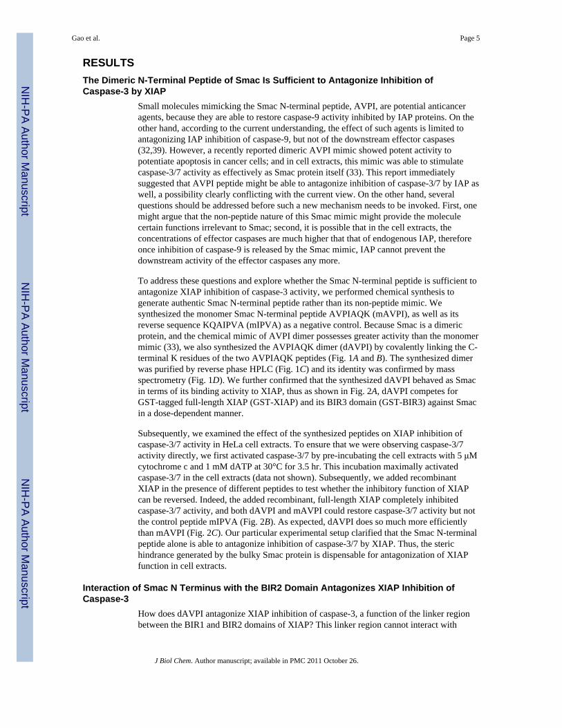
RESULTSThe Dimeric N-Terminal Peptide of Smac Is Sufficient to Antagonize Inhibition ofCaspase-3 by XIAP
Small molecules mimicking the Smac N-terminal peptide, AVPI, are potential anticanceragents, because they are able to restore caspase-9 activity inhibited by IAP proteins. On theother hand, according to the current understanding, the effect of such agents is limited toantagonizing IAP inhibition of caspase-9, but not of the downstream effector caspases(32,39). However, a recently reported dimeric AVPI mimic showed potent activity topotentiate apoptosis in cancer cells; and in cell extracts, this mimic was able to stimulatecaspase-3/7 activity as effectively as Smac protein itself (33). This report immediatelysuggested that AVPI peptide might be able to antagonize inhibition of caspase-3/7 by IAP aswell, a possibility clearly conflicting with the current view. On the other hand, severalquestions should be addressed before such a new mechanism needs to be invoked. First, onemight argue that the non-peptide nature of this Smac mimic might provide the moleculecertain functions irrelevant to Smac; second, it is possible that in the cell extracts, theconcentrations of effector caspases are much higher that that of endogenous IAP, thereforeonce inhibition of caspase-9 is released by the Smac mimic, IAP cannot prevent thedownstream activity of the effector caspases any more.
To address these questions and explore whether the Smac N-terminal peptide is sufficient toantagonize XIAP inhibition of caspase-3 activity, we performed chemical synthesis togenerate authentic Smac N-terminal peptide rather than its non-peptide mimic. Wesynthesized the monomer Smac N-terminal peptide AVPIAQK (mAVPI), as well as itsreverse sequence KQAIPVA (mIPVA) as a negative control. Because Smac is a dimericprotein, and the chemical mimic of AVPI dimer possesses greater activity than the monomermimic (33), we also synthesized the AVPIAQK dimer (dAVPI) by covalently linking the C-terminal K residues of the two AVPIAQK peptides (Fig. 1A and B). The synthesized dimerwas purified by reverse phase HPLC (Fig. 1C) and its identity was confirmed by massspectrometry (Fig. 1D). We further confirmed that the synthesized dAVPI behaved as Smacin terms of its binding activity to XIAP, thus as shown in Fig. 2A, dAVPI competes forGST-tagged full-length XIAP (GST-XIAP) and its BIR3 domain (GST-BIR3) against Smacin a dose-dependent manner.
Subsequently, we examined the effect of the synthesized peptides on XIAP inhibition ofcaspase-3/7 activity in HeLa cell extracts. To ensure that we were observing caspase-3/7activity directly, we first activated caspase-3/7 by pre-incubating the cell extracts with 5 μMcytochrome c and 1 mM dATP at 30°C for 3.5 hr. This incubation maximally activatedcaspase-3/7 in the cell extracts (data not shown). Subsequently, we added recombinantXIAP in the presence of different peptides to test whether the inhibitory function of XIAPcan be reversed. Indeed, the added recombinant, full-length XIAP completely inhibitedcaspase-3/7 activity, and both dAVPI and mAVPI could restore caspase-3/7 activity but notthe control peptide mIPVA (Fig. 2B). As expected, dAVPI does so much more efficientlythan mAVPI (Fig. 2C). Our particular experimental setup clarified that the Smac N-terminalpeptide alone is able to antagonize inhibition of caspase-3/7 by XIAP. Thus, the sterichindrance generated by the bulky Smac protein is dispensable for antagonization of XIAPfunction in cell extracts.
Interaction of Smac N Terminus with the BIR2 Domain Antagonizes XIAP Inhibition ofCaspase-3
How does dAVPI antagonize XIAP inhibition of caspase-3, a function of the linker regionbetween the BIR1 and BIR2 domains of XIAP? This linker region cannot interact with
Gao et al. Page 5
J Biol Chem. Author manuscript; available in PMC 2011 October 26.
NIH
-PA Author Manuscript
NIH
-PA Author Manuscript
NIH
-PA Author Manuscript
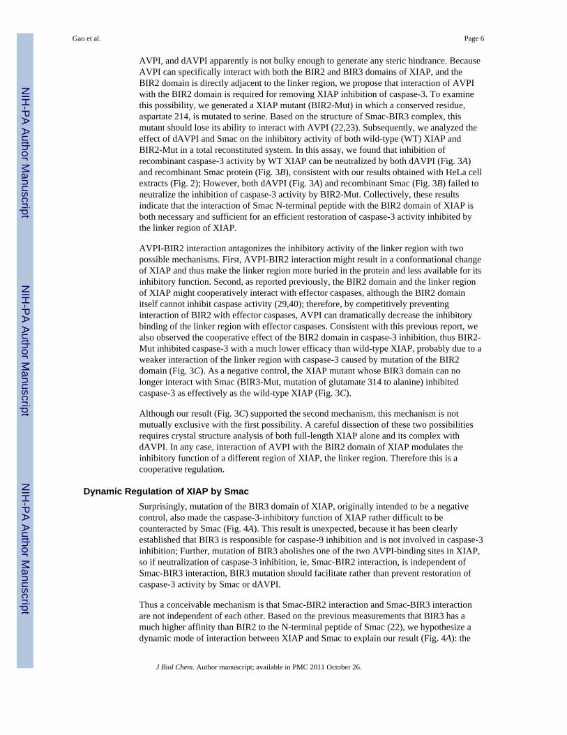
AVPI, and dAVPI apparently is not bulky enough to generate any steric hindrance. BecauseAVPI can specifically interact with both the BIR2 and BIR3 domains of XIAP, and theBIR2 domain is directly adjacent to the linker region, we propose that interaction of AVPIwith the BIR2 domain is required for removing XIAP inhibition of caspase-3. To examinethis possibility, we generated a XIAP mutant (BIR2-Mut) in which a conserved residue,aspartate 214, is mutated to serine. Based on the structure of Smac-BIR3 complex, thismutant should lose its ability to interact with AVPI (22,23). Subsequently, we analyzed theeffect of dAVPI and Smac on the inhibitory activity of both wild-type (WT) XIAP andBIR2-Mut in a total reconstituted system. In this assay, we found that inhibition ofrecombinant caspase-3 activity by WT XIAP can be neutralized by both dAVPI (Fig. 3A)and recombinant Smac protein (Fig. 3B), consistent with our results obtained with HeLa cellextracts (Fig. 2); However, both dAVPI (Fig. 3A) and recombinant Smac (Fig. 3B) failed toneutralize the inhibition of caspase-3 activity by BIR2-Mut. Collectively, these resultsindicate that the interaction of Smac N-terminal peptide with the BIR2 domain of XIAP isboth necessary and sufficient for an efficient restoration of caspase-3 activity inhibited bythe linker region of XIAP.
AVPI-BIR2 interaction antagonizes the inhibitory activity of the linker region with twopossible mechanisms. First, AVPI-BIR2 interaction might result in a conformational changeof XIAP and thus make the linker region more buried in the protein and less available for itsinhibitory function. Second, as reported previously, the BIR2 domain and the linker regionof XIAP might cooperatively interact with effector caspases, although the BIR2 domainitself cannot inhibit caspase activity (29,40); therefore, by competitively preventinginteraction of BIR2 with effector caspases, AVPI can dramatically decrease the inhibitorybinding of the linker region with effector caspases. Consistent with this previous report, wealso observed the cooperative effect of the BIR2 domain in caspase-3 inhibition, thus BIR2-Mut inhibited caspase-3 with a much lower efficacy than wild-type XIAP, probably due to aweaker interaction of the linker region with caspase-3 caused by mutation of the BIR2domain (Fig. 3C). As a negative control, the XIAP mutant whose BIR3 domain can nolonger interact with Smac (BIR3-Mut, mutation of glutamate 314 to alanine) inhibitedcaspase-3 as effectively as the wild-type XIAP (Fig. 3C).
Although our result (Fig. 3C) supported the second mechanism, this mechanism is notmutually exclusive with the first possibility. A careful dissection of these two possibilitiesrequires crystal structure analysis of both full-length XIAP alone and its complex withdAVPI. In any case, interaction of AVPI with the BIR2 domain of XIAP modulates theinhibitory function of a different region of XIAP, the linker region. Therefore this is acooperative regulation.
Dynamic Regulation of XIAP by SmacSurprisingly, mutation of the BIR3 domain of XIAP, originally intended to be a negativecontrol, also made the caspase-3-inhibitory function of XIAP rather difficult to becounteracted by Smac (Fig. 4A). This result is unexpected, because it has been clearlyestablished that BIR3 is responsible for caspase-9 inhibition and is not involved in caspase-3inhibition; Further, mutation of BIR3 abolishes one of the two AVPI-binding sites in XIAP,so if neutralization of caspase-3 inhibition, ie, Smac-BIR2 interaction, is independent ofSmac-BIR3 interaction, BIR3 mutation should facilitate rather than prevent restoration ofcaspase-3 activity by Smac or dAVPI.
Thus a conceivable mechanism is that Smac-BIR2 interaction and Smac-BIR3 interactionare not independent of each other. Based on the previous measurements that BIR3 has amuch higher affinity than BIR2 to the N-terminal peptide of Smac (22), we hypothesize adynamic mode of interaction between XIAP and Smac to explain our result (Fig. 4A): the
Gao et al. Page 6
J Biol Chem. Author manuscript; available in PMC 2011 October 26.
NIH
-PA Author Manuscript
NIH
-PA Author Manuscript
NIH
-PA Author Manuscript
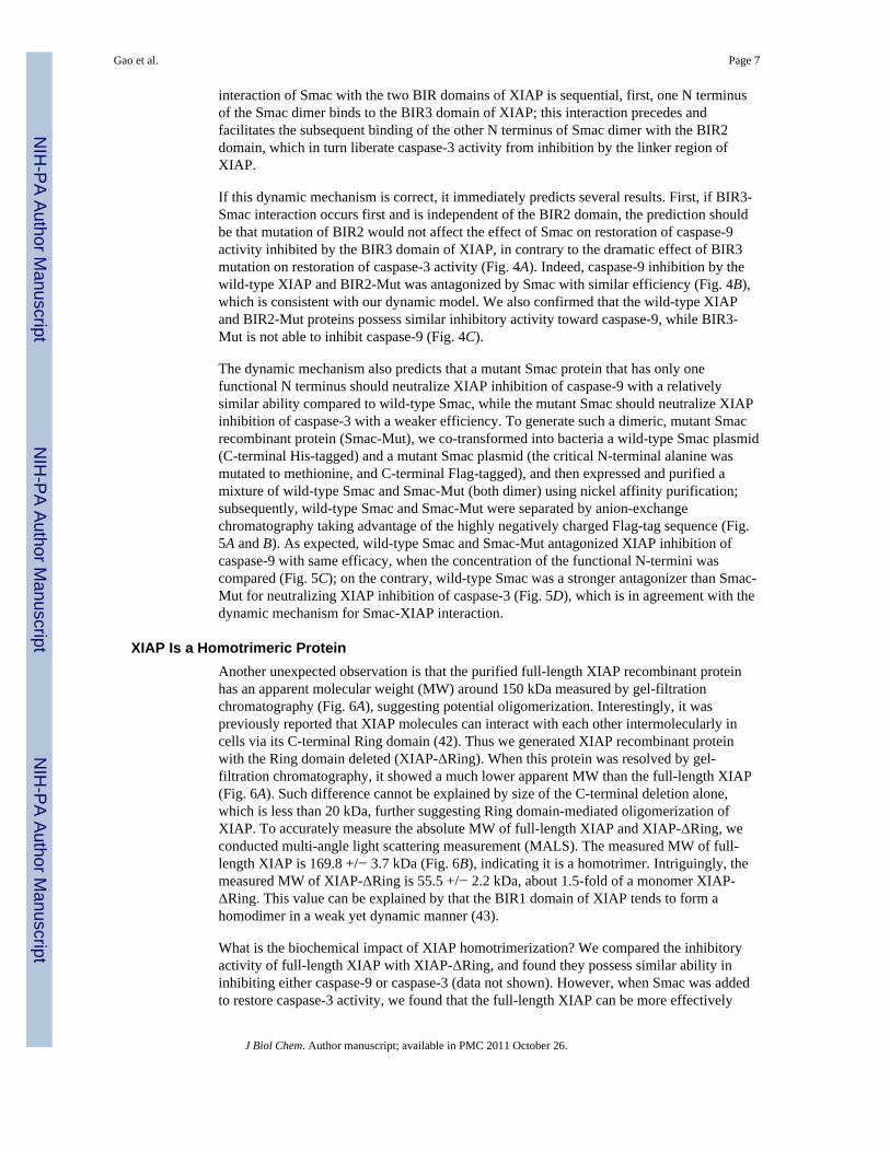
interaction of Smac with the two BIR domains of XIAP is sequential, first, one N terminusof the Smac dimer binds to the BIR3 domain of XIAP; this interaction precedes andfacilitates the subsequent binding of the other N terminus of Smac dimer with the BIR2domain, which in turn liberate caspase-3 activity from inhibition by the linker region ofXIAP.
If this dynamic mechanism is correct, it immediately predicts several results. First, if BIR3-Smac interaction occurs first and is independent of the BIR2 domain, the prediction shouldbe that mutation of BIR2 would not affect the effect of Smac on restoration of caspase-9activity inhibited by the BIR3 domain of XIAP, in contrary to the dramatic effect of BIR3mutation on restoration of caspase-3 activity (Fig. 4A). Indeed, caspase-9 inhibition by thewild-type XIAP and BIR2-Mut was antagonized by Smac with similar efficiency (Fig. 4B),which is consistent with our dynamic model. We also confirmed that the wild-type XIAPand BIR2-Mut proteins possess similar inhibitory activity toward caspase-9, while BIR3-Mut is not able to inhibit caspase-9 (Fig. 4C).
The dynamic mechanism also predicts that a mutant Smac protein that has only onefunctional N terminus should neutralize XIAP inhibition of caspase-9 with a relativelysimilar ability compared to wild-type Smac, while the mutant Smac should neutralize XIAPinhibition of caspase-3 with a weaker efficiency. To generate such a dimeric, mutant Smacrecombinant protein (Smac-Mut), we co-transformed into bacteria a wild-type Smac plasmid(C-terminal His-tagged) and a mutant Smac plasmid (the critical N-terminal alanine wasmutated to methionine, and C-terminal Flag-tagged), and then expressed and purified amixture of wild-type Smac and Smac-Mut (both dimer) using nickel affinity purification;subsequently, wild-type Smac and Smac-Mut were separated by anion-exchangechromatography taking advantage of the highly negatively charged Flag-tag sequence (Fig.5A and B). As expected, wild-type Smac and Smac-Mut antagonized XIAP inhibition ofcaspase-9 with same efficacy, when the concentration of the functional N-termini wascompared (Fig. 5C); on the contrary, wild-type Smac was a stronger antagonizer than Smac-Mut for neutralizing XIAP inhibition of caspase-3 (Fig. 5D), which is in agreement with thedynamic mechanism for Smac-XIAP interaction.
XIAP Is a Homotrimeric ProteinAnother unexpected observation is that the purified full-length XIAP recombinant proteinhas an apparent molecular weight (MW) around 150 kDa measured by gel-filtrationchromatography (Fig. 6A), suggesting potential oligomerization. Interestingly, it waspreviously reported that XIAP molecules can interact with each other intermolecularly incells via its C-terminal Ring domain (42). Thus we generated XIAP recombinant proteinwith the Ring domain deleted (XIAP-ΔRing). When this protein was resolved by gel-filtration chromatography, it showed a much lower apparent MW than the full-length XIAP(Fig. 6A). Such difference cannot be explained by size of the C-terminal deletion alone,which is less than 20 kDa, further suggesting Ring domain-mediated oligomerization ofXIAP. To accurately measure the absolute MW of full-length XIAP and XIAP-ΔRing, weconducted multi-angle light scattering measurement (MALS). The measured MW of full-length XIAP is 169.8 +/− 3.7 kDa (Fig. 6B), indicating it is a homotrimer. Intriguingly, themeasured MW of XIAP-ΔRing is 55.5 +/− 2.2 kDa, about 1.5-fold of a monomer XIAP-ΔRing. This value can be explained by that the BIR1 domain of XIAP tends to form ahomodimer in a weak yet dynamic manner (43).
What is the biochemical impact of XIAP homotrimerization? We compared the inhibitoryactivity of full-length XIAP with XIAP-ΔRing, and found they possess similar ability ininhibiting either caspase-9 or caspase-3 (data not shown). However, when Smac was addedto restore caspase-3 activity, we found that the full-length XIAP can be more effectively
Gao et al. Page 7
J Biol Chem. Author manuscript; available in PMC 2011 October 26.
NIH
-PA Author Manuscript
NIH
-PA Author Manuscript
NIH
-PA Author Manuscript
antagonized by Smac than XIAP-ΔRing (Fig. 6C); while the difference of the full-lengthXIAP vs XIAP-ΔRing in restoration of caspase-9 activity by Smac is less obvious (data notshown).
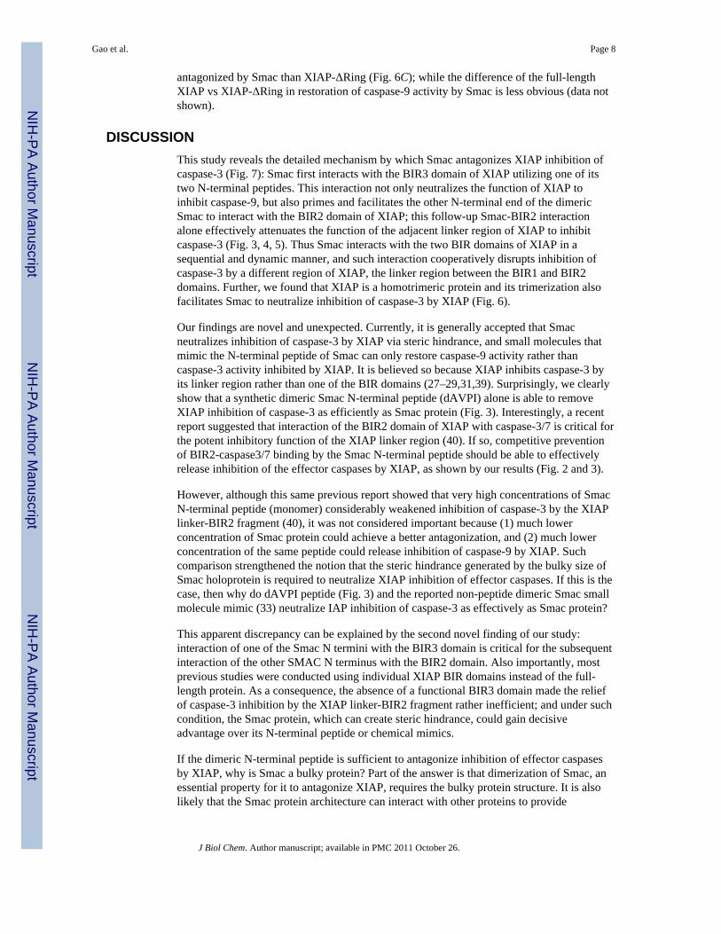
DISCUSSIONThis study reveals the detailed mechanism by which Smac antagonizes XIAP inhibition ofcaspase-3 (Fig. 7): Smac first interacts with the BIR3 domain of XIAP utilizing one of itstwo N-terminal peptides. This interaction not only neutralizes the function of XIAP toinhibit caspase-9, but also primes and facilitates the other N-terminal end of the dimericSmac to interact with the BIR2 domain of XIAP; this follow-up Smac-BIR2 interactionalone effectively attenuates the function of the adjacent linker region of XIAP to inhibitcaspase-3 (Fig. 3, 4, 5). Thus Smac interacts with the two BIR domains of XIAP in asequential and dynamic manner, and such interaction cooperatively disrupts inhibition ofcaspase-3 by a different region of XIAP, the linker region between the BIR1 and BIR2domains. Further, we found that XIAP is a homotrimeric protein and its trimerization alsofacilitates Smac to neutralize inhibition of caspase-3 by XIAP (Fig. 6).
Our findings are novel and unexpected. Currently, it is generally accepted that Smacneutralizes inhibition of caspase-3 by XIAP via steric hindrance, and small molecules thatmimic the N-terminal peptide of Smac can only restore caspase-9 activity rather thancaspase-3 activity inhibited by XIAP. It is believed so because XIAP inhibits caspase-3 byits linker region rather than one of the BIR domains (27–29,31,39). Surprisingly, we clearlyshow that a synthetic dimeric Smac N-terminal peptide (dAVPI) alone is able to removeXIAP inhibition of caspase-3 as efficiently as Smac protein (Fig. 3). Interestingly, a recentreport suggested that interaction of the BIR2 domain of XIAP with caspase-3/7 is critical forthe potent inhibitory function of the XIAP linker region (40). If so, competitive preventionof BIR2-caspase3/7 binding by the Smac N-terminal peptide should be able to effectivelyrelease inhibition of the effector caspases by XIAP, as shown by our results (Fig. 2 and 3).
However, although this same previous report showed that very high concentrations of SmacN-terminal peptide (monomer) considerably weakened inhibition of caspase-3 by the XIAPlinker-BIR2 fragment (40), it was not considered important because (1) much lowerconcentration of Smac protein could achieve a better antagonization, and (2) much lowerconcentration of the same peptide could release inhibition of caspase-9 by XIAP. Suchcomparison strengthened the notion that the steric hindrance generated by the bulky size ofSmac holoprotein is required to neutralize XIAP inhibition of effector caspases. If this is thecase, then why do dAVPI peptide (Fig. 3) and the reported non-peptide dimeric Smac smallmolecule mimic (33) neutralize IAP inhibition of caspase-3 as effectively as Smac protein?
This apparent discrepancy can be explained by the second novel finding of our study:interaction of one of the Smac N termini with the BIR3 domain is critical for the subsequentinteraction of the other SMAC N terminus with the BIR2 domain. Also importantly, mostprevious studies were conducted using individual XIAP BIR domains instead of the full-length protein. As a consequence, the absence of a functional BIR3 domain made the reliefof caspase-3 inhibition by the XIAP linker-BIR2 fragment rather inefficient; and under suchcondition, the Smac protein, which can create steric hindrance, could gain decisiveadvantage over its N-terminal peptide or chemical mimics.
If the dimeric N-terminal peptide is sufficient to antagonize inhibition of effector caspasesby XIAP, why is Smac a bulky protein? Part of the answer is that dimerization of Smac, anessential property for it to antagonize XIAP, requires the bulky protein structure. It is alsolikely that the Smac protein architecture can interact with other proteins to provide
Gao et al. Page 8
J Biol Chem. Author manuscript; available in PMC 2011 October 26.
NIH
-PA Author Manuscript
NIH
-PA Author Manuscript
NIH
-PA Author Manuscript
additional regulation. In addition, it has not been ruled out that Smac might have non-apoptotic function inside of mitochondria. Further, because the linker region of XIAPpossesses very weak but still detectable inhibitory activity toward effector caspasesindependent of the BIR2 domain (27,28,40), one might argue that the steric hindrancecreated by the bulky Smac protein is necessary for a more complete restoration of caspase-3activity inhibited by XIAP, especially when the inhibitor concentration is very high.Although Smac protein indeed antagonizes high concentrations of XIAP more completely(data not shown), the cellular and physiological relevance of such a high IAP concentrationis questionable.
Our finding that XIAP is a homotrimeric protein (Fig. 6) is also intriguing. Structurally, thetrimerization is mediated by the C-terminal Ring domain of XIAP. However, the Ringdomain, which is a putative ubiquitin ligase catalytic domain (44) and was also reported tobe able to mediate protein dimerization (45), has not been shown to mediate proteintrimerization. Functionally, although trimerization of XIAP has no measurable effect on itscaspase inhibitory activity, it facilitates Smac to neutralize its own caspase-3 inhibitoryfunction, which might be physiologically relevant considering that the inhibitory function ofIAP should be readily removed to ensure effective apoptosis when needed. It is not clearmechanistically how trimerization makes XIAP more susceptible to Smac, and sterichindrance should not be the reason because Smac protein and dAVPI neutralize full-lengthXIAP with similar efficacy. On the other hand, trimerization of XIAP appears to have lessobvious effect on neutralization of its caspase-9 inhibitory activity by Smac. This scenariomight be more complicated in cells, because in cells the active caspase-9 is in a million-dalton protein complex, the apoptosome-caspase-9 holoenzyme (46,47). Speculatively,binding of sub-stoichiometric amount of bulky homotrimeric XIAP with the caspase-9holoenzyme might create a steric barrier to effectively prevent the holoenzyme from actingon the downstream effector caspases, thus trimerization makes XIAP a more efficientinhibitor; conversely, the sub-stoichiometric amount of XIAP should be more effectivelycounteracted by Smac when required. Furthermore, Ring domain-mediated XIAPtrimerization should be able to stabilize the dynamic dimerization of the BIR1 domain ofXIAP, a property essential for activation of NF-κB by XIAP (43).
In summary, this study delineated a mechanism by which Smac antagonizes the inhibitoryactivity of XIAP towards caspase-3, an essential regulatory event in the process of apoptosisand a promising target for cancer treatment.
AcknowledgmentsWe thank the members of XJ lab for insightful discussion and suggestions. This work was supported by NIH R01AI045937 (to HW), a Goodwin Experimental Therapeutic Center fund (to YML and XJ), R01 AG026660 (toYML), Alzheimer’s Association Zenith Fellows Award (to YML), American Health Assistance Foundation (toYML), and NIH R01 CA113890 (to XJ).
References1. Budihardjo I, Oliver H, Lutter M, Luo X, Wang X. Annu Rev Cell Dev Biol. 1999; 15:269–290.
[PubMed: 10611963]2. Thornberry NA, Lazebnik Y. Science. 1998; 281:1312–1316. [PubMed: 9721091]3. Jiang X, Wang X. Annu Rev Biochem. 2004; 73:87–106. [PubMed: 15189137]4. Liu X, Kim CN, Yang J, Jemmerson R, Wang X. Cell. 1996; 86:147–157. [PubMed: 8689682]5. Zou H, Henzel WJ, Liu X, Lutschg A, Wang X. Cell. 1997; 90:405–413. [PubMed: 9267021]6. Zou H, Li Y, Liu X, Wang X. J Biol Chem. 1999; 274:11549–11556. [PubMed: 10206961]7. Adams JM, Cory S. Trends Biochem Sci. 2001; 26:61–66. [PubMed: 11165519]
Gao et al. Page 9
J Biol Chem. Author manuscript; available in PMC 2011 October 26.
NIH
-PA Author Manuscript
NIH
-PA Author Manuscript
NIH
-PA Author Manuscript
8. Gross A, McDonnell JM, Korsmeyer SJ. Genes Dev. 1999; 13:1899–1911. [PubMed: 10444588]9. Harris MH, Thompson CB. Cell Death Differ. 2000; 7:1182–1191. [PubMed: 11175255]10. Yang J, Liu X, Bhalla K, Kim CN, Ibrado AM, Cai J, Peng TI, Jones DP, Wang X. Science. 1997;
275:1129–1132. [PubMed: 9027314]11. Jiang X, Kim HE, Shu H, Zhao Y, Zhang H, Kofron J, Donnelly J, Burns D, Ng SC, Rosenberg S,
Wang X. Science. 2003; 299:223–226. [PubMed: 12522243]12. Kim HE, Du F, Fang M, Wang X. Proc Natl Acad Sci U S A. 2005; 102:17545–17550. [PubMed:
16251271]13. Deveraux QL, Reed JC. Genes Dev. 1999; 13:239–252. [PubMed: 9990849]14. Du C, Fang M, Li Y, Li L, Wang X. Cell. 2000; 102:33–42. [PubMed: 10929711]15. Verhagen AM, Ekert PG, Pakusch M, Silke J, Connolly LM, Reid GE, Moritz RL, Simpson RJ,
Vaux DL. Cell. 2000; 102:43–53. [PubMed: 10929712]16. Hegde R, Srinivasula SM, Zhang Z, Wassell R, Mukattash R, Cilenti L, DuBois G, Lazebnik Y,
Zervos AS, Fernandes-Alnemri T, Alnemri ES. J Biol Chem. 2002; 277:432–438. [PubMed:11606597]
17. Martins LM, Iaccarino I, Tenev T, Gschmeissner S, Totty NF, Lemoine NR, Savopoulos J, GrayCW, Creasy CL, Dingwall C, Downward J. J Biol Chem. 2002; 277:439–444. [PubMed:11602612]
18. Suzuki Y, Imai Y, Nakayama H, Takahashi K, Takio K, Takahashi R. Mol Cell. 2001; 8:613–621.[PubMed: 11583623]
19. van Loo G, van Gurp M, Depuydt B, Srinivasula SM, Rodriguez I, Alnemri ES, Gevaert K,Vandekerckhove J, Declercq W, Vandenabeele P. Cell Death Differ. 2002; 9:20–26. [PubMed:11803371]
20. Verhagen AM, Silke J, Ekert PG, Pakusch M, Kaufmann H, Connolly LM, Day CL, Tikoo A,Burke R, Wrobel C, Moritz RL, Simpson RJ, Vaux DL. J Biol Chem. 2002; 277:445–454.[PubMed: 11604410]
21. Chai J, Du C, Wu JW, Kyin S, Wang X, Shi Y. Nature. 2000; 406:855–862. [PubMed: 10972280]22. Liu Z, Sun C, Olejniczak ET, Meadows RP, Betz SF, Oost T, Herrmann J, Wu JC, Fesik SW.
Nature. 2000; 408:1004–1008. [PubMed: 11140637]23. Wu G, Chai J, Suber TL, Wu JW, Du C, Wang X, Shi Y. Nature. 2000; 408:1008–1012. [PubMed:
11140638]24. Shiozaki EN, Chai J, Rigotti DJ, Riedl SJ, Li P, Srinivasula SM, Alnemri ES, Fairman R, Shi Y.
Mol Cell. 2003; 11:519–527. [PubMed: 12620238]25. Srinivasula SM, Hegde R, Saleh A, Datta P, Shiozaki E, Chai J, Lee RA, Robbins PD, Fernandes-
Alnemri T, Shi Y, Alnemri ES. Nature. 2001; 410:112–116. [PubMed: 11242052]26. Takahashi R, Deveraux Q, Tamm I, Welsh K, Assa-Munt N, Salvesen GS, Reed JC. J Biol Chem.
1998; 273:7787–7790. [PubMed: 9525868]27. Chai J, Shiozaki E, Srinivasula SM, Wu Q, Datta P, Alnemri ES, Shi Y. Cell. 2001; 104:769–780.
[PubMed: 11257230]28. Huang Y, Park YC, Rich RL, Segal D, Myszka DG, Wu H. Cell. 2001; 104:781–790. [PubMed:
11257231]29. Riedl SJ, Renatus M, Schwarzenbacher R, Zhou Q, Sun C, Fesik SW, Liddington RC, Salvesen
GS. Cell. 2001; 104:791–800. [PubMed: 11257232]30. Sun C, Cai M, Gunasekera AH, Meadows RP, Wang H, Chen J, Zhang H, Wu W, Xu N, Ng SC,
Fesik SW. Nature. 1999; 401:818–822. [PubMed: 10548111]31. Huang Y, Rich RL, Myszka DG, Wu H. J Biol Chem. 2003; 278:49517–49522. [PubMed:
14512414]32. Huang Y, Lu M, Wu H. Cancer Cell. 2004; 5:1–2. [PubMed: 14749118]33. Li L, Thomas RM, Suzuki H, De Brabander JK, Wang X, Harran PG. Science. 2004; 305:1471–
1474. [PubMed: 15353805]34. Verhagen AM, Coulson EJ, Vaux DL. 2001; Genome Biol. 2:REVIEWS3009. [PubMed:
11516343]
Gao et al. Page 10
J Biol Chem. Author manuscript; available in PMC 2011 October 26.
NIH
-PA Author Manuscript
NIH
-PA Author Manuscript
NIH
-PA Author Manuscript

35. Zender L, Spector MS, Xue W, Flemming P, Cordon-Cardo C, Silke J, Fan ST, Luk JM, Wigler M,Hannon GJ, Mu D, Lucito R, Powers S, Lowe SW. Cell. 2006; 125:1253–1267. [PubMed:16814713]
36. Schimmer AD, Dalili S. Hematology Am Soc Hematol Educ Program. 2005:215–219. [PubMed:16304383]
37. Schimmer AD, Welsh K, Pinilla C, Wang Z, Krajewska M, Bonneau MJ, Pedersen IM, Kitada S,Scott FL, Bailly-Maitre B, Glinsky G, Scudiero D, Sausville E, Salvesen G, Nefzi A, Ostresh JM,Houghten RA, Reed JC. Cancer Cell. 2004; 5:25–35. [PubMed: 14749124]
38. Wang Z, Cuddy M, Samuel T, Welsh K, Schimmer A, Hanaii F, Houghten R, Pinilla C, Reed JC. JBiol Chem. 2004; 279:48168–48176. [PubMed: 15337764]
39. Riedl SJ, Shi Y. Nat Rev Mol Cell Biol. 2004; 5:897–907. [PubMed: 15520809]40. Scott FL, Denault JB, Riedl SJ, Shin H, Renatus M, Salvesen GS. Embo J. 2005; 24:645–655.
[PubMed: 15650747]41. Yin Q, Park HH, Chung JY, Lin SC, Lo YC, da Graca LS, Jiang X, Wu H. Mol Cell. 2006;
22:259–268. [PubMed: 16630893]42. Silke J, Hawkins CJ, Ekert PG, Chew J, Day CL, Pakusch M, Verhagen AM, Vaux DL. J Cell
Biol. 2002; 157:115–124. [PubMed: 11927604]43. Lu M, Lin SC, Huang Y, Kang YJ, Rich R, Lo YC, Myszka D, Han J, Wu H. Mol Cell. 2007;
26:689–702. [PubMed: 17560374]44. Joazeiro CA, Weissman AM. Cell. 2000; 102:549–552. [PubMed: 11007473]45. De P, Rodgers KK. Immunol Rev. 2004; 200:70–82. [PubMed: 15242397]46. Jiang X, Wang X. J Biol Chem. 2000; 275:31199–31203. [PubMed: 10940292]47. Rodriguez J, Lazebnik Y. Genes Dev. 1999; 13:3179–3184. [PubMed: 10617566]
Gao et al. Page 11
J Biol Chem. Author manuscript; available in PMC 2011 October 26.
NIH
-PA Author Manuscript
NIH
-PA Author Manuscript
NIH
-PA Author Manuscript
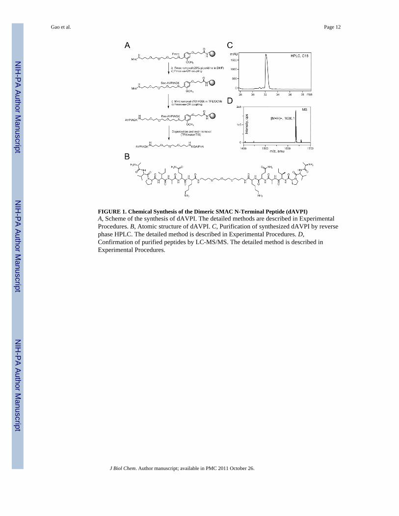
FIGURE 1. Chemical Synthesis of the Dimeric SMAC N-Terminal Peptide (dAVPI)A, Scheme of the synthesis of dAVPI. The detailed methods are described in ExperimentalProcedures. B, Atomic structure of dAVPI. C, Purification of synthesized dAVPI by reversephase HPLC. The detailed method is described in Experimental Procedures. D,Confirmation of purified peptides by LC-MS/MS. The detailed method is described inExperimental Procedures.
Gao et al. Page 12
J Biol Chem. Author manuscript; available in PMC 2011 October 26.
NIH
-PA Author Manuscript
NIH
-PA Author Manuscript
NIH
-PA Author Manuscript

FIGURE 2. dAVPI Antagonizes XIAP Inhibition of Caspase-3 in HeLa Cell ExtractsA, dAVPI competes with Smac for binding with BIR3 domain (GST-BIR3) or full-lengthXIAP (GST-XIAP). As described in Experimental Procedures, GST-BIR3 or GST-XIAPwas first conjugated to glutathione beads, then incubated with Smac in the presence ofvarious concentrations of dAVPI as indicated. Bound Smac was resolved by SDS-PAGEfollowed by Western blotting using an antibody against Smac (upper panel), and the totalGST-BIR3 and GST-XIAP were stained by Ponceau S (lower panel). B, HeLa cell cytosolicextracts (HS100) was activated with dATP and cytochrome c as described in ExperimentalProcedures. An aliquote of activated HS100 was then added into the pre-warmed reactionmixture without XIAP (−XIAP), or with 100 nM XIAP in the absence (+XIAP) or presenceof dAVPI (+XIAP/dAVPI), mAVPI (+XIAP/mAVPI), or the control mIPVA (+XIAP/mIPVA), as indicated. C, dAVPI is more potent than mAVPI in liberating caspase-3 activity
Gao et al. Page 13
J Biol Chem. Author manuscript; available in PMC 2011 October 26.
NIH
-PA Author Manuscript
NIH
-PA Author Manuscript
NIH
-PA Author Manuscript
from XIAP inhibition. Assays were performed as in panel B, with individual syntheticpeptides added with indicated concentration. Control indicates reactions in the absence ofpeptides. All data are presented as velocity of Ac-DEVD-AMC cleavage normalized againstthe maximal velocity (that is, in the absence of XIAP. Same for other figures). Error barsrepresent standard error calculated from 3 independent experiments (same for other figures).
Gao et al. Page 14
J Biol Chem. Author manuscript; available in PMC 2011 October 26.
NIH
-PA Author Manuscript
NIH
-PA Author Manuscript
NIH
-PA Author Manuscript
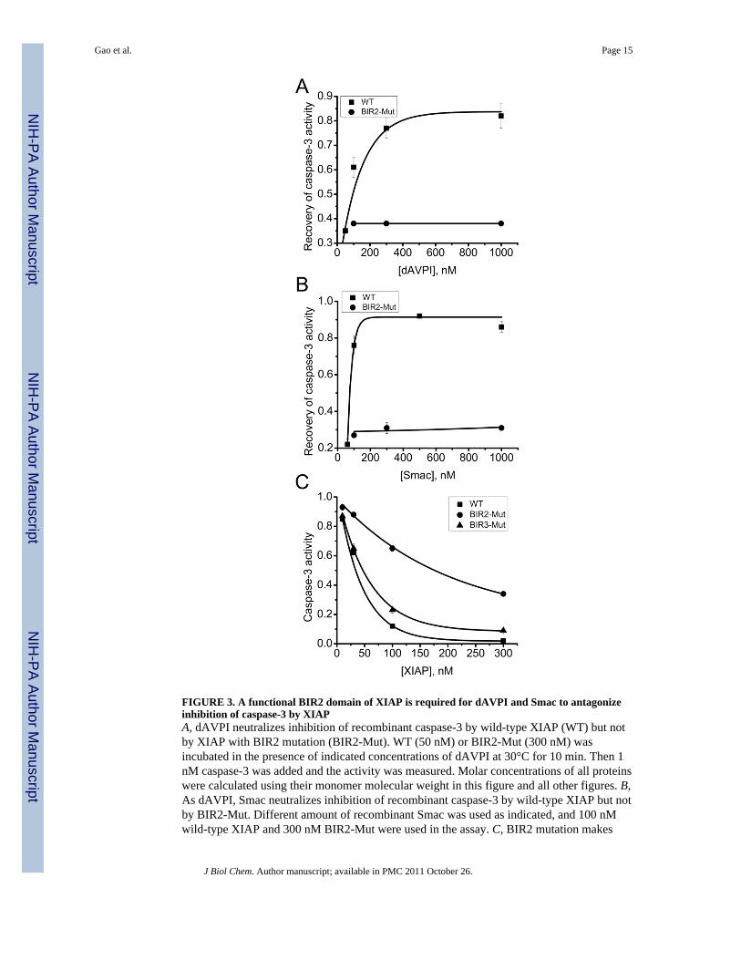
FIGURE 3. A functional BIR2 domain of XIAP is required for dAVPI and Smac to antagonizeinhibition of caspase-3 by XIAPA, dAVPI neutralizes inhibition of recombinant caspase-3 by wild-type XIAP (WT) but notby XIAP with BIR2 mutation (BIR2-Mut). WT (50 nM) or BIR2-Mut (300 nM) wasincubated in the presence of indicated concentrations of dAVPI at 30°C for 10 min. Then 1nM caspase-3 was added and the activity was measured. Molar concentrations of all proteinswere calculated using their monomer molecular weight in this figure and all other figures. B,As dAVPI, Smac neutralizes inhibition of recombinant caspase-3 by wild-type XIAP but notby BIR2-Mut. Different amount of recombinant Smac was used as indicated, and 100 nMwild-type XIAP and 300 nM BIR2-Mut were used in the assay. C, BIR2 mutation makes
Gao et al. Page 15
J Biol Chem. Author manuscript; available in PMC 2011 October 26.
NIH
-PA Author Manuscript
NIH
-PA Author Manuscript
NIH
-PA Author Manuscript

XIAP a weaker inhibitor for caspase-3. Indicated concentrations of WT XIAP, BIR2-Mut, orBIR3-Mut were mixed with 1 nM caspase-3 to measure caspase-3 activity.
Gao et al. Page 16
J Biol Chem. Author manuscript; available in PMC 2011 October 26.
NIH
-PA Author Manuscript
NIH
-PA Author Manuscript
NIH
-PA Author Manuscript
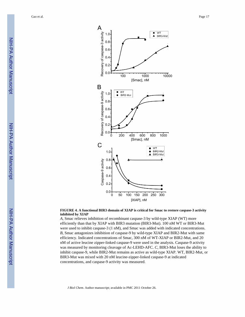
FIGURE 4. A functional BIR3 domain of XIAP is critical for Smac to restore caspase-3 activityinhibited by XIAPA, Smac relieves inhibition of recombinant caspase-3 by wild-type XIAP (WT) moreefficiently than that by XIAP with BIR3 mutation (BIR3-Mut). 100 nM WT or BIR3-Mutwere used to inhibit caspase-3 (1 nM), and Smac was added with indicated concentrations.B, Smac antagonizes inhibition of caspase-9 by wild-type XIAP and BIR2-Mut with sameefficiency. Indicated concentrations of Smac, 300 nM of WT-XIAP or BIR2-Mut, and 20nM of active leucine zipper-linked caspase-9 were used in the analysis. Caspase-9 activitywas measured by monitoring cleavage of Ac-LEHD-AFC. C, BIR3-Mut loses the ability toinhibit caspase-9, while BIR2-Mut remains as active as wild-type XIAP. WT, BIR2-Mut, orBIR3-Mut was mixed with 20 nM leucine-zipper-linked caspase-9 at indicatedconcentrations, and caspase-9 activity was measured.
Gao et al. Page 17
J Biol Chem. Author manuscript; available in PMC 2011 October 26.
NIH
-PA Author Manuscript
NIH
-PA Author Manuscript
NIH
-PA Author Manuscript
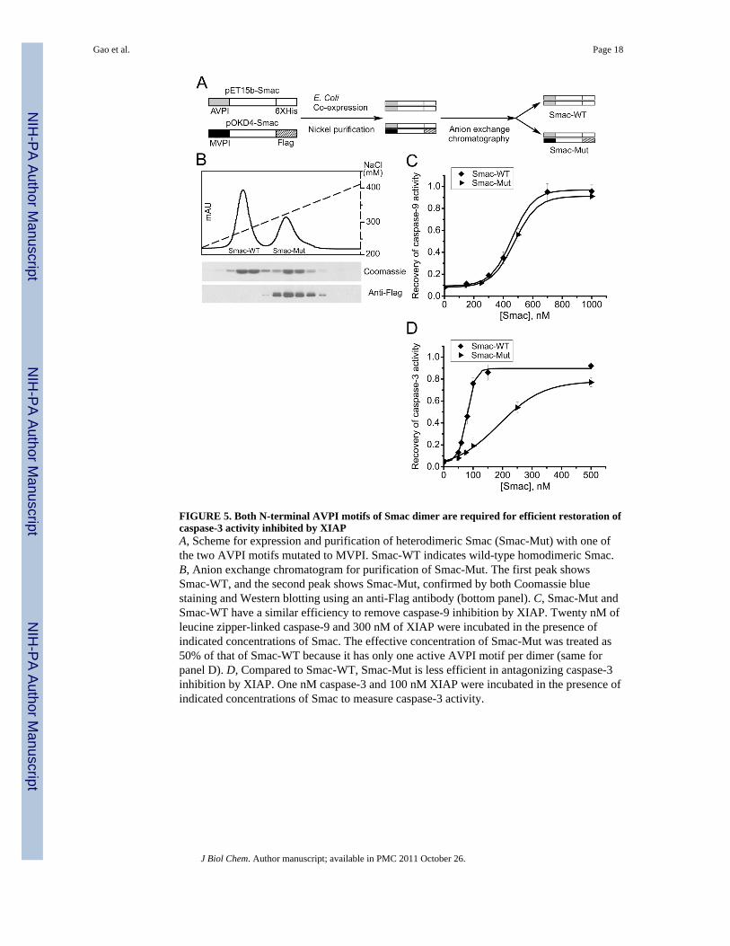
FIGURE 5. Both N-terminal AVPI motifs of Smac dimer are required for efficient restoration ofcaspase-3 activity inhibited by XIAPA, Scheme for expression and purification of heterodimeric Smac (Smac-Mut) with one ofthe two AVPI motifs mutated to MVPI. Smac-WT indicates wild-type homodimeric Smac.B, Anion exchange chromatogram for purification of Smac-Mut. The first peak showsSmac-WT, and the second peak shows Smac-Mut, confirmed by both Coomassie bluestaining and Western blotting using an anti-Flag antibody (bottom panel). C, Smac-Mut andSmac-WT have a similar efficiency to remove caspase-9 inhibition by XIAP. Twenty nM ofleucine zipper-linked caspase-9 and 300 nM of XIAP were incubated in the presence ofindicated concentrations of Smac. The effective concentration of Smac-Mut was treated as50% of that of Smac-WT because it has only one active AVPI motif per dimer (same forpanel D). D, Compared to Smac-WT, Smac-Mut is less efficient in antagonizing caspase-3inhibition by XIAP. One nM caspase-3 and 100 nM XIAP were incubated in the presence ofindicated concentrations of Smac to measure caspase-3 activity.
Gao et al. Page 18
J Biol Chem. Author manuscript; available in PMC 2011 October 26.
NIH
-PA Author Manuscript
NIH
-PA Author Manuscript
NIH
-PA Author Manuscript

FIGURE 6. Trimerization of XIAP improves the efficiency of Smac to remove its inhibition oncaspase-3A, Gel filtration profile of full-length XIAP (XIAP-WT) and XIAP with the Ring domaindeleted (XIAP-ΔRing). Coomassie blue staining shows the staining pattern correlated withthe eluted peaks. B, Molecular weight measurement of XIAP-WT and XIAP-ΔRing. Thetrace in thin line corresponds to light-scattering signals at 90°. The trace in thick line showsthe variation in molecular mass determination as a function of elution volume. C, Smacantagonizes inhibition of caspase-3 by XIAP-ΔRing less effectively than that by full-lengthXIAP. One nM of caspase-3 and 100 nM of XIAP-WT or XIAP-ΔRing were incubated inthe presence of indicated concentrations of Smac to measure caspase-3 activity.
Gao et al. Page 19
J Biol Chem. Author manuscript; available in PMC 2011 October 26.
NIH
-PA Author Manuscript
NIH
-PA Author Manuscript
NIH
-PA Author Manuscript

FIGURE 7. A model for dynamic and cooperative regulation of XIAP by SmacSee text for detailed description. XIAP is shown in the model as a monomer for simplicity.
Gao et al. Page 20
J Biol Chem. Author manuscript; available in PMC 2011 October 26.
NIH
-PA Author Manuscript
NIH
-PA Author Manuscript
NIH
-PA Author Manuscript