Buccal Fat Pad for Closure of Oroantral and Oronasal … PAVAN.pdf · 2018-10-08 · repair of...
5
IJOCR International Journal of Oral Care and Research, April-June 2018;6(2):27-31 27 ORIGINAL RESEARCH Buccal Fat Pad for Closure of Oroantral and Oronasal Communication: A Study of 16 Cases V V N Pavan Kumar 1 , Sandeep Kashyap 2 , Naqoosh Haidry 3 , Madhumathi Singh 4 , Shruthi Rangaswamy 5 , Narahari Ranganatha 6 ABSTRACT Background: Oroantral communications may develop as a complication of dental extractions but may also result from accidental or iatrogenic trauma, cyst, neoplasm, or infection. Some of the traditional methods that are being employed in the repair of oroantral communications include buccal advance- ment flaps, palatal rotation, palatal transposition flaps, tongue flaps, and nasolabial flaps. Buccal fat pad (BFP) is increas- ingly being employed in the repair of oroantral fistula (OAF) and other oral defects worldwide. Materials and Methods: A total of 16 patients with oroantral, oronasal, and OAF were randomly selected for this study. The defects were closed using BFP under general/local anesthe- sia. If it is OAF, the fistulous tract/polyp is excised and then communication is closed with BFP flap. Patient followed up on the 1 st post-operative day, 3 rd , 7 th , 21 st , 3 months, and 6 months to check for complete epithelialization and healing of operated site and post-operative complications were recorded. Success was accessed by complete closure of communication or fistula. Results: The efficacy of BFP evaluated based on post-oper- ative complications and clinical outcome results of epithelial- ization and healing procedure in 13 patients of 16 in closure of oroantral/oronasal communications. Statistically, there was 81.72% (13 patients) success rate out of 100% (16 patients) and failure in 18.25 (3 patients) where one patient was reoperated. Conclusions: The use of BFP in closure of small-to-medium sized oroantral/oronasal defects is a clinically effective, reli- able, and a quick method of reconstruction. Keywords: Buccal fat pad, Oroantral communication, Oronasal communication. 1,2 Assistant Professor, 3 Lecturer, 4 Professor and Head, 5 Reader, 6 Senior Lecturer 1 Department of Dental Surgery, Katuri Medical College and Hospital, Guntur, Andhra Pradesh, India 2 Department of Dental Surgery, Sikkim Manipal Institute of Medical Sciences, Gangtok, East Sikkim, India 3 Department of Oral and Maxillofacial Surgery, Patna Dental College and Hospital, Patna, Bihar, India 4-6 Department of Oral and Maxillofacial Surgery, Rajarajeshwari Dental College, Bengaluru, Karnataka, India Corresponding Author: Dr. Sandeep Kashyap, Assistant Professor, Department of Dental Surgery, Sikkim Manipal institute of Medical Sciences, Gangtok, East Sikkim, India. Phone: +91-8095845115. e-mail: [email protected] How to cite this article: Kumar VVNP, Kashyap S, Haidry N, Singh M, Rangaswamy S, Ranganatha N. Buccal Fat Pad for Closure of Oroantral and Oronasal Communication: A Study of 16 Cases. Int J Oral Care Res 2018;6(2):27-31. Source of support: Nil Conflict of interest: None INTRODUCTION Oroantral communications may develop as a compli- cation of dental extractions and may also result from accidental or iatrogenic trauma, cyst, neoplasm, or infection. Some of the traditional methods that are being employed in the repair of oroantral communica- tions (oroantral fistula [OAF]) include buccal advance- ment flaps, palatal rotation, palatal transposition flaps, tongue flaps, and nasolabial flaps. Buccal fat pad (BFP) is increasingly being employed in the repair of OAF and other oral defects. [1] The use of BFP as a pedicle graft was first reported by Egyedi. [2] Oral defect closure using the BFP has been increasingly employed because it is a fast surgical pro- cedure, is relatively easy to perform, has a high success rate, and is able to cover defects of up to 60 mm 3 × 50 mm 3 . The rich blood supply of the BFP explains its high success rate. It may be one reason for the quick epitheli- alization of the fat. [3-5] In 1732, Heister [6] made a mention of BFP as an ana- tomical element and named it “glandular molars.” This anatomical element was then described by Bichat [7] in 1801, and it came to be known as “lobule of Bichat.” It has been referred in the literature by different names such as the sucking pad, sucking cushion, masticatory fat pad, or buccal pad of fat. [8] The anatomic name of the BFP is corpus adiposum buc- cae. It is a biconvex structure surrounded by a thin but distinctive capsule, and it is situated in the buccal space between the buccinator muscle and the masseter mus- cle. The fat pad attains its greatest volume in this buc- cal space, and it is this part that should be used for the BFP flap. The anterior aspect of the BFP protrudes into the oral cavity ventral to the anterior border of the mas- seter muscle. From here, it extends between the masse- ter and buccinator muscles to continue posteriorly and superiorly into the fatty tissue that occupies the space
Transcript of Buccal Fat Pad for Closure of Oroantral and Oronasal … PAVAN.pdf · 2018-10-08 · repair of...

IJOCR
International Journal of Oral Care and Research, April-June 2018;6(2):27-31 27
ORIGINAL RESEARCH
Buccal Fat Pad for Closure of Oroantral and Oronasal Communication: A Study of 16 CasesV V N Pavan Kumar1, Sandeep Kashyap2, Naqoosh Haidry3, Madhumathi Singh4, Shruthi Rangaswamy5, Narahari Ranganatha6
ABSTRACT
Background: Oroantral communications may develop as a complication of dental extractions but may also result from accidental or iatrogenic trauma, cyst, neoplasm, or infection. Some of the traditional methods that are being employed in the repair of oroantral communications include buccal advance-ment flaps, palatal rotation, palatal transposition flaps, tongue flaps, and nasolabial flaps. Buccal fat pad (BFP) is increas-ingly being employed in the repair of oroantral fistula (OAF) and other oral defects worldwide.
Materials and Methods: A total of 16 patients with oroantral, oronasal, and OAF were randomly selected for this study. The defects were closed using BFP under general/local anesthe-sia. If it is OAF, the fistulous tract/polyp is excised and then communication is closed with BFP flap. Patient followed up on the 1st post-operative day, 3rd, 7th, 21st, 3 months, and 6 months to check for complete epithelialization and healing of operated site and post-operative complications were recorded. Success was accessed by complete closure of communication or fistula.
Results: The efficacy of BFP evaluated based on post-oper-ative complications and clinical outcome results of epithelial-ization and healing procedure in 13 patients of 16 in closure of oroantral/oronasal communications. Statistically, there was 81.72% (13 patients) success rate out of 100% (16 patients) and failure in 18.25 (3 patients) where one patient was reoperated.
Conclusions: The use of BFP in closure of small-to-medium sized oroantral/oronasal defects is a clinically effective, reli-able, and a quick method of reconstruction.
Keywords: Buccal fat pad, Oroantral communication, Oronasal communication.
1,2Assistant Professor, 3Lecturer, 4Professor and Head, 5Reader, 6Senior Lecturer1Department of Dental Surgery, Katuri Medical College and Hospital, Guntur, Andhra Pradesh, India2Department of Dental Surgery, Sikkim Manipal Institute of Medical Sciences, Gangtok, East Sikkim, India3Department of Oral and Maxillofacial Surgery, Patna Dental College and Hospital, Patna, Bihar, India4-6Department of Oral and Maxillofacial Surgery, Rajarajeshwari Dental College, Bengaluru, Karnataka, India
Corresponding Author: Dr. Sandeep Kashyap, Assistant Professor, Department of Dental Surgery, Sikkim Manipal institute of Medical Sciences, Gangtok, East Sikkim, India. Phone: +91-8095845115. e-mail: [email protected]
How to cite this article: Kumar VVNP, Kashyap S, Haidry N, Singh M, Rangaswamy S, Ranganatha N. Buccal Fat Pad for Closure of Oroantral and Oronasal Communication: A Study of 16 Cases. Int J Oral Care Res 2018;6(2):27-31.
Source of support: Nil
Conflict of interest: None
INTRODUCTION
Oroantral communications may develop as a compli-cation of dental extractions and may also result from accidental or iatrogenic trauma, cyst, neoplasm, or infection. Some of the traditional methods that are being employed in the repair of oroantral communica-tions (oroantral fistula [OAF]) include buccal advance-ment flaps, palatal rotation, palatal transposition flaps, tongue flaps, and nasolabial flaps. Buccal fat pad (BFP) is increasingly being employed in the repair of OAF and other oral defects.[1]
The use of BFP as a pedicle graft was first reported by Egyedi.[2] Oral defect closure using the BFP has been increasingly employed because it is a fast surgical pro-cedure, is relatively easy to perform, has a high success rate, and is able to cover defects of up to 60 mm3 × 50 mm3. The rich blood supply of the BFP explains its high success rate. It may be one reason for the quick epitheli-alization of the fat.[3-5]
In 1732, Heister[6] made a mention of BFP as an ana-tomical element and named it “glandular molars.” This anatomical element was then described by Bichat[7] in 1801, and it came to be known as “lobule of Bichat.” It has been referred in the literature by different names such as the sucking pad, sucking cushion, masticatory fat pad, or buccal pad of fat.[8]
The anatomic name of the BFP is corpus adiposum buc-cae. It is a biconvex structure surrounded by a thin but distinctive capsule, and it is situated in the buccal space between the buccinator muscle and the masseter mus-cle. The fat pad attains its greatest volume in this buc-cal space, and it is this part that should be used for the BFP flap. The anterior aspect of the BFP protrudes into the oral cavity ventral to the anterior border of the mas-seter muscle. From here, it extends between the masse-ter and buccinator muscles to continue posteriorly and superiorly into the fatty tissue that occupies the space
Kumar, et al.
International Journal of Oral Care and Research, April-June 2018;6(2):27-31 28
between the masticatory muscles. At the anterior border of the temporalis muscle, the fat pad extends upward into the temporal fossa between the anterior border of the temporalis muscle and the temporal surface of the zygomatic bone. Around the tendon of the temporalis muscle, an extension of the BFP communicates with the pterygomandibular space. The main blood supply courses through this pedicle. It is derived from branches of the buccinator artery, a branch of the internal max-illary artery. The buccinator artery communicates with the external maxillary and transverse facial arteries.[9,10]
Operative Technique
An incision through the mucosa on the buccal aspect of the vestibule in the molar region will readily expose the BFP. A vertical mucosal incision slightly lateral to the anterior margin of the ascending ramus also will result in a forward bulging of the fat pad. A third approach to the fat pad is the elevation of a mucoperiosteal flap in the molar region on the lateral aspect of the maxillary alveolar process and then incision of the periosteum at the level of the buccal sulcus. The choice of exposure depends on the requirements of the specific situation in which the flap is used. After exposure of the BFP, it can be grasped with tissue forceps and carefully teased out. Care should be taken to not severe the pedicle from which the blood supply is derived. The BFP then is drawn into the defect and sutured in place.
MATERIALS AND METHODS
The study was done on patients who reported to the Department of Oral and maxillofacial surgery, Rajarajeswari Dental College and Hospital, Bengaluru. BFP was used on 16 patients with oronasal fistula and OAF. The etiology, location, and size of the defect were recorded. Of 16 patients, 11 were operated under general anesthesia and 5 under local anesthesia. In case of OAF, the fistulous tract/polyp is excised, followed by measurement of the defect size, and then, communication was closed with BFP flap. Six cases of OAF, five cases of defect after removal of periapical cyst, and five defects after tumor resection were treated. Patients’ follow-up was done on post-operative 1, 3, 7, and 21 days, 3 months, and 6 months to check for complete epithelialization and healing of oper-ated site and post-operative complications. Success was accessed by complete closure of communication or fistula without any patency or wound dehiscence [Figures 1-7].
RESULTS
9 male and 7 female patients with the lowest age of 18 years and highest age of 70 years were included in the study. The mean age was 41.94 years.
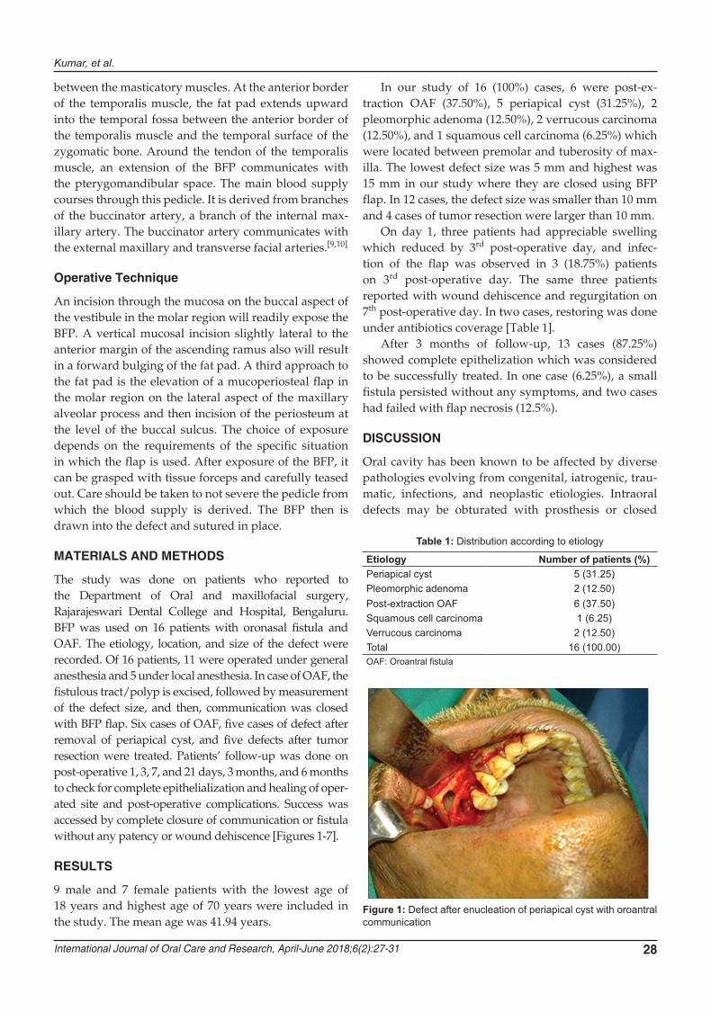
In our study of 16 (100%) cases, 6 were post-ex-traction OAF (37.50%), 5 periapical cyst (31.25%), 2 pleomorphic adenoma (12.50%), 2 verrucous carcinoma (12.50%), and 1 squamous cell carcinoma (6.25%) which were located between premolar and tuberosity of max-illa. The lowest defect size was 5 mm and highest was 15 mm in our study where they are closed using BFP flap. In 12 cases, the defect size was smaller than 10 mm and 4 cases of tumor resection were larger than 10 mm.
On day 1, three patients had appreciable swelling which reduced by 3rd post-operative day, and infec-tion of the flap was observed in 3 (18.75%) patients on 3rd post-operative day. The same three patients reported with wound dehiscence and regurgitation on 7th post-operative day. In two cases, restoring was done under antibiotics coverage [Table 1].
After 3 months of follow-up, 13 cases (87.25%) showed complete epithelization which was considered to be successfully treated. In one case (6.25%), a small fistula persisted without any symptoms, and two cases had failed with flap necrosis (12.5%).
DISCUSSION
Oral cavity has been known to be affected by diverse pathologies evolving from congenital, iatrogenic, trau-matic, infections, and neoplastic etiologies. Intraoral defects may be obturated with prosthesis or closed
Figure 1: Defect after enucleation of periapical cyst with oroantral communication
Table 1: Distribution according to etiology
Etiology Number of patients (%)Periapical cyst 5 (31.25)Pleomorphic adenoma 2 (12.50)Post-extraction OAF 6 (37.50)Squamous cell carcinoma 1 (6.25)Verrucous carcinoma 2 (12.50)Total 16 (100.00)OAF: Oroantral fistula

Buccal fat pad in oral reconstruction
IJOCR
International Journal of Oral Care and Research, April-June 2018;6(2):27-31 29
with local flaps such as buccal advancement flap, palatal pedicled flap, or double-layered closure flaps using buccal and palatal tissues. However, the afore-mentioned procedures produce large denuded areas, result in a decrease of vestibular sulcus, and cannot be used to close large defects.[5,11,12] Regional flaps such as tongue, temporalis muscle, or nasolabial flaps have also
been successfully used for intraoral reconstruction, but they are generally preferred for defects of much larger dimensions.[13]
Egyedi first recommended the use of buccal fat as a pedicled flap to repair oral defects, since then this flap has been used successfully for closure of OAF and repair of oral defects by many surgeons. Some authors
Figure 2: Buccal fat pad harvested
Figure 3: Closure of defect with buccal fat pad
Figure 4: 1 Month post-operative picture
Figure 5: Verrucous carcinoma of edentulous maxilla
Figure 6: Defect after excision
Figure 7: Healing after 3 months

Kumar, et al.
International Journal of Oral Care and Research, April-June 2018;6(2):27-31 30
reported a 100% success rate and found this technique capable of closing of large defects.[14]
Due to its anatomical situation, the ideal defects to be reconstructed with a BFP are the maxillary defects, from the premolar area to the posterior tuberosity. Furthermore, soft and hard palate, superior alveolar rim, cheek mucosa, and tonsillar fossa are suitable places to be employed as suggested by other authors.[5,10,15,16] Excessive traction of BFP toward the medium usually causes with a vestibular sulcus loss, which requires a secondary surgical procedure. Too much traction appears to be the cause of unfavorable results.[17,18]
In the present study, all the surgeries were performed by the same operator, and thus, there is no interopera-tor variability which may influence the post-operative swelling and healing. The technique of harvesting BFP is simple so that it has been performed by different sur-geons in a very highly successful way. Nevertheless, some important points must be remembered.
Perhaps, the most important is the careful manipula-tion of the flap to maintain its thin capsule. Mechanical suction must be avoided. Further, blunt dissection is preferable with two vascular clamps. Gently pull out the emergent part, and dissect the oral mucosa and other tissues surrounding the BFP.
Pain is an expected outcome of most surgery and all the oral procedures resulted in mild-to-moderate pain. Swelling may not become apparent until the day after surgery and will not reach its maximum until 2–3 days postoperatively and usually subsides completely by the day 7. We have observed more swelling in four cases, even though objective measurement was not done. The swelling can be related to the size of the defect and trauma during surgery rather than the selected flap.
In our study, we checked for complications such as swelling, bleeding, and hematoma on day 1, infection, dehiscence, and post-nasal drip on day 3, regurgitation, patency of communication, and flap necrosis on day 7, wound healing, flap necrosis, and epithelialization on day 21, and epithelialization and wound dehiscence at 3 and 6 months. Of 16 patients, 13 were successful in closure of the defect with BFP. In three patients, we observed infection with wound dehiscence, and all of them were treated with antibiotic therapy but they did not take up and lead to flap necrosis. One patient was reoperated for closure of the defect. Of these three cases, one was pleomorphic adenoma, one was squamous cell carcinoma, and a case of verrucous carcinoma. In all the three cases, the defect size was larger than 12 mm. Even though it cannot be proven statistically, closure of larger defects with BFP was the probable reason for failure.
Hao[15] used pedicled BFP flaps for reconstruction of medium-sized post-surgical oral defects, most of which
were malignant lesions. In that series, partial dehiscence of the graft was observed in two patients and failure in one patient who were possibly caused by too large amount of fat transfer which is comparable to our study.
Complications in large series range between 3.1 and 6.9%.[14,19,20] These included partial necrosis, infec-tion, excessive scarring, excessive granulation, and sul-cus obliteration or facial nerve injury. Colella et al.[21] reported complications with pedicled BFP flap, massive hemorrhage, limitation of mouth opening, excessive scarring, and facial nerve injury 38. In our study, we have not come across such complications.
The success criteria considered were complete epi-thelization of the graft and a definite covering of the defect. As observed by Chao et al.,[8] clinically in the typical course, the surface of orally exposed fat becomes yellowish-white in 3 days and then gradually became red within 1 week, which was likely so due to the for-mation of young granulation tissue during the 2nd week. It became completely epithelialized with slight contrac-tion of wound by 3rd–4th weeks after surgery. We had similar experience with complete epithelialization by 1 month. A study included a series of 31 patients of post-oncologic cases; they have commented that radio-therapy if necessary can begin early, due to fast epithe-lialization process.[22]
Limitations of the present study include smaller sample size which will not draw conclusions statisti-cally. A larger sample study is necessary with a longer follow-up. The etiology for the oroantral communica-tion and the size of the defect were varied. Randomized clinical trails with single etiology may give clear results. The disadvantages of BFP are as follows: It can be used only once, is not advocated for larger defects, and can-not be used for anterior maxillary defects.
CONCLUSIONS
BFP can be used small-to-medium intraoral defects, sim-ple and quick surgical technique with low rate of com-plications, and it is highly predictable but large-sized defects require more careful selection of flap. The use of the BFP for the reconstruction of appropriate surgical defects in the mouth is worthy of consideration.
REFERENCES
1. Adeyemo WL, Ogunlewe MO, Ladeinde AL, James O. Closure of oro-antral fistula with pedicled buccal fat pad — Case report and review of literature. Afr J Oral Health 2004;1:42-6.
2. Egyedi P. Utilization of the buccal fat pad for closure of oro-antral and/or oro-nasal communications. J Maxillofac Surg 1977;5:241-4.
3. Baumann A, Ewers R. Application of the buccal fat pad in
Buccal fat pad in oral reconstruction
IJOCR
International Journal of Oral Care and Research, April-June 2018;6(2):27-31 31
oral reconstruction. J Oral Maxillofac Surg 2000;58:389-92.4. Rapidis AD, Alexandridis CA, Eleftheriadis E, Angelopoulos AP
The use of the buccal fat pad for reconstruction of oral defects: Review of the literature and report of 15 cases. J Oral Maxillofac Surg 2000;58:158-63.
5. StajcićZ.Thebuccalfatpadintheclosureoforo-antralcom-munications: A study of 56 cases. J Craniomaxillofac Surg 1992;20:193-7.
6. Heister L. Compendium anatomicum. Applied surgical anatomy of the buccal fat pad. Oral Maxillofac Surg Clin North Am 1990;2:377.
7. Stuzin JM, Wagstrom L, Kawamoto HK, Baker TJ, Wolfe SA. The anatomy and clinical applications of the buccal fat pad. Plast Reconstr Surg 1990;85:29-37.
8. Chao CK, Chang LC, Liu SY, Wang JJ. Histologic examina-tion of pedicled buccal fat pad graft in oral submucous fibro-sis. J Oral Maxillofac Surg 2002;60:1131-4.
9. Dubin B, Jackson IT, Halim A, Triplett WW, Ferreira M. Anatomy of the buccal fat pad and its clinical significance. Plast Reconstr Surg 1989;83:257-64.
10. Tideman H, Bosanquet A, Scott J. Use of the buccal fat pad as a pedicled graft. J Oral Maxillofac Surg 1986;44:435-40.
11. Güven O. A clinical study on oroantral fistulae. J Craniomaxillofac Surg 1998;26:267-71.
12. Samman N, Cheung LK, Tideman H. The buccal fat pad in oral reconstruction. Int J Oral Maxillofac Surg 1993;22:2-6.
13. el-Hakim IE, el-Fakharany AM. The use of the pedicled buc-cal fat pad (BFP) and palatal rotating flaps in closure of oro-antral communication and palatal defects. J Laryngol Otol
1999;113:834-8.14. Nezafati S, Vafaii A, Ghojazadeh M. Comparison of pedicled
buccal fat pad flap with buccal flap for closure of oro-antral communication. Int J Oral Maxillofac Surg 2012;41:624-8.
15. Hao SP. Reconstruction of oral defects with the pedi-cled buccal fat pad flap. Otolaryngol Head Neck Surg 2000;122:863-7.
16. Martín-Granizo R, Naval L, Costas A, Goizueta C, Rodriguez F, Monje F, et al. Use of buccal fat pad to repair intraoral defects: Review of 30 cases. Br J Oral Maxillofac Surg 1997;35:81-4.
17. Fujimura N, Nagura H, Enomoto S. Grafting of the buc-cal fat pad into palatal defects. J Craniomaxillofac Surg 1990;18:219-22.
18. Loh FC, Loh HS. Use of the buccal fat pad for correction of intraoral defects: Report of cases. J Oral Maxillofac Surg 1991;49:413-6.
19. Hernando J, Gallego L, Junquera L, Villarreal P. Oroantral communications. A retrospective analysis. Med Oral Patol Oral Cir Bucal 2010;15:e499-503.
20. Chakrabarti J, Tekriwal R, Ganguli A, Ghosh S, Mishra PK. Pedicled buccal fat pad flap for intraoral malignant defects: A series of 29 cases. Indian J Plast Surg 2009;42:36-42.
21. Colella G, Tartaro G, Giudice A. The buccal fat pad in oral reconstruction. Br J Plast Surg 2004;57:326-9.
22. Giudice M, Giudice A, Colangeli W, Cristofaro MG. The buccal fat pad in recostruction of malignant lesions of the oral cavity: our experience on 31 cases. BMC Geriatr 2010;10 Suppl 1:A59.