Bryan Krantz - mcb.berkeley.edumcb.berkeley.edu/labs/krantz/members/krantz/krantz_cv.pdf · Bryan...
11
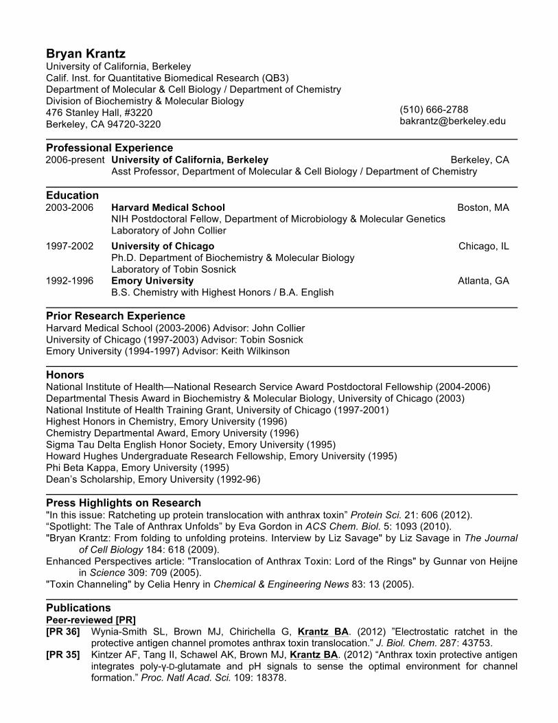
Bryan Krantz University of California, Berkeley Calif. Inst. for Quantitative Biomedical Research (QB3) Department of Molecular & Cell Biology / Department of Chemistry Division of Biochemistry & Molecular Biology 476 Stanley Hall, #3220 Berkeley, CA 94720-3220 (510) 666-2788 [email protected] Professional Experience 2006-present University of California, Berkeley Berkeley, CA Asst Professor, Department of Molecular & Cell Biology / Department of Chemistry Education 2003-2006 Harvard Medical School Boston, MA NIH Postdoctoral Fellow, Department of Microbiology & Molecular Genetics Laboratory of John Collier 1997-2002 University of Chicago Chicago, IL Ph.D. Department of Biochemistry & Molecular Biology Laboratory of Tobin Sosnick 1992-1996 Emory University B.S. Chemistry with Highest Honors / B.A. English Atlanta, GA Prior Research Experience Harvard Medical School (2003-2006) Advisor: John Collier University of Chicago (1997-2003) Advisor: Tobin Sosnick Emory University (1994-1997) Advisor: Keith Wilkinson Honors National Institute of Health—National Research Service Award Postdoctoral Fellowship (2004-2006) Departmental Thesis Award in Biochemistry & Molecular Biology, University of Chicago (2003) National Institute of Health Training Grant, University of Chicago (1997-2001) Highest Honors in Chemistry, Emory University (1996) Chemistry Departmental Award, Emory University (1996) Sigma Tau Delta English Honor Society, Emory University (1995) Howard Hughes Undergraduate Research Fellowship, Emory University (1995) Phi Βeta Kappa, Emory University (1995) Dean’s Scholarship, Emory University (1992-96) Press Highlights on Research "In this issue: Ratcheting up protein translocation with anthrax toxin” Protein Sci. 21: 606 (2012). “Spotlight: The Tale of Anthrax Unfolds” by Eva Gordon in ACS Chem. Biol. 5: 1093 (2010). "Bryan Krantz: From folding to unfolding proteins. Interview by Liz Savage" by Liz Savage in The Journal of Cell Biology 184: 618 (2009). Enhanced Perspectives article: "Translocation of Anthrax Toxin: Lord of the Rings" by Gunnar von Heijne in Science 309: 709 (2005). "Toxin Channeling" by Celia Henry in Chemical & Engineering News 83: 13 (2005). Publications Peer-reviewed [PR] [PR 36] Wynia-Smith SL, Brown MJ, Chirichella G, Krantz BA. (2012) ”Electrostatic ratchet in the protective antigen channel promotes anthrax toxin translocation.” J. Biol. Chem. 287: 43753. [PR 35] Kintzer AF, Tang II, Schawel AK, Brown MJ, Krantz BA. (2012) “Anthrax toxin protective antigen integrates poly-γ-D-glutamate and pH signals to sense the optimal environment for channel formation.” Proc. Natl Acad. Sci. 109: 18378.
Transcript of Bryan Krantz - mcb.berkeley.edumcb.berkeley.edu/labs/krantz/members/krantz/krantz_cv.pdf · Bryan...
Bryan Krantz University of California, Berkeley Calif. Inst. for Quantitative Biomedical Research (QB3) Department of Molecular & Cell Biology / Department of Chemistry Division of Biochemistry & Molecular Biology 476 Stanley Hall, #3220 Berkeley, CA 94720-3220
(510) 666-2788 [email protected]
Professional Experience 2006-present University of California, Berkeley Berkeley, CA
Asst Professor, Department of Molecular & Cell Biology / Department of Chemistry
Education 2003-2006 Harvard Medical School Boston, MA
NIH Postdoctoral Fellow, Department of Microbiology & Molecular Genetics Laboratory of John Collier
1997-2002 University of Chicago Chicago, IL Ph.D. Department of Biochemistry & Molecular Biology Laboratory of Tobin Sosnick
1992-1996 Emory University B.S. Chemistry with Highest Honors / B.A. English
Atlanta, GA
Prior Research Experience Harvard Medical School (2003-2006) Advisor: John Collier University of Chicago (1997-2003) Advisor: Tobin Sosnick Emory University (1994-1997) Advisor: Keith Wilkinson
Honors National Institute of Health—National Research Service Award Postdoctoral Fellowship (2004-2006) Departmental Thesis Award in Biochemistry & Molecular Biology, University of Chicago (2003) National Institute of Health Training Grant, University of Chicago (1997-2001) Highest Honors in Chemistry, Emory University (1996) Chemistry Departmental Award, Emory University (1996) Sigma Tau Delta English Honor Society, Emory University (1995) Howard Hughes Undergraduate Research Fellowship, Emory University (1995) Phi Βeta Kappa, Emory University (1995) Dean’s Scholarship, Emory University (1992-96)
Press Highlights on Research "In this issue: Ratcheting up protein translocation with anthrax toxin” Protein Sci. 21: 606 (2012). “Spotlight: The Tale of Anthrax Unfolds” by Eva Gordon in ACS Chem. Biol. 5: 1093 (2010). "Bryan Krantz: From folding to unfolding proteins. Interview by Liz Savage" by Liz Savage in The Journal
of Cell Biology 184: 618 (2009). Enhanced Perspectives article: "Translocation of Anthrax Toxin: Lord of the Rings" by Gunnar von Heijne
in Science 309: 709 (2005). "Toxin Channeling" by Celia Henry in Chemical & Engineering News 83: 13 (2005).
Publications Peer-reviewed [PR] [PR 36] Wynia-Smith SL, Brown MJ, Chirichella G, Krantz BA. (2012) ”Electrostatic ratchet in the
protective antigen channel promotes anthrax toxin translocation.” J. Biol. Chem. 287: 43753. [PR 35] Kintzer AF, Tang II, Schawel AK, Brown MJ, Krantz BA. (2012) “Anthrax toxin protective antigen
integrates poly-γ-D-glutamate and pH signals to sense the optimal environment for channel formation.” Proc. Natl Acad. Sci. 109: 18378.
Krantz-2 [PR 34] von Moltke J, Trinidad NJ, Moayeri M, Kintzer AF, Wang SM, Rooijen N, Brown CR, Krantz BA,
Leppla SH, Gronert K, Vance RE. (2012) "Rapid induction of lipid mediators is a novel effector function of the inflammasome in vivo" Nature. 490: 107.
[PR 33] Feld GK, Brown MJ, Krantz BA. (2012) "Ratcheting up protein translocation with anthrax toxin." Protein Sci. 21: 606.
[PR 32] Feld GK, Kintzer AF, Tang II, Thoren KL, Krantz BA. (2012) "Domain flexibility modulates the heterogeneous assembly mechanism of anthrax protective antigen." J. Mol. Biol. 415: 159.
[PR 31] Sterling HJ, Kintzer AF, Feld GK, Cassou CA, Krantz BA, Williams ER. (2012) "Supercharging protein complexes from aqueous solution disrupts their native conformations." J. Am. Soc. Mass. Spectrom. 23: 191.
[PR 30] Brown MJ, Thoren KL, Krantz BA. (2011) "Charge requirements for proton gradient-driven translocation of anthrax toxin." J. Biol. Chem. 286: 23189.
[PR 29] Thoren KL, Krantz BA. (2011) “The unfolding story of anthrax toxin translocation” Mol. Microbiol. 80: 588.
[PR 28] Sterling HJ, Cassou CA, Trnka MJ, Burlingame AL, Krantz BA, Williams ER. (2011) "The role of conformational flexibility on protein supercharging in native electrospray ionization" Phys. Chem. Chem. Phys. 13: 18288.
[PR 27] Sterling HJ, Daly MP, Feld GK, Thoren KL, Kintzer AF, Krantz BA, Williams ER. (2010) “Effects of supercharging reagents on noncovalent complex structure in electrospray ionization from aqueous solutions.” J. Am. Soc. Mass. Spectrom. 21: 1762.
[PR 26] Feld GF, Thoren KL, Kintzer AF, Sterling HJ, Tang II, Greenberg SG, Williams ER, Krantz BA. (2010) "Structural basis for the unfolding of anthrax lethal factor by the protective antigen oligomers." Nature Struct. Mol. Biol. 17:1383.
[PR 25] Kintzer AF, Sterling HJ, Williams ER, Krantz BA. (2010) "Anthrax toxin receptor drives protective antigen oligomerization and stabilizes the heptameric and octameric oligomer by a similar mechanism." PLoS ONE. 5(11):e13888.
[PR 24] Kintzer AF, Sterling HJ, Tang II, Abdul-Gader A, Miles AJ, Wallace BA, Williams ER, Krantz BA. (2010) "Role of the protective antigen octamer in the molecular mechanism of anthrax lethal toxin stabilization in plasma." J. Mol. Biol. 399: 741.
[PR 23] Thoren KL, Worden EJ, Yassif JM, Krantz BA. (2009) " Lethal factor unfolding is the most force-dependent step of anthrax toxin translocation." Proc. Natl Acad. Sci. 106: 21555.
[PR 22] Kintzer AF, Thoren KL, Sterling HJ, Dong KC, Feld GK, Tang II, Zhang TT, Williams ER, Berger JM, Krantz BA. (2009) "The protective antigen component of anthrax toxin forms functional octameric complexes." J. Mol. Biol. 292: 614.
[PR 21] Sosnick TR, Krantz BA, Dothager RS, Baxa M. (2006) "Characterizing the protein folding transition state using Ψ analysis." Chem. Rev. 106: 1862.
[PR 20] Krantz BA, Finkelstein A, Collier RJ. (2006) “Protein translocation through the anthrax toxin transmembrane pore is driven by a proton gradient.” J. Mol. Biol. 355: 968.
[PR 19] Christensen KA, Krantz BA, Collier RJ (2006). "The assembly and disassembly kinetics of anthrax toxin complexes." Biochemistry. 45: 2380.
[PR 18] Wolfe JT, Krantz BA, Rainey GJA, Young JAT, Collier RJ. (2005) “Whole-cell voltage clamp measurements of anthrax protective antigen pores.” J. Biol. Chem. 280: 39417.
[PR 17] Krantz BA, Melnyk RA, Zhang S, Juris SJ, Lacy DB, Wu Z, Finkelstein A, Collier RJ. (2005) “A phenylalanine clamp catalyzes protein translocation through the anthrax toxin pore.” Science. 309: 777.
[PR 16] Christensen KA, Krantz BA*, Melnyk RA, Collier RJ. (2004) “Interaction of the 20 kDa and 63 kDa fragments of anthrax protective antigen: kinetics and thermodynamics.” Biochemistry. 44: 1047. *Contributed equally to this work.
[PR 15] Krantz BA, Trivedi AD, Cunningham K, Christensen KA, Collier RJ. (2004) “Anthrax toxin’s lethal and edema factors unfold under acidic pH conditions.” J. Mol. Biol. 344: 739.
[PR 14] Sosnick TR, Dothager RS, Krantz BA. (2004) “Differences in the folding transition state of ubiquitin indicated by φ- and ψ-analyses.” Proc. Natl Acad. Sci. 101: 17377.
[PR 13] Wigelsworth DJ, Krantz BA*, Christensen KA, Lacy DB, Juris SJ, Collier RJ. (2004) “Binding stoichiometry and kinetics of the interaction of a human anthrax toxin receptor, CMG2, with protective antigen.” J. Biol. Chem. 279: 23349. *Contributed equally to this work.
Krantz-3 [PR 12] Pimental RL, Christensen KA, Krantz BA, Collier RJ. (2004) “Anthrax toxin complexes:
heptameric protective antigen can bind lethal factor and edema factor simultaneously.” Biochem. Biophys. Res. Comm. 322: 258.
[PR 11] Jacob J, Krantz B, Dothager RS, Thiyagarajan P, Sosnick TR. (2004) “Early collapse is not an obligate step in protein folding.” J. Mol. Biol. 338: 369.
[PR 10] Krantz BA, Dothager R, Sosnick TR. (2004) “Discerning the structure and energy of multiple transition states in protein folding using ψ-analysis.” J. Mol. Biol. 337: 463.
[PR 9] Krantz BA, Mayne L, Rumbley J, Englander SW, and Tobin R. Sosnick. (2002) “Fast and slow intermediate accumulation and the initial barrier mechanism in protein folding.” J. Mol. Biol. 324: 359.
[PR 8] Krantz BA, Srivastava AK, Nauli S, Baker D, Sauer RT, Sosnick TR. (2002) “Understanding protein hydrogen bond network formation with kinetic D/H amide isotope effects.” Nature Struct. Biol. 9: 458.
[PR 7] Shi Z, Krantz BA, Kallenbach N, Sosnick TR. (2002) “Contribution of hydrogen bonding to protein stability estimated from isotope effects.” Biochemistry. 41: 2120.
[PR 6] Krantz BA, Sosnick TR. (2001) “Engineered metal binding sites map the heterogeneous folding landscape of a coiled coil.” Nature Struct. Biol. 8: 1042.
[PR 5] Krantz BA, and Sosnick TR. (2000) “Distinguishing between two-state and three-state models for ubiquitin folding.” Biochemistry. 39: 11696.
[PR 4] Yin L, Krantz B, Russell NS, Deshpande S, Wilkinson KD. (2000) “Nonhydrolyzable diubiquitin analogues are inhibitors of ubiquitin conjugation and deconjugation.” Biochemistry. 39:10001.
[PR 3] Krantz BA, Moran LB, Kentsis A, Sosnick TR. (2000) “D/H amide isotope effects reveal when hydrogen bonds form during protein folding.” Nature Struct. Biol. 7: 62.
[PR 2] Larsen CN, Krantz BA, Wilkinson KD. (1998) “Substrate specificity of deubiquitinating enzymes: ubiquitin C-terminal hydrolases.” Biochemistry. 37: 3358.
[PR 1] Amerik A, Swaminathan S, Krantz BA, Wilkinson KD, Hochstrasser M. (1997) “In vivo disassembly of free polyubiquitin chains by yeast Ubp14 modulates rates of protein degradation by the proteasome.” EMBO J. 16: 4826.
Non-peer-reviewed [NPR] [NPR 2] Pandit AD, Krantz BA, Dothager RS, Sosnick TR. (2007) "Characterizing protein folding transition
States using Ψ-analysis." Methods Mol. Biol. 350: 83. [NPR 1] Krantz BA. (2002) Protein Folding: New Methods Unveil Rate-limiting Structures. Ph.D. diss.,
U. of Chicago. Patents [1] Krantz BA, Kintzer AF, von Moltke JH. (filed Jan. 10, 2012; recordation Aug. 15, 2012; refiled Jan. 20,
2013) "Poly-Glutamic Acid Anti-Anthrax Composition and Methods for Using the Same" US Application Serial Nos. 61/585,183 and 13/738,911.
Grant Support Ongoing Grant No.: NIH 2R01 AI077703-05 Title: "Physical Principles of bacterial toxin translocation across membranes" Total funding: $1,687,241. Begins: 01/10/2013 Ends on: 12/31/2017 Project Goals: Our continuing goal is to elucidate the molecular mechanism of transmembrane translocation using anthrax toxin as a model. We are specifically probing the role of secondary structure in the molecular mechanism of translocation, testing the observed structural requirements for peptide channel interactions, and investigating the allosteric communication between peptide binding sites in the toxin translocase. As the key PI on the grant, I organize the aims and collaborations, perform experiments, write manuscripts, and communicate results at meetings. Completed Grant No.: NIH R01 AI077703-01 Title: "Physical Principles of bacterial toxin translocation across membranes" Total funding: $1,455,606. Begins: 09/15/2008 Ends on: 09/01/2012 Project Goals: Our goal was to elucidate the molecular mechanism of transmembrane translocation using anthrax toxin as a model system. We specifically sought to obtain the molecular basis for
Krantz-4 translocation-coupled unfolding, to determine the underlying sequence features responsible for the proton gradient-dependent translocation, and to understand the structure and function of a novel octameric assembly-state of the anthrax toxin protective antigen. As the key PI on the grant, I organized the aims and collaborations, performed experiments, wrote manuscripts, and communicated the results.
Under consideration or in process Granting agency: National Institute of Health. Type: Research grant (R01) Title: “Bacterial virulence factor assembly, host countermeasures, and cellular trafficking” Amount requested: $360,000 per year (requesting 5 years). Project Goals: Our goals are to understand the molecular basis of ferric iron-mediated inhibition of anthrax toxin via a unique transferrin receptor 1 trafficking pathway. This pathway may be used to exploit an Achilles Heal of the toxin used during pathogenesis. We are investigating: (i) the unique high-affinity ferric iron binding sites in B. anthracis’ poly-γ-D-glutamate capsule, which can extract iron from transferrin; (ii) the structural basis of poly-γ-D-glutamate capsule interactions with anthrax toxin; and (iii) the Tfr1-mediated trafficking mechanism of ferric-poly-γ-D-glutamate-capsule and toxin-poly-γ-D-glutamate-capsule complexes. Collaborations are planned to investigate the pathophysiology of these newly discovered ferric iron interactions in an anthrax infection model.
Granting agency / type: University of California / UC Proof of Concept Grant Title: "Development of Iron-Poly-Glutamic Acid Anti-Anthrax Compositions" Total funding: $125,000. Project Goals: Our goal is to investigate the therapeutic potential of iron-poly-glutamic acid as an anti-anthrax countermeasure. We will develop derivatives of the natural product iron-poly-γ-D-glutamate and test these formulations in an anthrax lethal toxin mouse model.
Invited Seminars [1] “Anthrax toxin protective antigen promotes lethal factor unfolding and translocation using an α-helical clamp.” Southwest structural biology conference (SWSBC).Univ. of Bristol, UK. 2013-July-2. [2] “Molecular insights on anthrax toxin translocation.” Oxford University, UK. 2013-July-1. [3] “Molecular insights on anthrax toxin translocation and trafficking” École Polytechnique Fédérale De Lausanne. Lausanne, Switzerland. 2013-June-27. [4] “Molecular insights on anthrax toxin translocation and trafficking” Tufts Medical School. Boston, MA. 2013-June-12 [5] “Molecular Insights on Anthrax Toxin Unfolding and Translocation across Membranes” Membrane protein folding meeting. Biophysical Society. Seoul, South Korea. 2013-May-21. [6] “Molecular insights on anthrax toxin translocation and trafficking” National Institutes of Health. Bethesda, MD 2013-Feb-26. [7] “Molecular insights on anthrax toxin translocation and trafficking” Biosciences & Biotechnology Seminar. Lawrence Livermore National Laboratory. Livermore, CA. 2013-Jan-10. [8] “Molecular insights on anthrax toxin translocation and trafficking” Quantitative Biosciences Seminar. University of California, Berkeley. Berkeley, CA. 2012-Nov-19. [9] “Trafficking and transport: new molecular mechanisms of anthrax toxin regulation.” Gordon Research Conference. Microbial Toxins & Pathogenicity. Waterville Valley, NH. 2012-Jul-09. [10] “Anthrax toxin protein translocation powered by hydrogen+” Biophysical Society. San Diego, California. 2012-Feb-25. [11] “Insights on the molecular mechanism of transmembrane protein transport using anthrax toxin as a model system.” University of California, Los Angeles. 2012-Jan-17. [12] “Insights on the molecular mechanism of transmembrane protein transport using anthrax toxin as a model system.” University of Madison-Wisconsin. 2011-Dec-15. [13] “Insights on the molecular mechanism of transmembrane protein transport using anthrax toxin as a model system.” Northwestern University. 2011-Dec-08. [14] “Unfolding story of anthrax toxin protein translocation” Brandeis University. 2011-Nov-09. [15] “Unfolding story of anthrax toxin protein translocation.” New York University. 2011-Nov-08.
Krantz-5 [16] “Structural basis for the unfolding of anthrax lethal factor by protective antigen” At Albert Einstein College of Medicine. Bronx, NY. 2010-Nov-04. [17] “Structural basis for the unfolding of anthrax lethal factor by protective antigen” At University of Chicago. Chicago, IL. 2010-May-25. [18] “Structural basis for the pre-translocation unfolding of proteins by anthrax lethal toxin” At University of California, Davis School of Medicine. Davis, CA. 2009-Dec-07. [19] “Structural Basis for the Pre-Translocation Unfolding of Proteins by Anthrax Lethal Toxin.” At Children's Hospital Oakland Research Institute (CHORI). Oakland, CA. 2009-Oct-06. [20] “Anthrax toxin assembly & translocation: an interplay of form & function” Gordon Research Conference. Microbial Toxins & Pathogenicity. Proctor Academy, Andover, NH. 2008-Jul-13-18. [21] “Molecular mechanisms of anthrax toxin translocation” Bay Area Microbial Pathogenesis Symposium (BAMPS). University of California, San Francisco. 2008-Mar-29. [22] “How the anthrax toxin pore unfolds and then translocates its enzymatic factors across bilayer membranes.” Biopolymers Gordon Research Conference. Newport, RI. Jun 2006. [23] “Anthrax toxin translocation.” 50th Meeting of The Biophysical Society. Salt Lake City, UT. Feb 2006. [24] “Anthrax toxin's protective antigen pore: a protein translocase with a chaperone-like active site.” Harvard Medical School Dept. of Biological Chemistry & Molecular Pharmacology Seminar Series. Boston, MA. Oct 2005. [25] “Small molecules target protective antigen’s phenylalanine clamp—the site required to translocate lethal & edema factor into the host cell.” 2nd Annual Retreat of the New England Regional Center of Excellence for Biodefense and Emerging Infectious Disease Research. Durham, NH. Sep 2005. [26] “The protective antigen binding domains of the lethal and edema factors of anthrax toxin unfold under acidic pH conditions.” 49th Meeting of The Biophysical Society. Long Beach, CA. Feb 2005. [27] “Realizing protein hydrogen bond network formation with kinetic D/H amide isotope effects.” University of Chicago. Chicago, IL. Dec 2001. [28] “When Do Hydrogen Bonds Form? D/H Backbone Amide Isotope Effects in Protein Folding Kinetics.” University of Chicago. Lake Geneva, WI. Oct 1998.
Statement of Departmental & University Service Departmental committees. Graduate Student Recruitment (2007-08) – Dept. of Molecular & Cell Biology; Structural & Quantitative Biology Seminar Coordinator (2007-08) – Dept. of Chemistry; Graduate Student Recruitment (2008-09) – Dept. of Molecular & Cell Biology; Structural & Quantitative Biology Seminar Coordinator (2008-09) – Dept. of Chemistry; New Faculty Search Committee (2009) – Dept. of Chemistry; Graduate student 1st year advisor (2010) – Dept. of Molecular & Cell Biology; Undergraduate Chemical Biology advisor (2010-11) – Dept. of Chemistry; Graduate student 2nd year advisor (2012) – Dept. of Molecular & Cell Biology; Undergraduate Chemical Biology advisor (2011-12) – Dept. of Chemistry; Graduate student 2nd year advisor (2013) – Dept. of Molecular & Cell Biology; Undergraduate Chemical Biology advisor (2012-13) – Dept. of Chemistry; Graduate Chemical Biology Program advisor (2012-2013) – Dept. of Chemistry; Coordinator of Ethics Seminar Series for non-MCB graduate students (2012-2013) – Dept. of Chemistry Other service. Besides my informal interaction with the Chemistry graduate student recruitment, I am also active in the Biophysics graduate group recruitment. I perform reviews for academic manuscripts for the J. Mol. Biol., PNAS, J. Bacteriology, J. Gen. Phys., Cell, Biophysical Journal, Protein Science, PLOS ONE, PLOS Pathogens, and Biochemistry. I also provided expertise and training to students in other labs on campus to build in-house, custom bacterial fermenter apparatus (with J. Berger, A. Martin, S. Marqusee, and M. Marletta); these fermenters have been used in very creative ways beside run-of-the-mill aerobic bacterial protein expression, namely bulk C. elegans strain growth (with A. Dernburg) and anaerobic production of oxygen-sensitive proteins (with M. Marletta).
Krantz-6 Statement of Teaching Highlights and new directions. I have broad formal classroom instruction experiences in both the Depts. of Chemistry and Molecular & Cell Biology. These experiences are mainly at the upper-division undergraduate level. I have instructed in a wide array of courses and am highly adaptable to lecturing on physical chemistry, enzymology, bioenergetics, and membrane transport. In the future, I would like to develop new lectures by applying these basic principles to human disease and pathogenesis. CHEMC130. “Biophysical Chemistry: The Molecules of Life: Physical Principles and Cellular Functions,” a class of about 100-175, which I co-taught about 10-15 1.5 hr lectures per semester with Prof. John Kuriyan in both the Fall of 2007 and Fall of 2008; I taught the full course in the Spring of 2011; two thirds of the course in Spring 2012; and one third in the Spring 2013. The course is a unique offering at Berkeley. It exposes non-Chemistry majors to physical chemistry in a way that is immediately applicable to biological chemistry. The principles of thermodynamics are taught using an accessible statistical-mechanical model of protein folding. Later lectures delve into applications of these models to biophysical questions in the life sciences. This course is challenging in terms of keeping diverse backgrounds abreast of quantitative analysis and abstract principles. The very best students, who often visit my office hours, are very rewarding to teach and interact with. Overall I enjoy this course. MCB102. “Survey of the Principles of Biochemistry and Molecular Biology” in the Spring of 2008, Fall of 2011, and Fall of 2012; this course is a large lecture series (375-500 students), and I co-taught for 13-15 1-hr lectures. I issued homework and the final examination. I designed a compact set of lectures to bring the class through central catabolism. I rather enjoyed teaching the enzymology section of this course but found it had to move at a very brisk pace. The large size and wide array of backgrounds are challenging factors to consider. Overall, I thought my first effort was a success given these constraints. MCB110L. “General Biochemistry & Molecular Biology Laboratory” in the Spring of 2009 and Spring of 2010; this course is a ~70 person hands-on lab experience, which also requires 10-12 lectures, handouts, quizzes, final exam and two lab reports per semester. This course was a newly designed course when I first began teaching it; a new kinesin enzyme system was being investigated in the lab course. I designed my own lectures on quantitative RNA analysis, protein structure and methods to analyze structure as well as basic enzyme kinetics. The kinesin’s weak or nonexistent enzyme activity made certain aspects of this new course difficult to manage. In light of those challenges, the course was successful. MCB290. “Principles of Cellular Protein Unfolding and Translocation Across Membranes” is a literature survey and discussion course that I designed to focus on transmembrane protein translocation; this course was taught in the Fall of 2007 to 15 students, who were asked to present critical reviews of cutting-edge research papers. The students in this course were all well-engaged, providing thoughtful presentations, and I took that to mean the course was well received.
Statement of Research Introduction. As biology has transitioned into the post-genomic era, a key frontier area has emerged in molecular biophysics to study how life operates at the molecular level. My laboratory has committed itself to probe how molecular machines unfold, translocate, and traffic proteins in the cell. While these research areas have been traditionally difficult to characterize biophysically, my laboratory has discovered a new understanding of molecular machine-catalyzed protein unfolding and translocation across membranes. We functionally dissected the unfolding and translocation steps of anthrax toxin protein translocation, and found unfolding is the most driving force-dependent step in the mechanism. We structurally identified a new octameric oligomeric state of anthrax toxin, and we defined the octamer’s physiological role as a highly stable molecular configuration. Using the octamer, we co-crystallized and determined the structure of the anthrax lethal toxin core complex, which reveals an unfolding substrate protein on the surface of its unfoldase machinery. We also identified the requirements for proton-gradient driven translocation, which are consistent with an electrostatic ratchet model. Additionally, we developed and patented an anti-anthrax toxin therapy. In doing so, we discovered new host-side and pathogen-side ferric iron acquisition pathways. These results define a new function for a bacterial capsule, and reveal how ferric iron controls anthrax toxin trafficking and function. In general, our insights on the bioenergetics and molecular mechanics of protein transport and trafficking have enabled new understandings of cellular protein unfolding, molecular-ratchet mechanisms, host-pathogen interactions, and molecular immunology.
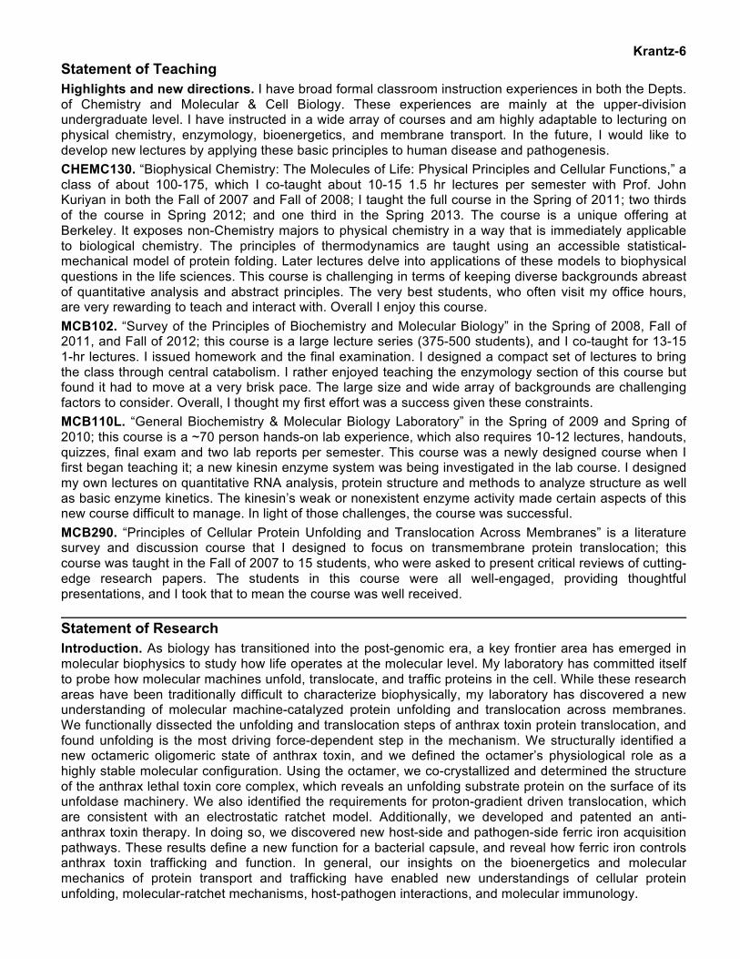
Krantz-7 I. Cellular protein unfolding and translocation across membranes. Transmembrane protein translocation plays a key role in infectious disease, since a host cell’s membrane bilayer functions as a front line of defense, isolating the pathogen from its cytosol. The cellular mechanism of protein unfolding is also of general relevance to processes that allow the cell to maintain protein quality control, regulate the cell cycle, and recover from protein-misfolding conditions. Using anthrax toxin as a model system, we have made progress in discerning the molecular bases of nonspecific substrate recognition, translocation-coupled protein unfolding, and ratchet-based mechanisms of directional transport. Bacillus anthracis secretes two virulence factors: a tripartite exotoxin, anthrax toxin and a poly-γ-glutamate capsule. Anthrax toxin is composed of: protective antigen (PA), lethal factor (LF), and edema factor (EF). PA assembles into an oligomer and then co-assembles with LF and/or EF. When these toxic complexes are properly trafficked, the PA oligomer converts into a transmembrane channel and creates a passageway across the host cell’s endosomal membrane. Through this passageway LF and EF translocate into the host cell’s cytosol. The internal diameter of the channel is narrow, requiring that LF and EF traverse the channel as unfolded polypeptide chains. Once inside the target cell’s cytosol, LF and EF refold and catalyze reactions that disrupt normal cellular physiology. These key mechanisms central to anthrax pathogenesis are probed by a broad array of biophysical and biochemical approaches. (See [PR29, PR33] for reviews). A. Functional analysis of protein unfolding during translocation [PR23]. We mapped the energy landscape of the unfolding and translocation reactions in the anthrax toxin translocation mechanism by carefully controlling the driving force, either the membrane potential (Δψ) or proton gradient (ΔpH), using planar bilayer electrophysiology [PR23]. We then identified two major barriers to translocation: one barrier is 10- to 20-fold more driving-force dependent than the other barrier. The more force-dependent barrier coincides with the major unfolding step. By examining the effects of destabilizing point mutations introduced into a variety of locations in LFN’s structure, we were then able to show how a protein unfolds during translocation. A β-sheet subdomain was identified as the most mechanically stable subdomain in agreement with single-molecule force-microscopy studies. This work marks an important advance, as we expect our formulism will be adopted in future translocation-coupled protein unfolding studies. Historically, prior efforts to examine translocation failed to identify the underlying barriers in the mechanism, and as a result they underestimated that substantial unfolding is rate-limiting to translocation. We have thus provided the first detailed analysis of the driving-force dependencies of translocation, showing unfolding is the most force-dependent step. Other more recent force-microscopy and electrophysiological efforts have now confirmed our view. B. Structural basis of translocation-coupled protein unfolding [PR26]. Protein translocation and unfolding machinery in the cell utilize general and nonspecific polypeptide binding sites to unfold substrate proteins and to interact with unfolded structure; these nonspecific sites share common features among many divergent transporters and unfoldases. Their function in protein translocation is poorly understood in part because structures of substrate proteins in complex with their unfoldase machinery are unknown. We determined the crystal structure of the core of a lethal toxin complex to 3.1-Å resolution; the structure contains a PA octamer bound to four copies of LF’s PA-binding domain (LFN) (i.e., PA8(LFN)4) (Fig. 1) [PR26]. We had identified the novel PA8 architecture in prior studies [PR22, PR24, PR25]. The
Fig. 1. Structure of the lethal toxin core complex. (A) (Left) Ribbons depiction of the PA2LFN ternary complex from the octameric lethal toxin core structure (PA8LF4). PAC (chain A, blue), PAN (chain B, green), LFN (chain C, red), and calcium ions (gray spheres). (Right) Slices through a surface rendering of the two LFN-binding subsites: (top) the carboxy-terminal binding subsite and (bottom) the α-clamp subsite. (B) Axial rendering of the biological unit, the PA8(LFN)4 complex, colored as in (A).
Krantz-8 first α helix and β strand (α1 and β1) of each LFN unfold and dock into a deep amphipathic cleft on the surface of the PA octamer, which we call the α clamp. Hence, the α clamp is a nonspecific protein denaturation site that facilitates LF unfolding.
The structure described in PR26 is a major advance, being both the first report revealing a lethal toxin core complex and the first example of a protein bound to an unfolding machine in a partially unfolded conformation. The functional characterization of the site also provides a compelling analysis of nonspecific binding sites in protein translocase machinery. The α-clamp binding site identified in this structure and the phenylalanine clamp (ϕ clamp) [PR17] identified in prior studies are paradigm-shifting discoveries in the field, because translocase channels were not thought to be able to bind to and form stable intermediate structures with unfolded protein during translocation. While many models imply that forces can be applied to denature a protein, there was little structural evidence that unfolding transitions/barriers could be stabilized and reduced via interactions with the translocase. We expect in light of this discovery that other related systems will be re-examined and similar nonspecific binding sites will be revealed. C. Electrostatic ratchet mechanism of protein translocation [PR30, PR36]. A proton gradient (ΔpH) can drive anthrax toxin translocation [PR20]; however, the molecular mechanism of ΔpH-mediated force generation and the enforcement of directionality during translocation are not understood. We have shown that the ΔpH is sufficient to drive translocation, and acidic residues are required in the substrate protein to harness the ΔpH [PR30]. The densest cluster of acidic residues is located amino-terminal to the folded domain of LF, and the spacing of this force-harnessing cassette relative to the folded domain is critical to drive the unfolding step of translocation. A deficiency in the density of the acidic-residue cassette defines the relatively poor translocation of EF’s highly homologous amino terminal domain [PR36]. Moreover, the acidic residue cassette is critical to membrane potential (Δψ)-driven translocation, showing that a key electrostatic feature resides in the PA channel. We computationally predicted the electrostatics of a β-barrel channel model and showed that disrupting these anion-repulsive features blocked Δψ and ΔpH-driven translocation. Hence for the first time, we have uniquely defined the substrate-side and channel-side features of an electrostatic ratchet in a protein translocase. II. Exotoxin control through macromolecular assembly and altered host-cell trafficking. The pathogenesis of B. anthracis is attributable to a known synergy amongst its two virulence factors: (i) anthrax toxin and (ii) poly-γ-D-glutamate capsule. These virulence factors must assemble and function in diverse environments that vary spatially and temporally during infection. Also the molecular basis of pathogenesis depends upon well-regulated and efficient iron scavenging by the host and pathogen. Hence, we are increasingly interested in the molecular basis of virulence factor assembly in diverse host environments; trafficking of exotoxins, capsular polypeptides, and complexes thereof; and particularly how ferric iron interactions may alter toxin uptake and trafficking. The complexity of anthrax toxin assembly has only been more recently appreciated. The prior literature believed the assembled toxin was a homogenous heptameric species, which tended to only form on cell surfaces. The older literature from the 1950s and 60s, however, revealed toxin activity was markedly different when purified from in vivo sources as compared to in vitro sources. As recent studies have shown, the toxin can assemble in bovine plasma as a stable octameric oligomer and high concentrations of toxin subunits and capsule. It is the octameric form and not the heptameric form that maintains toxin activity and function. Moreover, large quantities of capsule polymers are found secreted into the blood of anthrax-diseased animals, and these capsule polymers form complexes with anthrax toxin. Hence what is needed is a biochemical and biophysical description of these complexes, which are germane to the in vivo states of the toxin; and moreover, the in vivo toxin must be extracted from anthrax animal models to better understand the true nature of the toxin during pathogenesis. Below I summarize my results that paint the beginning pictures of this exciting story. A. Discovery of the stable octameric anthrax toxin architecture [PR22, PR24, PR25, PR26, PR32]. We discovered a novel, highly-stable octameric PA oligomer [PR22] used later as a self-symmetric platform to solve the lethal toxin core structure [PR26]. The result in PR22 was surprising given that the field had only previously recognized the toxin only forms a heptamer. We assessed the octamer’s biophysical properties by purifying it, crystallizing it, establishing its stability and translocation activity (with respect to the heptamer). To our surprise, the octamer is stable under physiological conditions (37 °C, pH 7.3) but the heptamer is not (Fig. 2). The stability of the octamer is functionally relevant as well [PR24]. The octameric lethal toxin maintains its macrophage cytolysis activity in bovine plasma at 37 °C for
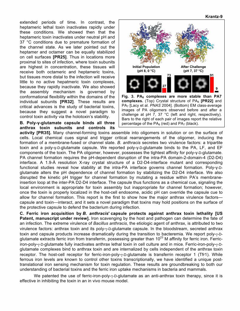
Krantz-9 extended periods of time. In contrast, the heptameric lethal toxin inactivates rapidly under these conditions. We showed then that the heptameric toxin inactivates under neutral pH and 37 °C conditions due to premature formation of the channel state. As we later pointed out the heptamer and octamer can be equally stabilized on cell surfaces [PR25]. Thus in locations more proximal to sites of infection, where toxin subunits are highest in concentration, these tissues will receive both octameric and heptameric toxins, but tissues more distal to the infection will receive little to no active hepatmeric toxin complexes, because they rapidly inactivate. We also showed the assembly mechanism is governed by conformational flexibility within the domains of the individual subunits [PR32]. These results are critical advances is the study of bacterial toxins, because they suggest a novel paradigm to control toxin activity via the holotoxin’s stability. B. Poly-γ-glutamate capsule binds all three anthrax toxin subunits and controls its activity [PR35]. Many channel-forming toxins assemble into oligomers in solution or on the surface of cells. Local chemical cues signal and trigger critical rearrangements of the oligomer, inducing the formation of a membrane-fused or channel state. B. anthracis secretes two virulence factors: a tripartite toxin and a poly-γ-D-glutamate capsule. We reported poly-γ-D-glutamate binds to the PA, LF, and EF components of the toxin. The PA oligomer, however, possesses the tightest affinity for poly-γ-D-glutamate. PA channel formation requires the pH-dependent disruption of the intra-PA domain-2-domain-4 (D2-D4) interface. A 1.9-Å resolution X-ray crystal structure of a D2-D4-interface mutant and corresponding functional studies reveal how stability at the intra-PA interface governs channel formation. Poly-γ-D-glutamate alters the pH dependence of channel formation by stabilizing the D2-D4 interface. We also disrupted the kinetic pH trigger for channel formation by mutating a residue within PA’s membrane-insertion loop at the inter-PA D2-D4 interface. The capsule thus functions as a chemical cue, signaling the local environment is appropriate for toxin assembly but inappropriate for channel formation; however, once the toxin is properly localized in the host-cell endosome, acidic pH can override the capsule cue to allow for channel formation. This report is the first to show how the major anthrax virulence factors—capsule and toxin—interact, and it sets a novel paradigm that toxins may hold positions on the surface of the protective capsule to defend the bacterium during infection. C. Ferric iron acquisition by B. anthracis' capsule protects against anthrax toxin lethality [US Patent, manuscript under review]. Iron scavenging by the host and pathogen can determine the fate of an infection. The extreme virulence of Bacillus anthracis, the etiologic agent of anthrax, is attributed to two virulence factors: anthrax toxin and its poly-γ-D-glutamate capsule. In the bloodstream, secreted anthrax toxin and capsule products increase dramatically during the transition to bacteremia. We report poly-γ-D-glutamate extracts ferric iron from transferrin, possessing greater than 1023 M affinity for ferric iron. Ferric-iron-poly-γ-D-glutamate fully inactivates anthrax lethal toxin in cell culture and in mice. Ferric-iron-poly-γ-D-glutamate complexes bind to anthrax toxin and are internalized by cells independent of the anthrax toxin receptor. The host-cell receptor for ferric-iron-poly-γ-D-glutamate is transferrin receptor 1 (Tfr1). While ferrous iron levels are known to control other toxins transcriptionally, we have identified a unique post-translational iron sensing mechanism for toxin regulation. These results are groundbreaking to both our understanding of bacterial toxins and the ferric iron uptake mechanisms in bacteria and mammals.
We patented the use of ferric-iron-poly-γ-D-glutamate as an anti-anthrax toxin therapy, since it is effective in inhibiting the toxin in an in vivo mouse model.
91
927
73
After Challenge(pH 7, 37 °C)
Initial Population(pH 8, 0 °C)
Fig. 3. PA8 complexes are more stable than PA7 complexes. (Top) Crystal structure of PA8 [PR22] and PA7 [Lacy et al. PNAS 2004]. (Bottom) EM class-average images of PA oligomers observed before and after a challenge at pH 7, 37 °C (left and right, respectively). Bars to the right of each pair of images report the relative percentage of the PA8 (red) and PA7 (black).
Krantz-10 III. Collaborations. I maintained several key collaborations and feel strongly about working with others to gain new insight. Evan Williams (UC Berkeley) and I have used state of the art mass spectrometry to understand the conformational heterogeneity of anthrax toxin [PR22, PR24, PR25, PR26, PR32] and to understand protein-folding transitions in the gas phase [PR27, PR28, PR31]; we are moving to examine anthrax toxin unfolding in the near future. With Russell Vance (UC Berkeley) we are using anthrax toxin as a delivery vehicle for the pathogen associated molecular pattern (PAMP) protein flagellin; these studies dissected the molecular basis of inflammasome activation and the lipid signals used during inflammation [PR34]. A similar collaboration is underway with Edward Miao at University of North Carolina, Chapel Hill. We are working with Gisou van der Goot at École Polytechnique Fédérale De Lausanne to investigate the cellular trafficking and protein turnover of anthrax toxin and ferric-iron-poly-γ-D-glutamate complexes. We are dissecting the pathophysiology of ferric-iron-poly-γ-D-glutamate complexes in animal models for the toxin with Russell Vance and anthrax infections with Ian Glomski (U. of Virginia). We are working with Paramjit (Bobby) Arora at New York University to study peptide translocation using substrates with unique backbone conformational constraints. We are working with Zhongchao Yin at Temasek Life Sciences Laboratory in Singapore to understand a pore-forming toxin pivotal in devastating bacteria infections in rice. We are working with Jeffrey Cox (UC San Francisco) to understand the process of pore formation by a key toxin in Mycobacterium tuberculosis, the causative agent of tuberculosis. We are working with Daniel Portnoy (UC Berkeley) to learn how Listeria monocytogenes senses cellular redox states. IV. Summary of Accomplishments. My laboratory has made key insights on molecular machine-catalyzed protein unfolding, translocation, and trafficking. We have shown that protein unfolding is the most force-dependent step of translocation. We described a novel octameric architecture of the anthrax toxin translocase; and we crystalized the core of the lethal toxin complex—a previously unknown structure. For the first time, the co-crystal structure has gained key insight on molecular machine catalyzed unfolding, as the structure trapped part of lethal factor in an unfolded conformation. We also discovered the molecular mechanism of proton-gradient driven translocation by not only identifying an electrostatic ratchet in the anthrax toxin channel but also by identifying the corresponding acidic-residue molecular teeth in the substrate. We also patented a new anti-anthrax toxin therapy, which is the ferric iron-chelate of poly-γ-glutamate. We discovered that ferric iron-poly-γ-glutamate is a potent anti-toxin in a mouse animal model. Discovery of the anti-toxin enabled my research group to describe a ferric iron-acquisition mechanism for encapsulated bacteria, since the capsule is able to extract ferric iron from transferrin. We also determined the eukaryotic host-side scavenging mechanism for ferric-iron-capsule chelates requires transferrin receptor 1. We now understand that these ferric iron interactions effectively alter normal anthrax toxin trafficking. Overall, our work on the bioenergetics and molecular mechanics of protein transport and trafficking have inspired new understandings of cellular protein unfolding, molecular-ratchet mechanisms, host-pathogen interactions, and nutrient immunity. V. Future directions. We are continuing to pursue the structure and function studies of anthrax toxin and other model systems, particularly those providing insight on the molecular basis of protein unfolding and translocation across membranes. We have been developing crystallographic tools to obtain the structure of the PA channel. We expect to extend these studies with cryoelectron microscopy given our new understanding of the complex oligomeric heterogeneity of the system. We are probing the role of the α-helical clamp in the PA translocase by developing a mechanism-based inhibitor of the site. We have also begun using optical-force microscopy to investigate the force-dependence of LF and EF unfolding. We would like to extend these studies to incorporate a soluble channel form of the PA translocase. We are interested in mapping the force-dependent barriers and translocation step sizes in the system. We have initiated a detailed single-channel electrophysiology study of the PA channel, examining the transport of smaller peptide substrates with backbone conformational constraints. These studies are providing new insight on the conformational states and dynamics of the substrate during translocation.
Ultimately, anthrax toxin is an in vivo toxin. Its study requires a new biochemical and biophysical approach to understand its physiology during infection. Purifying the toxin from sterilize blood samples will be a critical aim in the future. We want to know its composition and oligomeric states. Since poly-γ-D-Glu capsule polymers are prevalent in blood during infection, we have co-crystalized poly-γ-D-Glu capsule with the PA8 oligomer and obtained high-quality 2.6-Å X-ray diffraction. In our co-crystal structure, unique, contiguous, >2σ difference electron densities for the capsule are observed at the interface of PA’s domains 1 and 3 and domains 2 and 4. We are working to complete this structure and further test its
Krantz-11 newly defined interactions functionally. We are also investigating the molecular basis for how transferrin receptor 1 and 2 interact with unstructured ferric-iron chelates, including poly-γ-glutamate and other capsular polysaccharides. We are examining the pathophysiology of these ferric-iron interactions in collaboration with a laboratory capable of these biosecurity experiments. Ultimately, we will be looking at the ability of the bacillus to acquire iron and other metal ions directly from the capsule polymer. We believe the polymer is retrotranslocated back into the bacillus. We are looking to identify the ferric iron transport pathway. These studies and others are leading us to determine the molecular basis of ferric iron-uptake by B. anthracis and/or related species.