
Brainstem truncus encephali - Anatomický ústav 1. LF UKanat.lf1.cuni.cz/souhrny/lekls0702a.pdf ·...
101
Author: Ivo Klepáček Obor: general medicine, dentistry Univerzita Karlova v Praze – 1. lékařská fakulta Brainstem truncus encephali εγκεφαλικού στελέχους truncus encephalicus Anatomický ústav
Transcript of Brainstem truncus encephali - Anatomický ústav 1. LF UKanat.lf1.cuni.cz/souhrny/lekls0702a.pdf ·...

Author: Ivo Klepáček
Obor: general medicine, dentistry
Univerzita Karlova v Praze – 1. lékařská fakulta
Brainstem
truncus encephali
εγκεφαλικού στελέχους truncus encephalicus
Anatomický ústav
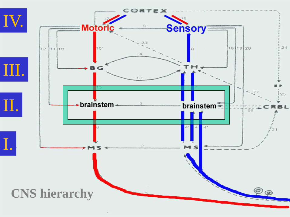
CNS hierarchy
III.
II.
I.
IV.
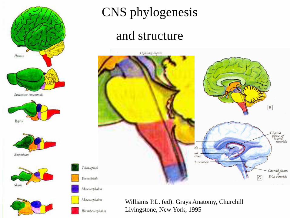
Williams P.L. (ed): Grays Anatomy, Churchill
Livingstone, New York, 1995
CNS phylogenesis
and structure
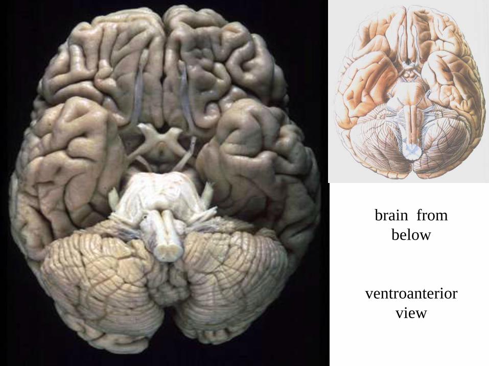
brain from
below
ventroanterior
view
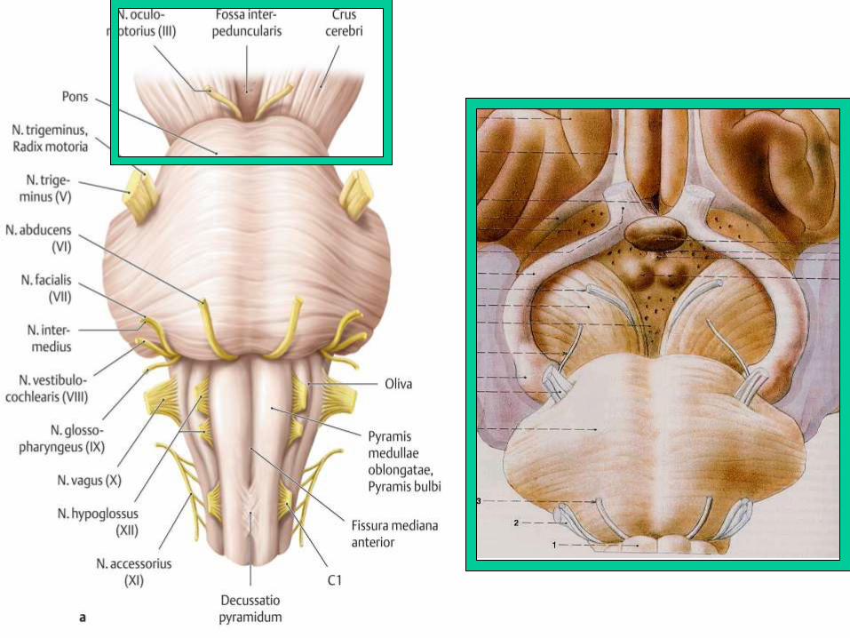
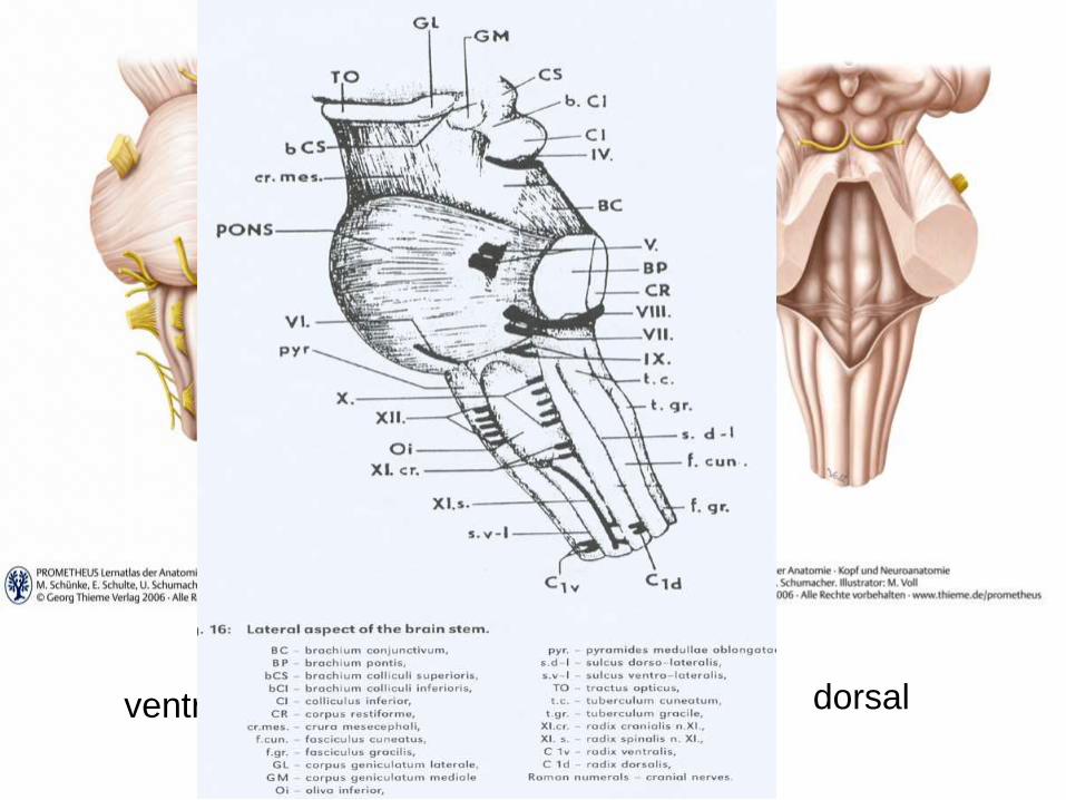
ventral lateral dorsal

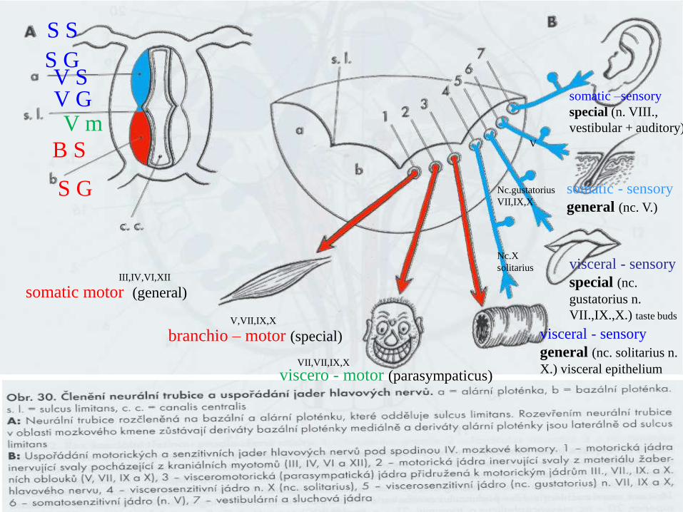
How the brainstem is formed :
dorsal view
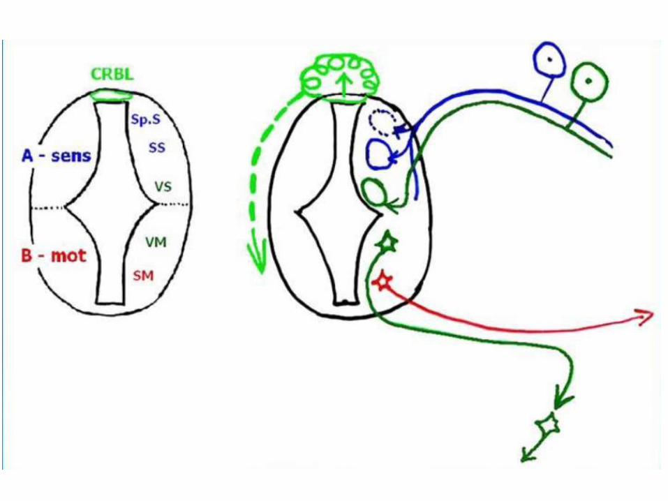
Viscero –
motor (parasympaticus)
visceral - sensory
general (nc. solitarius n.
X.) visceral epithelium
visceral -
sensory special (nc. gustatorius n.
VII.,IX.,X.) taste buds
somatic - sensory
general (nc. V.)
Branchio –
motor (special)
VII,VII,IX,X
Nc.gustatorius
VII,IX,X V
Nc.X
solitarius
Somatic -
motor (general)
V,VII,IX,X
III,IV,VI,XII
somatic –sensory
special (n. VIII.,
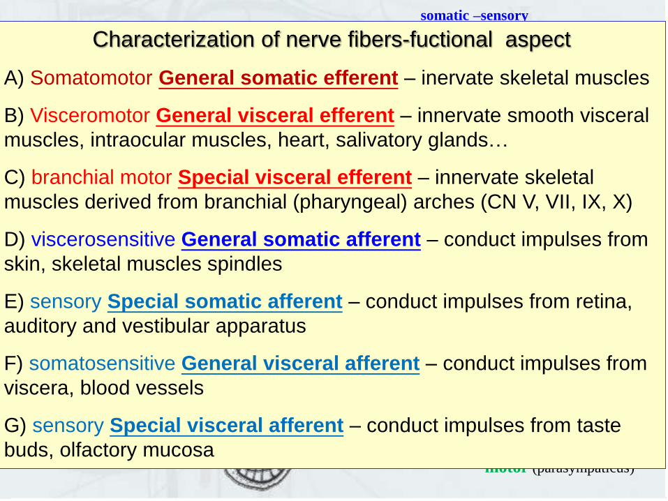
vestibular + auditory) Characterization of nerve fibers-fuctional aspect
A) Somatomotor General somatic efferent – inervate skeletal muscles
B) Visceromotor General visceral efferent – innervate smooth visceral
muscles, intraocular muscles, heart, salivatory glands…
C) branchial motor Special visceral efferent – innervate skeletal
muscles derived from branchial (pharyngeal) arches (CN V, VII, IX, X)
D) viscerosensitive General somatic afferent – conduct impulses from
skin, skeletal muscles spindles
E) sensory Special somatic afferent – conduct impulses from retina,
auditory and vestibular apparatus
F) somatosensitive General visceral afferent – conduct impulses from
viscera, blood vessels
G) sensory Special visceral afferent – conduct impulses from taste
buds, olfactory mucosa
viscero - motor (parasympaticus)
visceral - sensory
general (nc. solitarius n.
X.) visceral epithelium
visceral - sensory
special (nc.
gustatorius n.
VII.,IX.,X.) taste buds
somatic - sensory
general (nc. V.)
somatic –sensory
special (n. VIII.,
vestibular + auditory)
branchio – motor (special)
somatic motor (general) III,IV,VI,XII
V,VII,IX,X
VII,VII,IX,X
Nc.gustatorius
VII,IX,X
V
Nc.X
solitarius
S G
B S
V m V G V S
S G
S S
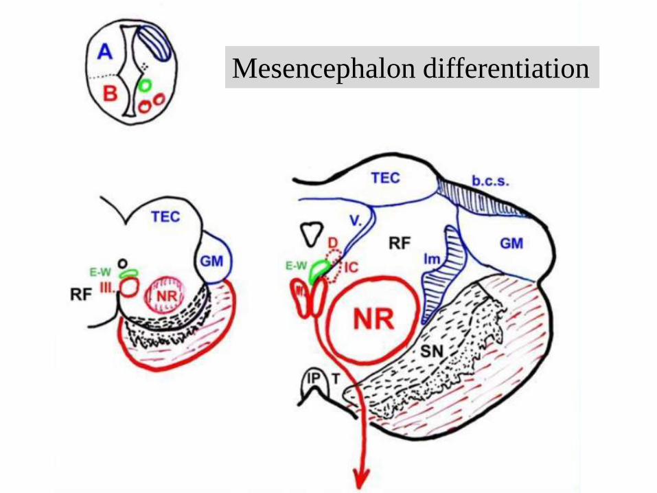
Mesencephalon differentiation
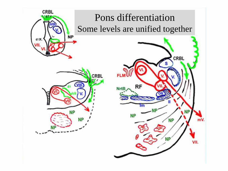
Pons differentiation Some levels are unified together
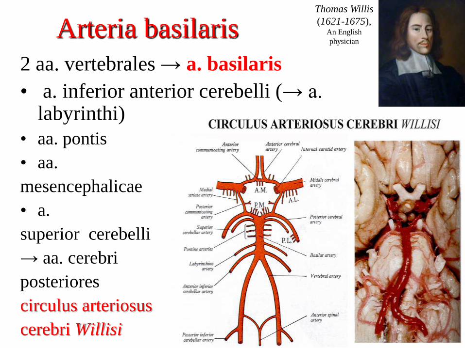
Arteria basilaris
2 aa. vertebrales → a. basilaris
• a. inferior anterior cerebelli (→ a. labyrinthi)
• aa. pontis
• aa.
mesencephalicae
• a.
superior cerebelli
→ aa. cerebri
posteriores
circulus arteriosus
cerebri Willisi
Thomas Willis
(1621-1675), An English
physician
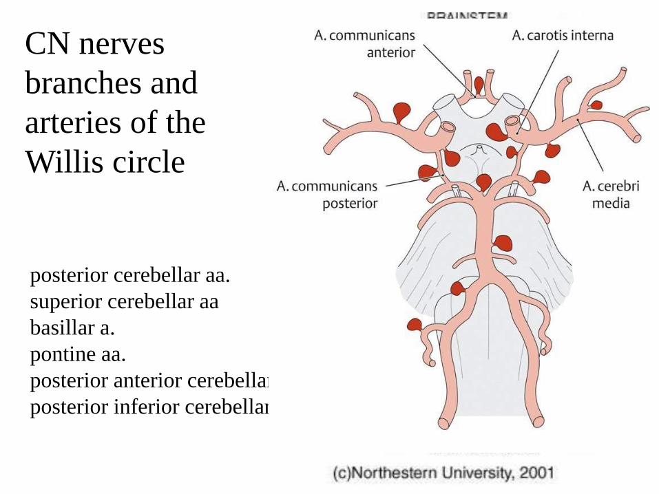
CN nerves
branches and
arteries of the
Willis circle
posterior cerebellar aa.
superior cerebellar aa
basillar a.
pontine aa.
posterior anterior cerebellar aa.
posterior inferior cerebellar aa.

veins draining brainstem
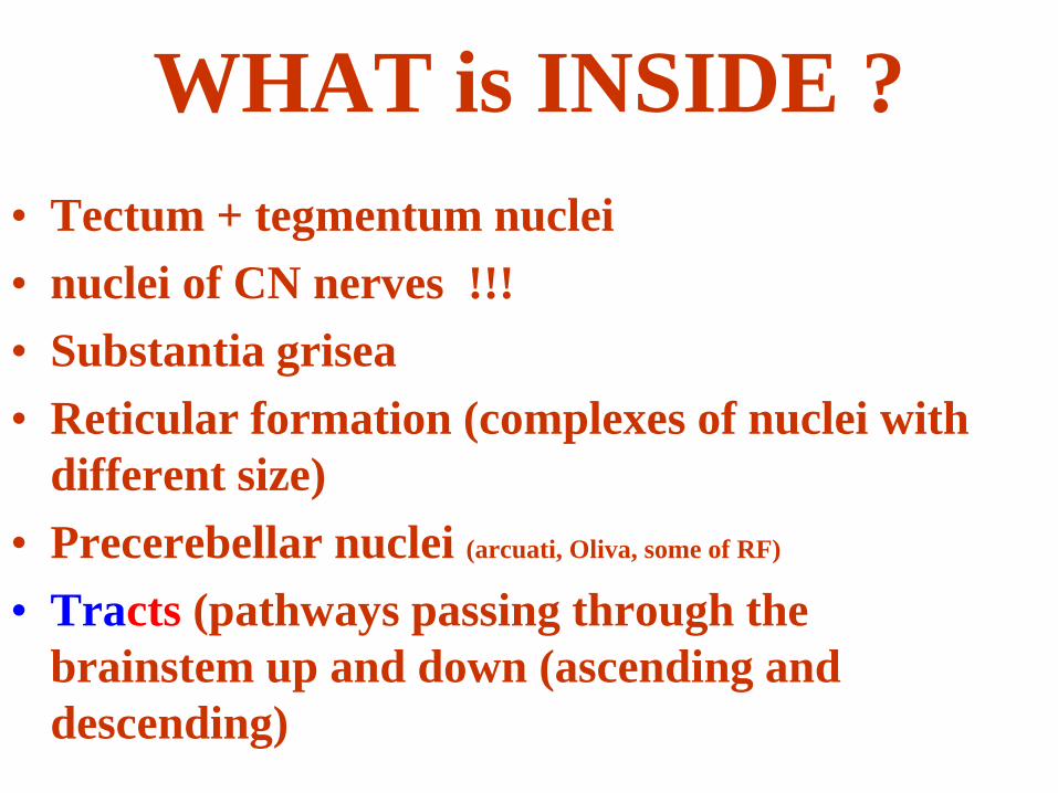
WHAT is INSIDE ?
• Tectum + tegmentum nuclei
• nuclei of CN nerves !!!
• Substantia grisea
• Reticular formation (complexes of nuclei with
different size)
• Precerebellar nuclei (arcuati, Oliva, some of RF)
• Tracts (pathways passing through the
brainstem up and down (ascending and
descending)
Chemical systems in the CNS
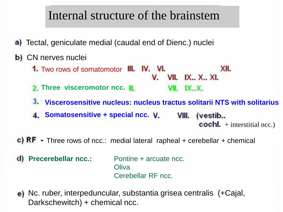
Internal structure of the brainstem
Tectal, geniculate medial (caudal end of Dienc.) nuclei
CN nerves nuclei
Two rows of somatomotor
Three visceromotor ncc.
Viscerosensitive nucleus: nucleus tractus solitarii NTS with solitarius
+ interstitial ncc.)
Two rows of somatomotor
Somatosensitive + special ncc.
Three rows of ncc.: medial lateral rapheal + cerebellar + chemical
Precerebellar ncc.:
Nc. ruber, interpeduncular, substantia grisea centralis (+Cajal,
Darkschewitch) + chemical ncc.
Pontine + arcuate ncc.
Oliva
Cerebellar RF ncc.
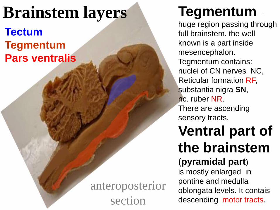
anteroposterior
section
Brainstem layers Tectum
Tegmentum
Pars ventralis
Tegmentum - huge region passing through
full brainstem. the well
known is a part inside
mesencephalon.
Tegmentum contains:
nuclei of CN nerves NC,
Reticular formation RF,
substantia nigra SN,
nc. ruber NR.
There are ascending
sensory tracts.
Ventral part of
the brainstem (pyramidal part) is mostly enlarged in
pontine and medulla
oblongata levels. It contais
descending motor tracts.
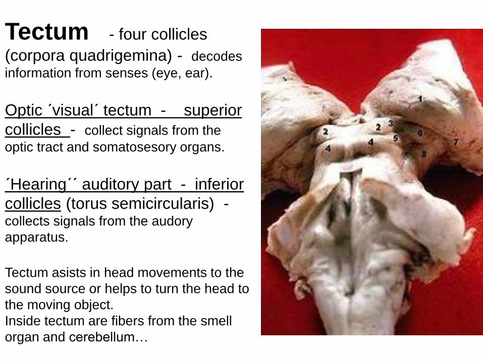
Tectum - four collicles
(corpora quadrigemina) - decodes
information from senses (eye, ear).
Optic ´visual´ tectum - superior
collicles - collect signals from the
optic tract and somatosesory organs.
´Hearing´´ auditory part - inferior
collicles (torus semicircularis) - collects signals from the audory
apparatus.
Tectum asists in head movements to the
sound source or helps to turn the head to
the moving object.
Inside tectum are fibers from the smell
organ and cerebellum…
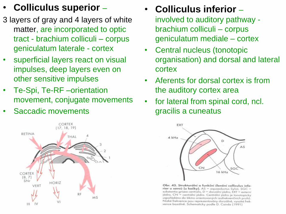
• Colliculus superior –
3 layers of gray and 4 layers of white
matter, are incorporated to optic
tract - brachium colliculi – corpus
geniculatum laterale - cortex
• superficial layers react on visual
impulses, deep layers even on
other sensitive impulses
• Te-Spi, Te-RF –orientation
movement, conjugate movements
• Saccadic movements
• Colliculus inferior –
involved to auditory pathway -
brachium colliculi – corpus
geniculatum mediale – cortex
• Central nucleus (tonotopic
organisation) and dorsal and lateral
cortex
• Aferents for dorsal cortex is from
the auditory cortex area
• for lateral from spinal cord, ncl.
gracilis a cuneatus
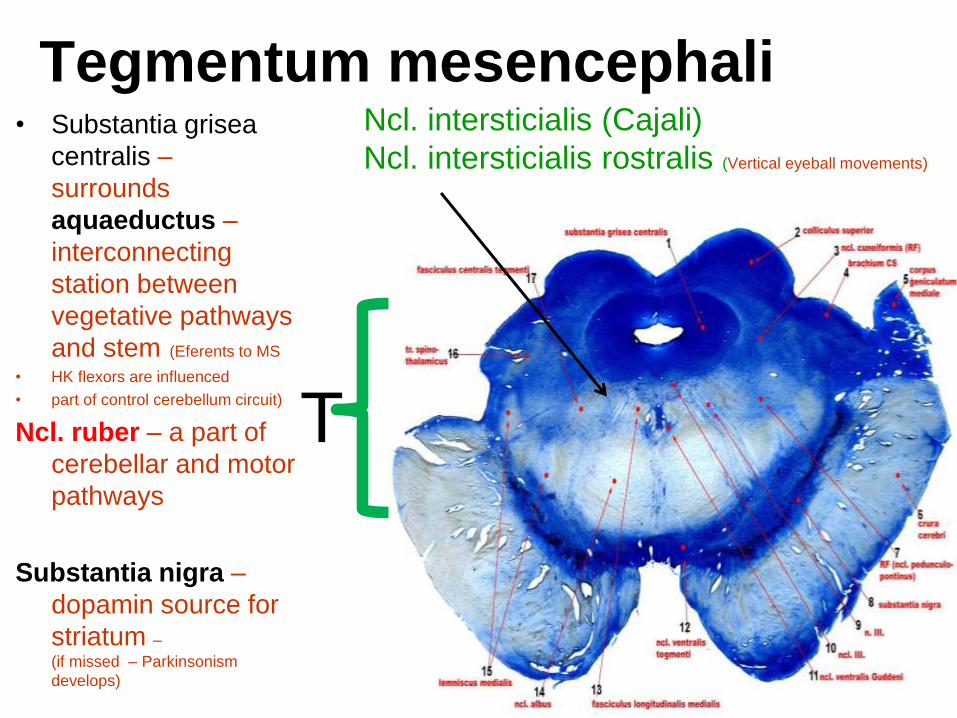
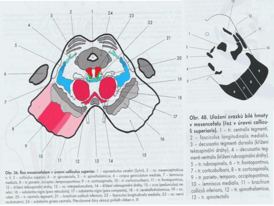
Tegmentum mesencephali • Substantia grisea
centralis –
surrounds
aquaeductus –
interconnecting
station between
vegetative pathways
and stem (Eferents to MS
• HK flexors are influenced
• part of control cerebellum circuit)
Ncl. ruber – a part of
cerebellar and motor
pathways
Substantia nigra –
dopamin source for
striatum –
(if missed – Parkinsonism
develops)
Ncl. intersticialis (Cajali)
Ncl. intersticialis rostralis (Vertical eyeball movements)
T
Ncl. intersticialis (Cajali), Ncl.
intersticialis rostralis
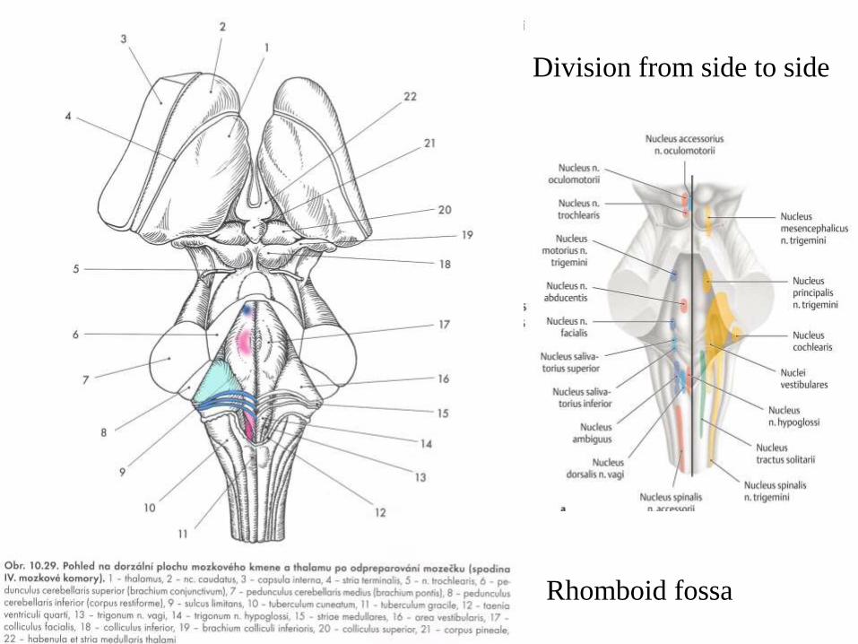
Division from side to side
Rhomboid fossa
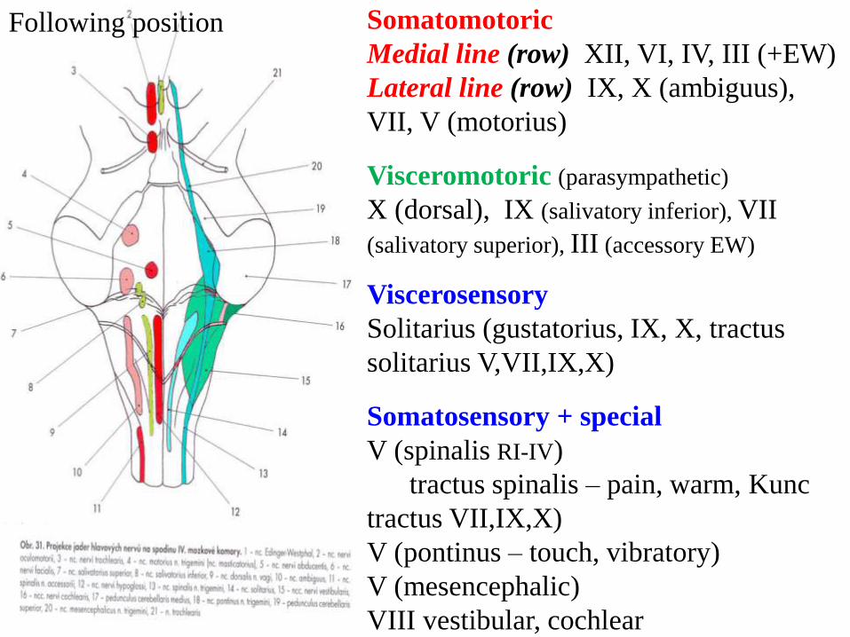
Somatomotoric
Medial line (row) XII, VI, IV, III (+EW)
Lateral line (row) IX, X (ambiguus),
VII, V (motorius)
Visceromotoric (parasympathetic)
X (dorsal), IX (salivatory inferior), VII
(salivatory superior), III (accessory EW)
Viscerosensory
Solitarius (gustatorius, IX, X, tractus
solitarius V,VII,IX,X)
Somatosensory + special
V (spinalis RI-IV)
tractus spinalis – pain, warm, Kunc
tractus VII,IX,X)
V (pontinus – touch, vibratory)
V (mesencephalic)
VIII vestibular, cochlear
Following position
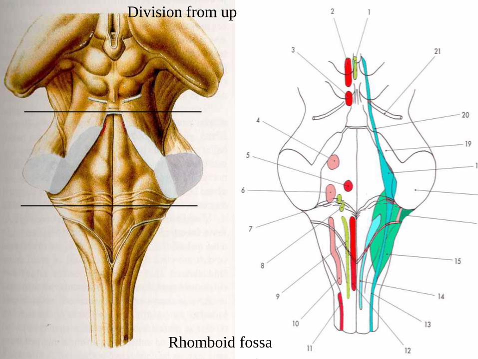
Division from up to down
Rhomboid fossa
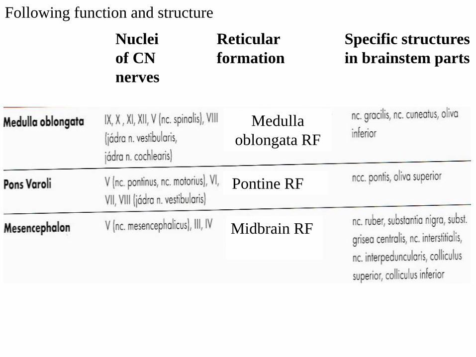
Nuclei
of CN
nerves
Reticular
formation
Specific structures
in brainstem parts
Pontine RF
Medulla
oblongata RF
Midbrain RF
Following function and structure
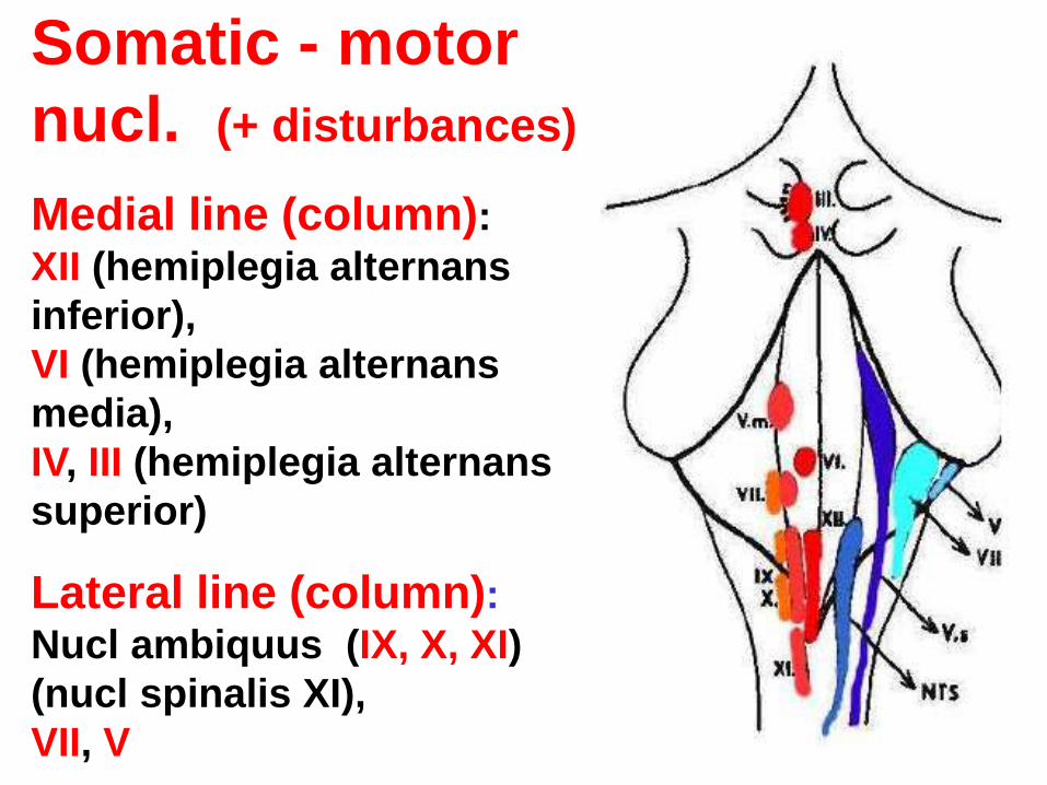
Somatic - motor
nucl. (+ disturbances)
Medial line (column): XII (hemiplegia alternans
inferior),
VI (hemiplegia alternans
media),
IV, III (hemiplegia alternans
superior)
Lateral line (column): Nucl ambiquus (IX, X, XI)
(nucl spinalis XI),
VII, V
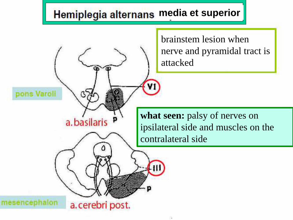
brainstem lesion when
nerve and pyramidal tract is
attacked
what seen: palsy of nerves on
ipsilateral side and muscles on the
contralateral side
media et superior
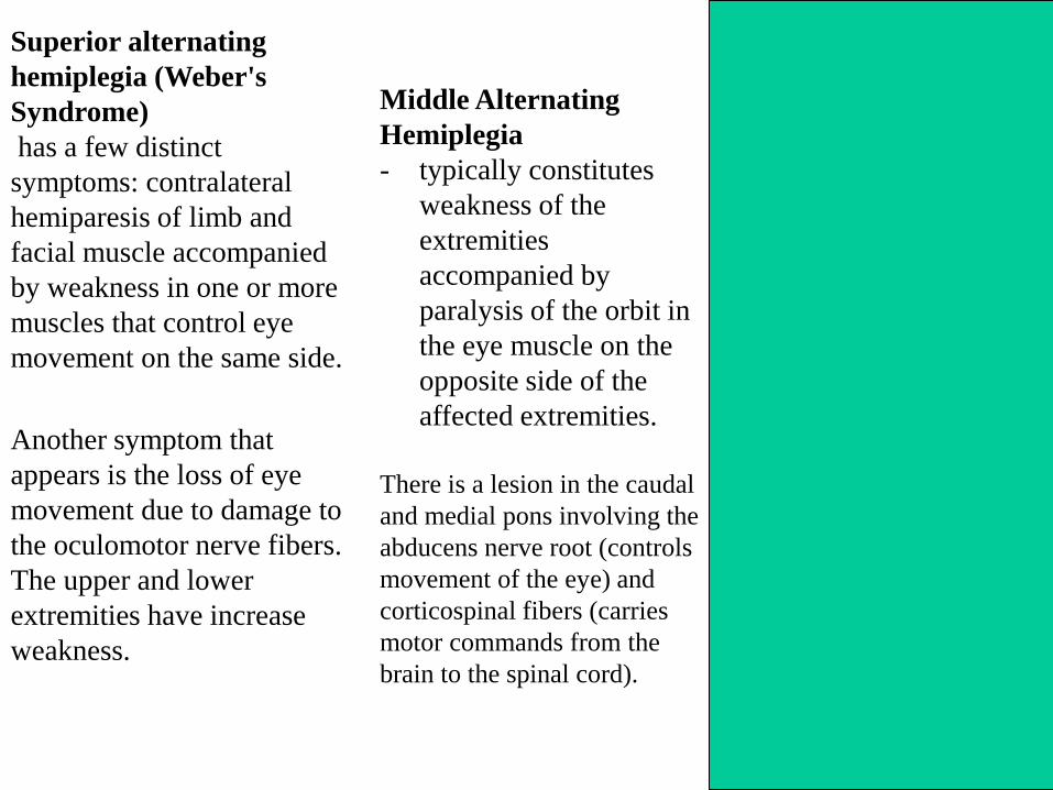
Superior alternating
hemiplegia (Weber's
Syndrome)
has a few distinct
symptoms: contralateral
hemiparesis of limb and
facial muscle accompanied
by weakness in one or more
muscles that control eye
movement on the same side.
Another symptom that
appears is the loss of eye
movement due to damage to
the oculomotor nerve fibers.
The upper and lower
extremities have increase
weakness.
Middle Alternating
Hemiplegia
- typically constitutes
weakness of the
extremities
accompanied by
paralysis of the orbit in
the eye muscle on the
opposite side of the
affected extremities.
There is a lesion in the caudal
and medial pons involving the
abducens nerve root (controls
movement of the eye) and
corticospinal fibers (carries
motor commands from the
brain to the spinal cord).
Inferior Alternating
Hemiplegia
(also known as medial
medullary syndrome) -
involves a “weakness of
the extremities
accompanied by
paralysis of muscles on
the ipsilateral side of the
tongue (seen as a
deviation of the tongue
on that side on
protrusion).
These symptoms indicate a
lesion in the medulla
involving the corticospinal
fibers in the pyramid and
the exiting hypoglossal
nerve roots).
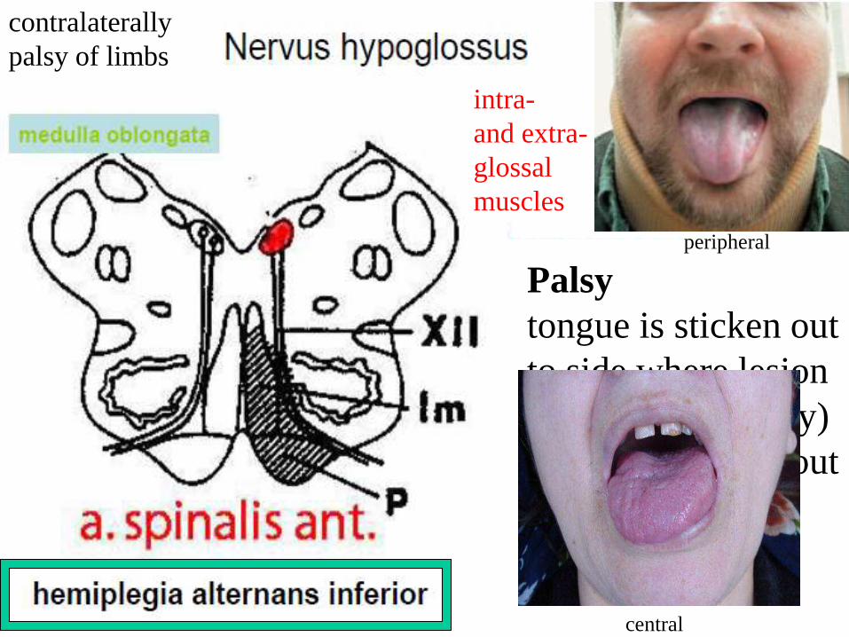
Palsy
tongue is sticken out
to side where lesion
is (peripheral palsy)
tongue is sticken out
to opposite side
(central palsy)
contralaterally
palsy of limbs
intra-
and extra-
glossal
muscles
peripheral
central
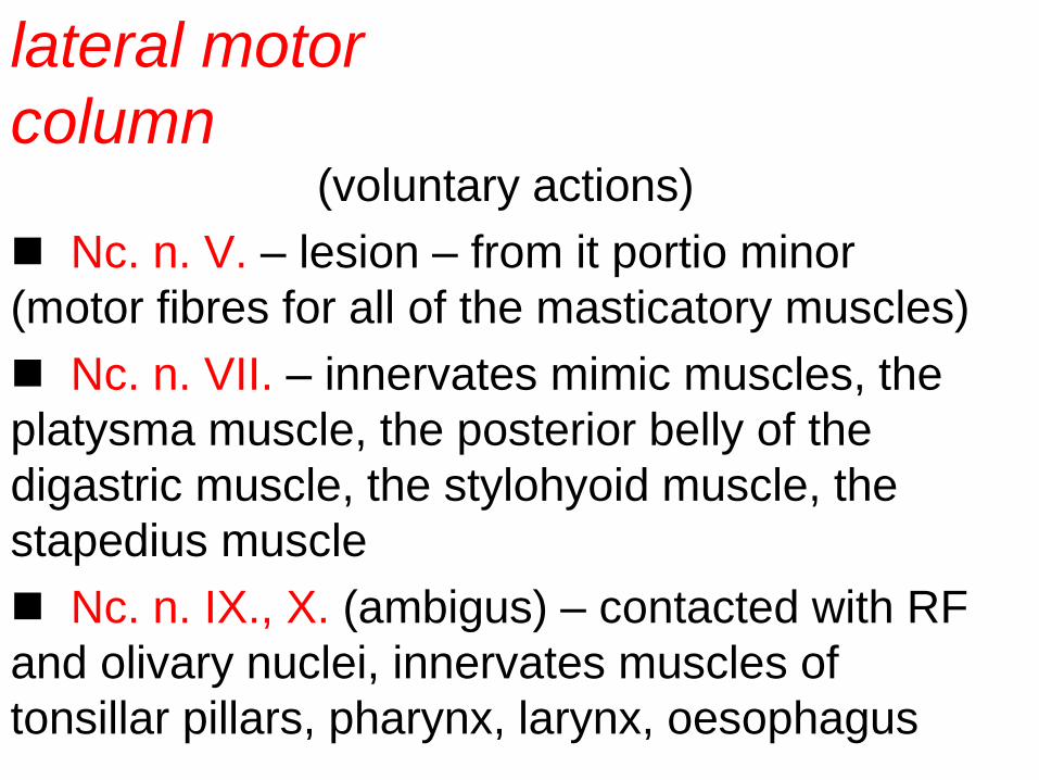
(voluntary actions)
Nc. n. V. – lesion – from it portio minor
(motor fibres for all of the masticatory muscles)
Nc. n. VII. – innervates mimic muscles, the
platysma muscle, the posterior belly of the
digastric muscle, the stylohyoid muscle, the
stapedius muscle
Nc. n. IX., X. (ambigus) – contacted with RF
and olivary nuclei, innervates muscles of
tonsillar pillars, pharynx, larynx, oesophagus
lateral motor
column
motor ___
touch -----
pain warm ________
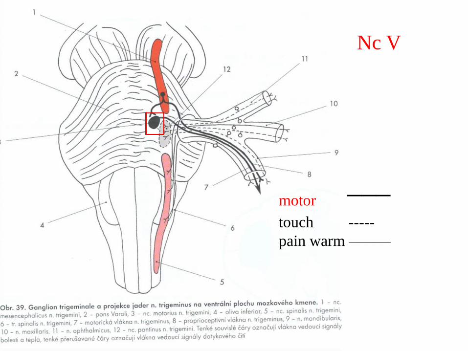
Nc V
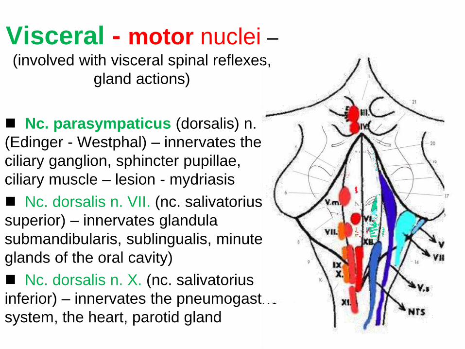
Visceral - motor nuclei – (involved with visceral spinal reflexes,
gland actions)
Nc. parasympaticus (dorsalis) n. III.
(Edinger - Westphal) – innervates the
ciliary ganglion, sphincter pupillae,
ciliary muscle – lesion - mydriasis
Nc. dorsalis n. VII. (nc. salivatorius
superior) – innervates glandula
submandibularis, sublingualis, minute
glands of the oral cavity)
Nc. dorsalis n. X. (nc. salivatorius
inferior) – innervates the pneumogastric
system, the heart, parotid gland
mixed nerves
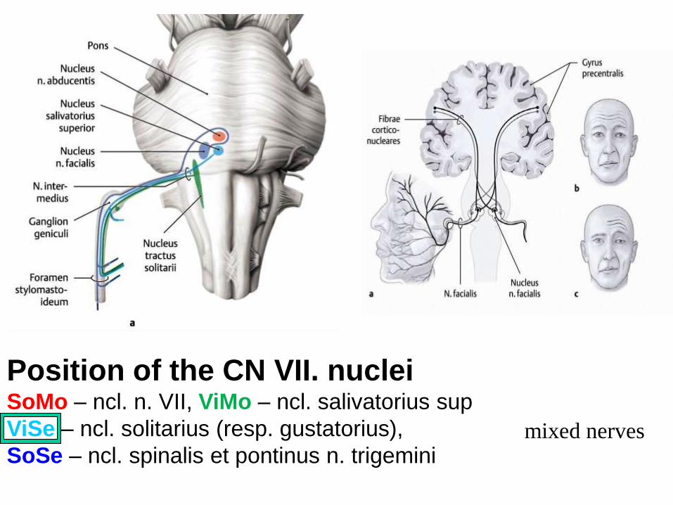
Position of the CN VII. nuclei SoMo – ncl. n. VII, ViMo – ncl. salivatorius sup
ViSe – ncl. solitarius (resp. gustatorius),
SoSe – ncl. spinalis et pontinus n. trigemini
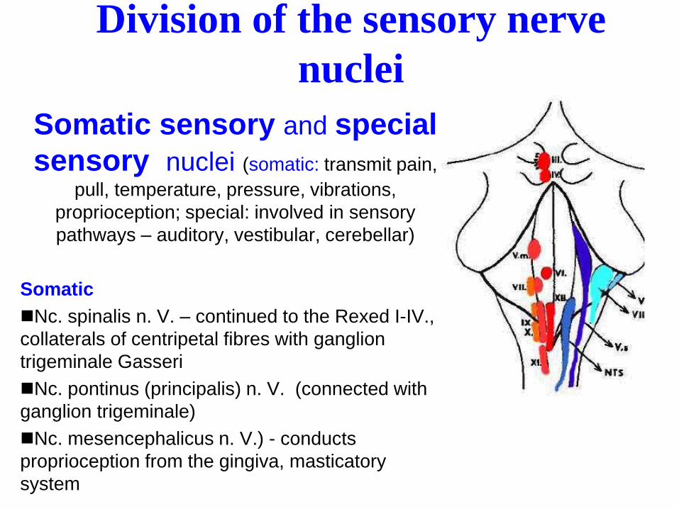
Division of the sensory nerve
nuclei
Somatic sensory and special
sensory nuclei (somatic: transmit pain,
pull, temperature, pressure, vibrations,
proprioception; special: involved in sensory
pathways – auditory, vestibular, cerebellar)
Somatic
Nc. spinalis n. V. – continued to the Rexed I-IV.,
collaterals of centripetal fibres with ganglion
trigeminale Gasseri
Nc. pontinus (principalis) n. V. (connected with
ganglion trigeminale)
Nc. mesencephalicus n. V.) - conducts
proprioception from the gingiva, masticatory
system
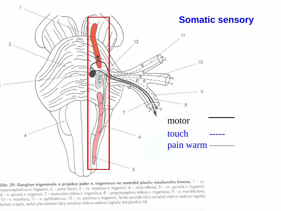
motor ___
touch -----
pain warm ________
Somatic sensory
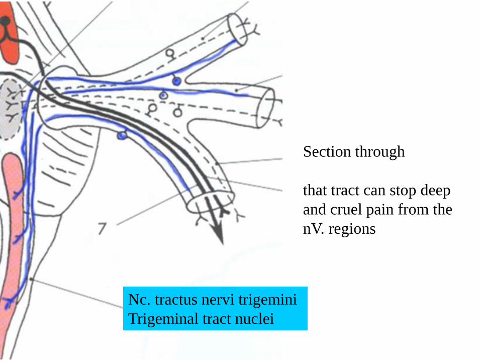
Section through
that tract can stop deep
and cruel pain from the
nV. regions
Nc. tractus nervi trigemini
Trigeminal tract nuclei
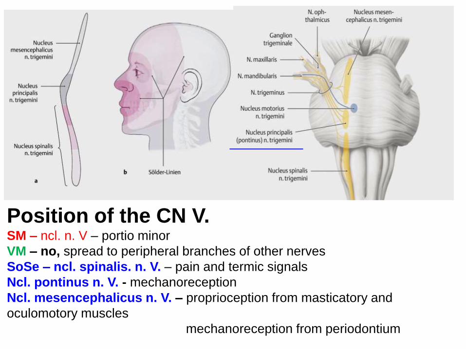
Position of the CN V. SM – ncl. n. V – portio minor
VM – no, spread to peripheral branches of other nerves
SoSe – ncl. spinalis. n. V. – pain and termic signals
Ncl. pontinus n. V. - mechanoreception
Ncl. mesencephalicus n. V. – proprioception from masticatory and
oculomotory muscles
mechanoreception from periodontium

Division of the sensory nerve
nuclei Somatic sensory and
special sensory nuclei (somatic: transmit pain, pull, temperature,
pressure, vibrations, proprioception;
special: involved in sensory pathways –
auditory, vestibular, cerebellar)
Special
Nc. vestibulares (medial- Schwalbe;
lateral-Deiters, cranial-Bechterew, caudal-
Roller) – connection with the cerebellar
nuclei
Nc. cochleares (caudal; cranial) –
involved in the auditory pathway,
connections with the corpora trapezoidea
and lemniscus

Division of the sensory nerve
nuclei
Visceral sensory nuclei (transmit visceral
sensations, involved in taste
reflex and in visceral reflex
arches)
Nc. n. V., VII., IX., X.) – nc.
solitarius surrounded by tractus
solitarius:
- rostral part – nc. gustatorius
- caudal part – nc. commisuralis
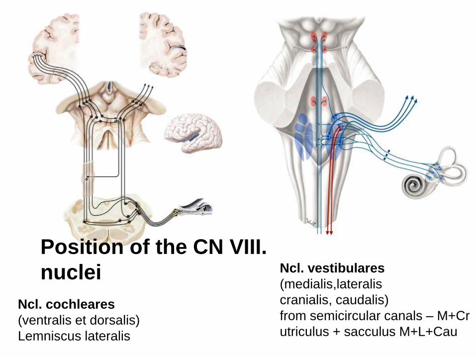
Ncl. cochleares
(ventralis et dorsalis)
Lemniscus lateralis
Ncl. vestibulares
(medialis,lateralis
cranialis, caudalis)
from semicircular canals – M+Cr
utriculus + sacculus M+L+Cau
Position of the CN VIII.
nuclei
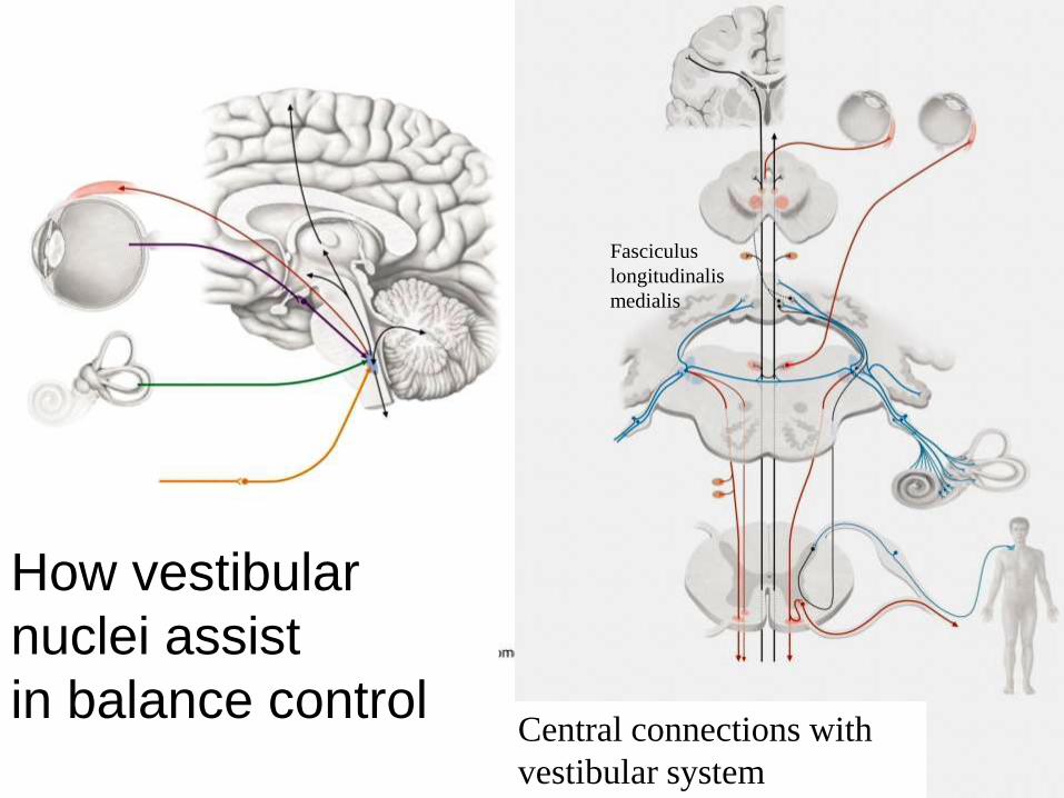
Central connections with
vestibular system
How vestibular
nuclei assist
in balance control
Fasciculus
longitudinalis
medialis
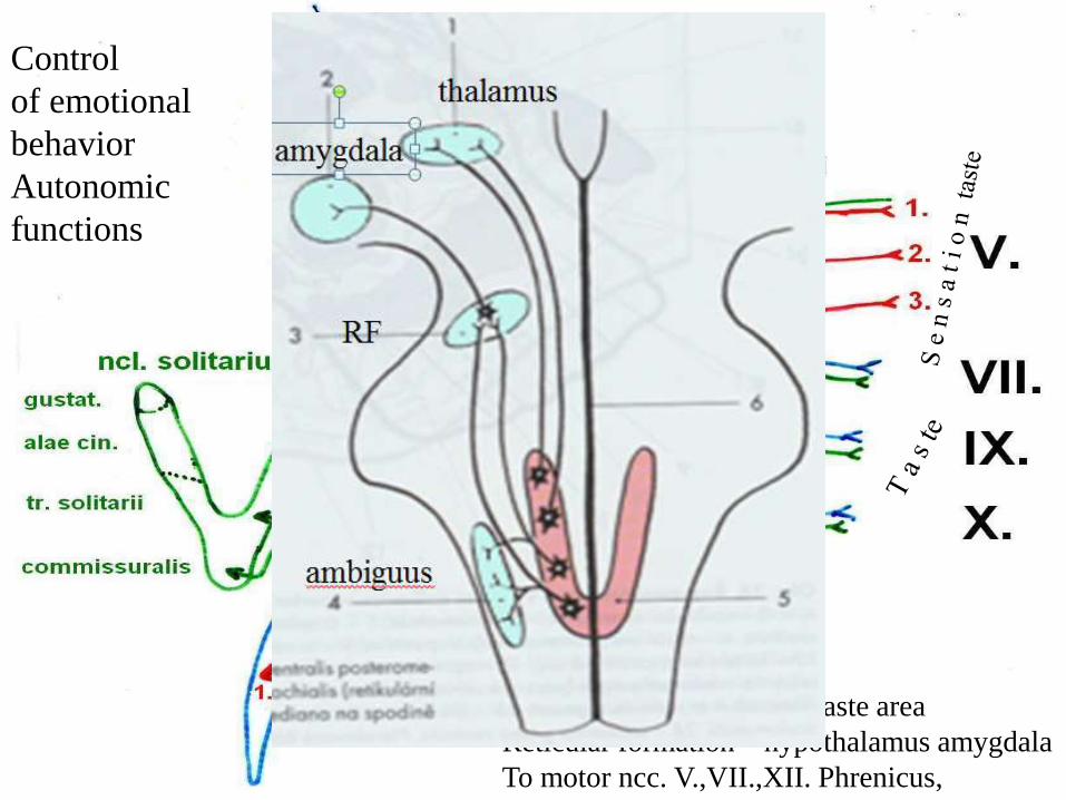
Afferent: Taste !
Respiratory digestive systems
Chemo+baroreceptors of vessels
Efferent: Thalamus – 43 taste area
Reticular formation – hypothalamus amygdala
To motor ncc. V.,VII.,XII. Phrenicus,
Control
of emotional
behavior
Autonomic
functions
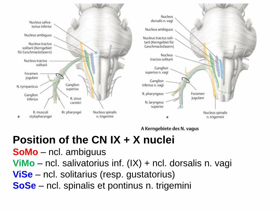
Position of the CN IX + X nuclei SoMo – ncl. ambiguus
ViMo – ncl. salivatorius inf. (IX) + ncl. dorsalis n. vagi
ViSe – ncl. solitarius (resp. gustatorius)
SoSe – ncl. spinalis et pontinus n. trigemini
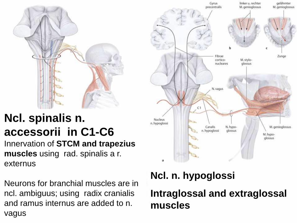
Ncl. spinalis n.
accessorii in C1-C6 Innervation of STCM and trapezius
muscles using rad. spinalis a r.
externus
Neurons for branchial muscles are in
ncl. ambiguus; using radix cranialis
and ramus internus are added to n.
vagus
Ncl. n. hypoglossi
Intraglossal and extraglossal
muscles
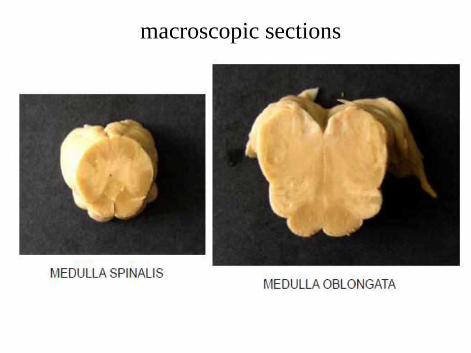
macroscopic sections
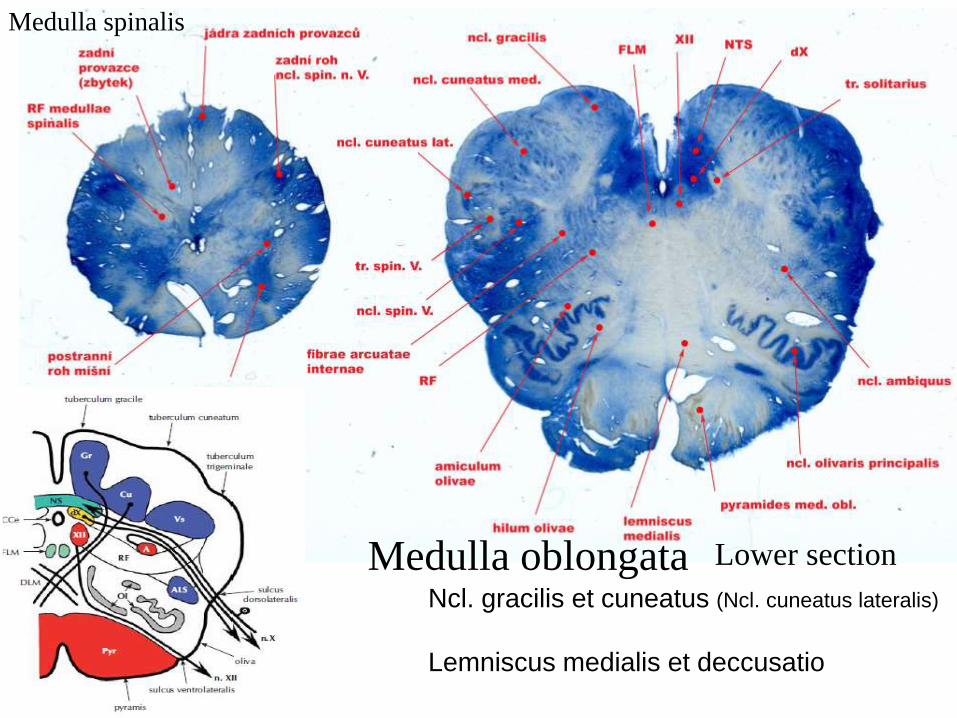
Ncl. gracilis et cuneatus (Ncl. cuneatus lateralis)
Lemniscus medialis et deccusatio
Lower section Medulla oblongata
Medulla spinalis

Ncl. olivaris – p.crbl. inf.
climbing fibers
Rytmic dyskinesis
Medulla
oblongata
Upper section
The ventral portion of the medulla oblongata contains
medullary pyramids. These two ridge-like structures travel along the length of the medulla oblongata and are
bordered medially by the anterior median fissure. They each have an anterolateral
sulcus along their lateral borders.
Also located laterally from each pyramid is a pronounced bulge known as an olive.
The medullary pyramids contain motor fibers that are known
as the corticobulbar and corticospinal tracts.
The corticospinal tracts are on the anterior surface of the
pyramids.
These tracts condust motor signals that originated in the precentral gyrus
and travelled through the internal capsule to the medulla oblongata and
pyramids. Extrapyramidal tracts are those motor tracts that do not
traverse the medullary pyramids.
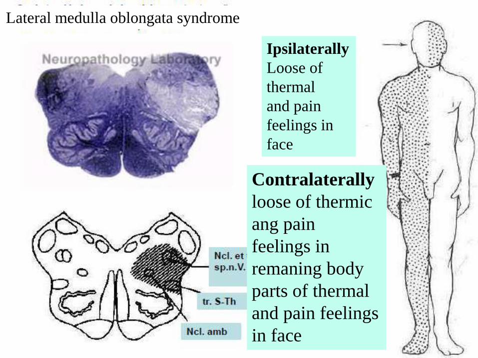
Lateral medulla oblongata syndrome
Ipsilaterally
Loose of
thermal
and pain
feelings in
face
Contralaterally
loose of thermic
ang pain
feelings in
remaning body
parts of thermal
and pain feelings
in face
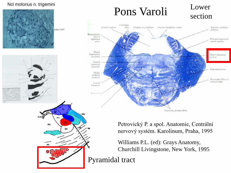
Pons Varoli
Petrovický P. a spol. Anatomie, Centrální
nervový systém. Karolinum, Praha, 1995
Williams P.L. (ed): Grays Anatomy,
Churchill Livingstone, New York, 1995
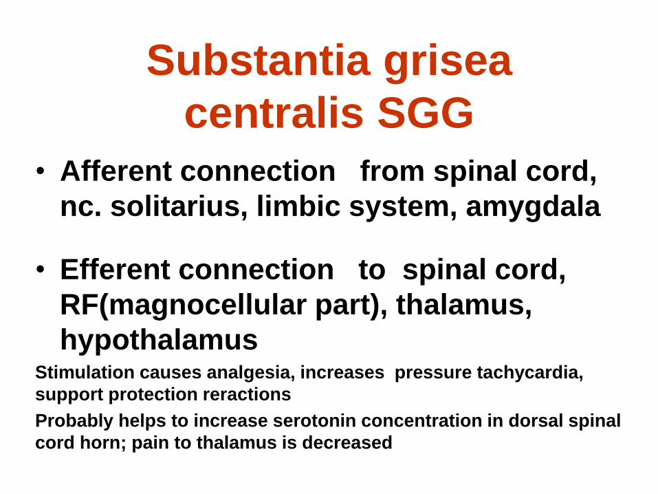
Pyramidal tract
Lower
section
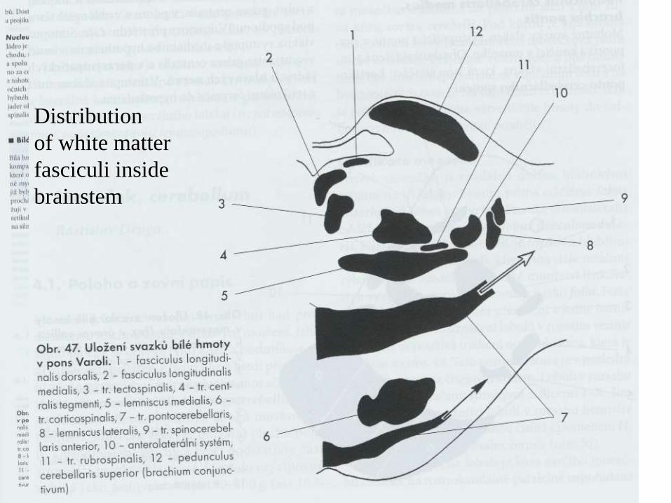
Distribution
of white matter
fasciculi inside
brainstem
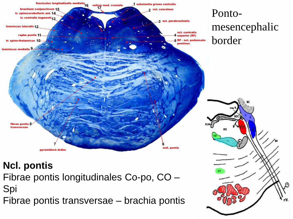
Ncl. pontis
Fibrae pontis longitudinales Co-po, CO –
Spi
Fibrae pontis transversae – brachia pontis
Ponto-
mesencephalic
border
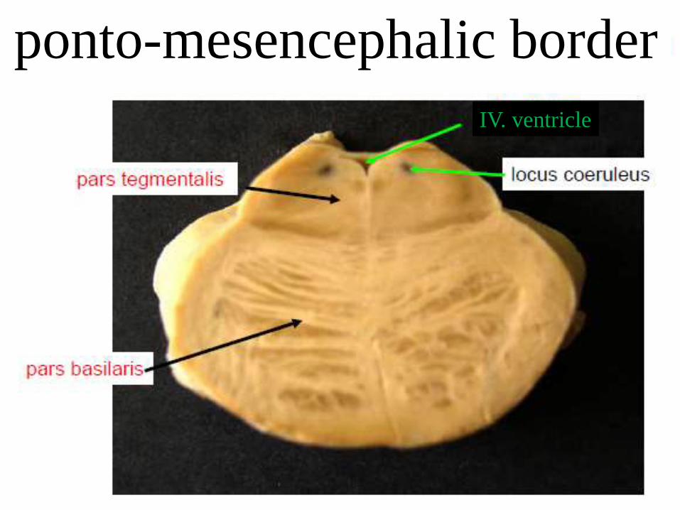
ponto-mesencephalic border
IV. ventricle
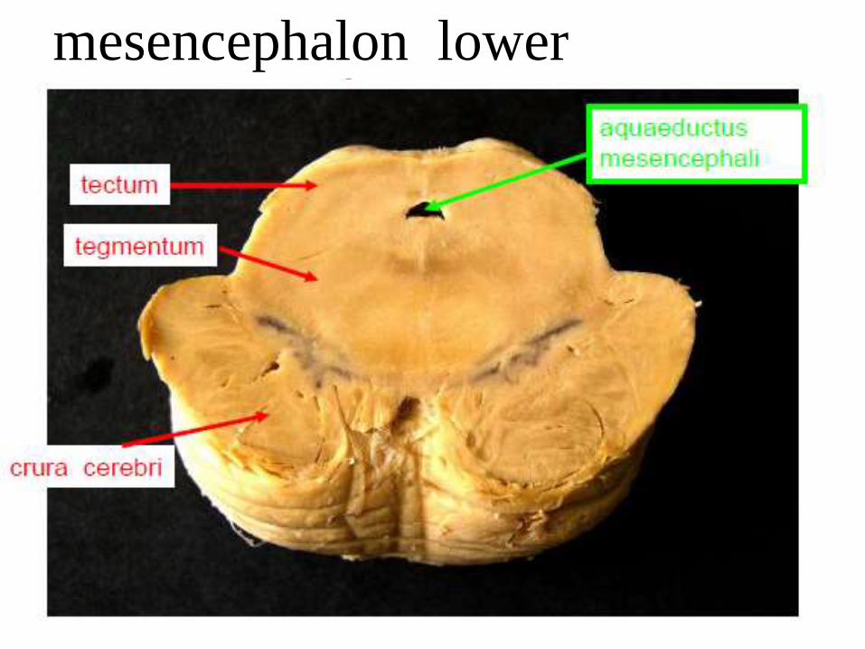
mesencephalon lower
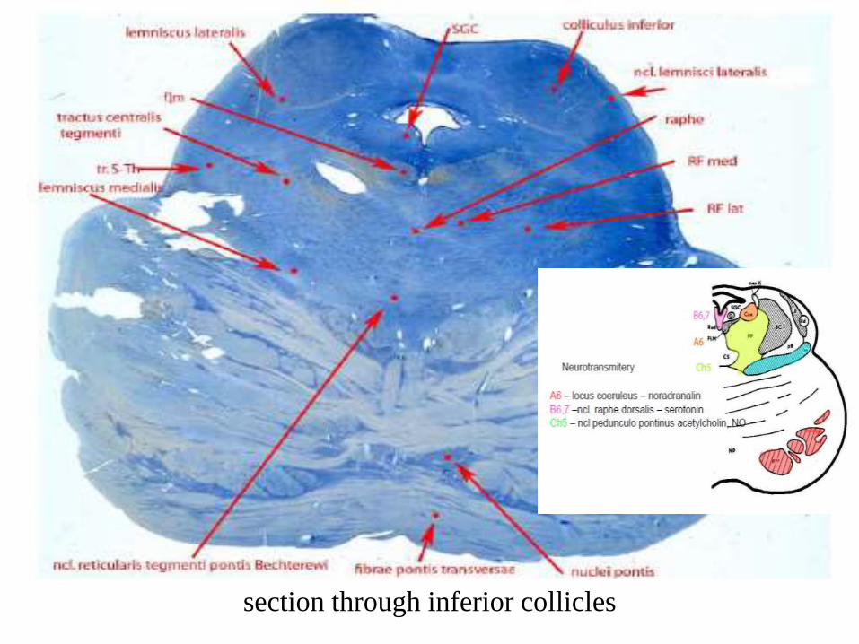
section through inferior collicles
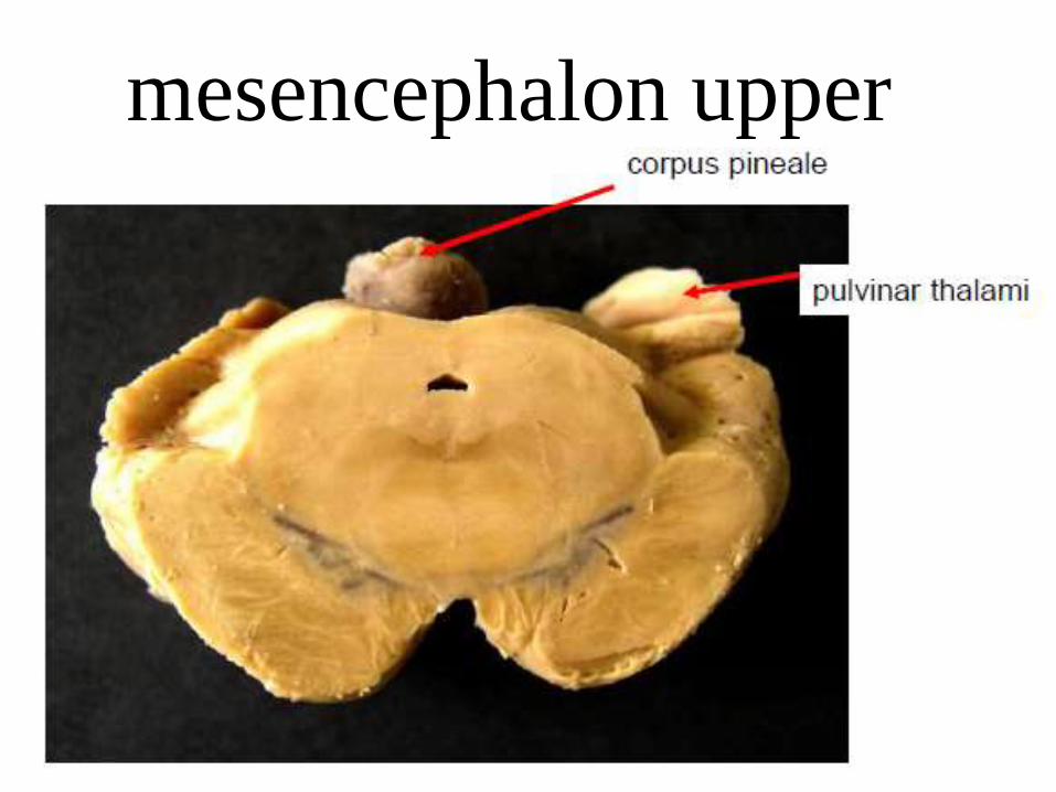
mesencephalon upper
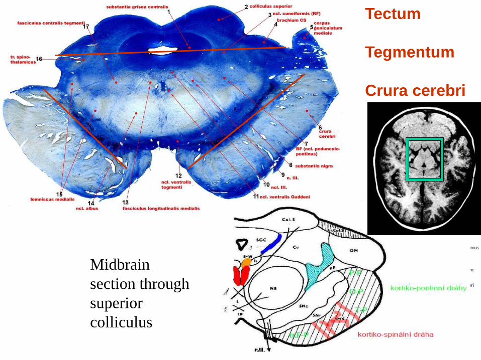
Tectum
Tegmentum
Crura cerebri
Midbrain
section through
superior
colliculus
cortico-pontine
pathways
corticospinal
pathways
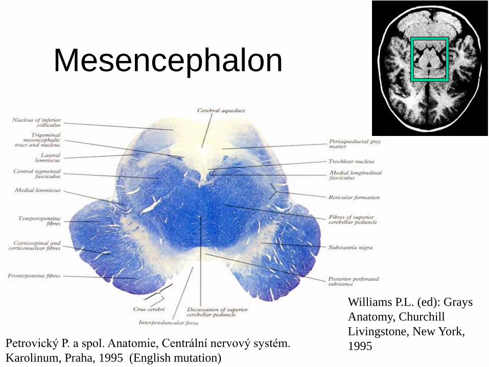
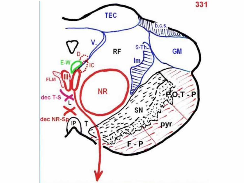
Mesencephalon
Williams P.L. (ed): Grays
Anatomy, Churchill
Livingstone, New York,
1995 Petrovický P. a spol. Anatomie, Centrální nervový systém.
Karolinum, Praha, 1995 (English mutation)
Substantia grisea
centralis SGG
• Afferent connection from spinal cord,
nc. solitarius, limbic system, amygdala
• Efferent connection to spinal cord,
RF(magnocellular part), thalamus,
hypothalamus Stimulation causes analgesia, increases pressure tachycardia,
support protection reractions
Probably helps to increase serotonin concentration in dorsal spinal
cord horn; pain to thalamus is decreased
Substantia nigra SN • Compact part - dopaminergic (Tsai group)
• Reticular part – GABA
• Afferent connection from striatum (basal
nuclei) smaller part from pallidum
• Efferent connection to striatum, amygdala +
neocortex
Loose of dopamin – Parkinsonism (hypokinesis, hypertonism, tremor)
Nucleus ruber NR • magnocellular part - dopaminergic (Tsai
group)
• parvicellular part – GABA
• Afferent connection from cingular cortex
(noncrossed)
• Efferent connection to lateral spina fasciculus
(rubrospinal tract)
to oliva inferior
to RF

Reticular formation
www.macalester.edu http://img132.imageshack.us
Mostly produce serotonin (B1-B6)
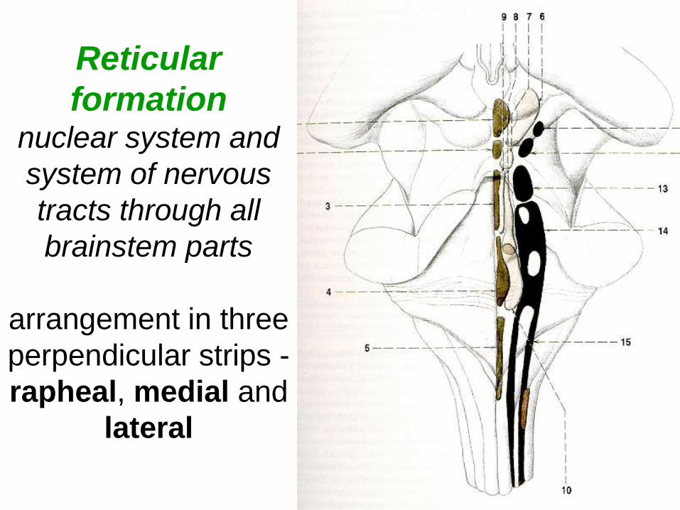
Reticular
formation nuclear system and
system of nervous
tracts through all
brainstem parts
arrangement in three
perpendicular strips -
rapheal, medial and
lateral

Rapheal – middle, unpaired
nuclear strip; many
connections with medial row
and with limbic system
Medial – paired, massive,
long connections
Lateral – only in medulla
oblongata and pons, small
cells, connections with
rapheal system,
+ nuclei showing
characteristic nervous
conections or typical mediator (cerebellar cholinergic adrenergic)
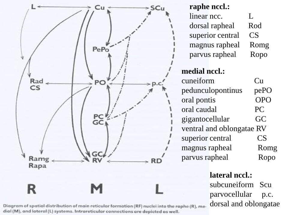
raphe nccl.:
linear ncc. L
dorsal rapheal Rod
superior central CS
magnus rapheal Romg
parvus rapheal Ropo
medial nccl.:
cuneiform Cu
pedunculopontinus pePO
oral pontis OPO
oral caudal PC
gigantocellular GC
ventral and oblongatae RV
superior central CS
magnus rapheal Romg
parvus rapheal Ropo
lateral nccl.:
subcuneiform Scu
parvocellular p.c.
dorsal and oblongatae
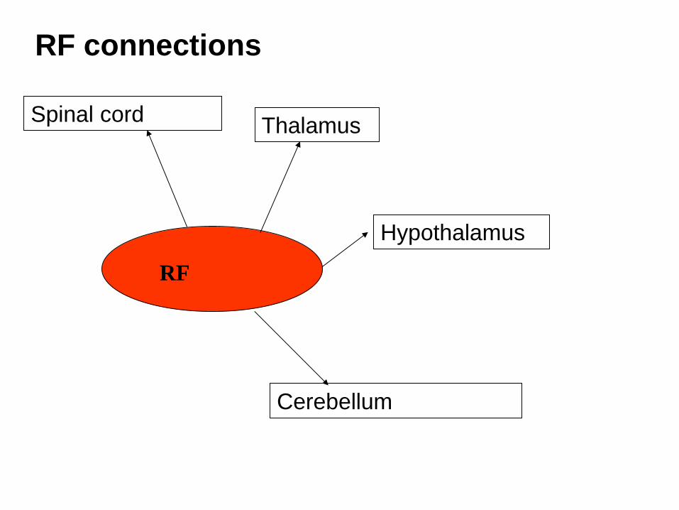
RF connections
RF
Spinal cord Thalamus
Hypothalamus
Cerebellum
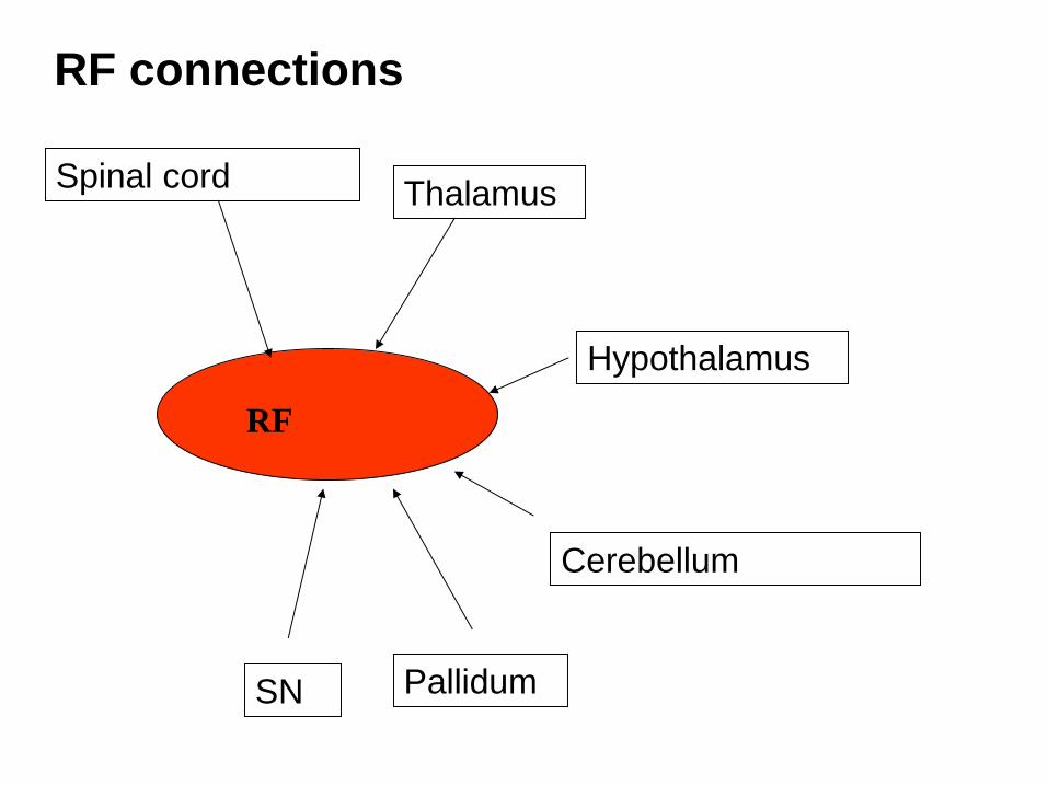
RF connections
RF
Spinal cord Thalamus
Hypothalamus
Cerebellum
Pallidum SN
RF connections
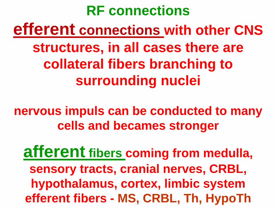
efferent connections with other CNS
structures, in all cases there are
collateral fibers branching to
surrounding nuclei
nervous impuls can be conducted to many
cells and becames stronger
afferent fibers coming from medulla,
sensory tracts, cranial nerves, CRBL,
hypothalamus, cortex, limbic system
efferent fibers - MS, CRBL, Th, HypoTh
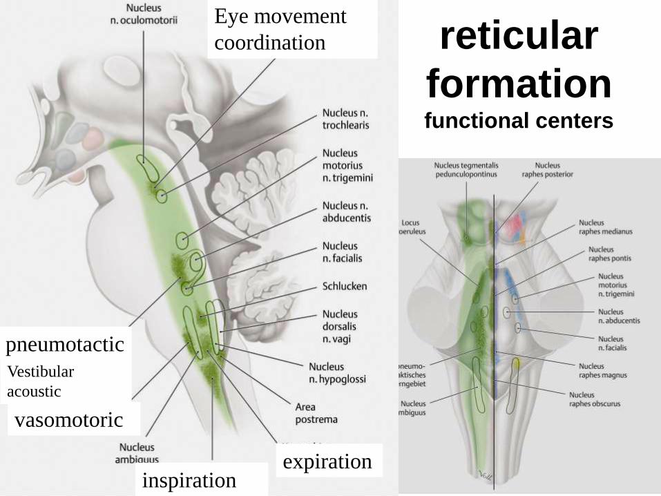
reticular
formation functional centers
pneumotactic
vasomotoric
inspiration expiration
Eye movement
coordination
Vestibular
acoustic
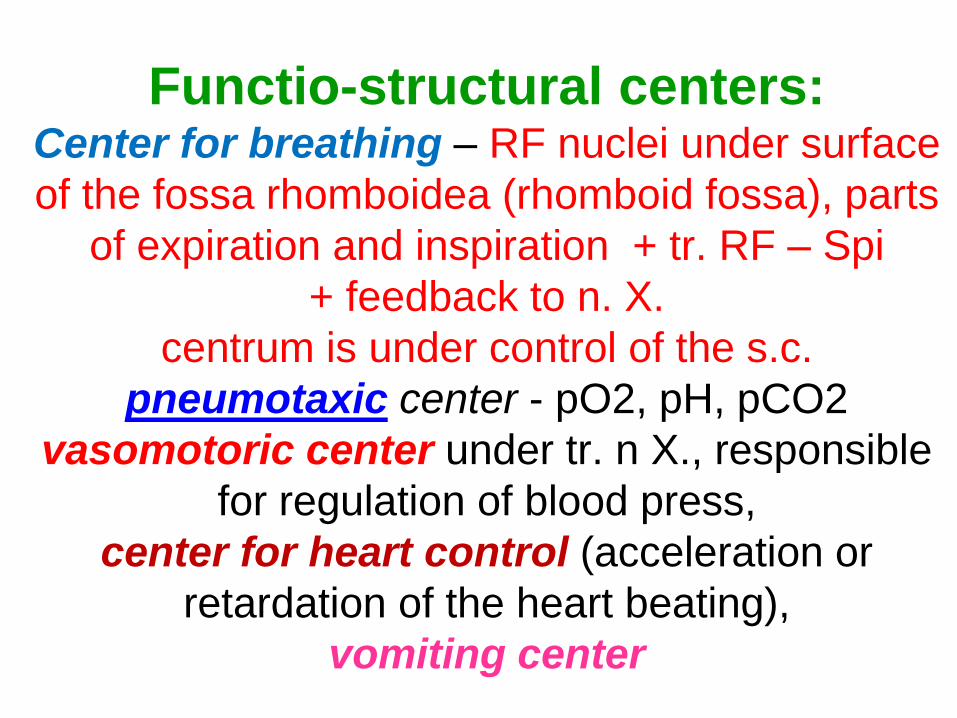
Functio-structural centers: Center for breathing – RF nuclei under surface
of the fossa rhomboidea (rhomboid fossa), parts
of expiration and inspiration + tr. RF – Spi
+ feedback to n. X.
centrum is under control of the s.c.
pneumotaxic center - pO2, pH, pCO2
vasomotoric center under tr. n X., responsible
for regulation of blood press,
center for heart control (acceleration or
retardation of the heart beating),
vomiting center
Reflexes of “resuscitation“:
swalloving reflex,
sucking reflex,
reflexes of salivary glands,
visceromotoric reflexes
Protecting reflexes:
ef + af: fibers NC, under RF control
twinking, blinking, corneal, tearing, coughing,
vomiting (suffocating), iridial

Frédéric Bremer
neurophysiologist
1892–1982
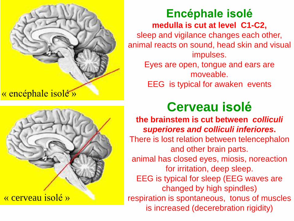
« encéphale isolé »
« cerveau isolé »
Encéphale isolé medulla is cut at level C1-C2,
sleep and vigilance changes each other,
animal reacts on sound, head skin and visual
impulses.
Eyes are open, tongue and ears are
moveable.
EEG is typical for awaken events
Cerveau isolé the brainstem is cut between colliculi
superiores and colliculi inferiores.
There is lost relation between telencephalon
and other brain parts.
animal has closed eyes, miosis, noreaction
for irritation, deep sleep.
EEG is typical for sleep (EEG waves are
changed by high spindles)
respiration is spontaneous, tonus of muscles
is increased (decerebration rigidity)
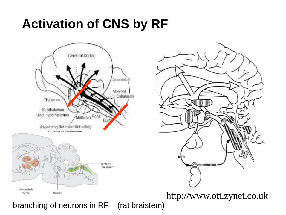
Activation of CNS by RF
http://www.ott.zynet.co.uk branching of neurons in RF (rat braistem)
RF -
Activation role increases intensity of impulses for
cortex and medullar motoneurons and
vice versa
Inhibitory role inhibites activity of spinal cord
motoneurons
cerveau isole, encephale isole
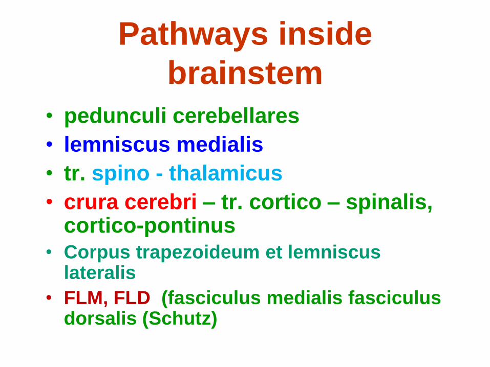
Pathways inside
brainstem
• pedunculi cerebellares
• lemniscus medialis
• tr. spino - thalamicus
• crura cerebri – tr. cortico – spinalis, cortico-pontinus
• Corpus trapezoideum et lemniscus lateralis
• FLM, FLD (fasciculus medialis fasciculus dorsalis (Schutz)
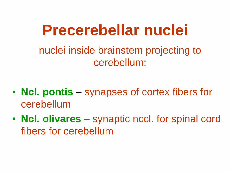
Precerebellar nuclei
nuclei inside brainstem projecting to
cerebellum:
• Ncl. pontis – synapses of cortex fibers for
cerebellum
• Ncl. olivares – synaptic nccl. for spinal cord
fibers for cerebellum

Chemical systems in the CNS
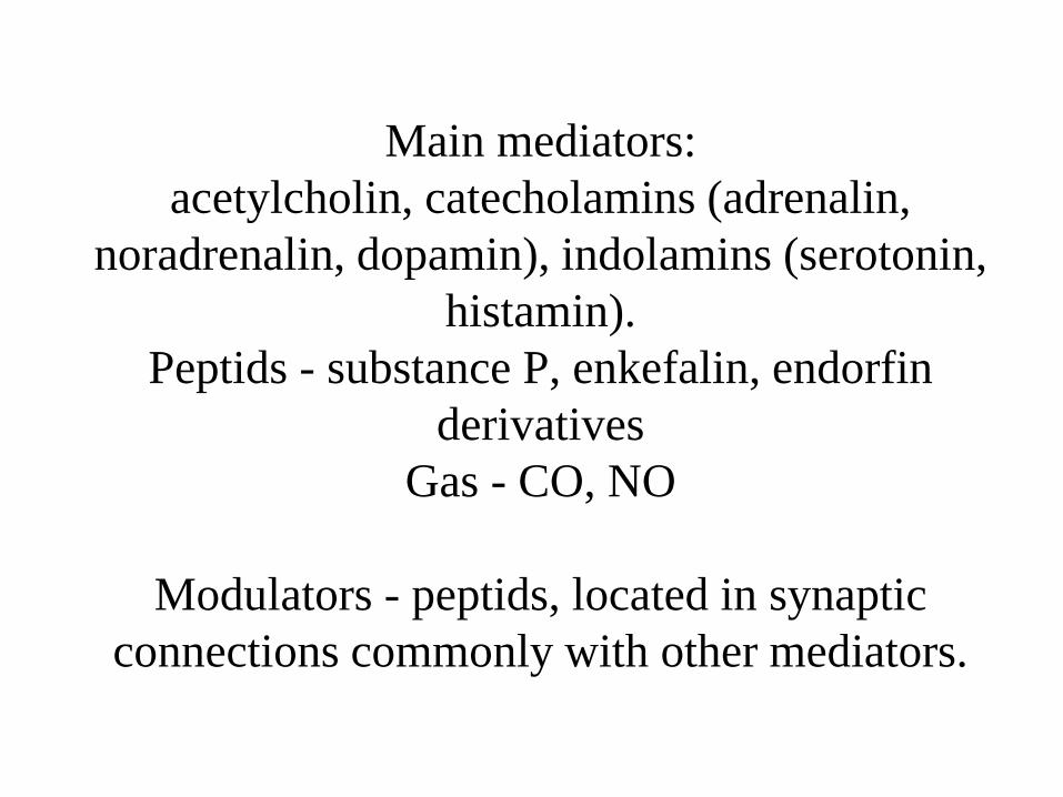
Main mediators:
acetylcholin, catecholamins (adrenalin,
noradrenalin, dopamin), indolamins (serotonin,
histamin).
Peptids - substance P, enkefalin, endorfin
derivatives
Gas - CO, NO
Modulators - peptids, located in synaptic
connections commonly with other mediators.
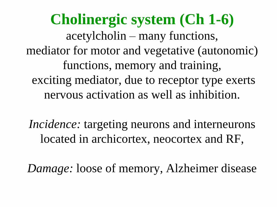
Cholinergic system (Ch 1-6) acetylcholin – many functions,
mediator for motor and vegetative (autonomic)
functions, memory and training,
exciting mediator, due to receptor type exerts
nervous activation as well as inhibition.
Incidence: targeting neurons and interneurons
located in archicortex, neocortex and RF,
Damage: loose of memory, Alzheimer disease
Monoamines catecholamines (adrenalin, noradrenalin, dopamin)
(A 1 – 16)
caudorostral arrangement, predominantly exciting
influence
containing pigment instead of adrenergic cells,
indolamines (serotonin, histamin) (B1 - 9)
predominantly exerting inhibition
Adrenergic system (C1 - 3)
located in RF
Noradrenergic system (A 1 - 7, )
localized in RF (A1-5) and under locus
coeruleus (A 6,7),
throung nervous conections conveys to all
CNS: significantly increases attention - cortical
arousal reaction
impulses for breathing and cardiovascular
systems
Degeneration in locus coeruleus - demention
Dopaminergic system (A 8 - 10)
dopamin is necessary for extrapyramidal motor
effects,
(A9) - subst. nigra compacta – through nigrostriatic
tracts to striatum,
Disturbance in striatum – Parkinson syndrome
(akinesis, tremor, rigor) due to degeneration of
nigrostriatic system.
Damage of the A10 system is followed with
demention.
(A10) - area tegmentalis ventralis – through
telencephalicus medialis tract activating frontal and
limbic core, amygdalar complex and brain septum
Dopamin distribution is basic for reward
mechanism:
gratifying cmfortable feeling following some
body function as “reward“,
some drugs (cannabis derivatives, opium,
cocain, alcohol) support dopamin distribution;
at the some time serotonin distribution is
blocked - this is principle of neurobiological
base for the drug dependence.
Serotoninergic system rapheal nuclei (B1 - 9) distribute its compounds to all
CNS, serotonin there is predominantly inhibiting
factor (due to receptor type)
Great function spectrum:
in medulla: systém inhibits sympathetic neurons and
pain transduction; all motoneurons are activated,
exerts influence temperature regulation, sexual
behavior, digestion, brain blood supply, relax (dream)
regulations, serotonin decreasing is followed with endogenous depressive
reactions
Reflex
irritation – answer cycle in organism; the simpliest event.in
CNS effectors = mostly muscles
Reflexes: uncoditional (congenital) stereotypical, permanent –
(sneezing, coughing, corneal, reflex extenzing limbs, patellar)
Newborn - grasping
Reflexes: conditional (not congenital obtained through life):
Depends on experiences because of selfteaching I.P.Pavlov (dog
bulb and food), develops on uncoditionalreflex if some conditional irritation is added.
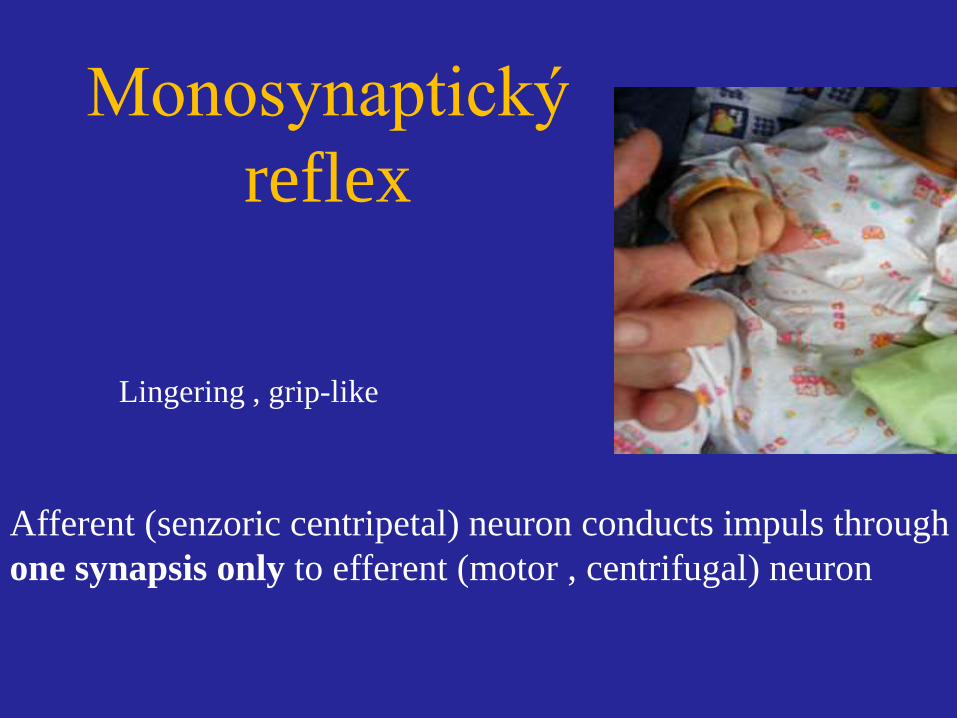
Monosynaptický
reflex
Afferent (senzoric centripetal) neuron conducts impuls through
one synapsis only to efferent (motor , centrifugal) neuron
Lingering , grip-like
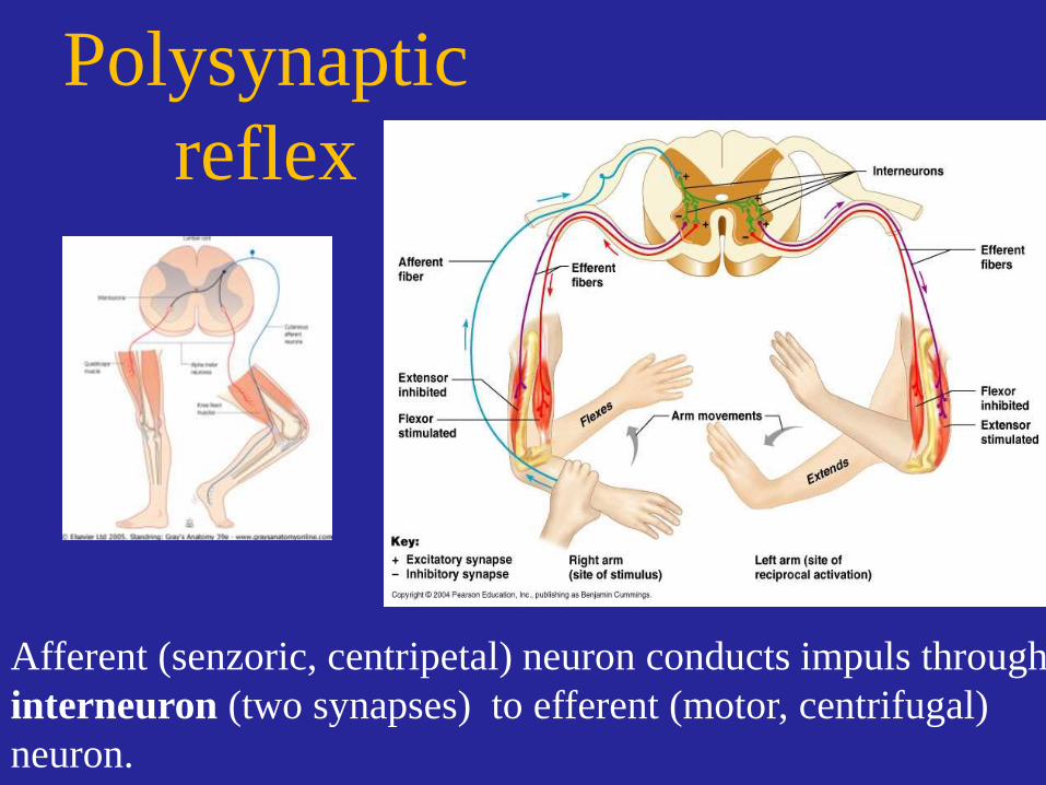
Polysynaptic
reflex
Afferent (senzoric, centripetal) neuron conducts impuls through
interneuron (two synapses) to efferent (motor, centrifugal)
neuron.
Česky: Korneální reflex, Rohovkový
reflex
English: Corneal reflex, Blink reflex
Corneal reflex arch:
Afferent:
Corneal exteroreceptors, afferent n.ophtalmicus (1. branch of n.V.)
efferent:
n.VII. tom. orbicularis oculi.
Cornea is touched - impuls is conducted through nervi cilliares a nervus ophthalmicus (V1.) to brainstem.
Efferentsugnal is conducted through VII . Nerve to m. orbicularis oculi (m. orbicularis oculi)

Česky: mrkací
reflex
English: blink
reflex
Blink reflex je relativně
relatively simple
Afferent part: (free nervous endings in
cornea, n. trigeminus, ganglion, radix and
tractus spinalis n. V.,
Continues to interneurons of RF, next
bilaterally to motor nucleus CN VII and to
m. orbicularis oculi.
Effect: lower eylid contracts
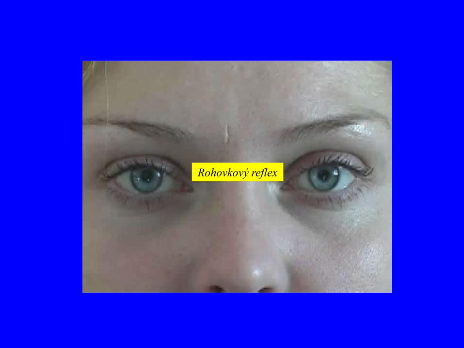
Rohovkový reflex
Česky: Faryngový reflex, Dávivý reflex
English: Pharyngeal reflex, Gag reflex
Irritation (e.g. Cotton of wool) of dorsal wall of pharynx; patient at the
same time pronounces „a" or „e".
Physiologic answer - elevation and constriction ofpharynx; tongue is
retracted.
Vyšetření zvlášť vlevo a vpravo umožní rozlišit lézi aferentní
(oboustranné snížení nebo vymizení odpovědi při dráždění jedné strany)
a eferentní části (jednostranné snížení nebo vymizení odpovědi při
dráždění obou stran) reflexního oblouku.
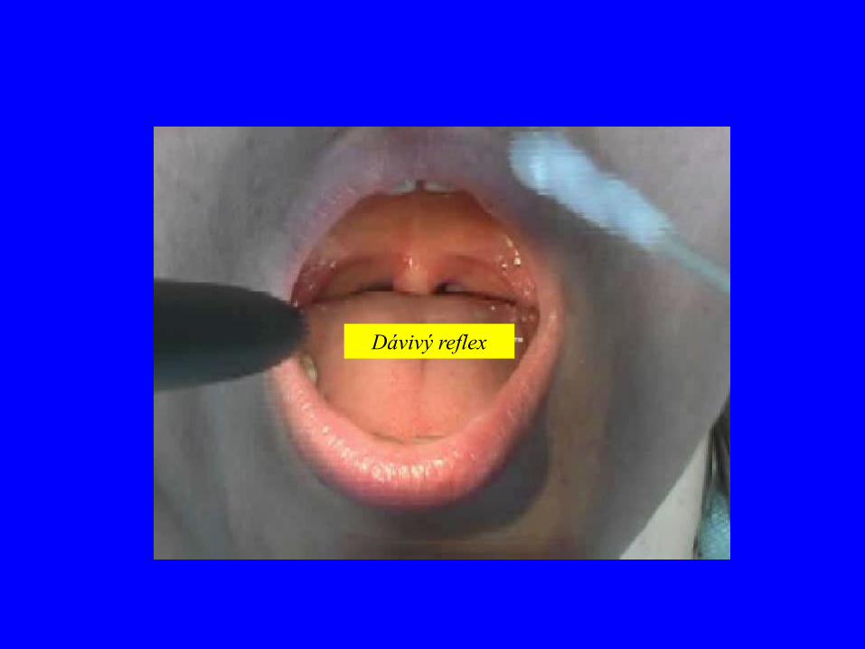
Dávivý reflex
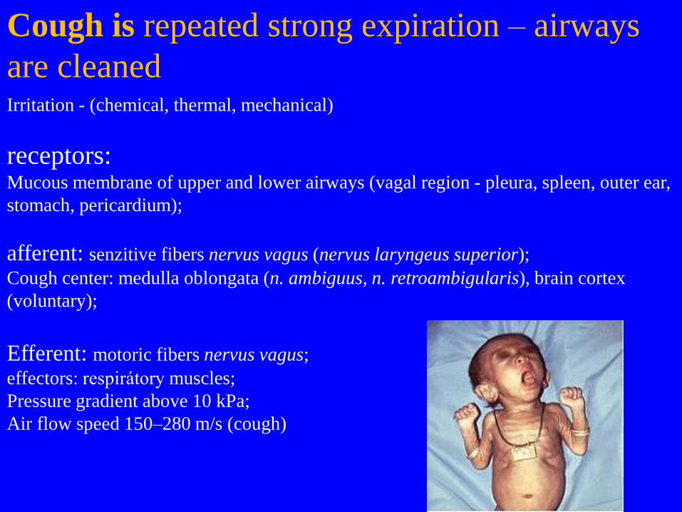
Cough is repeated strong expiration – airways
are cleaned Irritation - (chemical, thermal, mechanical)
receptors: Mucous membrane of upper and lower airways (vagal region - pleura, spleen, outer ear,
stomach, pericardium);
afferent: senzitive fibers nervus vagus (nervus laryngeus superior);
Cough center: medulla oblongata (n. ambiguus, n. retroambigularis), brain cortex
(voluntary);
Efferent: motoric fibers nervus vagus;
effectors: respirátory muscles;
Pressure gradient above 10 kPa;
Air flow speed 150–280 m/s (cough)
Some material used from various authors:
Petrovicky: CNS, PNS (paper texts), lectures
Petrovicky et al. Anatomy with topographic remarks
(Czech), Vol. III.
Druga, Grim et al.: Peripheral nervous system,
lectures
Ten Donkelaar: Clinical neuroanatomy
Moore, Dalley: Clinically oriented anatomy
Agur: Grant´s Atlas of Anatomy,
Grays´s Anatomy 39th ed.
Larsen´s Human embryology
Web sources
Own archive


