Antiproliferative Effects of Hibernating American Bullfrog ...

CHAPTER 9
Brain Dead: The DynamicNeuroendocrinological AdaptationsDuring Hypometabolism inMammalian HibernatorsSamantha M. Logan, Alex J. Watts, and Kenneth B. StoreyInstitute of Biochemistry & Department of Biology, Carleton University, Ottawa, Canada
Introduction
As seasonal temperatures fluctuate, endotherms must produce body heatendogenously when ambient temperatures drop below the level of theirown body temperature (Tb), in order to maintain cellular function across arange of newly established ambient temperatures. However, thermoregulatorymechanisms are fuel-expensive and require ever-increasing levels of metabolicactivity, to combat the parallel increase of heat transfer to the environmentby producing excess heat energy. For these reasons, animals that are exposedto extreme winter conditions must either find enough resources to maintainthermoregulatory mechanisms despite unfavorable conditions or must lowertheir own metabolic rate to survive without the need for massive energy stores.Furthermore, an animal’s ability to forage may be restricted by daily light-darkcycles, predation-risk and food-availability, while heat-production costs andsimilar resource shortages may be imposed on animals suffering throughfood-shortage or drought, even in warm climates. By lowering Tb during timesof rest an animal may preserve energy for times of greater need (e.g. duringincreased activity). Endothermic vertebrates that suppress energy usage andlower body temperature until conditions are more favourable for survival areconsidered to be in a state of “torpor,” and energy expenditure can be further
Model Animals in Neuroendocrinology: From Worm to Mouse to Man, First Edition.Edited by Mike Ludwig and Gil Levkowitz.© 2019 John Wiley & Sons Ltd. Published 2019 by John Wiley & Sons Ltd.Companion Website: www.wiley.com/go/ludwig/modelanimals
207

208 Model Animals in Neuroendocrinology
Figure 9.1 The Richardson’s ground squirrel is a model hibernating animal, capable ofwithstanding harsh environmental conditions during the winter months by entering a stateof reversible suspended animation. Source: J.M. Storey.
preserved by prolonging the time spent in torpor (model hibernator shown inFigure 9.1).
One classification scheme for heterothermic animals (i.e. those that can entertorpid states) relies on the amount of time spent in torpor. Heterothermic ani-mals may undergo daily torpor, in which they rely on heat loss to enter ahypometabolic torpid state for less than 24 hours, and after which they typ-ically continue foraging. Alternatively, for some animals, entering hibernationentails employing inhibitory mechanisms to lower their metabolic rate andremaining torpid for several days, while relying on internal energy stores orfood caches, built up in the preceding months. Animals that hibernate may bedifferentiated by the ‘trigger’ for entering torpor; animals that enter hiberna-tion seasonally, regardless of environmental conditions, are known as obligatehibernators, while those that enter hibernation bouts following uncharacteris-tically extreme seasons are known as facultative hibernators. Due to the lengthof time that hibernators remain hypometabolic and the phenotypic plasticityrequired to enter a prolonged hibernation, these animals have become charac-terized as the more extreme example of metabolic depression. For this reason,the following chapter will pertain mostly to examples of hibernating mammals,except when noted.
During hibernation, torpor bouts (which can last for hours to weeks) areinterrupted by periodic, active euthermic phases, in which basal metabolicrate and Tb are re-adopted for one to three days (see Figure 9.2). Althoughthe reasons for these arousal periods are debated, the process of transitioning

Brain Dead: The Dynamic Neuroendocrinological Adaptations 209
BASAL METABOLISM
• Consistency in heart and respiration rates
• Metabolic needs satisfied by foraging in environment
• Euthermic: constant temperature and metabolism maintained by thermogenesis
• Respiratory, heart rate severely depressed
• Metabolic needs fulfilled solely by stored food or energy
• Heterothermic: fluctuations in temperature and metabolism controlled by ambient temperature changes and intermittent thermogenesis
TORPID METABOLISM
40
30
20
10
00 6 12
Days
Tem
pera
ture
(°C
)
18 24
40
30
20
10
00 6 12
Days
Tem
pera
ture
(°C
)
18 24
Figure 9.2 A representative comparison of body temperature and physiology of hibernatorsduring euthermic (basal) and heterothermic (torpid) periods.
between torpor and arousal periods is characterized by extreme physiologicaland phenotypic changes, perhaps alluding to the necessity of arousals duringhibernation. Within the central nervous system, the hypothalamus appears tocontrol torpor-arousal transitions and seasonal rhythms, which are essentialfor physiological adaptations and the maintenance of energy stores. Thesympathetic nervous system also mediates transitions between torpor andarousal periods, namely through the activation of shivering thermogenesis inskeletal muscle and non-shivering thermogenesis within brown adipose tissue.
The past several decades have seen physiologists, biochemists, cell biologistsand neuroscientists alike come together to uncover the mysteries behind howthese organisms can intricately regulate their metabolic rate, Tb, heart rate andbreathing rate, as well as countless other physiological variables.
9.1 Hypothalamic regulation of hibernation
Studying the hypothalamus of hibernators is paramount for our understand-ing of metabolic suppression, since it is the control center for regulating bodytemperature (Tb) and energy homeostasis and is active over the entire temper-ature range of the torpor-arousal cycle, unlike the cerebral cortex (Schwartzet al., 2013). Hibernators, as opposed to pharmacologically induced hypother-mic and metabolically suppressed mice or rats, provide researchers with anexcellent model for studying natural metabolic rate depression and neuropro-tection without introducing many experimental manipulations that could con-found results.

210 Model Animals in Neuroendocrinology
Table 9.1 Examples of species that use obligate hibernation, facultative hibernation, or dailytorpor to survive changing environmental conditions and if they use hyperphagia for fatstorage or if they utilize food caches to supplement their energy stores throughout torpor.
TorporStrategy
Order TorporEnergyStorage
Example Genera ConfirmedHibernatingSpecies inGenera
Hibernating Species
Fat Allactaga (Jerboa) 3 A. euphratica, A.williamsi
Cynoms (Prarie dog) 3 C. leucurus, C.gunnisoni
Ictidomys,Spermophilus,Urocitellus (Groundsquirrel)
15 I. tridecemlineatus,S. lateralis
Marmota (Marmot) 3 M. marmota, M.monax
Rodentia Zapus, Napaeozapus(Jumping mice)
3 Z. hudsonius, N.insignis
ObligateHibernation
Fat / Food Eliomys,Muscardinus, Glis(Dormice)
3 E. quercinus, G. glis
Food Perognathus (Pocketmice)
2 P. longimembris
Tamias (Chipmunks) 2 T. amoenus, T.striatus
Carnivora Fat Ursus (Bear) 2 U. americanus, U. arctos
Chiroptera Fat Eptesicus, Myotis,Pipistrellus(Vespertilionid bats)
6 N. noctula, M.lucifugus
Eulipotyphia Fat Atelerix, Erinaceus(Hedgehog)
3 A. frontalis, E.europaeus
Monotremata Fat Tachyglossus(Echidna)
1 T. aculeatus
Primates Fat Cheirogaleus (Dwarflemur)
2 C. crossleyi, C.medius
FacultativeHibernation
Rodentia Fat Cynoms (Prarie dog) 1 C. ludovicianus
Food Cricetus,Mesocricetus(Hamster)
3 C. cricetus, M.brandti
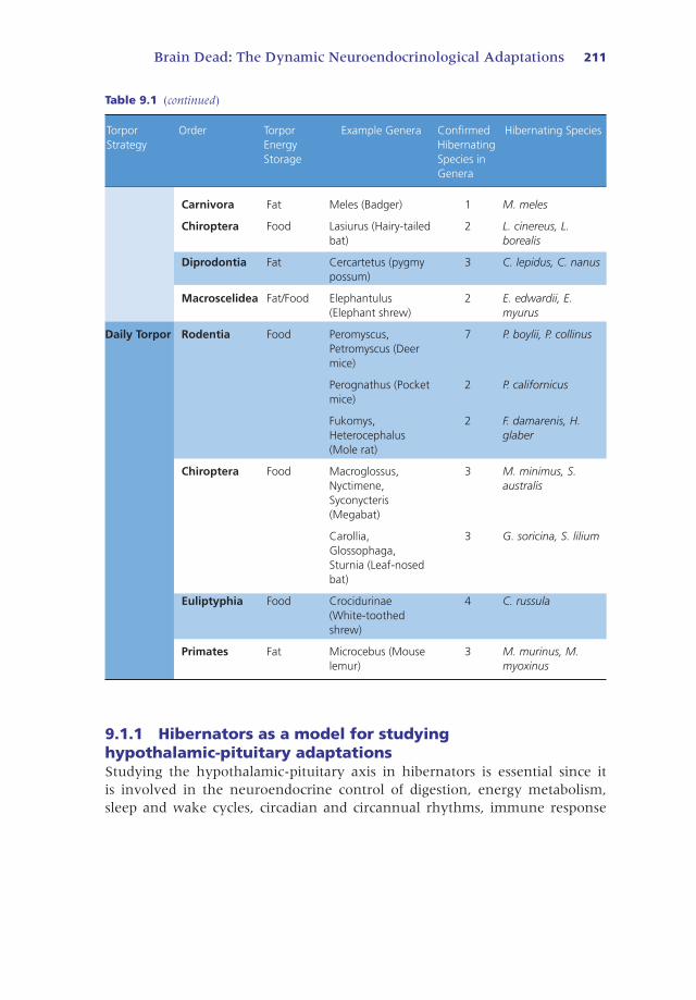
Brain Dead: The Dynamic Neuroendocrinological Adaptations 211
Table 9.1 (continued)
TorporStrategy
Order TorporEnergyStorage
Example Genera ConfirmedHibernatingSpecies inGenera
Hibernating Species
Carnivora Fat Meles (Badger) 1 M. meles
Chiroptera Food Lasiurus (Hairy-tailedbat)
2 L. cinereus, L.borealis
Diprodontia Fat Cercartetus (pygmypossum)
3 C. lepidus, C. nanus
Macroscelidea Fat/Food Elephantulus(Elephant shrew)
2 E. edwardii, E.myurus
Daily Torpor Rodentia Food Peromyscus,Petromyscus (Deermice)
7 P. boylii, P. collinus
Perognathus (Pocketmice)
2 P. californicus
Fukomys,Heterocephalus(Mole rat)
2 F. damarenis, H.glaber
Chiroptera Food Macroglossus,Nyctimene,Syconycteris(Megabat)
3 M. minimus, S.australis
Carollia,Glossophaga,Sturnia (Leaf-nosedbat)
3 G. soricina, S. lilium
Euliptyphia Food Crocidurinae(White-toothedshrew)
4 C. russula
Primates Fat Microcebus (Mouselemur)
3 M. murinus, M.myoxinus
9.1.1 Hibernators as a model for studyinghypothalamic-pituitary adaptationsStudying the hypothalamic-pituitary axis in hibernators is essential since itis involved in the neuroendocrine control of digestion, energy metabolism,sleep and wake cycles, circadian and circannual rhythms, immune response

212 Model Animals in Neuroendocrinology
and neurotransmission. The hypothalamus secretes trophic hormones into thebloodstream that stimulate receptors on the anterior pituitary, or it directlystimulates the posterior pituitary to induce hormone and neurotransmitterproduction and release from the pituitary. These hormones can influencehormone expression in other endocrine tissues, or affect non-endocrine tissuesthroughout the body.
9.1.1.1 The hypothalamic-pituitary-adrenal (HPA) axis coordinates ionhomeostasis, energy metabolism and behavior in hibernatorsSeveral lines of evidence support increased steroid synthesis in the adrenalglands during hibernation. Hibernation increases adrenal weight in severalspecies of ground squirrel (European, Arctic, and Columbian), implicatingincreased adrenal activity, but molecular studies provide more information asto which processes are more active during metabolic suppression. The HPA axisinvolves the production and release of vasopressin and corticotropin-releasinghormone, which stimulate the production/secretion of adrenocorticotropichormone (ACTH) in the pituitary. ACTH helps regulate ion homeostasis,glucose metabolism, reproduction, behavior and the stress response by causingthe adrenal glands to produce adrenaline and noradrenaline, corticosteroids(including cortisol and aldosterone) and androgens. Advances in our under-standing of the roles of these hormones in metabolic suppression will bediscussed.
During hibernation, renal blood flow decreases by 90%, urine output com-pletely stops, and creatinine builds up within the kidneys, yet organ structureand electrolyte balance are maintained, in several small hibernating mammalssuch as Columbian ground squirrels and jerboa (Ratigan and McKay, 2016).Control of plasma ion concentration and blood pressure during hibernationrequires increased responses of the renin-angiotensinogen-aldosterone andsympathetic nervous systems to reduce cardiac output while enhancingrenal vasoconstriction (Ratigan and McKay, 2016). Transmission electronmicroscopy of the zona glomerulus of the adrenal cortex, which secretesaldosterone, a mineralocorticoid that regulates kidney function, shows moremitochondrial cristae and a more prominent smooth endoplasmic reticulumin hibernating edible dormice (Glis glis) compared to euthermic and arousingdormice (Jani et al., 2013). This is consistent with reports of higher reninand aldosterone levels in hibernating organisms, and suggests an importantrole for the HPA axis in regulating ion homeostasis during torpor (Jani et al.,2013). Understanding the molecular mechanisms that stress-tolerant speciesuse to reduce metabolism in kidneys could be key to prolonging the viabilityof organs ready for transplant in humans.
The HPA axis also regulates androgen levels to control behavior in seasonalhibernators. Western blot analysis shows that androgen receptors decrease dur-ing hibernation (Boonstra et al., 2014). Summer-active Arctic ground squir-rels have more androgen receptor than Columbian ground squirrels, which

Brain Dead: The Dynamic Neuroendocrinological Adaptations 213
was postulated to be a species-specific protective mechanism in which Arcticground squirrels build muscle for catabolism during hibernation so that cellsare not starved. However, evidence from in-vitro tissue performance assays,muscle morphology analysis, 2D gel electrophoresis and Western blot analy-sis suggest that 13-lined ground squirrels prevent muscle degeneration duringhibernation (Tessier and Storey, 2016). Instead, pre-hibernation accumulationof muscle mass, which is possibly regulated by seasonal variation in androgenreceptor expression, is likely to be important for shivering thermogenesis duringinterbout arousals in Arctic ground squirrels (and less so in Columbian groundsquirrels), since they are the only species of ground squirrel that can supercooltheir body temperature to −2.9∘C. More research needs to be done to untan-gle the relationship between seasonally expressed hormones and hibernationas well as their roles in muscle accumulation and energy homeostasis.
9.1.1.2 The hypothalamic-pituitary-thyroid (HPT) axis may have rolesin depressing metabolism during hibernation and increasing bodytemperature upon arousalWhen low levels of triiodothyronine (T3) and thyroxine (T4) are sensed in theblood, the hypothalamus secretes thyrotropin-releasing hormone (TRH) lead-ing to the production and secretion of thyrotropin-stimulating hormone (TSH)by the pituitary. The thyroid glands then produce T3 and T4 which down-regulate processes during hibernation such as energy metabolism, heart andbreathing rate, body temperature and sleep.
Total T3 and T4 levels in the plasma are regulated by different life stages(circannual rhythm) in hibernators. Specifically, T3 levels are highest duringlactation and lowest during pre-hibernation hyperphagia (the period of intenseeating before hibernation) in female Arctic ground squirrels (Wilsterman et al.,2015). High T3 levels are used as an indicator of elevated basal metabolic rate(metabolic rate when animals are resting but not asleep) in both laboratoryand free-living animals (Wilsterman et al., 2015). Therefore, the notion thatT3 levels would be lowest during the pre-hibernation period is consistent withthe hibernator’s need to conserve metabolic fuel for the hibernation period.An activated HPT axis leads to increased energy expenditure, which is pos-sibly due to thyroid hormone acting as a transcription factor or its ability toincrease glycogen metabolism in the liver. Increased HPT activity is observed inthe hypothalamus of hibernators that are injected with TRH, which is supportedby increased brown adipose tissue (BAT) and rectal temperature and increasesthe turnover rate of norepinephrine, despite unchanging T3 levels (Shintaniet al., 2005). Cold-exposed Daurian ground squirrel BAT shows any increase inuncoupling protein 1 (UCP1) expression, which is likely involved in the mech-anism of increasing BAT temperature. Furthermore deiodinases, which convertT4 to T3 before they are released from the thyroid and in peripheral tissues,are more active in several cold-exposed hibernators, including Daurian and

214 Model Animals in Neuroendocrinology
Richardson’s ground squirrels and chipmunks, suggesting that BAT T3 avail-ability is regulated by temperature cues (Liu et al., 2001). Thyroid hormonesmight increase metabolism and thermogenesis by increasing the relative levelsof mitochondria although the impact of the thyroid on brown adipose tissue isstill incompletely explored (Wilsterman et al., 2015).
9.1.1.3 The hypothalamic-pituitary-gonadal (HPG) axis influencestorpor-arousal cycle length and seasonal reproductionIn response to the production and release of gonadotrophin-releasing hormonefrom the hypothalamus, the pituitary produces luteinizing hormone (LH) andfollicle-stimulating hormone (FSH), which influence the production of estro-gen and testosterone in the gonads. This signaling cascade is referred to as thehypothalamic-pituitary-gonadal (HPG) axis. Hormones associated with repro-ductive cycles in mammals are most likely to show circannual rhythms andhave served as the starting point for research into the timing involved in annualhibernation cycles.
A study measuring endogenous testosterone and dehydroepiandrosterone(DHEA, a hormone involved in regulating non-mating aggression) in Arcticground squirrels showed that testosterone levels are highest during the mat-ing season in the spring, and DHEA levels are highest in the late summer.Castrated ground squirrels can initiate hibernation around the same time ascontrol squirrels but arouse from heterothermy nearly one month later thannon-castrated squirrels (Richter et al., 2016). Evidence also shows that adminis-tration of testosterone following entry into torpor prevents cold-housed animalsfrom entering deep hibernation (Lee et al., 1990). Thus, testosterone appearsto have an important role in the inhibition of hibernation.
Most reproductive hormones such as prolactin and testosterone increase inabundance following arousal from torpor. An exception is that serum levelsof FSH, which are high during hibernation, seem to be regulated in a seasonalfashion. Turkish hamsters exposed to a short photoperiod have inhibited HPGsignaling, testicular regression, and decreased testosterone secretion, changesthat are absent in animals exposed to photoperiods that simulate summer.Serum levels of FSH increase about 40 days prior to the end of hibernation,or about 100 days after exposure to short photoperiods, both in hamsters thatremained euthermic in short-day cold conditions and in castrated hamstersthat lack gonadal signaling to the hypothalamus (Jarjisian and Zucker, 2011).A similar study conducted on golden-mantled ground squirrels also showsincreases in FSH levels within three days prior to arousal. Given that gonadalfeedback to the pituitary is influential within the HPG-axis, these resultssuggest that seasonal stimuli influence hypothalamic signaling to the pituitarygland and ultimately, secretion of reproductive endocrine factors. It is thereforeinteresting that the rhythmicity of seasonal FSH secretion seems to influencethe timing of arousal from hibernation and may serve as a seasonal signalingfactor within the context of hibernation, possibly via secretion of melatonin

Brain Dead: The Dynamic Neuroendocrinological Adaptations 215
from the pineal gland. However, there may be species-specific differences inHPG regulation. FSH-secreting cells within the hibernating little brown batbrain are less active during hibernation, evidenced by less developed roughendoplasmic reticulum and Golgi-apparati, as well as smaller numbers of secre-tory vesicles and melatonin receptors (Azzali et al., 2003). Overall, changesin FSH and testosterone levels may provide neuroendocrine control overhibernation timing but differences between hibernator species indicate there isstill much more research needed in the area of hibernator HPG signaling.
9.1.2 Hypothalamic-pineal regulation during hibernationThe control of many cyclical behaviors in mammals resides in the suprachi-asmatic nucleus (SCN), a region within the hypothalamus that can integrateenvironmental photoperiod cues from the retina with cycles in sleep and foodintake behavior. In turn, output pathways from the SCN influence endocrine(i.e. melatonin and arginine vasopressin) and neuronal signals that synchronizeperipheral oscillators located in most visceral organs and in several locationswithin the brain, serving to maintain metabolism, appetite and activity in acircadian (i.e. daily) fashion. On the other hand, circannual rhythms corre-late with seasonal changes in day length and ambient temperature and alterthe release of endocrine factors that entrain (or synchronize) the SCN and theperiphery to these changes. Predictably, circannual rhythm governs the onsetand cessation of torpor-arousal cycles in mammalian hibernators, even thosethat are kept in constant environmental conditions while in captivity (Williamset al., 2014).
9.1.2.1 Melatonin may control torpor-bout length and reduce braindamage during hibernationThrough the SCN, day-length signals are encoded as endocrine signals in theform of melatonin synthesized by the pineal gland and subsequently releasedinto the blood, most abundantly at night, during minimal photic input to theSCN. Melatonin synthesis appears to cease during hibernation in European andTurkish hamsters, ground squirrels and marmots, but is known to increase tor-por bout length in ground squirrels when injected intracerebroventricularly (Yuet al., 2002). Hibernators that have undergone surgical ablation of the SCN areexcellent models in which to study the effects of photoperiod input to the pinealgland during torpor-arousal cycles, although reports of their use for this purposehave so far been limited.
During arousal from hibernation melatonin levels peak. Melatonin injectionforces arousal from hibernation, suggesting that it is a key player in mediatingtorpor bout length. Melatonin may also be involved in the neuroprotectiveresponse to increasing levels of reactive-oxygen species, which are producedas the animal warms up and breathes more rapidly (Schwartz et al., 2015).Evidence for this includes more pro-apoptotic caspase-3 expression and

216 Model Animals in Neuroendocrinology
less mitochondrial respiration when ground squirrels are injected with amelatonin receptor antagonist during the mid-arousal phase. Furthermore,melatonin receptor Mel1a is elevated in hibernating ground squirrel brain,heart and brown adipose tissue, as is the activity of the rate-limiting enzyme formelatonin synthesis, arylalkylamine-N-acetyltransferase (AA-NAT) (Schwartzet al., 2015; Yu et al., 2002). Thus, it is possible that Mel1a and AA-NAT aresynthesized before arousal such that melatonin signaling is enhanced duringthe arousal phase. These studies suggest that melatonin may be important forneuroprotection upon arousal, possibly by ensuring optimal energy acquisitionor by promoting antioxidant and anti-apoptotic signaling (Schwartz et al.,2015), but more research needs to be done to confirm this.
9.1.2.2 Low temperatures may directly regulate hypothalamopinealsignaling in hibernatorsHypothalamic-pineal neuroendocrine signaling in hibernators may not be reg-ulated by circadian rhythm, but instead by decreased Tb. A recent study suggeststhat temperature may have an important role in regulating circadian rhythmsin Siberian hamsters (Phodopus sungorus), animals that can undergo daily torpor.Over a 48-hour period, researchers found rhythmic expression of Per1, Bmal1and Avp proteins in the SCN and AA-NAT in the pineal gland of both euther-mic and hypothermic hamsters, but protein expression was notably attenu-ated in hypothermic animals (Herwig et al., 2007). European hamsters exposedto a shorter photoperiod show a greater daily melatonin amplitude (highernight-time levels and lower day-time levels) as compared to hamsters exposedto a longer photoperiod, and like Siberian hamsters, this effect was exacerbatedin animals exposed to cold (Revel et al., 2007). European hamsters also consis-tently express clock-related genes (Per1, Per2, Bmal1) in the SCN and AA-NATin the pineal gland throughout torpor when Tb is suppressed, suggesting thatcircadian rhythm is lost in these animals (Revel et al., 2007). Whether this istrue for obligate hibernators such as the brown bear (Ursos arctos), which showelevated melatonin levels at night during both the summer and the winter, isstill undetermined. Day-time levels of melatonin during hibernation are higherthan summer levels, suggesting that the baseline level of melatonin increaseswith hibernation and this elevated baseline may mask any daily changes inmelatonin during the hibernation period, as opposed to these animals not hav-ing any circadian regulation of pineal signaling (Ware et al., 2013).
9.1.2.3 The signalling of seasonal changes is essential to hibernatorsObligate hibernators exhibit torpor-arousal cycles in the absence of changingtemperatures and photoperiod in their underground dens, which suggests thathibernation is regulated by an endogenous ‘clock’ that is not under the directcontrol of the circadian system or its inputs. Whether this seasonal timekeepingmechanism is the result of changes in environmental conditions leading upto winter or an endogenous circannual rhythm is currently unknown, and
Brain Dead: The Dynamic Neuroendocrinological Adaptations 217
differences in hibernation strategy (e.g. obligate vs facultative hibernators,hibernacula suitability) are likely to have evolved unique methods of time-keeping specific to the needs of the animal. Three lines of evidence supportthis conclusion.
First, European ground squirrels exhibit arrhythmic Tb upon arousal fromhibernation, which means that their Tb does not change with fluctuations inambient temperature. Importantly, Tb gradually re-entrains to daily Tb rhythms,suggesting muted circadian functioning even when animals are held in con-stant darkness and allowed to entrain to free-running cycles (Malan, 2010). Tb
arrhythmia either during or immediately following hibernation also occurs inhamsters and little brown bats. Developments in technology, including moresensitive Tb tracking devices and hibernation chambers that lower interferencefrom daily ambient temperature fluctuations, have allowed measurement of Tb
rhythms during torpor (diminished to <0.1∘C). As such, circadian Tb rhythmshave been shown to exist in some food-gathering species during arousals fromtorpor, or in captive animals measured at relatively higher ambient tempera-tures (∼12 ∘C) (Malan, 2010).
Second, the SCN and its underlying circadian rhythms do not modulate thetiming of hibernation bouts, as was shown by mutant hibernators displayingunaltered torpor rhythms. In an interesting study, three genotypes of Syrianhamsters with mutant endogenous circadian periods were developed in orderto determine the influence of circadian rhythms on the timing of torpor-arousalcycles. Hibernation cycles of mutant and wild-type hamsters hibernating in con-stant darkness are not different in torpor bout length or the timing of entry intoor emergence from torpor. Expecting that hamsters with shorter circadian peri-ods would have shorter torpor bouts, the authors concluded that regulationof the temporal organization of hibernation must not involve the endogenousperiods encoded within the circadian system (Malan, 2010).
Last, complete or partial ablation of the SCN was expected to disrupt thetiming of torpor entry and arousal in golden-mantled ground squirrels, andexperiments do show altered torpor bout duration, euthermic interval and tor-por re-entry rate. Further investigation of animals with only partial SCN lesionsshow that although these animals display alterations in hibernation rhythms,circadian Tb rhythms are in fact maintained. It would therefore appear thatchanges resulting from SCN ablation are the result of a loss of SCN function,suggesting possible non-circadian roles for the SCN in hibernators (Ruby, 2003).
These studies suggested that the time spent in torpor is not affected bycircadian rhythmicity, however daily Tb rhythms immediately following springarousal have been noted in some species, raising obvious questions about thecontinuation and possible roles of muted circadian rhythms during hiberna-tion. Furthermore, alternative methods of entrainment must be required forhibernating animals within their dens, due to their isolation from signals ofphotoperiod (daily light/dark rhythms). Given these facts, it seems possiblethat alternative endogenous clocks (i.e. circannual) have an influential role

218 Model Animals in Neuroendocrinology
in rhythmicity during torpor-arousal bouts (Malan, 2010; Ruby, 2003), incooperation with factors signaling metabolite buildup and extreme changes inambient temperature or surroundings that are indicative of seasonal changesin amount of daylight.
9.1.2.4 Circannual regulation of energy metabolism in seasonalhibernatorsAppetite and nutrient sensing can also be impacted by temperature, photope-riod and circannual rhythm in mammalian hibernators. Time of the year affectsthe arousal, appetite and food-seeking behavior of different species and sexes ofhibernator. Hamsters hibernate due to changes in photoperiod and arouse whenthey are hungry, to feed from food caches between torpor bouts. Male arcticground squirrels also have food caches, but do not eat until after spring arousal,possibly to manufacture sperm cells before the reproductive season while stillprotected in their dens (Florant and Healy, 2012). By contrast, female groundsquirrels and both male and female marmots arouse later in the season, whenfood is more readily available. Furthermore, Sciurid animals (ground squirrels,prairie dogs and marmots) given food at spring arousal in laboratory settingswill not eat, suggesting that they use an endogenous mechanism to regulateappetite following arousal (Florant and Healy, 2012).
9.1.3 Hypothalamic regulation of appetite and nutrientsensing in hibernatorsHibernators are excellent models in which to study controlled overeating andinsulin resistance when sampled during pre-hibernation, and also nutrientdeficiency, since they can survive up to 7 months of starvation during hiber-nation (Wu et al., 2013). Lesion studies suggest the hibernator hypothalamusis responsible for changes in food intake and body mass. Ventral medialhypothalamus (VMH) lesions increase feeding and obesity in ground squirrelsat all times of the year and lateral hypothalamus (LH) lesions decrease feeding,suggesting that these brain regions control satiety and hyperphagia, respec-tively. Close to the VMH and the LH, the arcuate nucleus (ARC) may be themost important area in regulating feeding response through nutrient sensing,since it is permeable to the bloodstream and allows the uptake of nutrientsand hormones from the blood. The ARC expresses neuropeptide Y (NPY) andagouti-related protein (AgRP) which stimulate feeding and energy storage,which is unsurprising since most neurons in the VMH and ARC are affectedby changes in fatty acid and glucose levels (Florant and Healy, 2012). At themolecular level, nutrients (glucose and free fatty acids), enzymes (AMPK andACC) and hormones (ghrelin, insulin, and leptin) have important roles incontrolling energy storage and usage in hibernators throughout the year.

Brain Dead: The Dynamic Neuroendocrinological Adaptations 219
9.1.3.1 Free fatty acids (FFAs) could suppress appetite at low Tb, butnoradrenaline does not increase lipolysis during hibernationHibernators endure prolonged periods of fasting with little or no food sup-ply, however, what controls appetite and lipolysis during hibernation is stillunknown. Entry into torpor is accompanied by an increase in serum FFAs,but to date no research has shown how FFA levels change within the hiber-nator brain (Florant and Healy, 2012). Circulating FFAs could regulate energymetabolism and food intake by triggering nutrient sensing in the hypothalamus,as other nutrients do. FFAs, often bound by fatty-acid binding proteins (FABPs),bind to insulin receptors in adipocytes and may regulate food intake this way(Florant and Geary, 1991). Northern blot analysis shows higher FABP levels inthe brown adipose tissue and skeletal muscle of hibernating little brown bats,compared to euthermic bats (Eddy and Storey, 2004). Furthermore, hiberna-tor FABPs have amino acid substitutions that increase their binding capacityat low temperatures (near 0∘C), especially compared to non-hibernators (Eddyand Storey, 2004). Increases in FABP protein levels and binding capacities dur-ing hibernation could suggest a role for FFAs in regulating energy metabolismduring hibernation.
Pharmacological studies suggest interesting theories about how hibernatorsregulate energy metabolism at extremely low body temperature. Followinginjection of noradrenaline in euthermic and hibernating ground squirrels,measurements of plasma glycerol levels, used to estimate relative changes inlipolysis of white and brown adipose tissue, show that lipolysis increases onlyin euthermic, noradrenaline-treated animals (Dark et al., 2003). Thus, lowtemperatures may inhibit noradrenaline-mediated lipolysis and hibernatorsuse alternative mechanisms to enhance fatty acid oxidation.
9.1.3.2 Ghrelin, AMP-activated protein kinase (AMPK) and acetyl-coAcarboxylase (ACC)Ghrelin is a hormone that binds to ghrelin receptors in the pituitary, activatesAMPK pathways to induce feeding behavior, and increases NPY and AgRPexpression in hypothalamic neurons to stimulate fatty acid synthesis (Healyet al., 2011). This is especially important in the pre-hibernation fatteningperiod. During hibernation, plasma ghrelin levels are much lower than sum-mer levels in grizzly bears and golden-mantled ground squirrels. Reduced butstill measurable ghrelin levels may inhibit the urge to arouse and feed or mayfacilitate non-rapid eye movement sleep, although the latter role for ghrelin isstill hotly debated (Healy et al., 2011). Major differences in ghrelin and otherhormone or enzyme levels in the brain between euthermia and hibernationare summarized in Figure 9.3.
Regardless of the season, ghrelin injections into golden-mantled groundsquirrels increase food intake and phosphorylation of AMP-activated pro-tein kinase (phospho-AMPK, the active form) compared to saline-injectedcontrols (Healy et al., 2011). AMPK is an energy-sensing enzyme that can

220 Model Animals in Neuroendocrinology
GhrelinGhrelin
POMC
NPY, AgRP NPY, AgRP
Brain stem Brain stem
Short term p-AMPKp-AMPK
Leptin Leptin
Increases food intake,energy storage
Increasesketogenesis?Food intake?
Decreases feeding
GHSR GHSRPituitary Pituitary
LH LH
VMH VMH
ARC ARC
SCN SCN
PVN PVN
(a) Euthermia, pre-hibernation (b) Hibernation
OC OC
Figure 9.3 Key molecular changes that occur between pre-hibernation euthermia andhibernation that could facilitate nutrient-sensing and regulation of appetite in hibernators.(a) During euthermia, ghrelin levels are much higher than during hibernation and thisinfluences the expression of neuropeptide Y (NPY) and agouti-related protein (AgRP), aswell as phosphorylated AMP-activated protein kinase (AMPK) levels to increase food intakeand energy storage during hyperphagia. High leptin levels during euthermia suppress NPY,AgRP, and AMPK activity following hyperphagia to suppress appetite. (b) Duringhibernation, low ghrelin levels and high proopiomelanocortin (POMC) are associated withlow NPY and AgRP levels possibly to suppress appetite, but phospho-AMPK levels remainelevated, perhaps to increase fatty acid metabolism.
simultaneously increase ATP production while inhibiting energy-expensivepathways, likely by inhibiting the mechanistic target of rapamycin (mTOR),which is essential for metabolic suppression (Healy et al., 2011; Zhang et al.,2015). Yellow-bellied marmots infused with an AMPK agonist also increasefood intake, emphasizing the importance of this enzyme in regulating appetite(Florant et al., 2010). In naturally hibernating ground squirrels, immunoblot-ting and enzyme activity assays show that the activity and phosphorylation ofAMPK do not increase in brain, skeletal muscle, brown adipose tissue or liver,but increase in white adipose tissue relative to euthermic controls (Hormanet al., 2005). ELISAs show AMPK to be activated in gray mouse lemur heartduring daily torpor, compared to aroused animals but not in five other lemurtissues. Interestingly, acetyl-CoA carboxylase (ACC) phosphorylation (whichoccurs due to elevated phospho-AMPK and inhibits of fatty acid synthesis)does not increase in winter in ghrelin-injected animals, suggesting that ACCis already maximally phosphorylated during winter, to facilitate fatty acidoxidation. Together these studies suggest that AMPK may be important in foodintake and nutrient sensing, thus limiting energy expenditure, but only incertain hibernator tissues (Zhang et al., 2015). AMPK activity is also regulatedby insulin, leptin, glucose, FFAs and AMP/ATP ratio, which fluctuate withdifferent times of the torpor-arousal cycle and time of year. To determinehow ghrelin and AMPK influence energy metabolism in models of naturalhibernation, relative total protein, phosphorylation and activity levels willneed to be assessed in animals at various points of the torpor-arousal cycle and

Brain Dead: The Dynamic Neuroendocrinological Adaptations 221
using tissues from specific brain regions. Although unexplored in hibernators,it is possible that ghrelin regulates energy metabolism through the orexinpathway, since this peptide also plays roles in appetite, thermoregulation andregulation of the sleep-wake cycle.
9.1.3.3 Leptin, NPY and AgRPThe level of serum leptin, a hormone produced by white adipose tissue,is predictive of fat mass. To suppress appetite, leptin binds to leptin recep-tors in the hypothalamus, increasing proopiomelanocortin (POMC) andalpha-melanocyte stimulating hormone expression, which reduces NPY andAgRP expression in the hibernator brain (Florant and Healy, 2012). Leptin andinsulin levels are elevated in serum during the pre-hibernation period, promot-ing hyperphagia, and decrease during torpor, when adiponectin levels increase,suppressing appetite and mobilizing fat stores (Florant and Healy, 2012).
Neuropeptides NPY and AgRP are produced by the same neurons in theARC. Both stimulate appetite and are inhibited by leptin. NPY has beenshown to reduce the anxiety associated with food foraging, resulting in morefood acquisition and AgRP influences food preference. A study using qPCRshows that in greater mouse-tailed bats hypothalamic NPY mRNA levels, butnot AgRP mRNA levels, increase during the pre-hibernation period, whenit is essential to increase fat intake (Levin et al., 2013). Transcriptomes of13-lined ground squirrels reveal low NPY and AgRP mRNA levels in thepre-hibernation period after hyperphagia has ceased, and high NPY and AgRPlevels during post-hibernation hyperphagia. By contrast, genes involved insuppressing appetite, such as those for cocaine- and amphetamine-regulatedtranscript prepropeptide (CARTPT) and thyrotropin-releasing hormone (TRH),are expressed at high levels during hibernation (Schwartz et al., 2013).Although the identities of appetite-inducing and suppressing genes are stillbeing resolved, what stimulates their expression and their mechanisms ofaction during hibernation have been little studied.
9.1.3.4 Adenosine as an energy-signaling molecule with rolesin thermogenesisAdenosine, an important neuromodulator in the hibernating brain, is pro-duced within neurons when ATP reserves are low. It is postulated that thebalance between intracellular and extracellular pools of adenosine controlskey metabolic and neuromodulatory processes such as food intake, sleep andTb. Specifically, extracellular adenosine may regulate torpor by binding toreceptors that inhibit arousal from torpor and shivering and non-shiveringthermogenesis. Extracellular accumulation could be due to inhibited adenosinekinase in astrocytes, which would prevent adenosine from being convertedinto AMP and would promote its passage across the plasma membrane, down aconcentration gradient. In humans, brain injury that increases seizure activityis associated with low extracellular adenosine levels, probably increasing

222 Model Animals in Neuroendocrinology
adenosine kinase activity, which decreases intracellular adenosine levels.Alternatively, tanycytes (nutrient-sensing glial cells) may release ATP intothe extracellular space to be converted into adenosine (Drew et al., 2016).Thirteen-lined ground squirrel tanycytes are more activated during torporand early arousal, so this is a possible mechanism of adenosine build-up inthe extracellular space, however, more research is required to determine thetriggering stimuli for this event.
Neural accumulation of adenosine can occur following sleep depriva-tion. Electroencephalographic experiments confirm that hibernators aresleep-deprived and may arouse periodically to get restful sleep. Adenosinerepresses arousal by inhibiting cell groups involved in arousal and disin-hibiting pro-sleep cell groups through adenosine receptor (AR) stimulation.Adenosine can bind to ARs from four subgroups, including A1AR and A2AAR,which are responsible for dopamine and glutamate release, and A2BAR andA3AR, which regulate immune response. A1AR stimulation, which inhibitsshivering and non-shivering thermogenesis, seems to be necessary for entryinto torpor. Hibernating hamsters are aroused during entrance (but not fromlate torpor) by A1AR antagonists such as 8-cyclopentyl theophylline, whileA1AR agonists such as 6N cyclohexyladenosine can induce torpor, but drugsaffecting A2AR or A3AR do not alter wakefulness. Similar results are seen inobligate hibernators if A1AR is stimulated in the winter season (Drew et al.,2016). Together, these results suggest that seasonal gene expression of A1ARor temperature sensitivity to adenosine binding may be key regulatory factorsdriving torpor/arousal.
9.2 Hibernating species as a model for preventingneural damage
In response to an approximately 90% decrease in blood supply to the brainduring torpor, hibernators decrease metabolism to match demand with supply,so as to preserve neural structure and function and retain pertinent memory.Even euthermic hibernators are more resistant to brain insults compared tonon-hibernators, making studying these stress-resistant model organisms a keyto better understanding of natural neuroprotection. Region-specific changes inbrain size, synaptic regression and gene expression have been studied in severalhibernating species. Seasonal differences in brain size have been documentedfor birds and mammals, in which summer brains are heavier than winterbrains, showing seasonal brain plasticity (Arendt and Bullmann, 2013). Spatiallearning involves synaptic signaling followed by second-messenger activation,transcription-factor translocation, gene expression and changes in neuronstructure. During artificial hypothermia, comparison of hibernation-capableand non-hibernating animals attempting spatial (maze) conditioning (feedingmachine or foot shock) or recognition (social or habitat) tasks, showed that

Brain Dead: The Dynamic Neuroendocrinological Adaptations 223
hibernation either had no effect or improved memory retention, compared tonon-hibernating species, which were either unaffected or showed decreasedmemory retention. However, species-specific differences in memory reten-tion following hibernation have been documented. Hibernation impairs theEuropean ground squirrel’s ability to perform spatial and operant tasks, whilegreater mouse-eared bats (Myotis myotis) could perform spatial tasks withoutproblems following torpor (Arendt and Bullmann, 2013). These results, aswell as the molecular data to be introduced, suggest that hibernating speciesuse synaptic regression in all brain regions to preserve brain structure, butperhaps the reestablishment of these neural connections is accelerated in theregions of the brain necessary for survival immediately following arousal. Forinstance, the cerebral cortex, which is the first region to lose brain activityand the last to regain activity, upregulates the expression of genes involvedin remodeling and plasticity, whereas in the hypothalamus, which remainsactive throughout hibernation, genes involved in damage response and proteinturnover, feeding and satiety, seasonal timing and fuel utilization pathways areexpressed (Schwartz et al., 2013).
9.2.1 The protective effects of neural regressionHibernators must protect their brains from ischemia-reperfusion damage,cold-stress and hypoxia, among other torpor-related stressors. One way bywhich their brains adapt to metabolic suppression is changing the structureand biochemical composition of their neurons. Research has shown synapticregression in animals that undergo torpor. Dendrites and axons shrink uponcold stress, decreasing the number and size of neuron branches, until arousal,when synaptic connections are re-established with no detectable loss in cogni-tive ability. Although this phenomenon is seen in the cortex and the thalamusof hibernating ground squirrels, the best evidence for loss of synaptic contactduring torpor comes from studying the hippocampal mossy fiber terminalsfrom the CA3 region. Tau phosphorylation increases in these neurons, sug-gesting a role for tau-microtubule interactions in regulating neuronal structure(Arendt et al., 2015). Transcriptomics approaches have shown more geneexpression associated with cytoskeletal rearrangement in the cerebral cortexof hibernating mammals compared to the constitutively active hypothalamicregion. Proteomics applied to the forebrain (encompassing the cerebellum,the thalamus, and the hypothalamus) show elevated levels of microtubuleand tubulin-interacting proteins in torpid (LT) and early arousal (EA) groundsquirrels. Neurofilament heavy chain (NEFH), stathmin 1 (STMN1) anddihydropyrimidinase-related protein 2 (DPYSL2) are upregulated duringLT and EA but are likely deactivated during this time by post-translationalmodifications like phosphorylation until arousal, when synaptic regenerationby microtubule assembly is essential. Importantly, reversible phosphoryla-tion regulates flux through metabolic pathways, and can influence neuron

224 Model Animals in Neuroendocrinology
structure via changes in cytoskeletal networks. Various isoforms of tubulin(TUBA1C, TUBB2A, TUBB4B) are winter-depressed (Hindle and Martin,2013). It is possible that these proteins are insoluble, similar to aggregates ofhyperphosphorylated tau or proteins such as sorcin that change conformationand increase membrane-association at low Tb, and are not detected in thesoluble protein extracts used for proteomics studies. Based on their lowtubulin levels and elevated levels of stathmin, which can bind tubulin dimersto remove them from the pool of soluble proteins, hibernators may formreservoirs of structural proteins that can be rapidly mobilized upon arousal toensure synapse reformation and neuroprotection, although this mechanism ofneuron plasticity during hibernation needs to be further explored (Hindle andMartin, 2013).
9.2.2 Tau phosphorylation prevents neural damage duringhibernationTau interacts with microtubules within axons to regulate the structural stabil-ity of neurons and neuronal plasticity. Hyper-phosphorylation of tau decreasesits association with microtubules, which increases the formation of neurofib-rillary tangles and neuronal cell death in many models of ’taupathies’, includ-ing patients with Alzheimer’s or frontotemporal dementia with Parkinsonismlinked to chromosome 17 (FTDP-17) (Stoothoff and Johnson, 2005). Notably,phosphorylation of tau at certain residues can decrease paired-helical forma-tion and tau aggregation. Reversible phosphorylation of tau proteins has beendocumented in the brains of several hibernating species, including Europeanground squirrels (Spermophilus citellus), Arctic ground squirrels (Spermophilusparryii), black bears (Ursus americanus) and Syrian hamsters (Mesocricetus aura-tus). During torpor, both obligate and facultative hibernators show elevated tauphosphorylation that is fully reversed following arousal. In black bears, obligatehibernators that do not periodically arouse, the same trend is observed. Com-pared to summer and winter euthermic controls, Arctic ground squirrels wereshown to have elevated tau phosphorylation at 6 of the 30 identified tau phos-phorylation sites during hibernation or arousal, including S199, T205, S214,S262, S396, but phosphorylation was reversible at only half of these sites (Suet al., 2008). Interestingly, a unique conformation of tau was discovered in thecortex of hibernating black bears, suggesting a means of preventing the aggre-gation of hyper-phosphorylated tau. It is essential to investigate which residuesare phosphorylated in various hibernating species, so as to reveal how theseanimals can retain neuronal integrity throughout torpor, which residues maycontribute to non-aggregating phenotype and how protective mammalian tor-por differs from pathological taupathies.
Phospho-tau levels increase when body temperature decreases, includingduring cold-stress, starvation and anaesthesia (Arendt and Bullmann, 2013).This can be explained by the altered kinetics of tau phosphatases compared

Brain Dead: The Dynamic Neuroendocrinological Adaptations 225
to kinases at low temperatures, resulting in more phospho-tau. One principlephosphatase, protein phosphatase 2A (PP2A), and numerous kinases, includingGSK3-β, SAPK/JNK, MAP, and cdk5, are involved in regulating tau phos-phorylation. Compared to summer-active Arctic ground squirrels, forebrainsfrom torpid and aroused animals have elevated levels of inactive GSK3 β (i.e.phospho-GSK3-β (S9)), decreased levels of active phospho-GSK3-β (Y216)and less GSK3-β activity, suggesting that tau is phosphorylated before deeptorpor. An environmental activity assay showed peak GSK3-β activity at 20∘C,suggesting that this kinase could phosphorylate tau during entrance into torpor,when these animals are rapidly decreasing their Tb and neuronal protectionis essential (Stieler et al., 2009). This interesting finding warrants a morecomplete time-course study on the role of GSK3β during the torpor-arousalcycle. Studies show Cdk5 is unlikely to be involved in tau phosphorylationduring hibernation, because of low Cdk5 activity levels and no detection ofp25, an activator of Cdk5 (Stieler et al., 2009; Su et al., 2008). Low activityof MAPKs (extracellular signal-regulated kinase, c-Jun N-terminal kinaseand p38) suggests they are not involved in tau phosphorylation during deeptorpor. This is seen in the brains of hibernating 13-lined ground squirrels,where phospho-p44/42 MAPK (T202/Y204) and phospho-S6K (T421/S424)levels decreased. Studying tau phosphorylation in hibernators is importantbecause it may allow us to discern natural protective responses to stressand disease-related physiology and pathology. Similarities between patientssuffering from taupathies and hibernators include synaptic reorganization,changes in neuronal connectivity and the types of neurons involved, and adecline in cognitive function, as well as hyper-phosphorylation of tau andan imbalance in the activities of tau-targeting kinases and phosphatases.In contrast to hibernating species, the brains of Alzheimer’s patients showelevated activation of GSK3-β, SAPK/ JNK, cdk5, and S6K phosphorylationat T421/Y424 (Sonoda et al., 2016). Importantly, differences between modelsystems could indicate pathophysiological mechanisms of taupathies.
9.2.3 Glucose-oxygen deprivation and excitotoxityGlucose-oxygen deprivation would be a real concern for hibernators if therewere no molecular mechanisms in place to compensate for the approximate90% decrease in blood perfusion to the brain during torpor. In animalsthat cannot adapt to glucose-oxygen deprivation (e.g. following strokeor as a result of neurodegenerative disease), decreases in cellular energyhalts Na+,K+-ATPase activity, depolarizes cells, causes glutamate releaseand binding to receptors (such as N-methyl-D-aspartate (NMDA) receptors,Ca2+-conducting α-amino-3-hydroxy-5-methyl-4-isoxazolepropionic acid(AMPA) receptors or nicotinic acetylcholine receptors), and increases calciuminflux and consequent excitotoxicity, which can lead to neural cell death(Henry et al., 2007; Ross et al., 2006; Zhao et al., 2014).
226 Model Animals in Neuroendocrinology
Both facultative and obligate hibernators are more capable of dealingwith excitotoxic stress than are non-hibernators, possibly because Na+–Ca2+
exchangers (NCX) are more active in hibernating species, consistent with theirenhanced ability to export intracellular Ca2+(Zhao et al., 2014). Specifically,overexpression of ground squirrel NCX2 but not NCX1 or NCX3 in fetal ratbrain cells increases non-hibernator survival following excitotoxicity (Zhaoet al., 2014).
Cell depolarization leads to glutamate release and changes in glutamate lev-els may promote excitotoxicity tolerance in hibernator brains. A unique study,performed using in vivo 1H-NMR spectroscopy to determine relative metabo-lite levels in summer-active, torpid and aroused 13-lined ground squirrel brain,showed that glutamate and glutamine levels remain low while relative levels ofthe inhibitory neurotransmitter GABA increase during hibernation, suggestinghibernator neuroprotection may be the result of coordinated changes in GABAand glutamate levels (Henry et al., 2007). It is possible that torpid animals do notexperience the damaging effects of excitotoxicity compared to aroused hiberna-tors and non-hibernators because glutamate is derived from glucose, which islimited in availability during this time (Drew et al., 2016). Interestingly, faculta-tive hibernators have shorter glutamate-initiated, non-NMDAR currents thando rats, suggesting shorter periods of Ca2+ influx in species capable of hiberna-tion (Zhao et al., 2014).
Released glutamate binds to receptors that control Ca2+ influx. Recently,it was determined that the degree of excitotoxicity depends on the amountof Ca2+ that enters the cell, but also the identity of the activated receptor,where influx of Ca2+ is more damaging if its influx is mediated by NMDARs(Zhao et al., 2014). In line with evidence showing hibernating organisms to bemore tolerant of excitotoxicity, NMDAR from ground squirrel brain slices wasresponsible for less Ca2+ influx during torpor compared to interbout arousal(Ross et al., 2006). Overall, hibernators regulate ion homeostasis and avoid cellstress incurred by excitotoxicity and glucose-oxygen deprivation by increasingsodium pump activity, controlling glutamate and GABA levels, and decreasingNMDA activity.
9.2.4 Other neuroprotective moleculesHibernation induction trigger (HIT) is a peptide that is more abundant in win-ter hibernators. Targeting opioid receptors with HIT or delta opioid (D-Ala 2,D-Leu 5) enkephalin (DADLE) induces hibernation (Oeltgen et al., 1982). BothHIT and DADLE increase organ viability during stress, including neuron sur-vival. Even in non-hibernators, these molecules prove to have anti-apoptoticroles in hypoxic and ischemic brain tissue, emphasizing the importance of iden-tifying and studying hibernation-inducible molecules for the development ofnovel, natural therapeutics for damaged human nervous systems (Staples et al.,2013).

Brain Dead: The Dynamic Neuroendocrinological Adaptations 227
9.3 Conclusions and perspectives
9.3.1 Hibernators as models for seasonal gene expressionand controlled metabolic regulationHibernators experience downregulation of metabolism, followed by a decreasein body temperature, which can produce a further decrease in metabolism.Understanding the process of metabolic suppression, including how Tb is regu-lated, what the purpose of periodic arousals is, and whether circadian or circan-nual rhythm is essential to inducing torpor, is essential if this natural adaptationis to be applied to humans. For instance, metabolic suppression of organs fortransplant could prolong their lifespan long enough for them to be transportedto patients in need or stored indefinitely and properly matched to the rightindividual, reducing the number of organ rejections. Ultimately this knowledgecould be applied to whole organisms, including astronauts needing to conserveenergy until they reach their targeted destination, or to comatose patients sothat their peripheral organs are not damaged from disuse. Over the decades,we have gained insight into some overarching themes of hibernator neuroen-docrinology. For instance, metabolic processes coordinated by the HPA and HPTaxes help to decrease metabolism and Tb during torpor and increase them uponarousal. Current theory suggests that torpor and arousal bouts are ‘timed’ byan additional oscillator that would allow altered endogenous periods to controltiming of torpor entry and arousal, probably through control of rhythmic pro-cesses by the SCN. However, there are many aspects of hibernation that are stillpoorly studied. Cold exposure seems to increase the expression of deiodinasesthat increase T3 levels as well as UCP1 levels in many hibernators, suggestinga role of the HPT in non-shivering thermogenesis, but how thyroid hormonesdirectly influence gene expression in hibernators is still incompletely under-stood. Many molecules are involved in energy-sensing and have metabolic rolesin the brain, but which pathways they turn on or off is not completely under-stood. Preliminary research is proving promising as researchers begin to teaseout which targets could be used for therapeutic hibernation, such as agonists ofA1AR which can induce a hibernation-like state in non-hibernators.
9.3.2 Hibernators as models of reversible insulin resistanceType-2 diabetics have cells that are unable to absorb glucose from theblood because they are insensitive to the hormone insulin. Although themolecular mechanisms of type-2 diabetes are commonly studied in insulinresistance-induced human cell lines and obese mouse/rat models, hiber-nating ground squirrels are a naturally insulin-resistant model and can beused to study the same problems. Hibernators are also excellent models forovereating and starvation without accumulating tissue damage or disease (Wuet al., 2013). As previously mentioned, ground squirrels rapidly accumulateadipocytes during hyperphagia, the intense period of eating before torpor,

228 Model Animals in Neuroendocrinology
which is probably facilitated by pre-hibernation insulin resistance. Insulinresistance is probably a mechanism for suppressing glucose metabolism andpromoting fat oxidation during torpor, and is gradually reversed during thebeginning of the hibernation period without affecting overall health. Type-2diabetic humans have alterations in glucose metabolism, Akt signaling andperoxisome proliferator-activated receptor-c (PPAR-c)/PPAR-c coactivator1-alpha (PGC-1a) signaling, which are all differentially regulated duringhibernation in a reversible manner (Wu et al., 2013). Recent studies haveshown that protein succinylation (a protein modification resulting directlyfrom an accumulation of fumarate in tissues) increases in models of type-2diabetic mice and diet-induced obese mice but not in ground squirrels, sug-gesting it to be a biomarker of mitochondrial stress in animals without theproper coping mechanisms for overeating (Thomas et al., 2012). Studying thenatural neuroendocrine control of appetite and reversible insulin resistancein hibernators, perhaps by identifying post-translational modifications of keyproteins other than phosphorylation, could highlight therapeutic targets inmodels of type-2 diabetes metabolic dysregulation to provide insights into howto treat diabetes at the molecular level going forward.
9.3.3 Hibernators as models of extreme neural stressand protectionAlthough some of the mechanisms that hibernators use to defend against reduc-tions in cerebral perfusion, cold-stress and glucose-oxygen deprivation havebeen elucidated, how these events are triggered is still poorly understood. Forinstance, full-time course studies need to be made to determine which kinasesand phosphatases are influencing tau hyperphosphorylation and neural regres-sion. It is likely that core body temperature influences the expression and activ-ities of the enzymes involved in neural regression, but more research is neededto be able to confirm this as the triggering stimulus. Alzheimer’s and dementiapatients suffer from memory loss as their brain deteriorates and synaptic con-nections are lost. Hibernators are remarkable models for the study of neuralregression and the mechanisms that they use to restore synapses (and essentialmemory) could provide novel treatment options for those with brain disease.Hibernators also avoid brain damage by preventing ion overload in neurons.They have more active NCXs, less NMDAR-dependent influx of calcium, lowerglutamine and glutamate levels and higher GABA levels during hibernation,but how these protective responses are stimulated is still unclear. Characteri-zation of hibernator molecular mechanisms of neuroprotection could lead toclinical applications including the prevention of secondary brain damage fol-lowing ischemia stroke or physical brain trauma, as well as protecting neuraltissue before premeditated cerebral insults (e.g. brain surgery or a procedurethat may impede blood flow throughout the body) (Frerichs, 1999).

Brain Dead: The Dynamic Neuroendocrinological Adaptations 229
Cited references
Arendt, T., Bullmann, T., 2013. Neuronal plasticity in hibernation and the proposed role ofthe microtubule-associated protein tau as a “master switch” regulating synaptic gain inneuronal networks. Am. J. Physiol. 305, R478–89.
Arendt, T., Stieler, J., Holzer, M., 2015. Brain hypometabolism triggers PHF-like phospho-rylation of tau, a major hallmark of Alzheimer’s disease pathology. J. Neural Transm. 122,531–539.
Azzali, G., Arcari, M. L., Cacchioli, A., Toni, R., 2003. Fine structure and photoperiodicalseasonal changes in PARS tuberalis of hibernating bats. Italian J. Anat. Embryol. 108, 49–64.
Boonstra, R., Mo, K., Monks, D. A., 2014. Managing anabolic steroids in pre-hibernatingArctic ground squirrels: obtaining their benefits and avoiding their costs. Biol. Lett. 10,20140734.
Dark, J., Miller, D., Lewis, D., Fried, S., Bunkin, D., 2003. Noradrenaline-induced lipolysisin adipose tissue is suppressed at hibernation temperatures in ground squirrels. J. Neuroen-
docrinol. 15, 451–458.Drew, K. L., Frare, C., Rice, S. A., 2016. Neural signaling metabolites may modulate energy
use in hibernation. Neurochem. Res. 42, 141–150.Eddy, S. F., Storey, K. B., 2004. Up-regulation of fatty acid-binding proteins during hiberna-
tion in the little brown bat, Myotis lucifugus. Biochim. Biophys. Acta 1676, 63–70.Florant, G. L., Fenn, A., Healy, J., Wilkerson, G., Handa, R., 2010. To eat or not to eat: the
effect of AICAR on food intake regulation in yellow-bellied marmots (Marmota flaviventris).J. Exp. Biol. 213, 2031–2037.
Florant, G. L., Geary, N., 1991. Is brain insulin a satiety signal in hibernators? Appetite 17,240.
Florant, G. L., Healy, J., 2012. The regulation of food intake in mammalian hibernators: areview. J. Comp. Physiol. B 182, 451–467.
Frerichs, K., 1999. Neuroprotective strategies in nature - novel clues for the treatment ofstroke and trauma. Acta Neurochir. Suppl. 73, 57–61.
Healy, J., Bateman, J., Ostrom, C., Florant, G., 2011. Peripheral ghrelin stimulates feedingbehavior and positive energy balance in a sciurid hibernator. Horm. Behav. 59, 512–519.
Henry, P., Russeth, K., Tkac, I., Drewes, L., Andrews, M., Gruetter, R., 2007. Brain energymetabolism and neurotransmission at near-freezing temperatures: in vivo (1)H MRS studyof a hibernating mammal. Journal of Neurochemistry 101: 1505–1515.
Herwig, A., Ivanova, E. A., Lydon, H., Barrett, P., Steinlechner, S., Loudon, A. S., 2007. His-tamine H3 receptor and orexin A expression during daily torpor in the Djungarian hamster(Phodopus sungorus). J. Neuroendocrinol. 19, 1001–1007.
Hindle, A., Martin, S., 2013. Cytoskeletal regulation dominates temperature-sensitive pro-teomic changes of hibernation in forebrain of 13-lined ground squirrels. PLoS ONE 8,e71627.
Horman, S., Hussain, N., Dilworth, S., Storey, K., Rider, M., 2005. Evaluation of the roleof AMP-activated protein kinase and its downstream targets in mammalian hibernation.Comp. Biochem. Physiol. B Biochem. Mol. Biol. 142, 374–82.
Jani, A., Martin, S. L., Jain, S., Keys, D., Edelstein, C. L., 2013. Renal adaptation duringhibernation. Am. J .Physiol. 305, F1521–F1532.
Jarjisian, S. G., Zucker, I., 2011. Elimination of short-day melatonin signaling acceleratesgonadal recrudescence but does not break refractoriness in male Turkish hamsters. J. Biol.
Rhythms 26, 130–135.
230 Model Animals in Neuroendocrinology
Lee, T., Pelz, K., Licht, P., Zucker, I., 1990. Testosterone influences hibernation ingolden-mantled ground squirrels. Am. J. Physiol. 259, R760–R767.
Levin, E., Yom-Tov, Y., Hefetz, A., Kronfeld-Schor, N., 2013 Changes in diet, body mass andfatty acid composition during pre-hibernation in a subtropical bat in relation to NPY andAgRP expression. J. Comp. Physiol. B 183, 157–166.
Liu, X., Li, Q., Sun, R., 2001. Uncoupling protein1 mRNA, mitochondrial GTP-binding, andT4 5’-deiodinase of brown adipose tissue in euthermic Daurian ground squirrel during coldexposure. Comp. Biochem. Physiol. A Mol. Integr. Physiol. 128, 827–835.
Malan, A., 2010. Is the torpor-arousal cycle of hibernation controlled by anon-temperature-compensated circadian clock? J. Biol. Rhythms 25, 166–175.
Oeltgen, P. R., Walsh, J. W., Hamann, S. R., Randall, D. C., Spurrier, W. A., Myers, R. D.,1982. Hibernation “trigger”: opioid-like inhibitory action on brain function of the monkey.Pharmacol. Biochem.Behav. 17, 1271–1274.
Ratigan, E., McKay, D., 2016. Exploring principles of hibernation for organ preservation.Transplant Rev. (Orlando) 30, 13–19.
Revel, F. G., Herwig, A., Garidou, M.-L., Dardente, H., Menet, J. S., Masson-Pévet, M., Simon-neaux, V., Saboureau, M., Pévet, P., 2007. The circadian clock stops ticking during deephibernation in the European hamster. Proc. Nat. Acad. Sci. U. S. A. 104, 13816–13820.
Richter, M., Barnes, B., O’Reilly, K., Fenn, A., Buck, C., 2016. The influence of androgens onhibernation phenology of free-living male arctic ground squirrels. Horm. Behav. 89, 92–97.
Ross, A., Christian, S., Zhao, H., Drew, K., 2006. Persistent tolerance to oxygen and nutri-ent deprivation and N-methyl-D-aspartate in cultured hippocampal slices from hibernatingArctic ground squirrel. J. Cereb. Blood Flow Metab. 26, 1148–1156.
Ruby, N. F., 2003. Hibernation: when good clocks go cold. J. Biol. Rhythms 18, 275–286.Schwartz, C., Ballinger, M., Andrews, M.T., 2015. Melatonin receptor signaling contributes to
neuroprotection upon arousal from torpor in thirteen-lined ground squirrels. Am. J. Physiol309, R1292–R12300.
Schwartz, C., Hampton, M., Andrews, M. T., 2013. Seasonal and Regional Differences in GeneExpression in the Brain of a Hibernating Mammal. PLoS ONE 8, e58427.
Shintani, M., Tamura, Y., Monden, M., Shiomi, H., 2005. Thyrotropin-releasing hormoneinduced thermogenesis in Syrian hamsters: site of action and receptor subtype. Brain Res.1039, 22–29.
Sonoda, Y., Tooyama, I., Mukai, H., Maeda, K., Akiyama, H., Kawamata, T., 2016. S6 kinasephosphorylated at T229 is involved in tau and actin pathologies in Alzheimer’s disease.Neuropathology 36, 325–332.
Staples, M., Acosta, S., Tajiri, N., Pabon, M., Kaneko, Y., Borlongan, C. V., 2013. Deltaopioid receptor and its peptide: A receptor-ligand neuroprotection. Int. J. Mol. Sci. 14,17410–17419.
Stieler, J., Bullmann, T., Kohl, F., Barnes, B. M., Arendt, T., 2009. PHF-like tau phosphory-lation in mammalian hibernation is not associated with p25-formation. J. Neural Transm.116, 345–350.
Stoothoff, W. H., Johnson, G. V. W., 2005. Tau phosphorylation: physiological and pathologicalconsequences. Biochim. Biophys. Acta 1739, 280–297.
Su, B., Wang, X., Drew, K. L., Perry, G., Smith, M. A., Zhu, X., 2008. Physiological regulationof tau phosphorylation during hibernation. J. Neurochem. 105, 2098–2108.
Tessier, S. N., Storey, K. B., 2016. Lessons from mammalian hibernators: molecular insightsinto striated muscle plasticity and remodeling. Biomol. Concepts 7, 69–92.
Thomas, S., Storey, K., Baynes, J., Frizzell, N., 2012. Tissue distribution of S-(2-succino)cysteine (2SC), a biomarker of mitochondrial stress in obesity and diabetes. Obesity (SilverSpring) 20, 263–269.

Brain Dead: The Dynamic Neuroendocrinological Adaptations 231
Ware, J. V., Nelson, O. L., Robbins, C. T., Carter, P., Sarver, B., Jansen, H. T., 2013. Endocrinerhythms in the brown bear (Ursus arctos): Evidence supporting selection for decreased pinealgland size. Physiol. Rep. 1, e00048.
Williams, C. T., Barnes, B. M., Kenagy, G. J., Buck, C. L., 2014. Phenology of hibernationand reproduction in ground squirrels: Integration of environmental cues with endogenousprogramming. J. Zool. 292, 112–124.
Wilsterman, K. C. L. B., Barnes, B. M., Williams, C. T., 2015. Energy regulation in context:Free-living female arctic ground squirrels modulate the relationship between thyroid hor-mones and activity among life history stages. Horm. Behav. 75, 111–119.
Wu, C.-W., Biggar, K.K., Storey, K. B., 2013. Biochemical adaptations of mammalian hiber-nation: exploring squirrels as a perspective model for naturally induced reversible insulinresistance. Brazilian J Med. Biol. Rese 46, 1–13.
Yu, E. Z., Hallenbeck, J. M., Cai, D., McCarron, R. M., 2002. Elevated arylalkylamine-N-acetyltransferase (AA-NAT) gene expression in medial habenular and suprachiasmaticnuclei of hibernating ground squirrels. Mol. Brain Res. 102, 9–17.
Zhang, J., Tessier, S. N., Biggar, K. K., Wu, C.-W., Pifferi, F., Perret, M., Storey, K. B., 2015.Regulation of torpor in the grey mouse lemur: Transcriptional and translational controlsand role of AMPK signaling. Genomics Proteomics Bioinformatics 13, 103–110.
Zhao, J., Gao, S., Jing, J., Zhu, M., Zhou, C., Chai, Z., 2014. Increased Na+/Ca2+ exchangeractivity promotes resistance to excitotoxicity in cortical neurons of the ground squirrel (ahibernator). PLoS ONE 9, e113594.