Bowel Obstruction
33
CHAPTER 1 Current Management of Small-Bowel Obstruction Awori J. Hayanga, MD General Surgery Resident, Johns Hopkins Hospital, Department of Surgery, Baltimore, Maryland Kirsten Bass-Wilkins, MD Attending Surgeon, Associated Colon & Rectal Surgeons, P.A., Edison, New Jersey Gregory B. Bulkley, MD Ravitch Professor of Surgery, Department of Surgery, Johns Hopkins University School of Medicine, Baltimore, Maryland F ew clinical problems remain as common yet as controversial as mechanical small-bowel obstruction. This condition accounts for as many as 12% to 16% of surgical admissions annually. 1 There is general agreement that most patients should be aggressively resus- citated during an initial 12- to 24-hour period, but the heated debate between the advocates of primary (ie, early) surgery and those of primary nonoperative management persist. This is largely fueled by the apparent paradox of large series of patients reportedly managed successfully without surgery set against clear evidence that intesti- nal strangulation is clinically undetectable at a reversible stage, which makes such an approach potentially dangerous. Fortunately, the combination of a simple discriminate paradigm with modern im- aging techniques allows the formulation of a straightforward and ra- tional algorithm for the management of these patients. PATHOGENESIS Small-bowel obstruction may be caused by a variety of intrinsic or extrinsic lesions (Table 1). In technologically advanced countries, the predominant cause is adhesions from a prior laparotomy, which account for up to 50% to 80% of the cases in many centers. 2,3 In less Advances in Surgery®, vol 39 1 Copyright 2005, Mosby, Inc. All rights reserved.
-
Upload
luis-peraza-aguirre -
Category
Documents
-
view
16 -
download
2
Transcript of Bowel Obstruction

CHAPTER 1
Current Management ofSmall-Bowel Obstruction
Awori J. Hayanga, MDGeneral Surgery Resident, Johns Hopkins Hospital, Department ofSurgery, Baltimore, Maryland
Kirsten Bass-Wilkins, MDAttending Surgeon, Associated Colon & Rectal Surgeons, P.A., Edison,New Jersey
Gregory B. Bulkley, MDRavitch Professor of Surgery, Department of Surgery, Johns HopkinsUniversity School of Medicine, Baltimore, Maryland
Few clinical problems remain as common yet as controversial asmechanical small-bowel obstruction. This condition accounts
for as many as 12% to 16% of surgical admissions annually.1 Thereis general agreement thatmost patients should be aggressively resus-citated during an initial 12- to 24-hour period, but the heated debatebetween the advocates of primary (ie, early) surgery and those ofprimary nonoperative management persist. This is largely fueled bythe apparent paradox of large series of patients reportedly managedsuccessfully without surgery set against clear evidence that intesti-nal strangulation is clinically undetectable at a reversible stage,which makes such an approach potentially dangerous. Fortunately,the combination of a simple discriminate paradigmwithmodern im-aging techniques allows the formulation of a straightforward and ra-tional algorithm for the management of these patients.
PATHOGENESISSmall-bowel obstruction may be caused by a variety of intrinsic orextrinsic lesions (Table 1). In technologically advanced countries,the predominant cause is adhesions from a prior laparotomy, whichaccount for up to 50% to 80% of the cases in many centers.2,3 In less
Advances in Surgery®, vol 39 1Copyright 2005, Mosby, Inc. All rights reserved.

developed nations, advanced hernias, volvuli, and intussusceptionare the predominant causes.4,5
Adhesions are responsible for approximately 60% of all cases ofintestinal obstruction in the United States. In a retrospective analy-sis of 144 cases of small-bowel obstruction from adhesions, Cox etal6 found that the most common reasons for previous laparotomiesbeing associated with obstruction were appendectomy (23%), colo-rectal resection (21%), gynecologic procedures (12%), and uppergastrointestinal (eg, gastric, biliary, splenic) (9%) and small-bowel(8%) surgery. The remaining 24% of the patients had had multiplelaparotomies: the most common combination was an appendec-tomy and 1 or more gynecologic procedures (10%). Thus, a total of80% had had prior operations within the pelvis. The authors alsoconfirmed the widely held clinical impression that single-band ad-hesions were most commonly found in cases of strangulating ob-structions and that multiple, matted adhesions were found moreoften in cases of simple (nonstrangulating) obstructions. Significant-ly, band adhesions were found most commonly after a prior appen-dectomy, colorectal surgery, or gynecologic resection.6 In a retro-spective study7 of 567 patients undergoing the aforementionedprocedures at the Yale NewHavenHospital, the overall incidence ofsmall-bowel obstruction within 5 years after a laparotomy was re-ported to be 11% after an appendectomy and 6%after a cholecystec-tomy. These and many other studies indicate that lower abdominaland pelvic operations are more likely than upper gastrointestinaltract procedures to be associated with the development of subse-quent small-bowel obstruction. One explanation is that the bowel isnormally tetheredmore cephalad at the root of themesentery and is,therefore, more mobile caudad within the pelvis. Adhesions form-ing in the pelvis, where the intestine is normally more mobile, ap-pear to be more likely to produce an obstructing torsion.
TABLE 1.Etiology of Small-Bowel Obstruction
Etiology Approximate Incidence, %
Adhesions 60Malignant Tumor 20Hernia 10Inflammatory Bowel Disease 5Volvulus 3Miscellaneous 2
2 A. J. Hayanga, K. Bass-Wilkins, and G. B. Bulkley
The continuing development of laparoscopic techniques,coupled with the growing indications for elective minimally inva-sive surgery,may ormay not prove to decrease the incidence of post-operative obstruction. Laparoscopy has been reported to cause fewerintra-abdominal adhesions than open surgery,8 but because fewbowel obstructions result from adhesions to the underside of the ab-dominal incision, it remains to be seen whether the uncritical pro-motion of laparoscopy for the prevention of bowel obstruction by itsproponents will be justified by rigorously controlled studies usinglong-term follow-up.
Malignant tumors account for approximately 20% of cases ofsmall-bowel obstruction. However, few are primary small-bowelneoplasms; most are secondary malignant foci.3 Several mecha-nisms of malignant spreading can produce obstruction. Direct intra-abdominal extension of a colonic, gastric, pancreatic, or ovarian can-cer may produce lesions that extrinsically compress the bowellumen or obstruct by direct invasion. Spreading to lymph nodesonly occasionally produces masses large enough and in the right lo-cation to impinge on adjacent bowel. Perhaps the most commoncause of small-bowel obstruction from a malignancy is secondaryperitoneal implants that have spread across the peritoneal cavityfrom an intra-abdominal primary tumor that is typically ovarian,pancreatic, gastric, or colonic. Less often, malignant cells from dis-tant sites may spread hematogenously and subsequently transcoelo-micallywithin the abdomen. For example, breast or lung cancermaymetastasize hematogenously to the ovary or adrenal gland and thenspread transcoelomically to produce peritoneal carcinomatosis andsubsequent bowel obstruction. Occasionally, amalignantmelanomawill metastasize to the submucosa of the small bowel, but this isusually seen as gastrointestinal bleeding rather than an obstruction.
Hernias account for about 10% of all cases of small-bowel ob-struction in the United States but are more often associated withstrangulation than are adhesions.9,10 These hernias include umbili-cal, ventral, incisional, inguinal, and internal hernias, as might oc-cur if the mesentery is not adequately reapproximated after bowelresection or a colostomy. Femoral hernias must not be overlookedclinically, especially in obese females. Uncommon, but oftenmissed, is an obturator hernia. Indeed, obturator hernias have beenreported to account for 1% of all hernia repairs and 1.6% of cases ofsmall-bowel obstruction at the Queen Mary Hospital in Hong Kong.The most common patient population affected is elderly, emaciatedwomen with multiple chronic diseases.11 With an increasingly ag-
Current Management of Small-Bowel Obstruction 3

ing population with chronic diseases, obturator hernias may be-come more prevalent.
Crohn’s disease is increasingly being recognized in the surgicalliterature as a leading cause of small-bowel obstruction, which is aconcept that has been long entertained in clinical radiology.12 It ac-counts for approximately 5% of all cases of small-bowel obstruc-tion. This subclass of patients often has a chronic, subacute, or in-termittent form of partial obstruction that is usually approacheddifferently from the more acute forms of small-bowel obstruction.
Miscellaneous causes represent only 2% to 3%of cases of small-bowel obstruction. For example, gallstone ileus is rare in the generalpopulation butmore common in the elderly.13 Small-bowel obstruc-tion is also uncommon during pregnancy, but it has been reportedwith an incidence of 1 in 16,709 deliveries. Most of these womenhave undergone previous surgery, and 50% had had a previous ap-pendectomy. Obstruction most commonly appears during the firstpregnancy after surgery. The fetal mortality rate is reported to be ashigh as 38%.14 In the pediatric population, congenital intestinalatresia, pyloric stenosis, and intussusception are commonly en-countered. Other causes in adult patients include phytobezoar inpatients with a history of previous gastric surgery15 and familialMediterranean fever,16 a disease characterized by recurring, self-limiting attacks of febrile inflammation of the peritoneum, pleura,and synovium, of which small-bowel obstruction has been found tobe the most frequent complication.
An important cause of small-bowel obstruction, especially par-tial obstruction, that is rarely listed in most clinical series is a local-ized intra-abdominal abscess from any cause but commonly from aruptured appendix or diverticulum or an anastomotic leak. At sur-gery, these patients often do not exhibit actualmechanical occlusionof the bowel lumen; rather, it appears that their clinical obstructionis caused by an intense local ileus in the bowel directly adjacent tothe abscess that obstructs functionally.
PATHOPHYSIOLOGYMechanical small-bowel obstruction is accompanied first by the de-velopment of mild, proximal intestinal distension that results fromthe accumulation of normal gastrointestinal secretions and gasabove the obstructed segment. Initially, this distension physiologi-cally stimulates peristalsis above and below the point of the obstruc-tion. This distal peristalsis accounts for the frequent loose bowelmovements that may accompany partial or even complete small-bowel obstruction in the early hours after onset. This distension also
4 A. J. Hayanga, K. Bass-Wilkins, and G. B. Bulkley
stimulates the physiologic secretion of fluid, electrolytes, and suc-cus entericus into the bowel lumen.17,18 Indeed, this initial responsemerely represents the normal physiologic response to feeding. If thebowel lumen remains occluded distally, increased distension oc-curs, and a positive feedback relationship evolves between secre-tion, peristalsis, and distension.
As the distension becomesmore severe, the intraluminal hydro-static pressure increases to the point (only a few centimeters of wa-ter) whereby the compression of the intestinal mucosal villus lym-phatics, the terminal lacteals, results in obstruction of the normallysubstantial level of lymphatic flow and the consequent develop-ment of bowel wall lymphedema. The accumulation of fluid in thebowel wall and subsequently within the lumen further increases in-traluminal hydrostatic pressure. Consequent compression of thepostcapillary venules eventually results in elevated hydrostaticpressure at the venous end of the capillary; this increased hydro-static pressure disrupts the Starling relationship of capillary fluidexchange, and the net filtration of fluid, electrolytes, and proteinacross the capillary bed into the bowel wall and lumen is increasedmassively. This “third space” loss of extracellular fluid from the in-travascular space results in dehydration and hypovolemia that cansometimes be severe. If the obstruction is proximal, the dehydrationmay be accompanied by hypochloremic, hypokalemic metabolic al-kalosis secondary to the vomiting of gastric juice. Prolonged dehy-dration may result in oliguria, azotemia, and hemoconcentration.Eventually, hypotension and hypovolemic shock may ensue. In-creasing abdominal distension may also lead to increased intra-abdominal pressure, which may impair ventilation by diaphrag-matic elevation and may further reduce venous return from thelower extremities by caval compression, thereby potentiating the ef-fects of hypovolemia.
Venous hypertension and ischemia may occasionally progressdirectly to arterial occlusion and subsequent frank ischemia at themicrovascular level. However, it ismore common for the loop of dis-tended bowel to further twist on itself and its associated mesenteryand result inmacrovascular arterial occlusion of themesenteric vas-cular branches at the root of the mesentery. Bowel ischemia and ne-crosis then progress rapidly and, if left untreated, may lead to bowelperforation, peritonitis, and death from sepsis.
Normally, the mucosa of the gastrointestinal tract acts as a barri-er to the systemic circulation of bacteria that normally reside withinthe gut lumen. However, the gastrointestinal tract may suffer failureof this barrier function under a number of conditions.19,20 Normally,
Current Management of Small-Bowel Obstruction 5

the proximal segment of intestine contains relatively few bacteria.However, during periods of intestinal stasis, these bacteria prolifer-ate rapidly. Many studies have found that indigenous bacteria colo-nizing the gastrointestinal tract can cross the mucosal epithelium toinfect mesenteric lymph nodes and even the systemic organs.21,22 Itremains likely that this process has a precise role in the develop-ment of frank clinical sepsis and/or the systemic inflammatory re-sponse syndrome; however, it has yet to be proved.
Simple intestinal obstruction is associated with increased bac-terial translocation to mesenteric lymph nodes, even in patientswithout an intra-abdominal infection. In 1 series,23 59% of the pa-tients undergoing laparotomy for simple small-bowel obstructionhad bacteria cultured from the mesenteric lymph nodes comparedwith only 4%of the patients operated on intra-abdominally for otherreasons. Escherichia coli was the most common species. If this oc-curs so often in simple small-bowel obstruction, it seems likely thatthis process would be greatly amplified in cases of strangulation,especially after detorsion (reperfusion). Nevertheless, it remains un-provenwhether antibiotics have a definitive role in the preoperativemanagement of simple (nonstrangulating) small-bowel obstruction.
CLINICAL PRESENTATIONThe diagnostic and therapeutic approach to small-bowel obstruc-tion should be systematic and lends itself to classification into 4phases: (1) recognizing mechanical obstruction, (2) distinguishingpartial from complete obstruction, (3) distinguishing simple fromstrangulating obstruction, and (4) identifying the underlying cause.This illustrates that the initial approach to bowel obstruction is ge-neric, and attention to the underlying cause is usually a secondaryconsideration.
RECOGNIZING SMALL-BOWEL OBSTRUCTIONIn most cases, identification of a patient with small-bowel obstruc-tion is straightforward and based on the characteristic symptoms,physical signs, and supine and upright plain abdominal radio-graphs. The patient’s history is often remarkable for previous, usu-ally pelvic, abdominal surgery. The patient typically has a variableperiod of abdominal pain (usually colicky, especially in the earlyperiod), nausea, vomiting, obstipation, or perhaps “diarrhea,” thatis, the passage of several small loose stools (distally, to the point ofobstruction). The nature of the pain may be helpful because colickypain tends to be encountered most frequently in cases of simple ob-struction, whereas constant pain has been attributed to late or stran-
6 A. J. Hayanga, K. Bass-Wilkins, and G. B. Bulkley

gulating obstruction. Diarrhea, if present, is secondary to the in-creased peristalsis distal to an early, complete obstruction or tomostpartial obstructions. Patients who come to the emergency depart-ment with crampy abdominal pain, nausea, vomiting, and diarrheawith hyperactive bowel sounds are often correctly given a diagnosisof gastroenteritis, but a bowel obstruction may be missed if supineand upright plain abdominal films are not obtained.
On physical examination, the patient will usually have abdomi-nal distension, and the degree often varies with the level of obstruc-tion. A duodenal or high proximal small-bowel obstruction may oc-cur with little evident distension. Bowel sounds may be eitherhyperactive early or hypoactive if the patient is seen late in thecourse of simple obstruction or has a strangulating lesion. Mild ab-dominal tendernessmay be presentwith orwithout a palpablemass.The presence of peritoneal signs may again point toward a late,strangulating obstruction. The importance of a careful examinationto rule out an obvious incarcerated hernia in the groin, the femoraltriangles, or the obturator foramina (palpable on digital rectal exam-ination) cannot be overemphasized. A rectal examination shouldalso be performed to screen for intraluminal masses and to check forthe presence of gross or occult blood.
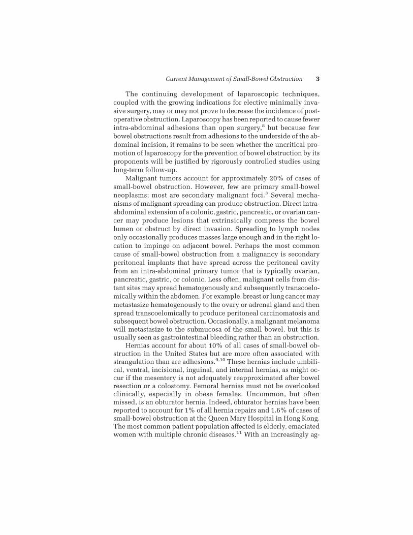
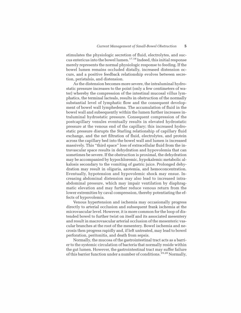
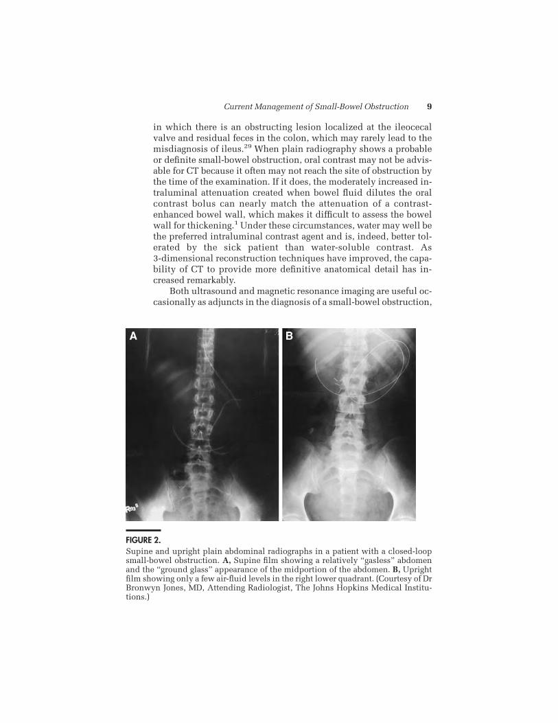
On initial plain-film examination, the findings of distendedloops of small bowel with air–fluid levels (on upright views) and apaucity of colonic air are characteristic (Fig 1). However, plain filmsmay be diagnostic only 45% to 60% of the time.24-27 For example, apatientmay have a gasless abdomen on plain films in the presence ofcomplete obstruction. This may be caused by a closed-loop obstruc-tion that precludes the accumulation of gas within the obstructedloop. Closer evaluation of such a film may reveal a “ground-glass”haziness in the midabdomen or displacement of adjacent bowel bythe “invisible,” dilated, closed loop (Fig 2).25 In an analysis of plainfilm findings reported by experienced gastrointestinal radiologists,a sensitivity of only 66% was found in proven cases of small-bowelobstruction. Twenty-one percent of patients reported to have normalresults did, in fact, have obstructions. Of those patients whose filmfindings were interpreted as abnormal but nonspecific, 13% hadlow-grade and 9% had high-grade obstruction.28 Despite these limi-tations, plain film radiography remains a cornerstone in the diag-nosis of small-bowel obstruction, largely because of its widespreaddiagnostic capability, availability, accessibility, and low cost. How-ever, when the diagnosis is in doubt, computed topography (CT)willhelp clarify the situation.
Current Management of Small-Bowel Obstruction 7

The CT diagnosis of a bowel obstruction and its discriminationfrom an adynamic ileus are based on the detection of fluid, luminalcontent, and/or air-filled loops of bowel proximal to the obstruction,the presence of a definite, localized transition zone, and the pres-ence of collapsed loops of small bowel or colon distal to the obstruc-tion. The exact point of obstruction can sometimes be visualized as abeaklike narrowing in patients with adhesions as the cause. An ad-vantage of CT is that extrinsic lesions such as hematomas, abscesses,inflammation, and extraluminal tumors, which cannot be visual-ized directly on plain-film or conventional intraluminal contraststudies, are often better visualized on CT.29 The use of intravenous(IV) contrast is recommended so that the bowel wall can be imagedin contrast to its luminal contents.30-33 Although oral contrast is notabsolutely essential for the identification of an obstruction becausefluid and air can easily be distinguished within the bowel loops,34 itis quite helpful in discriminating partial from complete obstructionand in localizing the level of obstruction. Certain limitations to theuse of CT in the setting of a small-bowel obstruction include the case
FIGURE 1.Supine and upright plain abdominal radiographs in a patient with small-bowel ob-struction.A, Supine film showing characteristic dilated loops of small bowel and apaucity of colonic air. B, Upright film revealing air-fluid levels and the “string ofpearls” sign in the right lower quadrant. (Courtesy of Dr Bronwyn Jones, MD, At-tending Radiologist, The Johns Hopkins Medical Institutions.)
8 A. J. Hayanga, K. Bass-Wilkins, and G. B. Bulkley
in which there is an obstructing lesion localized at the ileocecalvalve and residual feces in the colon, which may rarely lead to themisdiagnosis of ileus.29 When plain radiography shows a probableor definite small-bowel obstruction, oral contrast may not be advis-able for CT because it often may not reach the site of obstruction bythe time of the examination. If it does, the moderately increased in-traluminal attenuation created when bowel fluid dilutes the oralcontrast bolus can nearly match the attenuation of a contrast-enhanced bowel wall, which makes it difficult to assess the bowelwall for thickening.1 Under these circumstances, water may well bethe preferred intraluminal contrast agent and is, indeed, better tol-erated by the sick patient than water-soluble contrast. As3-dimensional reconstruction techniques have improved, the capa-bility of CT to provide more definitive anatomical detail has in-creased remarkably.
Both ultrasound and magnetic resonance imaging are useful oc-casionally as adjuncts in the diagnosis of a small-bowel obstruction,
FIGURE 2.Supine and upright plain abdominal radiographs in a patient with a closed-loopsmall-bowel obstruction. A, Supine film showing a relatively “gasless” abdomenand the “ground glass” appearance of the midportion of the abdomen. B, Uprightfilm showing only a few air-fluid levels in the right lower quadrant. (Courtesy of DrBronwyn Jones, MD, Attending Radiologist, The Johns Hopkins Medical Institu-tions.)
Current Management of Small-Bowel Obstruction 9
but the continuing evolution of multiphasic CT scanning has lim-ited this usefulness considerably.35-37 CT is faster, more available,less contingent on technical expertise, and capable of providing amore global evaluation of the abdomen and gastrointestinal tract.1
DISCRIMINATING PARTIAL FROM COMPLETE OBSTRUCTIONBecause themanagement of complete obstruction should usually beoperative and that of partial small-bowel obstruction, at least ini-tially, almost always nonoperative, discrimination between the 2 isimportant. The patient’s historymay provide a clue because the con-tinued passage of flatus or stool, 6 to 12 hours after the onset of symp-toms, is more consistent with a partial obstruction. However, even acomplete small-bowel obstruction can be accompanied early byloose stools secondary to peristalsis distal to the obstruction. Onplain films, the persistence of residual colonic gas after 6 to 12 hoursis also suggestive of a partial obstruction. Of importance, rectal ex-amination of supine patients does not introduce significant rectalair, whereas flexible or rigid sigmoidoscopy may well do so.
Despite the foregoing information, some patients can present areal diagnostic challenge because early complete obstruction can bedifficult to distinguish from partial, high-grade obstruction on plainfilms. For their discrimination, the use of oral, contrast-enhancedCThasmarkedly improved on, and often supplanted, traditional im-aging, small-bowel series, and enteroclysis. This may be attributedto the improvement in speed and resolution of current CT imaging.CT with IV contrast material is superior to barium studies in show-ing the bowelwall and extraluminalmasses and in revealing inflam-matory lesions, as well as features of strangulation.1,12
Modern CT may also provide strikingly detailed views of themesenteric vasculature. Moreover, images taken at intervals closelytimed to the injection of the IV contrast material can be used toevaluate mucosal perfusion by estimating the rapidity of the dyewashout. Oral contrast, either Hypaque or, increasingly, just wateralone is particularly useful in evaluating the size, patency, and pro-gression of luminal contents.
CT has proven particularly useful in discriminating a completefrom a partial obstruction by determining the degree of collapse andthe amount of residual air and fluid in the collapsed (distal) intesti-nal segment.12,29 A limitation of CT for the discrimination of a par-tial obstruction is that a mild partial obstruction may not reveal aclear transition zone on CT, which could lead to a misdiagnosis ofileus if there is not a close correlation between the history and phys-ical findings. In most cases, however, the presence or absence of re-
10 A. J. Hayanga, K. Bass-Wilkins, and G. B. Bulkley

sidual contrast within the colon on a plain abdominal radiographobtained 12 to 24 hours later will serve to definitively discriminate apartial from a complete small-bowel obstruction.
DISCRIMINATING A SIMPLE FROM A STRANGULATINGOBSTRUCTIONEarly recognition of strangulation in patients with mechanicalsmall-bowel obstruction has always been controversial. This issuehas been greatly confused by the indiscriminate mixing of patientswith partial and complete obstruction in many reports. Except forthe rare patient with a strangulated Richter’s hernia that has goneundetected on physical examination, patients with partial obstruc-tion can be considered to be at a minimal risk of strangulation.
On the other hand, patients with complete obstruction are atsubstantial risk of strangulation. In operative series,2,3,9 this risk hasbeen consistently reported to be between 20%and 40%. The “5 clas-sic signs” of strangulation obstruction have been variously cited toinclude continuous (vs colicky) abdominal pain, a fever, tachycar-dia, peritoneal signs, leukocytosis, acidosis, the presence of a pain-ful mass, the absence of bowel sounds, and blood in the stool. How-ever, it has been found consistently in both retrospective3,10,25,38 andprospective9 studies that these signs are not sensitive, specific, oraccurately predictive of strangulation. Elevated serum levels of amy-lase, potassium, phosphate, alkaline phosphatase, aspartate amino-transferase, alanine aminotransferase, lactate dehydrogenase, andcreatinine phosphokinase have no practical significance in diagnos-ing strangulation.9 Furthermore, no combination of these signs canaccurately predict vascular compromise.9,25 Moreover, despite fre-quent assurances to the contrary by surgeons convinced of their owndiagnostic acumen, a senior operating surgeon’s ability to prospec-tively recognize strangulation in operative cases of small-bowel ob-struction is no better than chance alone.9 Indeed, reversible strangu-lation (ie, viable bowel) is almost never recognized preoperatively.The reason for this is evident: the signs that have been used to indi-cate strangulation are largely signs of the body’s inflammatory re-sponse to (irreversible) tissue necrosis. Althoughmost surgeons cancorrectly identify advanced ischemic bowel in a patient with sepsisand a rigid abdomen, early, reversible ischemia is simply not clini-cally discernible. These factors contribute to the high mortality rateof patients with a strangulated bowel. Indeed, nearly half of alldeaths from small-bowel obstructions occur secondary to strangula-tion and its complications,25 and in most series,2,10 the presence ofstrangulation doubles the mortality rate (from about 10% to 20%)
Current Management of Small-Bowel Obstruction 11

FIGURE 3.(continued)

associated with small-bowel obstruction. The morbidity rate ofstrangulation obstruction is also as high as 42%, and wound infec-tions and urinary and pulmonary complications aremost frequentlyseen.10
CT has been reported to be useful specifically for the diagnosisof strangulation (Fig 3). IV contrast is recommended because the pat-tern of bowel wall enhancement can be useful in recognizing edemasecondary to ischemia. The CT signs of strangulation include thick-ening of the bowel wall (Fig 3,A), with or without a “target sign”;pneumatosis intestinalis (Fig 3,B and C); portal venous gas; mesen-teric haziness, fluid, or hemorrhaging and ascites; a serrated beaksign; and nonenhancement (or, rarely, increased bowel wall en-hancement due to prolonged venous phasewashout of intravascularcontrast material) after an IV contrast bolus.29 Once again, however,some of these signs (eg, pneumatosis intestinalis) usually indicateirreversible necrosis rather than reversible ischemia.
In summary, given the present state of the art, no clinical indica-tor, combination of indicators, diagnostic test, or “experienced clin-ical judgment” can reliably discriminate reversible strangulating ob-struction from simple obstruction. In the only prospective study9 ofoverall diagnostic capability, the (often confident) diagnosis of “non-strangulating obstruction” was wrong 31% � 15% of the time.
IDENTIFYING THE UNDERLYING CAUSE OF OBSTRUCTIONIn most situations, management decisions, including surgery, aremade on the basis of the aforementioned factors, regardless of thesuspected cause of the obstruction. Several situations, however,warrant special attention and possible modification of this ap-proach. These include the patient with a small-bowel obstructionsecondary to an incarcerated hernia, recurrent malignant tumor, in-flammatory bowel disease, intra-abdominal abscess, radiation en-teritis, acute postoperative obstruction, and multiple recurrentsmall-bowel obstructions, each of which will be discussed in more
FIGURE 3. (continued)Computed tomography in a patient with signs of strangulation. A, Note the mas-sively thickened bowel wall from edema. B and C, Note the areas of pneumatosisintestinalis, a late sign of ischemic necrosis. Note the “bull’s eye” on the sagittalview indicative of the massive amounts of air within the bowel wall. (Courtesy ofDr Elliot Fishman, MD, Attending Radiologist, The Johns Hopkins Medical Institu-tions)
Current Management of Small-Bowel Obstruction 13
detail later. Although the most important clues to this particularcomponent of the diagnosis are the history and physical examina-tion, often a CT scan or an enteroclysis study can be helpful. In mostcases, however, identification of the underlying cause can be madeat surgery with little disadvantage to the patient.
MANAGEMENTSYSTEMATIC RESUSCITATIONPatients with small-bowel obstructions are usually intravascularlydepleted, often massively, because of a decreased oral intake, vom-iting, and the sequestration of fluid from the intravascular spacewithin the bowel wall and lumen. This requires aggressive replace-ment with an IV saline solution such as Ringer’s lactate. Routinelaboratory measurements of serum sodium, potassium, chloride, bi-carbonate, and creatinine levels should be obtained. Serial measure-ments of the hematocrit level, white blood cell count, and serumelectrolyte levels are monitored closely to assess the adequacy offluid repletion and as a possible indication of late tissue necrosis.Serum lactic acid levels are usually obtained; however, a normal lac-tate level does not rule out early ischemia, and elevated lactate lev-els can be seen in a number of circumstances. Thus, this test is nei-ther sensitive nor specific but may sometimes be helpful. Becauseof their large fluid requirements, many patients will need either cen-tral venous pressure monitoring or the placement of a pulmonaryartery catheter. Almost all patients will need the placement of a Fo-ley catheter so that hourly urine output may be monitored. Broad-spectrum antibiotics are also often given in consideration of theevidence for bacterial translocation occurring in even simple ob-struction, or they are given as prophylaxis for resection or an inad-vertent enterotomy at surgery. However, this is a practice that variesgreatly and has not been subject to definitive study.
MANAGEMENT OF OBSTRUCTIONVirtually all patients with small-bowel obstructions benefit from theuse of nasoenteric suctioning, whether it be via a nasogastric or longintestinal tube such as a Baker tube. This provides almost immedi-ate symptomatic relief from the nausea and vomiting and, often to asignificant degree, the abdominal pain. It allows the administrationof radiographic contrast material to these nauseated patients. It alsohelps prevent aspiration at the time of induction of anesthesia. Insome situations, a long tube may provide a postoperative splint toprevent a recurrent obstruction. Sometimes it provides definitivetreatment in lieu of surgery. However, the decision to use a nasoen-
14 A. J. Hayanga, K. Bass-Wilkins, and G. B. Bulkley

teric tube must be made without regard to whether or when surgeryis to be performed.
A prospective, randomized but underpowered trial39 of short(nasogastric) versus long (nasointestinal) tubes detected no signifi-cant difference with regard to the decompression achieved and thesuccess or the morbidity after surgical intervention. Other studies2
also report similar success, regardless of whether short or long tubeswere used. The primary advantages of a nasogastric tube includeeasy placement and rapid, more effective gastric decompression,which is especially essential in the setting of anesthetic induction,in which the risk of aspiration is increased.40,41 The use of a naso-gastric tube is not associated with some of the rare complications oflong tubes, including perforation and intussusception of the smallbowel, either over the tubewhile it is in place or over adjacent bowelon removal of the tube.42,43
Nonetheless, the use of long tubes also has several advantages.Some surgeons believe that the tip of the long tube will open ob-structed loops of bowel as it passes more distally, although little di-rect evidence exists to support this. A long tube also provides suc-tion close to the area of obstructionwhen positioned correctly.39 Thepresence of a long intestinal tube also greatly enhances bowel de-compression at surgery, often facilitating primary closure of the ab-dominal wall without the need for an enterotomy.39 The alternativemethod of decompression at surgery is retrograde stripping of thesmall-bowel contents into the stomach with subsequent nasogastricsuction. An enterotomy is usually contraindicated. In rats, manipu-lation of the bowel either by stripping or enterotomy significantlyincreased the incidence of E coli bacteremia.44 Therefore, effectivepreoperative decompression with a long tube may decrease theamount of bowel manipulation required in the operating room andconsequent bacteremia.
The most controversial aspect of this disease is the role of earlysurgery versus a trial of nonoperative management in patients withsmall-bowel obstruction. On the 1 hand, there is noway to clinicallydiscern which patients have early reversible strangulation. On theother hand, a number of large, retrospective series report successwith nonoperative management in patients without signs of stran-gulation, followed by surgery only in select patients. For example,in a retrospective analysis45 of 123 admissions with adhesive small-bowel obstruction, the obstruction resolved in 85 patients withoutsurgery. In 88% of these patients, the obstruction resolvedwithin 48hours. Resolution of the obstruction in the remaining patients oc-curred within 72 hours. These authors reported no untoward effects
Current Management of Small-Bowel Obstruction 15

in patients who did require surgery after initial nonoperative treat-ment. Another retrospective series46 reported a 73% rate of resolu-tion of adhesive obstruction without a significant increase in themortality rate or the rate of strangulated bowel when comparedwithoutcomes in other series. In this series, “a trial of tube decompres-sion” (ie, nonoperative management) for more than 5 days was inef-fective. These authors argue that a trial of nonoperative nasoentericdecompression of 2 to 3 days’ duration, even up to 5 days in selectpatients, is reasonable in most patients who show no clinical evi-dence of strangulation. The problem with these and similar studiesis that they include a large, undefined population that is usually amix of patients with either complete or partial small-bowel obstruc-tions. (Indeed, there is little controversy that partial obstructionshould bemanaged nonoperatively initially.) The studies fail, there-fore, to definitely resolve the controversy over the correct manage-ment of complete small-bowel obstruction, but they do indicate thatsuch an approach is safe in patients with partial obstructions.
If initial nonoperative management fails, several operative ap-proaches are available via conventional laparotomy. Often, the ob-struction is caused by the presence of 1 or more constricting adhe-sive bands, and the obstruction is relieved through simple lysisof the adhesions and detorsion. An obstructing lesion may also bepresent and may require local bowel resection with primary reanas-tomosis. A side-to-side intestinal bypass or, rarely, the placement ofenterocutaneous stomata may be the appropriate management ofend-stage malignant obstructing lesions or radiation enteritis.
Advances in laparoscopic surgery have modified the approachto many general surgical problems, and laparoscopic managementof acute small-bowel obstruction is an option that is gaining advo-cacy. Franklin et al47 reported 23 patients with acute obstructionevaluated initially with laparoscopy (after an initial trial of conser-vative management had failed). Twenty patients had successful lap-aroscopic resolution of their obstruction, and 3 required laparoto-my. The 3 patients who were converted to laparotomy had severeadhesions, anatomy that precluded complete examination of the en-tire length of the bowel, or suspected ischemic necrosis, respec-tively. The authors47 emphasized the importance of using nontrau-matic bowel clamps when manipulating the dilated, friable bowelduring laparoscopy to avoid injury. Similar studies advocate thema-nipulation of the mesentery rather than the bowel wall wheneverpossible, particularly when “running the bowel.”8 Lerard et al,48 ina multicenter retrospective study, reported that laparoscopic treat-ment for small bowel obstruction was, in their series, of greatest
16 A. J. Hayanga, K. Bass-Wilkins, and G. B. Bulkley

benefit to those patients who had undergone less than 3 previousoperations, those who had been seen early after the onset of the ob-struction (particularly those who had previously undergone onlyappendectomy), and those in whom the probable cause of obstruc-tion was bands.
There is also a growing interest in the pharmacologic preventionof adhesion formation. For example, the laparoscopic placement ofa biosyntheticmembrane such as Seprafilm, amixture of hyaluronicacid and carboxymethylcellulose,49,50 has been found to reduce theincidence of postoperative adhesions to the underside of the ab-dominal scar.51 Few other adhesion barriers have been evaluated ascarefully in a clinical setting.49,52
During exploration, whether by laparotomy or laparoscopy, it issometimes difficult to evaluate bowel viability after the release ofstrangulation. The conventional clinical criteria used include thereturn of normal color, peristalsis, and arterial pulsations. A pro-spective, controlled trial comparing standard clinical judgmentwiththe use of a Doppler probe and with fluorescein for the intraopera-tive discrimination of viability found that the Doppler ultrasonicflow probe was less accurate than the conventional clinical judg-ment of the surgeon, which was usually correct if thought to be so.53
On the other hand, the pattern of fluorescein fluorescence was sig-nificantly more reliable than either clinical judgment alone or theuse of a Doppler probe in assessing intestinal viability. In difficultbowel segments of borderline viability, this is the only method ofviability assessment that has been formally evaluated in a prospec-tive, controlled clinical trial. Because clinical judgment is usuallyaccurate in this assessment, the use of fluorescein is recommendedin those cases in which bowel segments of borderline viability aredifficult to evaluate clinically.54
Another approach to the assessment of bowel viability is a“second-look” laparotomy or laparoscopy 18 to 48 hours after theinitial procedure. Most advocates of this approach suggest that thedecision to perform a second-look laparotomy be made before clo-sure, at the time of the initial procedure.55 However, carefully con-trolled, well-documented studies in animals have found that thefluorescein technique, when used correctly, is more accurate than asecond look at 24 hours after the initial procedure.54,56 One clinicalstudy57 (which looked at a small number of patients in an inconsis-tently controlled, incompletely defined fashion) reported that fluo-rescein fluorescence, pulse palpation, and Doppler analysis duringthe initial laparotomy were not accurate predictors of bowel viabil-ity in their handswhen comparedwith findings (in a few patients) at
Current Management of Small-Bowel Obstruction 17
a second-look laparotomy. Unfortunately, no details of the viabilityassessment techniques are given; if they were as superficial, as de-scribed in the article, they would not represent a fair assessment ofthe state of the art of these techniques. In the absence of controlledclinical trials, it remains unclear whether or when a second-looklaparotomy significantly enhances the assessment of intestinal vi-ability. However, because of the intestine’s particular vulnerabilityto the vasoconstrictive, hemodynamic response to shock, sepsis,and severe physiologic stress, a second-look laparotomy is clearlyindicated in a patient whose systemic condition deteriorates afterthe initial viability assessment.58,59
Almost all patients, except those in frank septic shock, benefitfrom an initial period of 12 to 24 hours of nasoenteric suctioning,fluid and electrolyte resuscitation, and, often, the administration ofantibiotics before laparotomy. This allows not only resuscitation butalso completion of the diagnostic studies described earlier, includ-ing, in almost all cases, definitive discrimination of partial fromcomplete obstruction. At this point, the role of early surgery (ie, afterthe initial 12-24 hours) versus a trial of nonoperative managementremains controversial. Much of the controversy, however, may beobviated if one discriminates partial from complete obstruction.
MANAGEMENT OF PARTIAL OBSTRUCTIONThe role of early surgery versus expectant management of completeobstruction remains controversial in some circles, but there is littlecontroversy with respect to partial obstruction. Most of these pa-tients benefit from an extension of the initial 12- to 24-hour period ofnonoperative management for up to several days. A number ofstudies60-62 indicate that 60% to 85% of these patients will ulti-mately resolve their obstruction and be dischargedwithout the needfor surgery.
Even those who do not respond are better prepared for surgerybecause of better mechanical bowel decompression, often even anantibiotic bowel preparation, a longer period of resuscitation to al-low better intercompartmental fluid and electrolyte equilibration,and usually the benefits of planned surgery by a fresh operative teamduring daylight hours. Sometimes amore definite idea about the un-derlying cause can be obtained. If the total period without oral nu-tritional intake is prolonged more than a few days, parenteral nutri-tion should be provided. In summary, most patients with partialsmall-bowel obstructions accrue many benefits and few disadvan-tages from an initial trial of nonoperative management.
18 A. J. Hayanga, K. Bass-Wilkins, and G. B. Bulkley

A substantial adjuvant to the management of partial small-bowel obstruction is the enteroclysis study, whereby graded vol-umes of dilute barium andmethyl cellulose are given through a longtube localized either by peristalsis or direct fluoroscopic positioningin the small bowel just proximal to the site of the obstruction. Thisstudy, in the hands of an experienced radiologist, can often help de-fine the degree of obstruction, its location, and its progression (ie,improvement or lack thereof) over time. Enteroclysis can objectivelygauge the severity of the intestinal obstruction, which is an impor-tant advantage over other modalities.63 For a low-grade partialsmall-bowel obstruction, there is no delay in the arrival of contrastto the point of the obstruction and there is sufficient flow of contrastthrough this point such that fold patterns in the postobstructiveloops are readily defined. A high-grade partial small-bowel obstruc-tion is diagnosed when the presence of retained fluid dilutes thebarium, which results in inadequate contrast density above the siteof obstruction and allows only small amounts of contrast material topass through the obstruction into the collapsed distal loops. Com-plete obstruction is diagnosed when there is no passage of contrastmaterial beyond the point of the obstruction, as seen on delayed ra-diographs obtained up to 24 hours after the start of the examina-tion.27,64 This may be useful in deciding whether to intervene surgi-cally or to wait longer for resolution. Sometimes, the underlyingcause can be inferred (eg, an adhesion can be discriminated from aneoplasm).28
To help resolve partial small-bowel obstructions nonoperative-ly, some have advocated the use of hyperosmolar water-soluble gas-trointestinal contrast agents as therapeutic as well as diagnostic mo-dalities. In a prospective randomized trial65 looking at the effect ofGastrograffin in the nonoperative management of partial small-bowel obstructions, among the patients managed successfully non-operatively, those who received 100 mL of Gastrograffin had a sig-nificant reduction in the number of days until the first stool and inthe length of their overall hospital stay, from approximately 4 to 2days. However, this trial found no significant difference in the pro-portion of patients who eventually required surgical intervention.Stordahl et al33 have also reported water-soluble contrast agents tobe useful as therapeutic agents. However, therewas no control grouptreated with nasogastric suction alone. Others66 have reported thatno advantage over conventional nonoperative management of par-tial small-bowel obstructionswas found, although administration ofsuch hyperosmolar contrast materials was safe in patients with par-tial small-bowel obstructions. On the other hand, there are 2 signifi-
Current Management of Small-Bowel Obstruction 19

cant drawbacks to this approach: most importantly, elderly patientsand patients with obtundation and bowel obstructions are quiteprone to aspiration, and the aspiration of some contrast agents, es-pecially Gastrograffin, can produce severe, often lethal aspirationpneumonia.Moreover, hyperosmolar agents do stimulate peristalsisand can cause severe pain in the patient with an obstruction.
MANAGEMENT OF COMPLETE OBSTRUCTIONFew other issues in surgery have generated such heated controversyfor such a long period as the question of primary operative versusprimary nonoperative treatment of patients with small-bowel ob-struction. Despite a preponderance of good evidence (but not a ran-domized, prospective trial), experienced surgeons often expressstrongly polarized opinions. The conventional argument of thosewho advocate primary nonoperative management is that it is oftensuccessful and that, with careful monitoring and “experienced clin-ical judgment,” they can recognize those patients with early stran-gulation in time to operate on them before the bowel becomes non-viable. The 60% to 85% success rates cited in several larger seriesare undeniable; however, as noted, these series predominantly con-tain patients with partial obstruction, and these are patients whorarelymanifest strangulation. As discussed, patientswith partial ob-struction should usually be treated nonoperatively, at least initially,and there is little controversy about this. Patients with complete ob-struction are another matter. The incidence of strangulation in thisgroup varies from 20% to 40% (admittedly, in operative series thatnecessarily exclude patients successfullymanaged nonoperatively).Moreover, prospective and retrospective studies are unequivocal inindicating that early, reversible strangulation is simply not discern-ible on clinical grounds. Therefore, the risk of deciding to manage apatient nonoperatively based on a clinical assessment of “simple(nonstrangulating) obstruction” necessarily entails the substantial(about 30%) risk that one is delaying the treatment of intestinalstrangulation until after that injury becomes irreversible. It is ratio-nal, therefore, to weigh the benefits and risks of each approach, aswe do in every clinical decision. However, 1 of the risks of primarynonoperative management that must be taken into account is thissubstantial risk of strangulation. In a severely unstable patient, suchas someone with acute myocardial infarction, treatable arrhythmia,hypovolemia, or shock, this risk is a reasonable one in return for thebenefits of improved systemic stability in the nonoperative period,even if it proves to be a preoperative period. On the other hand, apatient with long-term, irreversible risk factors mandates a correct
20 A. J. Hayanga, K. Bass-Wilkins, and G. B. Bulkley

decision, not necessarily a nonoperative one. In these situations andin most patients with complete small-bowel obstruction, the sub-stantial risk of unrecognized (indeed, unrecognizable) strangulationmandates primary operative management after the initial 12 to 24hours of resuscitation. Indeed, although several retrospective stud-ies60,67 report that a 12- to 24-hour delay of surgery in patients withcomplete small-bowel obstruction is safe, the incidence of strangu-lation and other complications significantly increases after longerperiods of nonoperative management.67
MANAGEMENT OF SPECIFIC LESIONSAdhesionsThe pathophysiologic process of adhesion formation has been stud-ied extensively and is clearly initiated by the formation of a fibrinclot (from transudated fibrinogen activated by tissue factor, regard-less of bleeding). Peritoneal trauma is a well-known cause.57 Theperitoneum (mesothelium) has been found to possess fibrinolytic ac-tivity via plasminogen activation.58,68-70 Ischemia, a known stimu-lus of adhesion formation, causes a marked reduction in plasmino-gen activator activity levels through the release of plasminogenactivator inhibitors.59,60 This pathway for adhesion formation lendssupport to the use of fibrinolytics in the prevention of adhesions. Inthe past, streptokinase and urokinase have been used with varyingdegrees of success in animal models.71,72 Their use in human beingshas usually been precluded for this purpose for fear of bleeding com-plications, especially in patientswho have undergone extensive dis-section and who are also at the greatest risk for adhesion formation.(Such agents are rapidly absorbed systemically from the perito-neum.) Nonsteroidal anti-inflammatory agents, including ketorolactromethamine, have also been found to be useful in inhibiting adhe-sion formation in pigs.73 Once again, however, steroids and antime-tabolites, although effective in animal models, are not usually usedin patients for fear of inhibiting wound healing, especially after ex-tensive dissection and in the presence of distension.
Hyaluronic acid, a product of a strain of Streptococcus, is highlylubricating and nonimmunogenic and can coat and protect serosalsurfaces.52 It seems to be effective in keeping traumatized surfacesseparate, thereby hindering the formation of connecting fibrousbands.74 Becker et al49 conducted the first prospective study of post-operative abdominal adhesion formation by using standardized di-rect peritoneal visualization. In this study, 183 patients with ulcer-ative colitis or familial polyposis who underwent open colectomy
Current Management of Small-Bowel Obstruction 21

and ileal pouch–anal anastomosis with diverting loop ileostomywere randomly assigned to receive or not receive a bioresorbablemembrane of hyaluronic acid and carboxymethylcellulose (Sepra-film) placed directly beneath themidline abdominal incision. At thetime of the subsequent ileostomy closure, laparoscopy revealed thatthe number of patients who had adhesions to the underside of theabdominal incision was reduced by more than 50% in those treatedwith the bioresorbable membrane. The extent and severity of theseadhesions were also reduced significantly in the treatment group.49
Although the implications of this study are limited by the fact thatobstructing adhesions do not usually form at the old incision sitebut within the pelvis, the feasible extension of this technology toserosal surfaces could represent a significant advance.49,75-77
Independent of adjuvant therapy for the prevention of adhesionformation, several operative steps should be taken at any laparoto-my, especially at 1 for lysis of adhesions, to help minimize the ex-tent of future adhesion formation. These include gentle handling ofthe bowel to reduce serosal trauma, avoidance of unnecessary dis-section, exclusion of foreign material from the peritoneal cavity (ie,the use of absorbable suture material when possible, the avoidanceof excessive use of gauze sponges, and the removal of starch fromgloves), adequate irrigation and removal of infectious and ischemicdebris, the use and preservation of the omentum around the site ofsurgery or in the denuded pelvis, and avoidance of lysis of adhe-sions that do not involve the small bowel.78
Patients who are initially seen with acute small-bowel obstruc-tion from adhesions usually benefit from early operative lysis. Usu-ally, a technically simple laparotomy (or laparoscopy) is all that isrequired. Except under exceptional circumstances, an enterotomyshould be avoided. In a retrospective analysis, Strickland et al79 re-ported that the use of laparoscopy obviated formal laparotomy in 40of their patients (68%). Laparoscopic adhesiolysis was also reportedto result in a shorter hospital stay, faster resumption of normal bowelfunctioning, decreased morbidity, and fewer complications, al-though these comparisons were with historical control subjects.64,79
The increased expertise of surgeons in advanced laparoscopy mayallow this option to become more widely adopted.79
Incarcerated HerniaWhen an inguinal, umbilical, incisional, or incarcerated abdominalwall hernia is the cause of the obstruction, the obstruction can oftenbe managed initially by simple manual reduction, sometimes aidedby sedation. However, the patient should be admitted for close ob-
22 A. J. Hayanga, K. Bass-Wilkins, and G. B. Bulkley

servation. During this hospitalization, elective hernia repair shouldbe performed to prevent recurrent incarceration and the possibilityof strangulation. A severely incarcerated, irreducible hernia is aclear indication for primary early operative management, often by atransabdominal approach.
Malignant TumorA small-intestinal obstruction caused by a primarymalignant tumoris rare; much more often, it is caused by a neoplasm from anotherorgan, such as the colon or ovary. The disease of these patients ismanaged like that of patients with simple small-bowel obstructionfrom adhesions, in combination with resection of the obstructingtumor, whenever feasible.
Most challenging, from a therapeutic standpoint, are patientswith intestinal obstructions who have been previously treated forcancer or who have known peritoneal carcinomatosis. In a retro-spective analysis80 of 81 episodes of small-bowel obstruction in 61patientswith previously treatedmalignancies, 69 episodes involvedthe small bowel, and 24 of these were diagnosed as complete ob-structions. Eight percent of these patients had concurrent small- andlarge-bowel obstructions. In 59 cases, the cause was established:61% of these obstructions were due to metastatic tumors, and 39%were due to benign causes (eg, adhesion, irradiation, and stricture).Forty-five percent of the cases of partial obstruction that were man-aged conservatively resolved without surgery. On the other hand,only 4%of the cases of complete obstructionwere successfullyman-aged without operative intervention.80 One of the most importantlessons from this study is that patients with a history of cancer whohave an obstruction should not necessarily be assumed to have car-cinomatosis as the cause of their obstruction. The surgeon shouldnot always avoid operating on a patient with obstruction from carci-nomatosis, although the management of such patients must be indi-vidualized, with the desires of the patient taken into account. Ofcourse, not every terminally ill patient is an operative candidate,and parenteral nutrition combined with percutaneous endoscopicgastrostomy offers the advantage of terminal care at home for thosepatients who either have obstructions not amenable to surgery orwho have chosen not to undergo surgery.80
Inflammatory Bowel DiseaseCrohn’s disease is now recognized as the third leading cause ofsmall-bowel obstructions in technologically advanced countries.12
Patients with Crohn’s disease of the small intestine can have com-
Current Management of Small-Bowel Obstruction 23

plete, partial, or intermittent small-bowel obstruction. The obstruc-tion may be secondary to the primary inflammatory process itself orto the gradual development of a fibrotic stricture as a sequela of re-peated episodes of inflammation and healing, with or without treat-ment. These patients, often with partial obstruction, can frequentlybe managed initially nonoperatively with tube decompression2 incombinationwith pharmacologic treatment of the inflammatory pro-cess (eg, with high-dose steroids). Parenteral nutrition should beprovided because the period of required bowel rest may be pro-longed. On the other hand, if fibrotic strictures are the primary causeof the obstruction, primary bowel resection may be necessary to re-lieve the obstruction. This does not imply that a nonoperative trialshould not be attempted; the obstruction related to the stricturesmay prove to be partial as the associated inflammation resolves.Over the past decade, a number of articles81 have reported the suc-cess of operative strictureplasty, with or without concomitant bowelresection in other areas, for multiple, short strictured segments inpatients with Crohn’s disease.
Intra-Abdominal AbscessOften, an acute intra-abdominal abscess may produce a clinical pic-ture that is indistinguishable from complete, mechanical, small-bowel obstruction. This is often due not to intraluminal obstructionor even to external compression of the bowel lumen but to a severelocalized ileus secondary to local inflammation and edema. Drain-age of the abscess is often sufficient to relieve the obstruction. Thisdoes not necessarily require a laparotomy because the abscess maybe accessible with the use of ultrasound- or CT-guided percutaneousdrainage. However, if the obstruction persists, a laparotomy may berequired.
Radiation EnteritisOf importance in the current management of malignancies of manytypes is the use of radiotherapy. In a retrospective analysis82 of pa-tients at the University of California Los Angeles undergoing radicalhysterectomy, a 5% incidence of subsequent small-bowel obstruc-tion was reported in those undergoing surgery alone, but a 20% in-cidence was reported in patients receiving adjuvant radiotherapy.Small-bowel obstruction is a recognized late complication of radio-therapy instituted for the treatment of rectosigmoid and rectal can-cer after low anterior resection and abdominoperineal resection. Therate has been reported to be as high as 30% in patients treated withdaily extended-field radiotherapy, 21% in those receiving single
24 A. J. Hayanga, K. Bass-Wilkins, and G. B. Bulkley

pelvic-field radiotherapy, and 9% in those with multiple pelvicfields in a retrospective review83 of 224 patients at M. D. AndersonCancer Center. In this study, patients whose radiation was givenwith small-bowel exclusion, achieved by the use of the open table-top device technique, had an incidence of obstruction of only 3%.The incidence of recurrent small-bowel obstruction was also signif-icantly correlated with the incidence of postsurgical small-bowelobstruction in these patients. Another technique for small-bowel ex-clusion that has been explored is the use of intraperitoneal saline-filled tissue expanders84 to keep the bowel out of a specific radiationfield, such as the pelvis, during radiotherapy.
Despite these precautions, however, cases of acute and chronicradiation enteritis do occur and are sometimes accompanied bybowel obstruction. If obstruction occurswithin a fewweeks of radio-therapy, it is often useful to treat it nonoperatively with tube decom-pression and steroids. However, complications from irradiationmaynot appear for many years after the completion of therapy and areusually progressive thereafter. When the obstruction occurs in thislate setting, nonoperative management is rarely effective, and a lap-arotomy is usually required. The surgeon may choose to either lo-cally resect the irradiated bowel or bypass the affected area. Wheth-er one resects or bypasses, it is essential to avoid anastomosis ofirradiated bowel.
Acute Postoperative ObstructionSmall-bowel obstruction that occurs in the immediate postoperativeperiod presents a challenge for both diagnosis and treatment. Thediagnosis is often difficult because the primary symptoms of ab-dominal pain and vomiting may often be masked and/or attributedto incisional pain and postoperative ileus. A careful history may re-veal pain that is colicky in nature, as opposed to pain that is dull andconstant.
Abdominal plain films may be helpful in distinguishing ileusfrom obstruction, but they are often not diagnostic. CT has beenfound to be especially useful in distinguishing postoperative ileusfrom obstruction. In fact, Frager et al85 reported 100% sensitivityand specificity when CT was used to distinguish early (within 10days of laparotomy) postoperative ileus from small-bowel obstruc-tion. Furthermore, some common causes of postoperative obstruc-tion such as intra-abdominal abscesses are easily visualized on CTscans. Upper gastrointestinal series with contrast may be quite use-ful in revealing not only the presence but also the degree of obstruc-
Current Management of Small-Bowel Obstruction 25
tion. Barium contrast should usually be used, unless there is a dan-ger of perforation or anastomotic leakage.
Once the diagnosis of obstruction has been established, it shouldbe managed like an obstruction that occurs otherwise in the postop-erative period. Specifically, partial obstruction may be afforded atrial of tube decompression. In fact, in this situation, the opportu-nity to temporarily stabilize the patient and delay surgery a whilelonger into the postoperative periodmay be an advantage. Completeobstruction is a relatively clear indication for early exploration.However, in the postoperative setting, it is not uncommon for thesurgeon to prefer an initial trial of nonoperative management. Cau-tion must be taken, however, because several series25 have reportedan especially high rate ofmissed strangulation in patients with earlypostoperative obstruction. Moreover, an initial delay can move thetiming of surgery to 10 to 14 days postoperatively, which is a time atwhich new, vascularized, dense adhesion formation can make theoperative dissection difficult and dangerous.
Recurrent ObstructionPatients with multiple recurrent adhesive obstructions represent adifficult management problem. (Various studies3-5 report recurrencerates of approximately 10% to 30%.) Recurrent obstruction seems tobe a particular problem for patients with extensive, dense intraper-itoneal adhesions. An initial nonoperative trial is usually desirableand is often safe. However, a retrospective study86 found that a re-currence happened sooner andmore frequently in patientsmanagedconservatively than in patients managed operatively after their sec-ond episode of a recurrence. This does not mean that every patientwith recurrent obstruction should be managed operatively. Patientsmust be evaluated as individuals, and their previous responses toparticular interventionsmust be taken into accountwhen theirman-agement plan is formulated.
Bowel fixation procedures have been used at surgery in an at-tempt to splint the bowel in a nonobstructive configuration whilethe inevitable adhesions form. There are 2 categories of bowel fixa-tion, external and internal. External plication procedures includethe Noble87 and the Childs-Phillips88 procedures and other varia-tions of these techniques, whereby the small intestine or its mesen-tery is sutured in large, gently curving loops. Variable success in pre-venting recurrent obstruction has been reported87-90 when thesetechniques are used. Common complications are the developmentof enteroenteric, enterocolic, and enterocutaneous fistulas, grossleakage, peritonitis, and death.87-90 For this reason and because of
26 A. J. Hayanga, K. Bass-Wilkins, and G. B. Bulkley

the low overall success rate, these procedures have largely beenabandoned.
Internal fixation or stenting procedures use a long intestinal tubeinserted via the nose, a gastrostomy, or even a jejunostomy to splintthe bowel in gentle, unobstructing curves. The intestinal tube is thenleft in place for at least 1 week postoperatively, even after nasoen-teric suctioning has been discontinued. The hope is that adhesionswill form in such a manner that future torsion of loops about bandadhesions is less likely. Several series91-94 have reported moderatesuccess with the use of this approach. Complications associatedwith the use of internal stenting tubes include intussusception ofthe bowel, either over the tube while it is in place or after tube re-moval, and difficult removal of the tube, whichmay require surgicalre-exploration.91-95 Close and Christensen96 have looked at the rateof recurrent obstruction in patients undergoing Childs-Phillips pli-cation or Baker tube stenting versus enterolysis alone in a retrospec-tive series. They found that the rate of recurrent obstruction wasrelatively low after all 3 interventions; the highest recurrence oc-curred after enterolysis alone (6.5%). These authors recommendthat enterolysis alone is adequate for single-band adhesions or forfew adhesions. In cases of severe, multiple adhesions, they advocatethe use of either Childs-Phillips plication or Baker tube stenting.They further suggest that the Baker tube should be used in cases ofmassive bowel distension because of its capability to decompressthe bowel as well as provide a means of plication. They also preferBaker tube stenting over external plication in cases of peritonitis be-cause the transmesenteric sutures may provide a nidus of infection.In the absence of studies controlled by comparable groups, however,it is problematic to advocate dogmatically; fewmodern surgeons useany method of stenting, and most of those that do use internal tubefixation.
SUMMARYThe most significant advances in the management of small-bowelobstruction are developments in imaging modalities available to as-sist in the diagnosis itself, as well as to possibly assist in the earlyidentification of those cases requiring urgent operative decompres-sion. The most marked of these have been in the use and interpreta-tion of contrast-enhanced CT. This has decreased the use of bariumstudies and has largely supplanted ultrasound and magnetic reso-nance imaging in the management of these patients.
Diagnostic and therapeutic laparoscopic techniques are alsogrowing in both capability and popularity. Laparoscopic adhesioly-
Current Management of Small-Bowel Obstruction 27

sis and the adjuvant of bioresorbable membranes each hold promisebut have yet to become established as standard treatment.
Further progress is needed in the detection of early, reversiblestrangulation. As a consequence, the fundamentals of the surgicalmanagement of small-bowel obstruction have evolved little over thepast 15 years.With our persistent inability to detect reversible ische-mia, a substantial risk of progression to irreversible ischemia re-mains when surgery is delayed, particularly in the setting of sus-pected complete obstruction.
REFERENCES1. Maglinte DDT, Heilkamp DE, Howard TJ, et al: Current concepts in im-
aging of small bowel obstruction. Radiol Clin North Am 41:262-283,2003.
2. Bizer LS, Liebling RW, DelaneyHM, et al: Small bowel obstruction: Therole of nonoperative treatment in simple intestinal obstruction and pre-dictive criteria for strangulation obstruction. Surgery 89:407-413, 1981.
3. Mucha P Jr: Small intestinal obstruction. Surg Clin North Am 67:597-620, 1987.
4. Chiedozi LC, Aboh IQ, Piserchia NE: Mechanical bowel obstruction:Review of 316 cases in Benin City. Am J Surg 139:389-393, 1980.
5. Holcombe C: Surgical emergencies in tropical gastroenterology.Gut 36:9-11, 1995.
6. CoxMR, Gunn IF, EastmanMC, et al: The operative aetiology and typesof adhesions causing small bowel obstruction. Aust N Z J Med 63:848-852, 1993.
7. Zbar RIS, CredeWB,McKhann CF, et al: The postoperative incidence ofsmall bowel obstruction following standard, open appendectomy andcholecystectomy:A six-year retrospective cohort study at Yale-NewHa-ven Hospital. Conn Med 57:123-127, 1993.
8. Garrard CL, Clements RH, Nanney L, et al: Adhesion formation is re-duced after laparoscopic surgery. Surg Endosc 13:10-13, 1999.
9. Sarr MG, Bulkley GB, Zuidema GD: Preoperative recognition of intesti-nal strangulation obstruction: Prospective evaluation of diagnostic ca-pability. Am J Surg 145:176-182, 1983.
10. Shatila AH, Chamberlain BE,WebbWR: Current status of diagnosis andmanagement of strangulation obstruction of the small bowel.Am J Surg132:299-303, 1976.
11. Lo CY, Lorentz TG, Lau PWK: Obturator hernia presenting as smallbowel obstruction. Am J Surg 167:396-398, 1994.
12. Miller G, Boman J, Shrier L, et al: Etiology of small bowel obstruction.Am J Surg 180:33-36, 2000.
13. Reisner RM, Cohen JR: Gallstone ileus: A review of 1001 reported cases.Am Surg 60:441-446, 1994.
28 A. J. Hayanga, K. Bass-Wilkins, and G. B. Bulkley

14. Meyerson S, Holtz T, Ehrinpreis M, et al: Small bowel obstruction inpregnancy. Am J Gastroenterol 90:299-302, 1995.
15. Lo CY, Lau PWK: Small bowel phytobezoars: An uncommon cause ofsmall bowel obstruction. Aust N Z J Med 64:187-189, 1994.
16. Ciftci AO, Tanyel FC, Büyükpamukcu N, et al: Adhesive small bowelobstruction caused by familial Mediterranean fever: The incidence andoutcome. J Pediatr Surg 30:577-579, 1995.
17. Shields R: The absorption and secretion of fluid and electrolytes by theobstructed bowel. Br J Surg 52:774-779, 1965.
18. Wright HK, O’Brien JJ, Tilson MD: Water absorption in experimentalclosed segment obstruction of the ileum in man. Am J Surg 121:96-99,1971.
19. Alexander JW, Boyce ST, Babcock GF, et al: The process of microbialtranslocation. Ann Surg 212:496-510, 1990.
20. Wells CL, Maddaus MA, Simmons RL: Proposed mechanisms for thetranslocation of intestinal bacteria. Rev Infect Dis 10:958-979, 1988.
21. Berg RD, Garlington AW: Translocation of certain indigenous bacteriafrom the gastrointestinal tract to the mesenteric lymph nodes and otherorgans in a gnotobiotic mouse model. Infect Immun 23:403-411, 1979.
22. Reed LL,MartinM,Manglano R, et al: Bacterial translocation followingabdominal trauma in humans. Circ Shock 42:1-6, 1994.
23. Deitch EA: Simple intestinal obstruction causes bacterial translocationin a man. Arch Surg 124:699-701, 1989.
24. Frager DH, Baer JW: Role of CT in evaluating patients with small-bowelobstruction. Semin Ultrasound CT MR 16:127-140, 1995.
25. Silen W, Hein MF, Goldman L: Strangulation obstruction of the smallintestine. Arch Surg 85:121-129, 1962.
26. Maglinte DDT, Reyes BL, Harmon BH: Reliability and the role of plainfilm radiography and CT in the diagnosis of small bowel obstruction.Am J Roentgenol 167:1451-1455, 1996.
27. Nadrowski LF: Pathophysiology and current treatments of intestinalobstruction. Rev Surg 31:381-407, 1974.
28. Shrake PD, Rex DK, Lappas JC, et al: Radiographic evaluation of sus-pected small bowel obstruction. Am J Gastroenterol 86:175-178, 1991.
29. Balthazar EJ: CT of small-bowel obstruction.Am J Roentgenol 162:255-261, 1994.
30. Sandikcioglu TG, Torp-Madsen S, Pedersen IK, et al: Contrast radiogra-phy in small bowel obstruction: A randomized trial of barium sulfateand a nonionic low-osmolar contrast medium. Acta Radiol 35:62-64,1994.
31. Joyce WP, Delaney PV, Gorey TF, et al: The value of water-soluble con-trast radiology in the management of acute small bowel obstruction.Ann R Coll Surg Engl 74:422-425, 1992.
32. Stordahl A, Laerum F: Water-soluble contrast media compared withbarium in enteric follow-through. Urinary excretion and radiographicefficacy in rats with intestinal ischemia. Invest Radiol 23:471-477,1988.
Current Management of Small-Bowel Obstruction 29

33. Stordahl A, Laerum F, Gjolberg T, et al: Water-soluble contrast media inradiography of small bowel obstruction: Comparison of ionic and non-ionic contrast media. Acta Radiol 29:53-56, 1988.
34. BlakeMP,Mendelson RM: Computed tomography in acute small bowelobstruction. Australas Radiol 38:298-302, 1994.
35. Ko YT, Lim JH, Lee DH, et al: Small bowel obstruction: Sonographicevaluation. Radiology 188:649-653, 1993.
36. Chou C-K, Liu G-C, Chen L-T, et al: The use of MRI in bowel obstruc-tion. Abdom Imaging 18:131-135, 1993.
37. Ogata M, Imai S, Hosotani R, et al: Abdominal ultrasonography for thediagnosis of strangulation in small bowel obstruction. Br J Surg 81:421-424, 1994.
38. Snyder EN Jr, McCranie D: Closed loop obstruction of the small bowel.Am J Surg 111:398-402, 1966.
39. Fleshner PR, Siegman MG, Slater GI, et al: A prospective, randomizedtrial of short versus long tubes in adhesive small-bowel obstruction.Am J Surg 170:366-370, 1995.
40. Zimmerman JE: Fatality following metallic mercury aspiration duringremoval of a long intestinal tube. JAMA 208:2158-2160, 1969.
41. Dzau VJ, Szabo S, Chang YC: Aspiration of metallic mercury. A 22-yearfollow-up. JAMA 238:1531-1532, 1977.
42. Sower N, Wratten GP: Intussusception due to intestinal tubes: Case re-ports and review of literature. Am J Surg 110:441-444, 1965.
43. Hunter TB, Fon GR, Silverstein ME: Complications of intestinal tubes.Am J Gastroenterol 76:256-261, 1981.
44. Merrett ND, Jorgenson J, Schwartz P, et al: Bacteremia associated withoperative decompression of a small bowel obstruction. J Am Coll Surg179:33-37, 1994.
45. CoxMR, Gunn IF, EastmanMC, et al: The safety and duration of nonop-erative treatment for adhesive small bowel obstruction.Aust N Z J Med63:367-371, 1993.
46. Seror D, Feigin E, Szold A, et al: How conservatively can postoperativesmall bowel obstruction be treated? Am J Surg 165:121-126, 1993.
47. Franklin ME Jr, Dorman JP, Pharand D: Laparoscopic surgery in acutesmall bowel obstruction. Surg Laparosc Endosc 4:289-296, 1994.
48. Lerard H, Boudel MJ, Msita S, et al: Laparoscopic treatment of acutesmall bowel obstruction: A multicentre retrospective study. Aust N Z JSurg 71:6641-6646, 2001.
49. Becker JM, Dayton MT, Fazio VW, et al: Prevention of postoperativeabdominal adhesions by a sodium hyaluronate-based bioresorbablemembrane: A prospective, randomized, double-blind multicenterstudy. J am Coll Surg 183:297-306, 1996.
50. Khaitan L, Scholz S, Richards WO: Laparoscopic adhesiolysis andplacement of Seprafilm™: A new technique and novel approach to pa-tients with intractable abdominal pain. J Laparoendosc Adv Surg TechA 12:241-247, 2002.
30 A. J. Hayanga, K. Bass-Wilkins, and G. B. Bulkley
51. Khaitan L, Scholz S, Houston HL, et al: Results after laparoscopic lysisof adhesions and placement of seprafilm for intractable abdominalpain. Surg Endosc 17:247-253, 2003.
52. Diamond MP: Seprafilm Adhesion Study Group: Reduction of adhe-sions after uterine myomectomy by Seprafilm® membrane (Hal-F): Ablinded, prospective, randomized, multicenter clinical study. FertilSteril 66:904-910, 1996.
53. Bulkley GB, Zuidema GD, Hamilton SR, et al: Intraoperative determi-nation of small intestinal viability following ischemic injury: A pro-spective, controlled trial of two adjuvant methods (Doppler and fluo-rescein) comparedwith standard clinical judgment.Ann Surg 193:628-637, 1981.
54. Bulkley GB, Wheaton LG, Strandberg JD, et al: Assessment of small in-testinal recovery from ischemic injury after segmental, arterial, venous,and arteriovenous occlusion. Surg Forum 30:210-213, 1979.
55. Schneider TA, Longo WE, Ure T, et al: Mesenteric ischemia: Acute ar-terial syndromes. Dis Colon Rectum 37:1163-1174, 1994.
56. Gorey TF: The recovery of intestine after ischemic injury. Br J Surg 67:699-702, 1980.
57. Ballard JL, Stone WM, Hallett JW, et al: A critical analysis of adjuvanttechniques used to assess bowel viability in acutemesenteric ischemia.Am Surg 59:309-311, 1993.
58. Bastidas JA, Reilly PM, Bulkley GB: Mesenteric vascular insufficiency,in Yamada T (ed): Textbook of Gastroenterology. Philadelphia, JB Lip-pincott, 1995, pp 2490-2523.
59. Reilly PM, Peters JH, Merine DS: Vascular insufficiency, in Yamada T(ed): Atlas of Gastroenterology. Philadelphia, JB Lippincott, 1992, pp415-430.
60. Peetz DJ Jr, Gamelli RL, Pilcher DB: Intestinal intubation in acute, me-chanical small-bowel obstruction. Arch Surg 117:334-336, 1982.
61. Brolin RE: The role of gastrointestinal tube decompression in the treat-ment of mechanical intestinal obstruction. Am Surg 49:131-137, 1983.
62. Wolfson PJ, Bauer JJ, Gelernt IM, et al: Use of the long tube in the man-agement of patients with small-intestinal obstruction due to adhesions.Arch Surg 120:1001-1006, 1985.
63. Shiate PD, Rex DK, Lappas JC: Radiographic evaluation of suspectedsmall bowel obstruction. Am J. Gastroenterol 86:175-178, 1991.
64. Franklin ME Jr, Gonzalez JJ Jr, Miter DB, et al: Laparoscopic diagnosisand treatment of intestinal obstruction. Surg Endosc 18:26-30, 2004.
65. Assalia A, Schein M, Kopelman D, et al: Therapeutic effect of oral Gas-trografin in adhesive, partial small-bowel obstruction: A prospectiverandomized trial. Surgery 115:433-437, 1994.
66. Feigin E, Seror D, Szold A, et al: Water-soluble contrast material has notherapeutic effect on postoperative small-bowel obstruction: Results ofa prospective, randomized clinical trial. Am J Surg 171:227-229, 1996.
67. Sosa J, Gardner B: Management of patients diagnosed as acute intesti-nal obstruction secondary to adhesions. Am Surg 59:125-128, 1993.
Current Management of Small-Bowel Obstruction 31

68. Porter JM, McGregor FH Jr, Mullen DC, et al: Fibrinolytic activity ofmesothelial surfaces. Surg Forum 20:80-82, 1969.
69. Buckman RF, Woods M, Sargent L, et al: A unifying pathogeneticmechanism in the etiology of intraperitoneal adhesions. J Surg Res 20:1-5, 1976.
70. Buckman RF Jr, Buckman PD, Hufnagel HV, et al: A physiologic basisfor the adhesion-free healing of deperitonealized surfaces. J Surg Res21:67-76, 1976.
71. Gervin AS, Puckett CL, Silver D: Serosal hypofibrinolysis: A cause ofpostoperative adhesions. Am J Surg 125:80-88, 1973.
72. JamesDCO, Ellis H, HughTB: The effect of streptokinase on experimen-tal intraperitoneal adhesion formation. J Pathol Bacteriol 90:279-287,1965.
73. Montz FJ, Monk BJ, Lacy SM, et al: Ketorolac tromethamine, a non-steroidal anti-inflammatory drug: Ability to inhibit post-radical pelvicsurgery adhesions in a porcine model. Gynecol Oncol 48:76-77, 1993.
74. Holzman S, Connolly RJ, Schwaitzberg SD: Effect of hyaluronic acidsolution on healing of bowel anastomoses. J Invest Surg 7:431-437,1994.
75. Landercasper J, Cogbill TH, Merry WH, et al: Long-term outcome afterhospitalization for small-bowel obstruction. Arch Surg 128:765-770,1993.
76. Beck DL, Cohen MD, Fleishman MD, et al: A prospective, randomized,multicenter controlled study of the safety of Seprafilm® adhesion bar-rier in abdominopelvic surgery of the intestine. Dis Colon Rectum 46:1310-1319, 2003.
77. Tzianabos AO, Cisneros RL, Gershkovich J: Effect of surgical adhesionreduction devices on the propagation of experimental intraabdominalinfection. Arch Surg 134:1254, 1999.
78. Menzies D: Postoperative adhesions: Their treatment and relevance inclinical practice. Ann R Coll Surg Engl 75:147-153, 1993.
79. Strickland P, Lourie DJ, Sudd Deson EA, et al: Is laparoscopy safe andeffective for treatment of acute small bowel obstruction? Surg Endosc13:695-698, 1999.
80. Tang E, Davis J, Silberman H: Bowel obstruction in cancer patients.Arch Surg 130:832-837, 1995.
81. Tjandra JJ, Faxio VW: Strictureplastywithout concomitant resection forsmall bowel obstruction in Crohn’s disease. Br J Surg 81:561-563, 1994.
82. Montz FJ, Holschneider CH, Solh S, et al: Small bowel obstruction fol-lowing radical hysterectomy: Risk factors, incidence, and operativefindings. Gynecol Oncol 53:114-120, 1994.
83. Mak AC, Rich TA, Schultheiss TE, et al: Late complications of postop-erative radiation therapy for cancer of the rectum and rectosigmoid. IntJ Radiat Oncol Biol Phys 28:597-603, 1994.
84. Hoffman JP, Lanciano R, Carp NZ, et al: Morbidity after intraperitonealinsertion of saline-filled tissue expanders for small bowel exclusion
32 A. J. Hayanga, K. Bass-Wilkins, and G. B. Bulkley

from radiotherapy treatment fields: A prospective four year experiencewith 34 patients. Am Surg 60:473-483, 1994.
85. Frager DH, Baer JW, Rothpearl A, et al: Distinction between postopera-tive ileus and mechanical small-bowel obstruction: Value of CT com-pared with clinical and other radiographic findings. AJR Am J Roent-genol 164:891-894, 1995.
86. Barkan H, Webster S, Ozeran S: Factors predicting the recurrence ofadhesive small-bowel obstruction. Am J Surg 170:361-365, 1995.
87. Ferguson AT, Reihmer VA, Gaspar MR: Transmesenteric plication forsmall intestinal obstruction. Am J Surg 114:203-208, 1967.
88. ChildsWA, Phillips RB: Experience with intestinal plication and a pro-posed modification. Ann Surg 152:258-265, 1960.
89. Noble TB Jr: Plication of small intestine as prophylaxis against adhe-sions. Am J Surg 35:41-44, 1937.
90. McCarthy JD: Further experience with the Childs-Phillips plication op-eration. Am J Surg 130:15-19, 1975.
91. Baker JW: A long jejunostomy tube for decompressing intestinal ob-struction. Surg Gynecol Obstet 109:519-520, 1959.
92. Baker JW, Ritter KJ: Complete surgical decompression for late obstruc-tion of the small intestine, with reference to a method. Ann Surg 157:759-769, 1963.
93. Ramsey-Stewart G, Shun A: Nasogastrointestinal intraluminal tubestenting in the prevention of recurrent small bowel obstruction.Aust NZ J Surg 53:7-11, 1983.
94. Weigelt JA, Snyder WH III, Norman JL: Complications and results of 10Baker tube plications. Am J Surg 140:810-815, 1980.
95. Kieffer RW,Neshat AA, Perez LM, et al: Indications for internal stentingin intestinal obstruction.Mil Med 158:478-479, 1993.
96. Close MB, Christensen NM: Transmesenteric small bowel plication orintraluminal tube stenting: Indications and contraindications. Am JSurg 138:89-96, 1979.
Current Management of Small-Bowel Obstruction 33