BONES. Functions 1.Provides framework for form and shape of the body (Prevents soft tissue from...
17
BONES
-
Upload
mallory-stille -
Category
Documents
-
view
218 -
download
0
Transcript of BONES. Functions 1.Provides framework for form and shape of the body (Prevents soft tissue from...

BONES
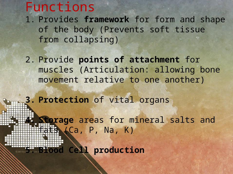
Functions1. Provides framework for form and shape of the body
(Prevents soft tissue from collapsing)
2. Provide points of attachment for muscles (Articulation: allowing bone movement relative to one another)
3. Protection of vital organs
4. Storage areas for mineral salts and fats (Ca, P, Na, K)
5. Blood Cell production

Macroscopic Structure• Diaphysis is a hollow cylinder of compact bone
containing yellow bone marrow (fat storage) in the centre
• Epiphyses contain spongy (cancellous bone) in centre. Large spaces in spongy bone are often filled with red bone marrow. (Red blood cell production)
• Periosteum: dense white fibrous covering
• Articular Cartilage: cartilage covering each epiphysis.

Microscopic Structure• Bone is a connective tissue (cells separated by large
amounts of non-cellular matrix) with inorganic salt deposits. (increase strength / rigidity)
• Composed of bones cells (osteocytes)
1. Haversian System/ Osteons (compact bone): • Projections from bone cells enter canaliculi and make
contact with adjacent cells (Material transfer)• Haversian/Central canal contains at least one blood
capillary. (may also contain nerves / lymph capillary)• Haversian system runs parallel to long axis of bone
(adds strength)• Lamellae: layers of bony matrix• Lacunae: pockets (spaces) between lamellae

Microscopic Structure
2. Trabeculae (spongy bone): • Irregular arrangement of thin, bony plates.• Contains bone cells but not in concentric layers• Nerves and blood vessels pass through irregular spaces in
matrix

CARTILAGE

Microscopic Structure
• A connective tissue• Perichondrium: fibrous protective layer• Only external blood vessels (in perichondrium), relies on
diffusion through matrix.• Consists of protein fibres (Collagen) which are embedded
in Protein-carb complex (chondrin).
• Chondroblasts: immature cartilage cells in the spaces in the matrix (produce matrix which will eventually surround it)
• Chondrocytes: mature cartilage cells which are inside a pocket (lacunae) surrounded by chondrin

CLASSIFICATOIN• Three types (based on thickness of fibres in matrix)1. Hyaline: Very fine and very dense for strength. E.g. larynx
(part), trachea, bronchi, articular cartilage, nose (part).
2. Elastic: medium thickness with elastic properties. E.g. ear, larynx (part), epiglottis, nose (part)
3. Fibrocartilage: thick and less dense, allowing for slight compression ideal for areas which withstand high pressure. E.g intervertebral discs, knee & pelvic joint.

joints

1. Fixed (Fibrous joints)• No movement occurs between the bones
involved.• Held in place by fibrous connective tissue• On impact bone fracture rather that joint
damage.• E.g. skull, teeth/jaw
2. Slightly movable (cartilaginous joints)• Allows very limited movement• Held in place by fibrous cartilage• eg symphysis pubis, vertebrae joints, joints
between ribs and sternum
3. Freely movable (synovial joints)• Amount of movement is limited only by
ligaments, muscles, tendons and adjoining bones.
• Highly mobile but equally weak

Ball-and-socket jointsSpherical head of one bone fits into cup-like head of another•Only occur in two places: shoulder (humerus/scapula) & hip (femur/pelvis)
Hinge JointAllows movement in one plane only.•Convex surface of one bone fits into concave surface of another
• E.g. elbow (ulna/Humerus), wrist (radius/carpals) , knee (femur/tibia), ankle (tibia/tarsals), fingers & toes (phalanges)

Pivot Joint•Rounded, pointed or conical end of one bone articulates with a ring (part bone, part ligament)•E.g. 1st vertebrae (head) / 2nd vertebrae & radius / ulna
Gliding JointGliding movement in any direction (back/forth, side/side), limited only by ligaments or bony processes.•E.g. carpals, tarsals, sternum/clavicle, scapula/clavicle

Saddle jointTwo saddle shaped joints•Allows side/side and back/forth movements•e.g. thumb (carpal/metacarpal)
Condyloid (ellipsoid) jointSlightly convex fits with slightly concave•Allows side/side or back/forth movements•e.g. radius/carpal, metacarpal/phalanges, metatarsals/phalanges

STRUCTURE OFA SYNOVIAL JOINT• Capsule: surrounding and enclosing the joint (2 layers): • 1) fibrous capsule (outer layer), dense, fibrous
connective tissue attached to periosteum . Flexibility allows movement but strength prevents dislocation
• 2) Synovial membrane (inner layer) , vascular, loose connective tissue
• Synovial Fluid (0.5mL): Secreted by synovial membrane, fills synovial cavity. Lubricates, nourishes & contains phagocytic cells.
STRUCTURE OFA SYNOVIAL JOINT• Articular Cartilage: provides smooth surface for
movement• Articular Discs: (in knee - Menisci/meniscus)
fibrocartilage extending inward from articular Capsule. Divide synovial cavities into two cavities.
• Bursae: little sacs of synovial fluid. Prevent friction between a bone and a ligament/tendon/skin
• Accessory Ligaments: hold bones together

Factors keeping bones together:• fit of articulating bones• Strength of bone ligaments• Tension by surrounding muscles

Movement of a Joint:
•Flexion: (bending) decreases angle between articulating bones•Extension: (Straightening) increases angle between articulating bones•Abduction: movement away from the body•Adduction: movement towards from the body•Rotation: Movement of a bone around its long axis.
![Dream is Collapsing From Inception[1]](https://static.fdocuments.in/doc/165x107/577ce4f91a28abf1038f87c9/dream-is-collapsing-from-inception1.jpg)