BMB 170 Lecture 14 Lipids and Membranes, Nov...
46
BMB 170 Lecture 14 Lipids and Membranes, Nov 9th Electron tomographic reconstructions The Boulder Laboratory for 3D Electron Microscopy of Cells http://bio3d.colorado.edu Part of a yeast cell
Transcript of BMB 170 Lecture 14 Lipids and Membranes, Nov...

BMB 170 Lecture 14Lipids and Membranes, Nov 9th
Electron tomographic reconstructionsThe Boulder Laboratory for 3D Electron Microscopy of Cells
http://bio3d.colorado.edu
Part of a yeast cell

C. jejuni PglB
Knockout

Treponema tomogram (Jensen)

“Hydrophobic effect” and lipid bilayers
“Ben Franklin Stilled the Waves” C. Tanford, Oxford (2004)
• Oil on water experiments - Benjamin Franklin 1774, Lord Rayleigh 1890, Agnes Pockels 1892, Irving Langmuir 1917
• Cells– Have a barrier that contains cell contents
• Impermeable to large molecules and small polar molecules
• Permeable to Water• Some molecules must be able to cross the membrane!
– Contain two types of lipids (France early 1800s)• Constant – phospholipids• Variable – fats and oils (storage)

“Hydrophobic effect” and lipid bilayers
“Ben Franklin Stilled the Waves” C. Tanford, Oxford (2004)
• Charles Overton (1899)– Cells completely permeable to many neutral molecules (cell type
independent – anesthetics on tadpoles)– Made the connection that increasing hydrophilicity decreased
permeability– Compared to oil permeability (membranes are similar to oils!)
• Gorter and Grendel (1924)– Gorter was a famous pediatrician – “We propose to demonstrate in this paper that the chromocytes of
different animals are covered by a layer of lipoids just two molecules thick.” J Exp Med
• Measured red blood cell surface area by microscopy• Isolated membranes and measured lipid surface area • Argued that the membrane would be similar to fatty acid crystals (Bragg
1924)

“Hydrophobic effect” and lipid bilayers
“Ben Franklin Stilled the Waves” C. Tanford, Oxford (2004)
• Danielli and Davson (1935)
• Robertson (1959)

The Fluid Mosaic Model
Singer & Nicolson Science (1972) 175:720-31
• Phospholipids assemble into a bilayer through the “hydrophobic effect”– Interior is apolar (largely impermeable to polar molecules - water, ions…)
• Proteins required for transport of polar molecules across membranes– There are peripheral and integral membrane proteins– They have apolar surfaces

Lipid forms
Fig 4.1

Components of sphingomyelin
Fig. 4.6
Phosphocholine(w/o a ceramide)
Amino-alcohol sphingosine
Fatty acid (R-aliphatic chain)
#1#2
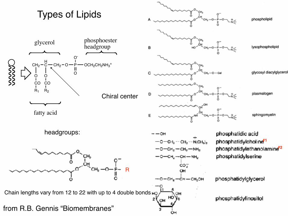
headgroups:
from R.B. Gennis “Biomembranes”
Types of Lipids
R
Chain lengths vary from 12 to 22 with up to 4 double bonds
Chiral center
CH2 CH2
O
HC
O
O P OCH2CH2NH3+
O-
O
CO CO
R1 R2
glycerol phosphoester headgroup
fatty acid
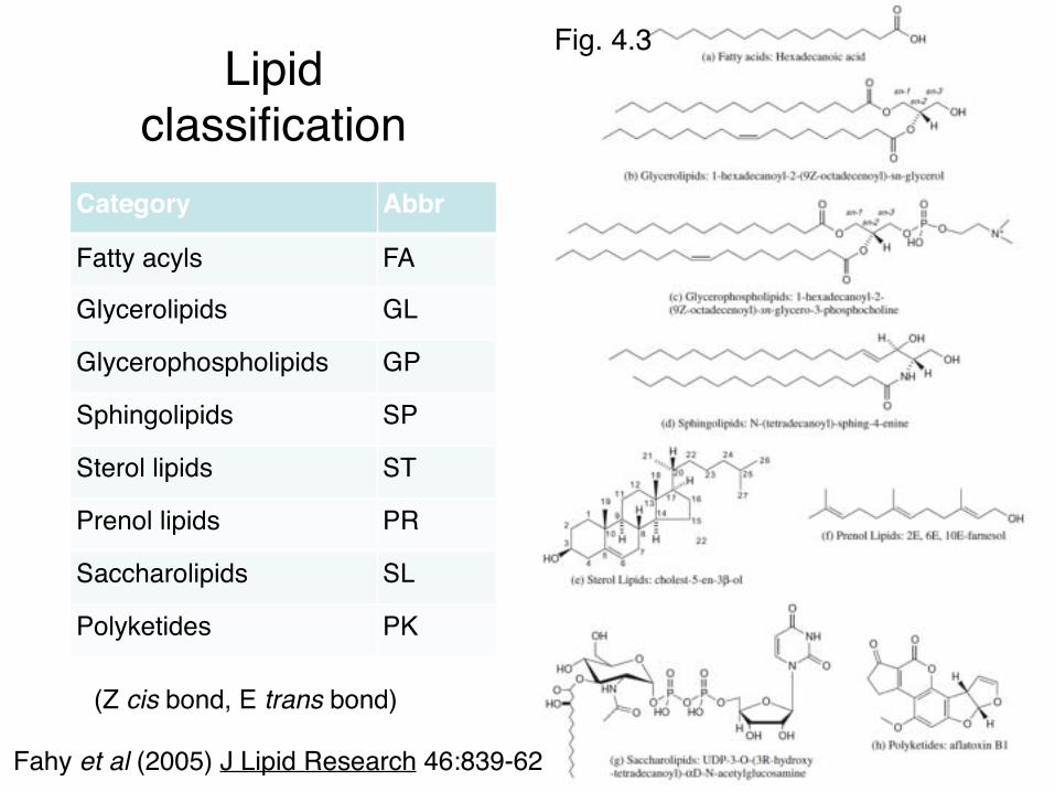
Lipid classification
Category Abbr
Fatty acyls FA
Glycerolipids GL
Glycerophospholipids GP
Sphingolipids SP
Sterol lipids ST
Prenol lipids PR
Saccharolipids SL
Polyketides PK
(Z cis bond, E trans bond)
Fahy et al (2005) J Lipid Research 46:839-62
Fig. 4.3

Common acyl chains
Fig. 4.5
DP-dipalmitoylPO-palmitoyloleoylPL-palmitollinoleoylPA-palmitoyarachidonylPD-palmitoyldocosahexaenoyl

• Phospholipid bilayer
• Thermophilic archaea– Increased hydrophobicity– Ethers (not esters) – more stable
• Hyperthermophilic archaea
Archaeal lipidsO
O
PO
OO
OOO
P OO
OOO
P OO
PO
OOO
P OO
PO
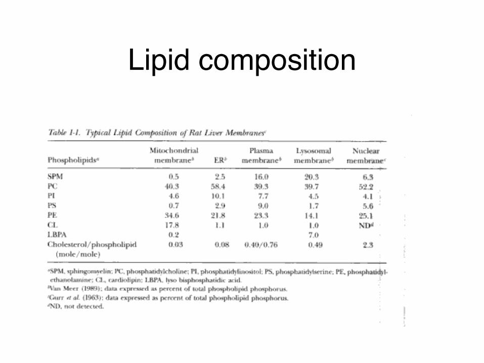
Lipid composition
L β L α
Nagle & Tristram-Nagle COSB (2000)10:474-80
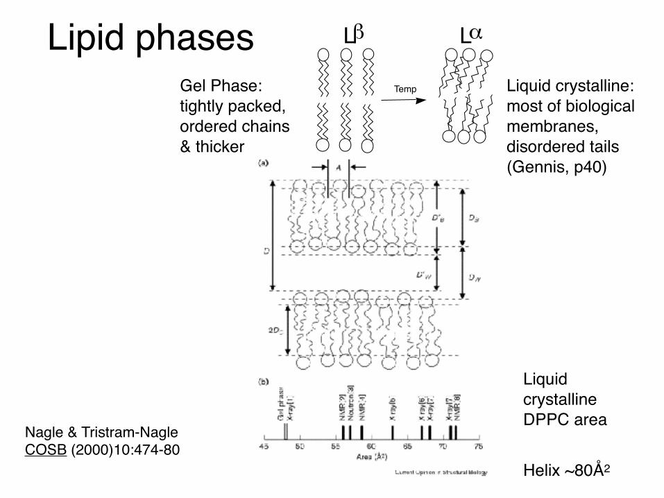
Lipid phasesTempGel Phase:
tightly packed, ordered chains & thicker
Liquid crystalline: most of biological membranes, disordered tails (Gennis, p40)
Liquid crystalline DPPC area
Helix ~80Å2

Liquid crystalline & gel phasesPhase Dimension Name Order
Lamellar One Lα Disordered, fluid
Ripple gel Two; oblique or centered Pβ’ Rippled
Gel One Lβ All trans chains
Normal hexagonal Two HI Disordered, fluid oil-in-water
Reversed hexagonal Two HII Disordered, fluid water-in-oil
Cubic Three I Disordered, fluid
Normal Cubic Three II Disordered, fluid
Reversed Cubic Three III Disordered, fluid
Table 4.2Fig. 4.8

Phase diagram• Thermotropic – phase
changes based on temperature
• Lyotropic – can form liquid crystal phasesPhase NameLamellar LαRipple gel Pβ’
Gel LβNormal hexagonal HI
Reversed hexagonal HII
Cubic I
Normal Cubic IIReversed Cubic III Fig. 4.7

Lipid shape affects curvature• Packing parameter: P=v/al• l – length of hydrophobic
chain• a – optimal cross-sectional
area of the head group• When P≅1 (cylinders) you
get bilayers• P>1 curve towards water• P<1 curve away
Fig. 4.11
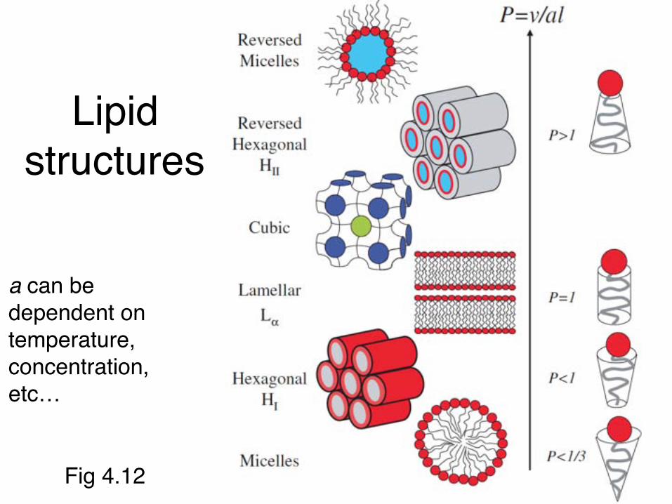
Lipid structures
a can be dependent on temperature, concentration, etc…
Fig 4.12

More structures
Fig. 4.14 & 4.15
Bicontinuous cubic phasesShape affects radius
Not empty space

Membrane fluidity• Pulling on the
membrane with laser tweezers
• Movie by Steven Block (Stanford)

Heller, Schaeffer & Schulten J Phys Chem (1993) 97:8343-60
20 Å
40 Å
Molecular dynamics simulation of 200 lipids in a bilayer

Bilayer distribution

Lewis & Engelman JMB (1983)166:211

20Å
32Å
capsid
bilayer
DNA
Stuart lab, Cockburn et al Nature (2004) 432:122
• Structure of bacteriophage PRD1– showed boundaries of membrane and DNA
• Dimensions of membrane– most hydrophobic region ~ 20Å– peak to peak distance ~ 32Å; expect ~36Å (10% thinning)– Phospholipid asymmetry
• zwitterionic PE in inner leaflet (DNA)• anionic PG and Cardiolipin in outer leaflet.
4Å Crystal structure of enveloped virus
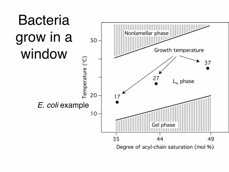
Bacteria grow in a window
E. coli example

Asymmetric layers
Devaux Annu Rev Biophys Biomol Struct (1992) 21:417-39

Asymmetry can drive structure
• Giant vesicles of phosphatidylcholine
• 1% phosphatidylglycerol and raise pH (4.5-9.5)
• 0.1% redistribution
Farge & Devaux Biophys J (1992) 61, 347-57

Lipids crossing membrane▪ Flipping is intrinsically slow
• Hours to weeks across artificial membranes• Tens of seconds or less across biogenic
membranes▪ Flippases▪ Glycosylated lipids may never cross
Sanyal & Menon (2009) Chem Bio 4:895

Flipping mechanisms
Pore model Slip-pop model for single TMs
Isoprenoid based flippases?
Review: Sanyal & Menon (2009) Chem Bio 4:895MsbA structure: Ward..Chang (2007) PNAS 104:19005

Membrane remodeling is dynamic!• Interplay between lipids and proteins
• Can be temporary or permanent• Can be local or wider region• Functions of membrane
curvature‣ Movement‣ Division‣ Vesicle trafficking/ viral budding/
tubule carrier formation
McMahon & Gallop Nature Review (2005) 438: 590

Lipids are dynamic systems
• Mixed saturated (blue), unsaturated (black) and cholesterol
• Phase separation of saturated lipids and cholesterol from the unsaturated lipids
Tristan Ursell, Phillips lab (now a Prof at U of Oregon)
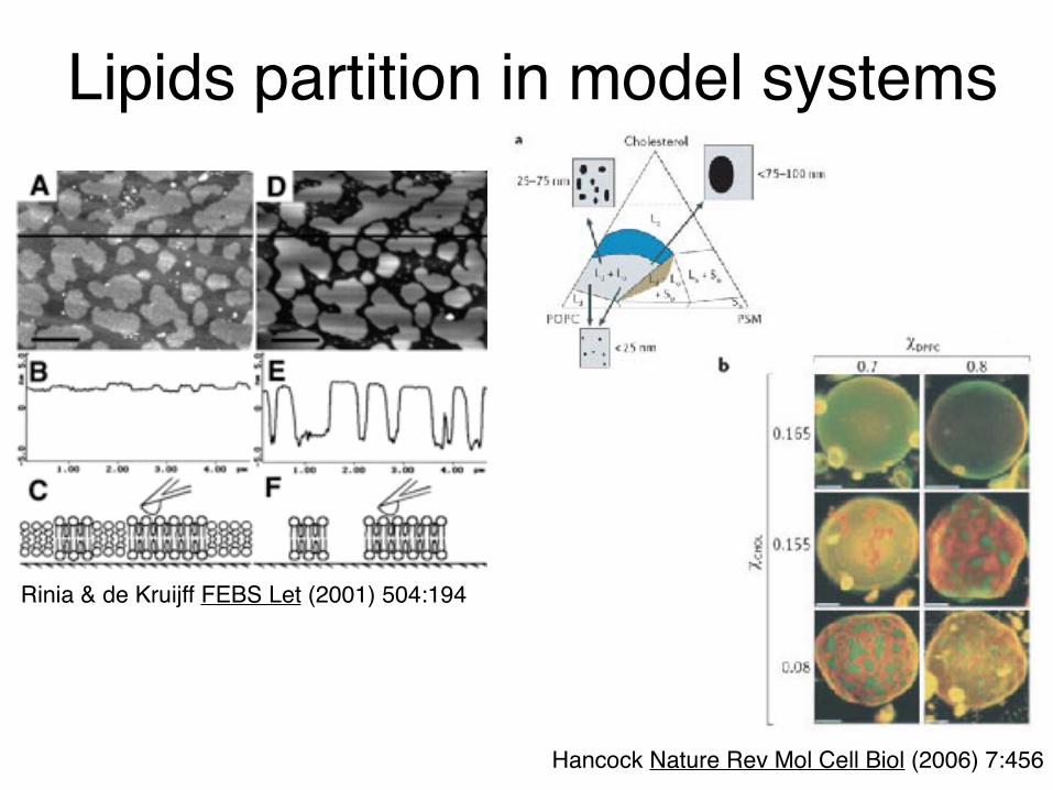
Lipids partition in model systems
Hancock Nature Rev Mol Cell Biol (2006) 7:456
Rinia & de Kruijff FEBS Let (2001) 504:194

Cholesterol-induced lateral phase separation and phases
Figure 4.28 & 4.29
▪ Saturated phospholipid, cholesterol and water▪ ld – liquid-disordered phase
▪ lo- liquid-ordered phase
▪ so- gel phase
▪ Tm – temperature at the phase transition from gel to lamellar

Lipid rafts▪ Compartmentalization of cellular membranes into lateral subdivision▪ Small (10–200 nm), heterogeneous, highly dynamic, sterol- and
sphingolipid-enriched domains that compartmentalize cellular processes▪ Enriched with cholesterol, sphingolipids, and proteins▪ Drive signal transduction, lipid trafficking, cytoskeletal organization, and
viral budding and entry▪ The true nature of membrane organization in living cells remains unknown
Levental & Veatch J Mol Biol (2016)Pike J Lipid Res (2003) 44:655
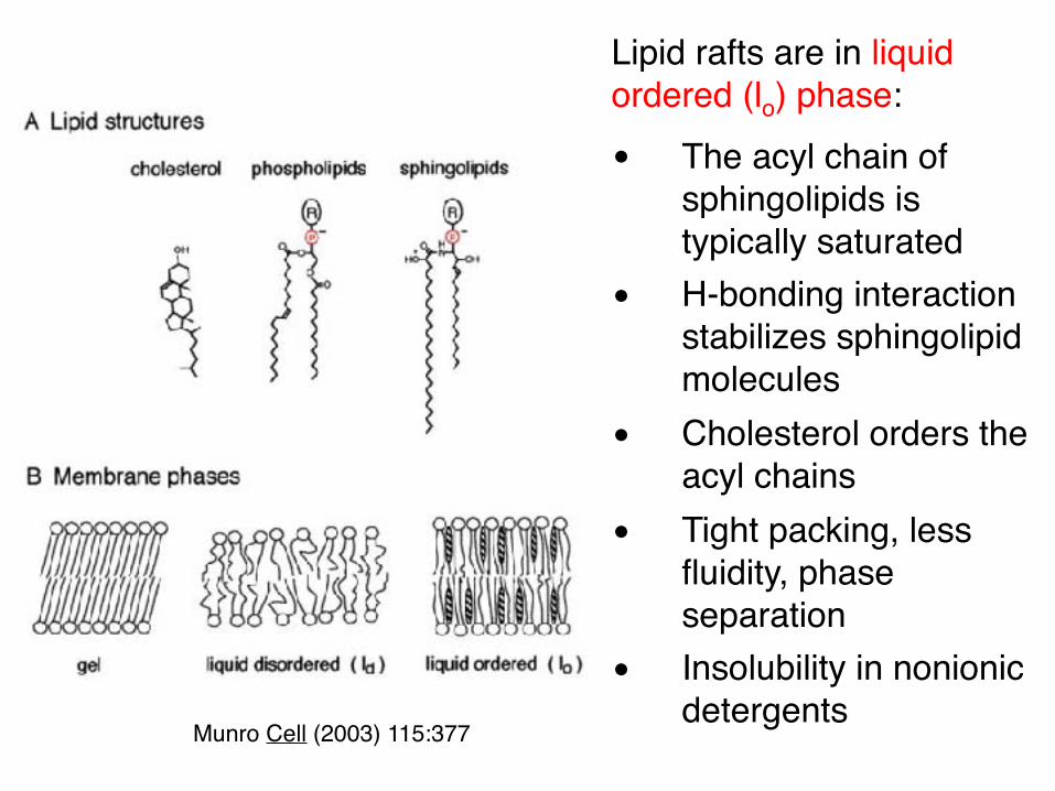
Munro Cell (2003) 115:377
Lipid rafts are in liquid ordered (lo) phase:• The acyl chain of
sphingolipids is typically saturated
• H-bonding interaction stabilizes sphingolipid molecules
• Cholesterol orders the acyl chains
• Tight packing, less fluidity, phase separation
• Insolubility in nonionic detergents

▪ Intrinsic factors: a, b, e ▪ Extrinsic factors: c, d
▪ Positive curvature: early stages of vesicle budding
▪ Negative curvature: viruses budding out of the cell
McMahon & Gallop Nature Review (2005) 438: 590

Sensing membrane curvatureBar Domains Reticulons
Amphiphysin BAR structure (1uru)
Class of proteins that drive membrane asymmetryrequired for the ER tubulation
McMahon lab: Peter et al Science (2004) 303:495 Rapoport lab: Voeltz et al Cell (2006) 124:573

Lateral pressure profile
Figure 4.17
At equilibrium, all the different forces exerted on the lipid bilayer have to cancel (total pressure = zero)
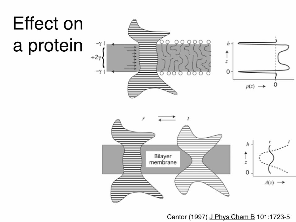
Effect on a protein
Cantor (1997) J Phys Chem B 101:1723-5

Effect on a protein
Perozo & Rees (2003) Curr Op Struct Biol 13:432
▪ A prokaryotic mechanosensitive channel (MscL)
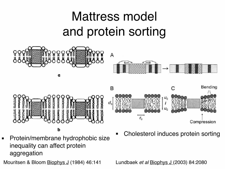
Mattress model and protein sorting
▪ Cholesterol induces protein sorting
Mouritsen & Bloom Biophys J (1984) 46:141 Lundbaek et al Biophys J (2003) 84:2080
▪ Protein/membrane hydrophobic size inequality can affect protein aggregation

€
D =kBT4πµh
ln µhµ'R#
$ %
&
' ( − γ
#
$ %
&
' (
Membrane diffusion
• First calculated by Saffman & Delbrück (1975) PNAS– Continuum hydrodynamic model– Diffusion weakly correlated to radius
• Confirmed by GUV measurements– Cell length = 2μm– Surface area 6 μm2
– Rafts/Oligomerization/cytoskeleton μ – viscosity of membraneμ' – viscosity of solventR – radius of moleculeh – bilayer thickness
Ramadurai..Poolman (2009) JACS 131:12650
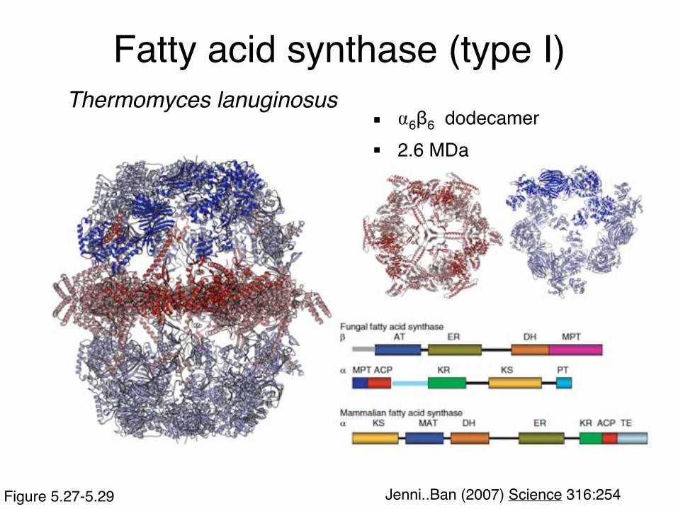
Fatty acid synthase (type I)Thermomyces lanuginosus
Jenni..Ban (2007) Science 316:254Figure 5.27-5.29
▪ ⍺6β6 dodecamer▪ 2.6 MDa

Domain organization
Fig. 5.29

Self contained reaction chamber
Figure 5.30 & 5.31 Jenni..Ban (2007) Science 316:254