01.20.10 Lecture 4 Cellular Building Blocks: Lipids and Membranes.
24
01.20.10 Lecture 4 Cellular Building Blocks: Lipids and Membranes
-
date post
19-Dec-2015 -
Category
Documents
-
view
220 -
download
2
Transcript of 01.20.10 Lecture 4 Cellular Building Blocks: Lipids and Membranes.

01.20.10Lecture 4
Cellular Building Blocks: Lipids and Membranes
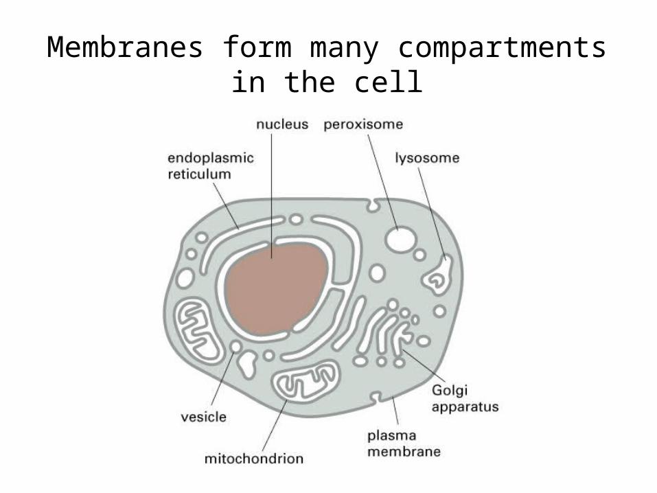
Membranes form many compartmentsin the cell

Biological membranes are composed of a lipid bilayer

Membrane lipids are amphipathic molecules
• Membrane lipids are amphipathic
• Hydrophilic heads (polar) form hydrogen bonds with water
• Hydrophobic tails (non-polar) are excluded by water molecules

Three classes of membrane lipids

Membrane lipids
Phosphatidylcholine is the most abundant phospholipid in cell membranes
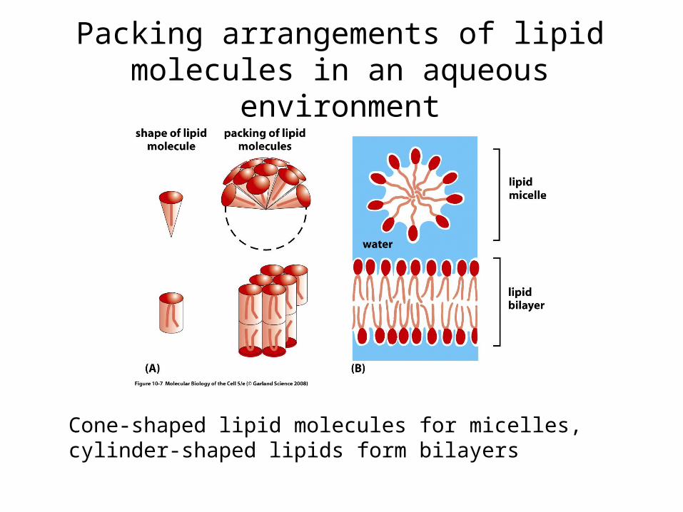
Packing arrangements of lipid molecules in an aqueous environment
Cone-shaped lipid molecules for micelles, cylinder-shaped lipids form bilayers

Phospholipid bilayers spontaneously close to form a sealed compartment

The membrane bilayer is a fluid
•Lateral diffusion occurs rapidly within the plane of the membrane
•Individual phospholipids may rotate axially
•Flip-flopping from one side to the other is very rare as it is energetically unfavorable

The membrane bilayer is a fluid: FRAP
1. A fluorescent probe is used to label membrane proteins
2. The probe is destroyed in a small region using intense laser light
3. Fluorescence microscopy is used to observe behavior of the unbleached probe
• Fluorescence microscopy of single GFP-labeled membrane proteins
• Diffusive movement within the plane of the lipid bilayer
QuickTime™ and aYUV420 codec decompressor
are needed to see this picture.
The membrane bilayer is a fluid: single molecule imaging

QuickTime™ and aMPEG-4 Video decompressor
are needed to see this picture.
The membrane bilayer is a fluid: laser tweezers
Beads trapped in a laser may be used to exert pulling forces on membranes
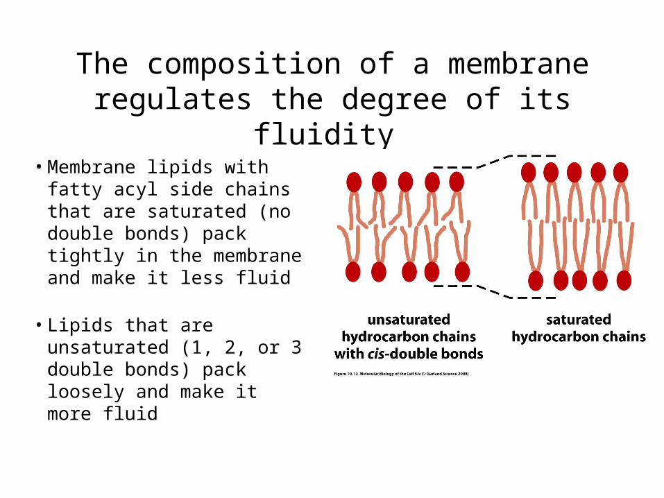
The composition of a membrane regulates the degree of its fluidity
• Membrane lipids with fatty acyl side chains that are saturated (no double bonds) pack tightly in the membrane and make it less fluid
• Lipids that are unsaturated (1, 2, or 3 double bonds) pack loosely and make it more fluid

The composition of a membrane regulates the degree of its fluidity
The presence of cholesterol in the membrane stiffens the bilayer making it more rigid

Cellular membranes are asymmetric
1. All lipids are synthesized on the cytosolic surface of the ER
2. Lipids in the outer leaflets are transported there by flippases
3. Continuity between organelle lumen & extracellular space

Membrane proteins
• Proteins compose ~50% of the membrane
• ~1 protein:50 lipid molecules
• Membrane proteins perform many functions
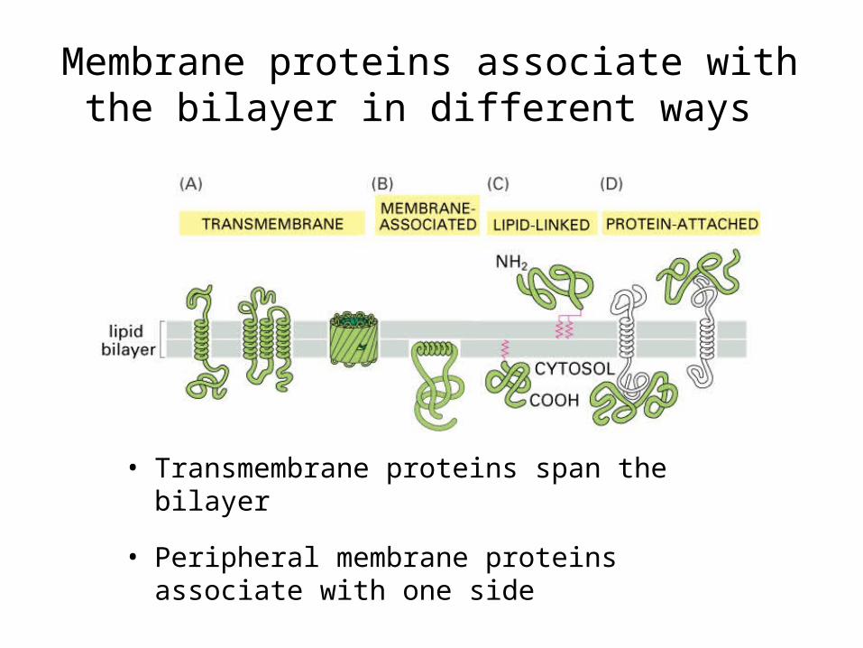
Membrane proteins associate with the bilayer in different ways
• Transmembrane proteins span the bilayer
• Peripheral membrane proteins associate with one side

Transmembrane proteins usually span the bilayer using alpha-helices

Some membrane proteins use beta-sheets to cross the bilayer
• Beta-sheets arranged in this cylindrical conformation are known as a “beta-barell”
• Hydrophilic amino acid residues face towards the pore, hydrophobics face the bilayer

The cytoplasmic side of the membrane is called the cell cortex
• Meshwork of transmembrane proteins and filaments (spectrin)
• Mechanical support for the membrane and cell shape

The extracellular surface of the membrane is coated with carbohydrate

Extracellular glycoproteins perform numerous functions
• Carbohydrate layer protects cells from chemical and mechanical damage
• Different cell types present different combinations of glycoproteins and proteoglycan on their surface - molecular signature
• Information in the carbohydrate layer aids in cell-cell recognition and communication

Cells use different mechanisms to restrict membrane protein movements
A. By tethering to elements inside of the cell (cortex)
B. By tethering to elements outside of the cell
C. By interacting with proteins on the surface of another cell
D. By diffusion barriers established within polarized cells

Epithelial cell polarity