BLOOD SUPPLY OF THE BRAIN DR.KUMAR.K.V 15/05/.2012.
33
BLOOD SUPPLY OF THE BRAIN DR.KUMAR.K.V 15/05/.2012
-
Upload
tracy-green -
Category
Documents
-
view
228 -
download
0
Transcript of BLOOD SUPPLY OF THE BRAIN DR.KUMAR.K.V 15/05/.2012.

BLOOD SUPPLY OF THE BRAIN
DR.KUMAR.K.V15/05/.2012
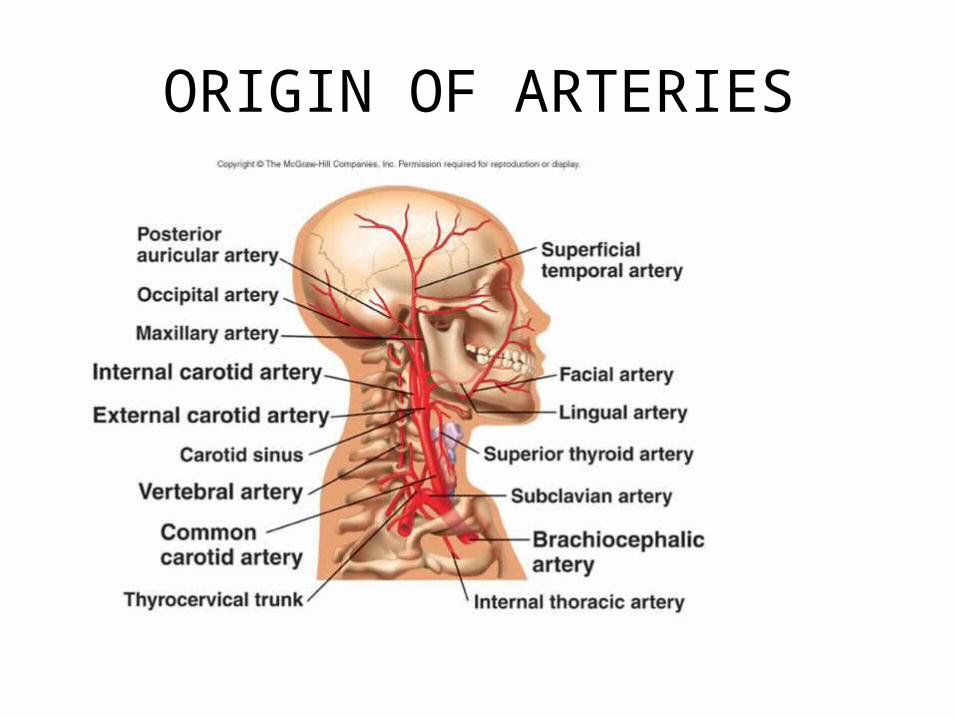
ORIGIN OF ARTERIES

ORIGIN OF INTERNAL AND VERTIBRAL ARTERIES
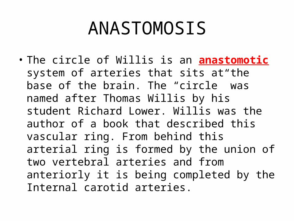
ANASTOMOSIS
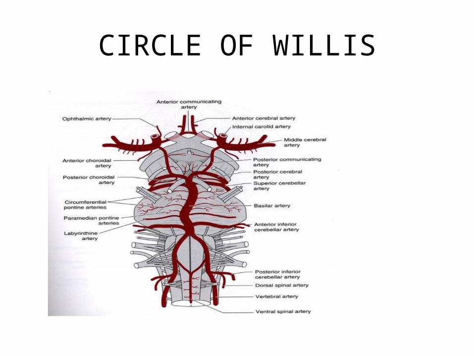
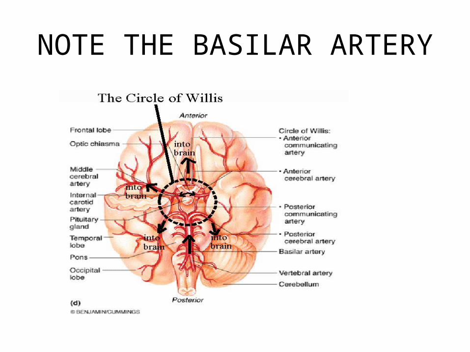
• The circle of Willis is an anastomotic system of arteries that sits at the base of the brain. The “circle” was named after Thomas Willis by his student Richard Lower. Willis was the author of a book that described this vascular ring. From behind this arterial ring is formed by the union of two vertebral arteries and from anteriorly it is being completed by the Internal carotid arteries.
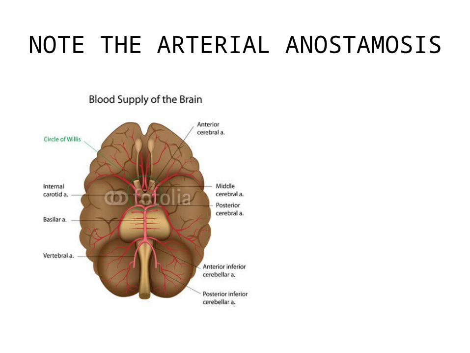
NOTE THE ARTERIAL ANOSTAMOSIS

ARTERIAL CIRCLE AT THE BASE
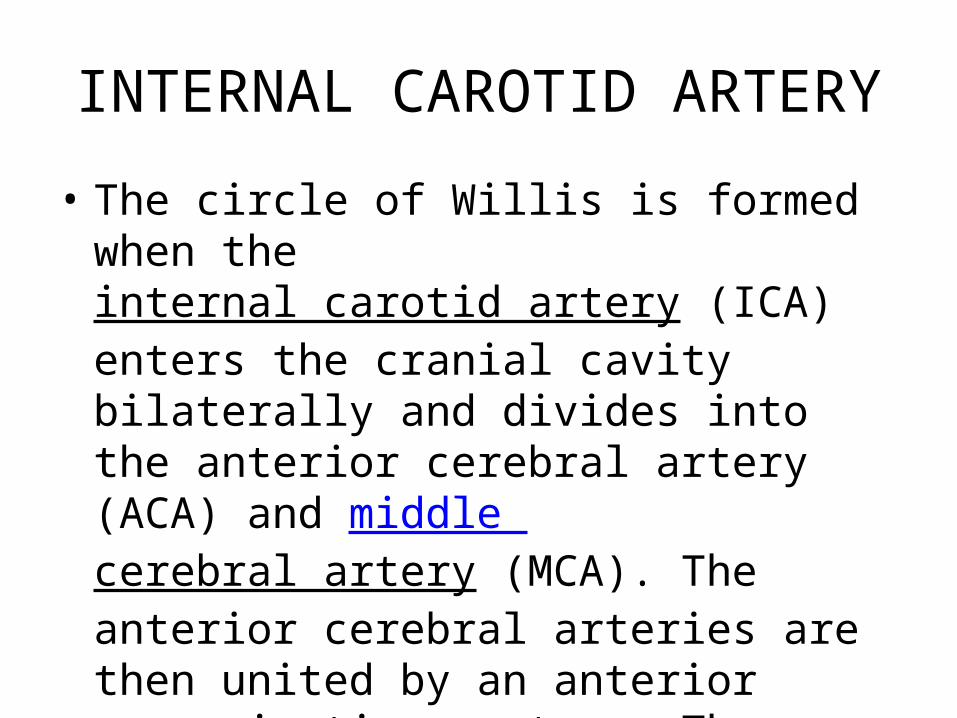
INTERNAL CAROTID ARTERY
• The circle of Willis is formed when the internal carotid artery (ICA) enters the cranial cavity bilaterally and divides into the anterior cerebral artery (ACA) and middle cerebral artery (MCA). The anterior cerebral arteries are then united by an anterior communicating artery. These connections form the anterior half (anterior circulation) of the circle of Willis
CIRCLE OF WILLIS

ARTERIAL CIRCLE OF WILLIS
• The cerebral arteries are derived from the internal carotid and vertebral, which at the base of the brain form a remarkable anastomosis known as the arterial circle of Willis. It is formed in front by the anterior cerebral arteries, branches of the internal carotid, which are connected together by the anterior communicating;

POSTERIOR AND THE MIDDLE CEREBRAL ARTERIES
• behind by the two posterior cerebral arteries, branches of the basilar, which are connected on either side with the internal carotid by the posterior communicating. The three trunks which together supply each cerebral hemisphere arise from the arterial circle of Willis. From its anterior part proceed the two anterior cerebrals, from its antero-lateral parts the middle cerebrals, and from its posterior part the posterior cerebrals.

VERTEBRAL AND THE BASILAR ARTERIES
• Posteriorly, the basilar artery, formed by the left and right vertebral arteries, branches into a left and right posterior cerebral artery (PCA), forming the posterior circulation. The PCAs complete the circle of Willis by joining the internal carotid system anteriorly via the posterior communicating (PCOM) arteries. (See the images below.)

ARTERIAL ANASTOMOSIS
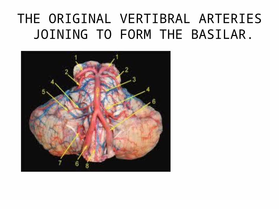
THE ORIGINAL VERTIBRAL ARTERIES JOINING TO FORM THE BASILAR.

SUPPLY TO SENSORY AND MOTOR CORTEX
• 9. The middle cerebral artery , the largest branch of the internal carotid, runs at the lateral cerebral or Sylvain fissure and then backward and upward , where it divides into a number of branches which are distributed to the lateral surface of the cerebral hemisphere and end up supplying most of lateral surface of the brain that include the motor and the sensory area
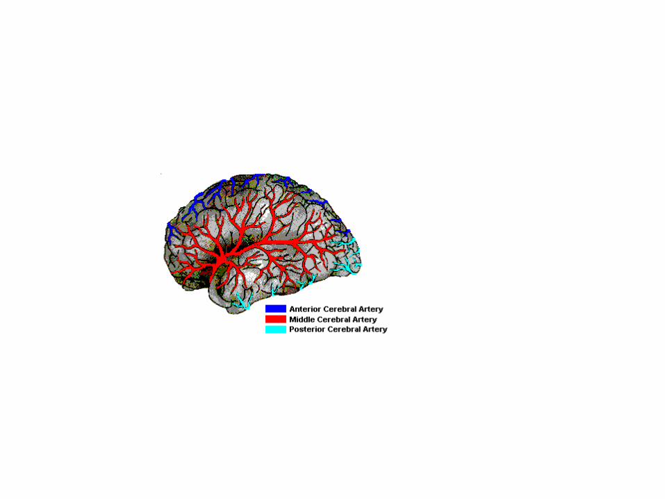

MIDDLE CEREBRAL ARTERY AND THE AREA SUPPLIED

ANTERIOR CEREBRAL ARTERY
• The Anterior Communicating Artery connects the two anterior cerebral arteries across the commencement of the longitudinal fissure.
• Most of the medial surface of the brain is covered by this artery that includes some part of the motor and the sensory cortex which lie on the medial surface called as PARACENTRAL LOBULE REPRESENTING THE LOWER LIMB.

THE PARACENTRAL LOBULE
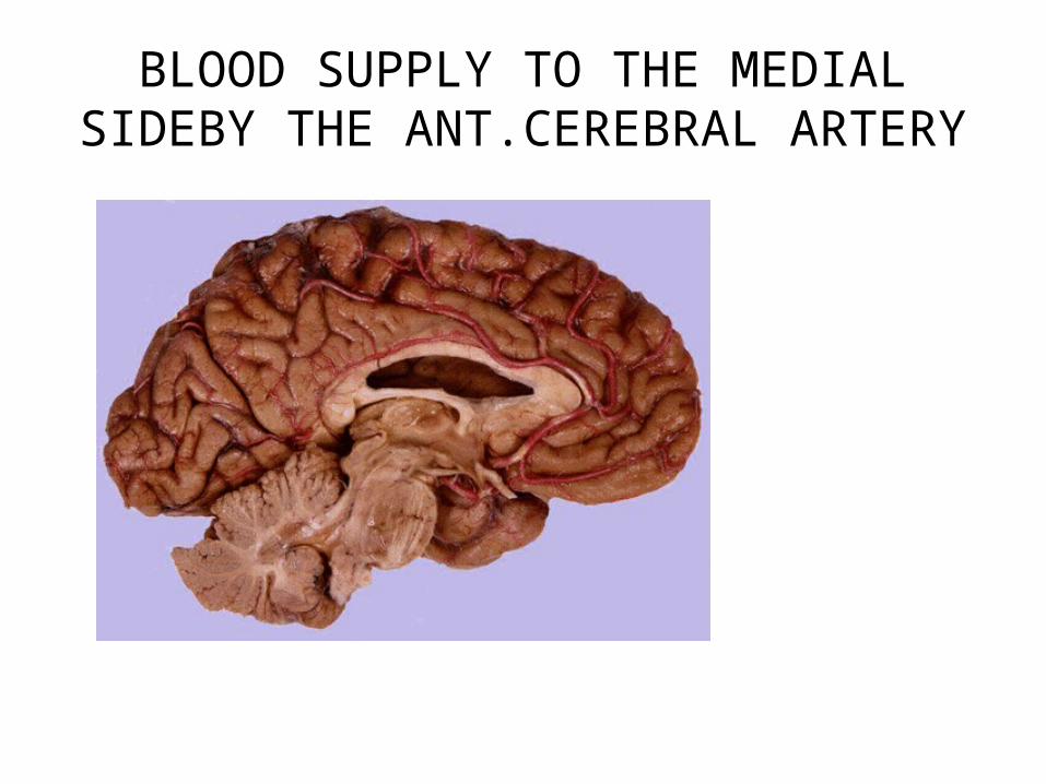
BLOOD SUPPLY TO THE MEDIAL SIDEBY THE ANT.CEREBRAL ARTERY

POSTERIOR COMMUNICATING ARTERY
• The posterior communicating artery runs backward from the internal carotid, and anastomoses with the posterior cerebral, a branch of the basilar.(BASILAR arteris are formed by the union of the vertebral arteries from behind) .
• The basilar artery lies on the pons in the basilar or pontine groove
NOTE THE BASILAR ARTERY
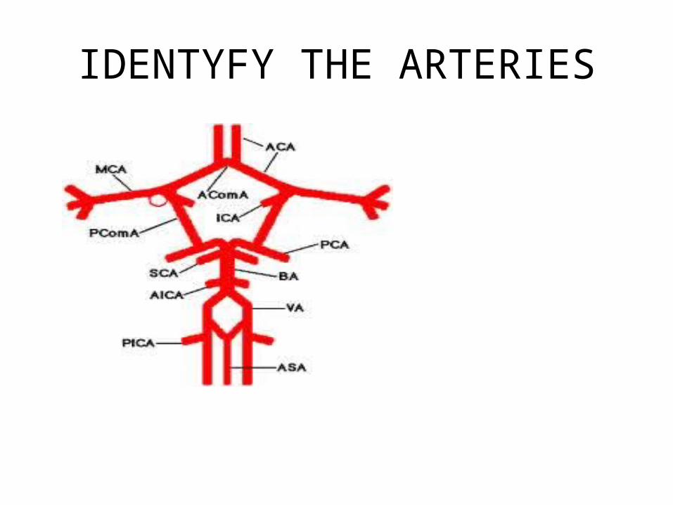
IDENTYFY THE ARTERIES

CIRCLE OF WILLIS

VEINS OF THE BRAIN
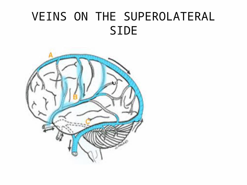
VEINS ON THE SUPEROLATERAL SIDE
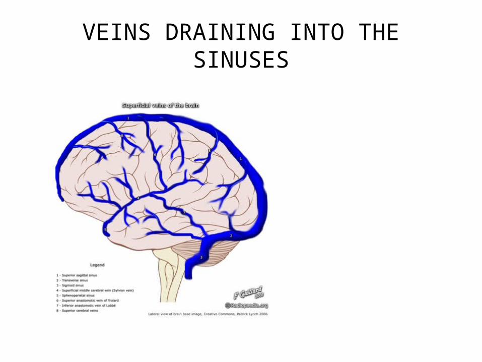
VEINS DRAINING INTO THE SINUSES
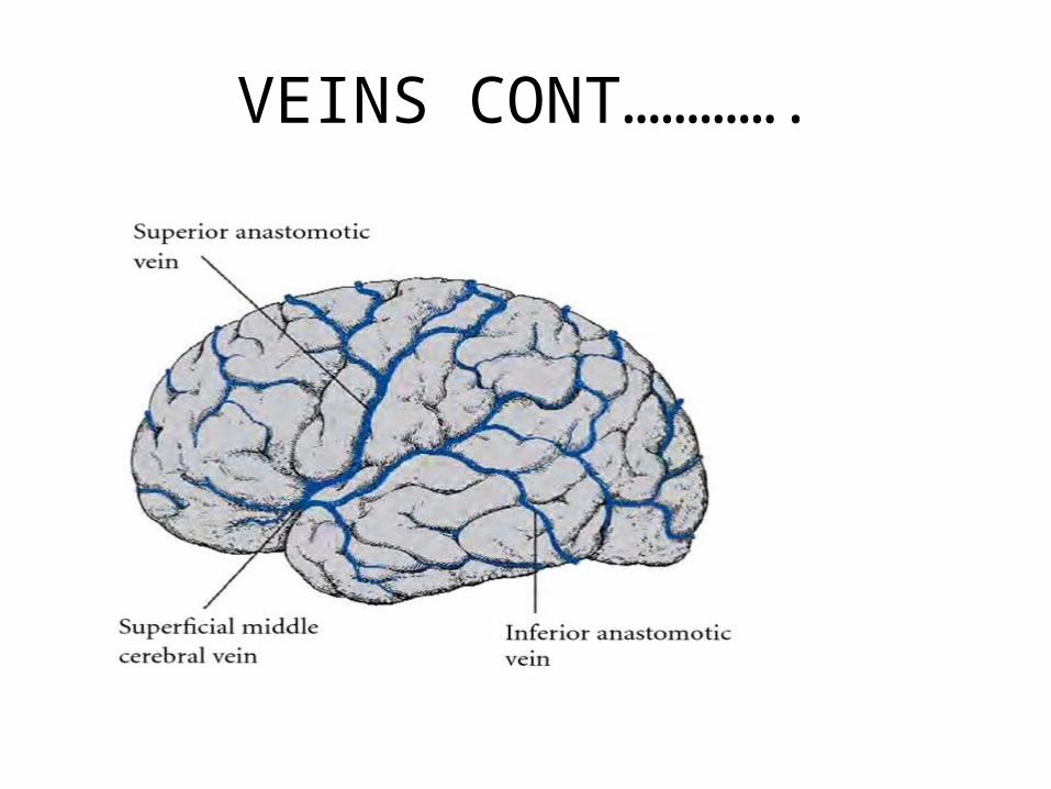
VEINS CONT………….
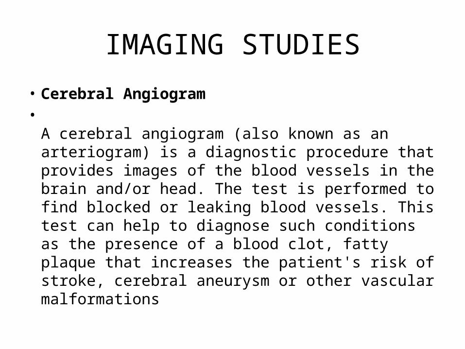
IMAGING STUDIES
• Cerebral Angiogram•
A cerebral angiogram (also known as an arteriogram) is a diagnostic procedure that provides images of the blood vessels in the brain and/or head. The test is performed to find blocked or leaking blood vessels. This test can help to diagnose such conditions as the presence of a blood clot, fatty plaque that increases the patient's risk of stroke, cerebral aneurysm or other vascular malformations

HOW IT IS DONE
• A cerebral angiogram requires that a special dye be injected into the arteries of the head or brain. Under the direction of an expert physician, this procedure is done by inserting a thin tube (a catheter) through a blood vessel, (most often starting in the patients thigh) all the way up to the head and/or brain. When the catheter is in the correct position the dye is them injected. At this point the cerebral angiogram can generate the images of the blood vessels

CEREBROVASCULAR ACCIDENTS
• What is a stroke?A person with a stroke has an interruption in the blood supply to an area of the brain, which causes the brain to malfunction. The brain is very sensitive to any interruption in blood flow. Brain cells begin to die within minutes of losing their supply of oxygen and glucose. The interruption of blood flow can occur by one of two mechanisms.

CVA cont……
• A hemorrhagic stroke is caused by bleeding from small blood vessels that supply blood to the brain. An ischemic stroke is caused by a blood clot that blocks the flow of blood through an artery that supplies blood to the brain. There are two types of ischemic stroke: thrombotic stroke and embolic stroke
C.V.A
• There are two types of ischemic stroke: thrombotic stroke and embolic stroke. A thrombotic stroke is caused by a blood clot that forms inside the brain artery. An embolic stroke is caused by a blood clot that forms outside the brain, travels through the bloodstream, and then becomes lodged in the brain artery. About 3 out of 2,000 adults over the age of 45 have a stroke each year.

AN ANGIOGRAM STUDY
• Try to identify• The arteries forming• The arterial anosta• Mosis or circle• Of Willis