Blood Supply and Sections of the Brain
44
Dr. Akram Jaffar Dr. Akram Jaffar The Brain II Blood Supply of the CNS and Sectional Anatomy of the Brain (for dental students) Akram Jaffar, Ph.D.
-
Upload
maryamalemadi -
Category
Documents
-
view
233 -
download
0
Transcript of Blood Supply and Sections of the Brain
Dr. Akram JaffarDr.
Akra
m J
affa
r
The Brain IIBlood Supply of the CNS
and Sectional Anatomy of the Brain(for dental students)
Akram Jaffar, Ph.D.

Dr. Akram JaffarDr.
Akra
m J
affa
rObjectives
The cerebellum:Describe the position of the cerebellum and the connection of its peducles with the brain stem.Define gross features of the cerebellum: folia, sulci, vermis, tonsil, valleculaDefine the main fissures and lobes. Correlate lobes with functions and connections of the cerebellum.Define cerebellar nuclei and outline their connections.Discuss cerebellar control and dysfunction
Blood supply of the brainEnumerate the branches of the internal carotid and vertebral arteries. Map the areas of supply of the anterior, middle and posterior cerebral arteries.Correlate areas of supply of cerebral arteries with functional cortical areas.Describe the position, significance and formation of the circle of Willis.Enumerate the veins draining the brain and the venous sinuses at which they drain.
Brain sectionsIdentify features of the cerebral hemispheres in coronal and axial sections at the level of the interventricular foramen.Identify the boundaries of the third ventricle in a mid-sagittal section.Describe the parts of the basal ganglia and their relation to the lateral ventricle.Identify the parts and functions of the limbic system.Describe the major functions of the thalamus and hypothalamus.Describe how the hypothalamus controls the functions of the pituitary gland.Identify the pineal gland, its function and clinical significance.Describe the formation, circulation, and absorption of the CSF.Discuss the properties and function of the CSF.Describe the location and discuss the significance of the brain barriers: blood brain barrier & blood CSF barrier.
Further reading• Snell RS (2010): Clinical neuroanatomy. 7th ed. Lippincott Williams and Wilkins.
Baltimore.

Dr. Akram JaffarDr.
Akra
m J
affa
r
The Cerebellum
Dr. Akram JaffarDr.
Akra
m J
affa
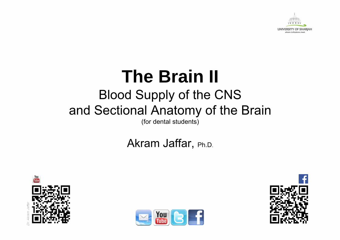
rPosition & peduncles of the cerebellum
• Lies in the posterior cranial fossa inferior to the tentorium cerebelli.
• Is attached to the back of the brain stem by three paired bundles of fibers: superior, middle, and inferior cerebellar peduncles.
Tentorium cerebelli
Superior cerebellarpeduncle
Middle cerebellarpeduncle
Inferior cerebellarpeduncle
Cerebellum
Cerebellum
4th ventricle

Dr. Akram JaffarDr.
Akra
m J
affa
rGross appearance of the cerebellum
• Two cerebellar hemispheres joined by a narrow median vermis.• The inferior surface shows a deep groove, the vallecula, the floor of which is
formed by the inferior aspect of the vermis.• The tonsil is a partly detached lobule overhanging the inferior vermis on each
side.
Dorsal view Ventral view
vermis vermis
vallecula
tonsil
Dr. Akram JaffarDr.
Akra
m J
affa
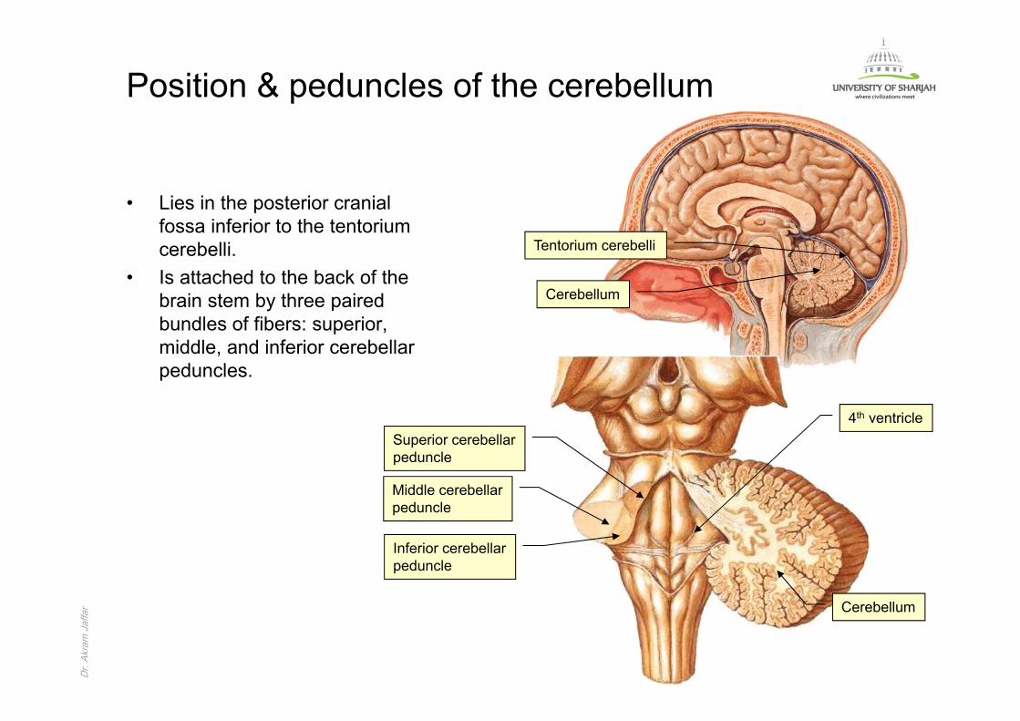
rGross appearance of the cerebellum
• The cortex, which is greatly convoluted.• The sulci are parallel and the ridges between them are called the folia.• In some places deep fissures are present.• The cerebellum forms the roof for the fourth ventricle.
Horizontal section of the cerebellum at the middle cerebellar peduncle
cortex
sulcus
folium
4th ventricle
Fissure
4th ventricle

Dr. Akram JaffarDr.
Akra
m J
affa
rCerebellar nuclei• Four pairs of nuclei. • The largest is the dentate nucleus.• Cerebellar afferents project to the cerebellar cortex, whose output is mostly to the
cerebellar nuclei in which efferent fibers originate.
Dentate nucleus

Dr. Akram JaffarDr.
Akra
m J
affa
rFissures & lobes of the cerebellum • Primary fissure:
– Located on the superior surface. – Separates the anterior lobe from
the middle (posterior) lobe.• Horizontal fissure
– Located posteriorly within the middle lobe.
• The uvulo-nodular fissure– Located on the inferior surface.– Separates the middle lobe from
the flocculo-nodular lobe.
Dorsal view
Ventral view
Primary fissure
Horizontal fissure
Uvulo-nodular fissure
Anterior lobe
Middle lobe
Flocculo-nodular lobe
Dr. Akram JaffarDr.
Akra
m J
affa
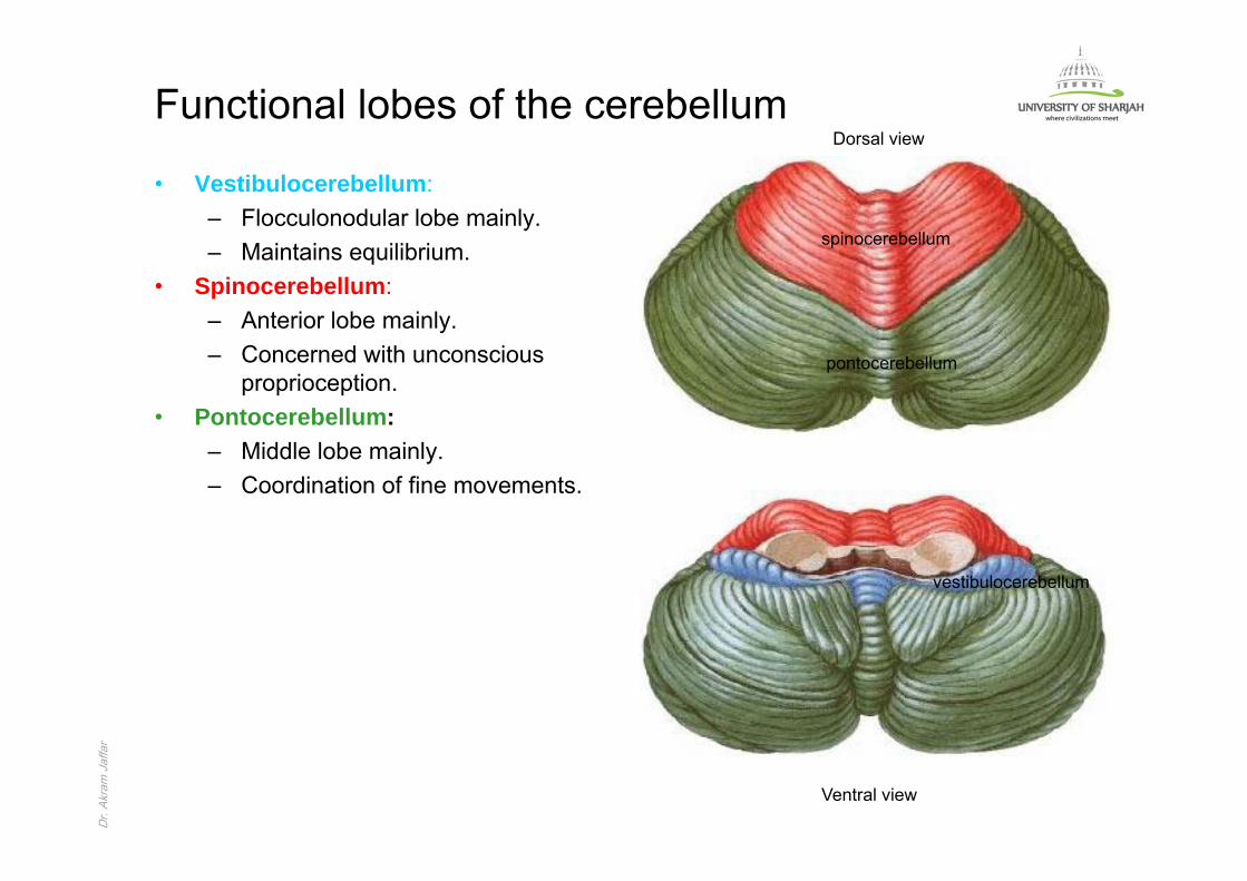
rFunctional lobes of the cerebellum
• Vestibulocerebellum:– Flocculonodular lobe mainly.– Maintains equilibrium.
• Spinocerebellum:– Anterior lobe mainly.– Concerned with unconscious
proprioception.• Pontocerebellum:
– Middle lobe mainly.– Coordination of fine movements.
Dorsal view
Ventral view
vestibulocerebellum
spinocerebellum
pontocerebellum
Dr. Akram JaffarDr.
Akra
m J
affa
rCerebellar control and dysfunction
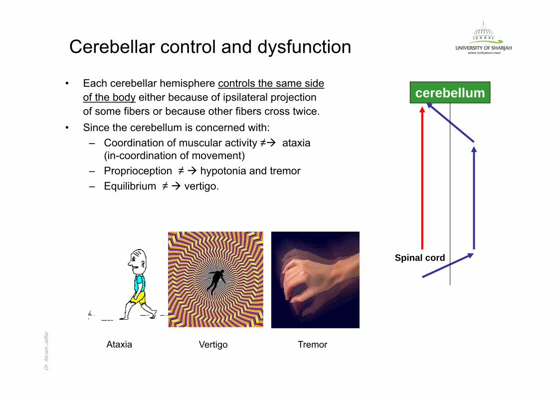
• Each cerebellar hemisphere controls the same side of the body either because of ipsilateral projection of some fibers or because other fibers cross twice.
• Since the cerebellum is concerned with:– Coordination of muscular activity ≠ ataxia
(in-coordination of movement)– Proprioception ≠ hypotonia and tremor– Equilibrium ≠ vertigo.
cerebellum
Spinal cord
Ataxia Vertigo Tremor
Dr. Akram JaffarDr.
Akra
m J
affa
r
Blood supply of the brain
Dr. Akram JaffarDr.
Akra
m J
affa
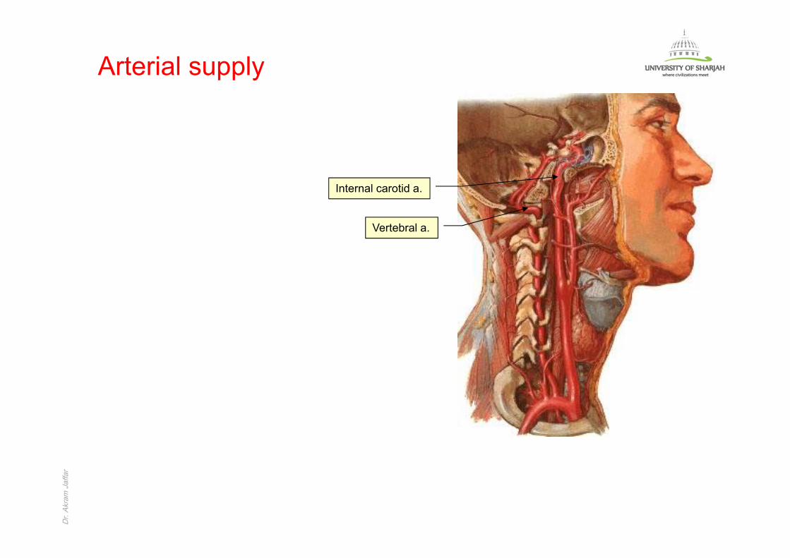
rArterial supply
Internal carotid a.
Vertebral a.
Dr. Akram JaffarDr.
Akra
m J
affa
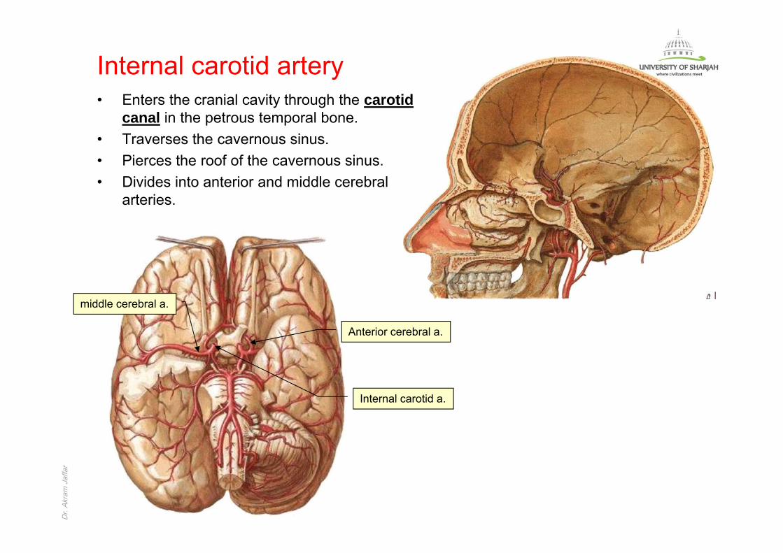
rInternal carotid artery• Enters the cranial cavity through the carotid
canal in the petrous temporal bone.• Traverses the cavernous sinus.• Pierces the roof of the cavernous sinus.• Divides into anterior and middle cerebral
arteries.
Anterior cerebral a.
middle cerebral a.
Internal carotid a.
Dr. Akram JaffarDr.
Akra
m J
affa
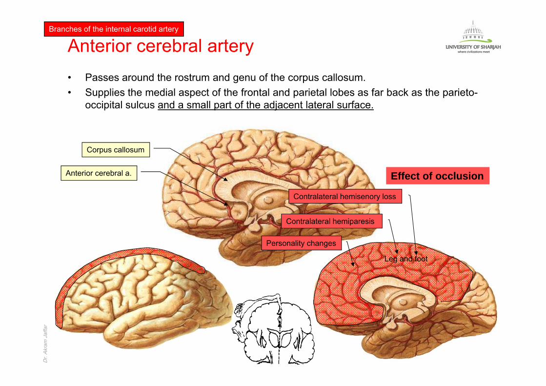
rAnterior cerebral artery• Passes around the rostrum and genu of the corpus callosum.• Supplies the medial aspect of the frontal and parietal lobes as far back as the parieto-
occipital sulcus and a small part of the adjacent lateral surface.
Branches of the internal carotid artery
Anterior cerebral a.
Corpus callosum
Effect of occlusion
Contralateral hemiparesis
Contralateral hemisenory loss
Personality changes
Leg and foot
Dr. Akram JaffarDr.
Akra
m J
affa
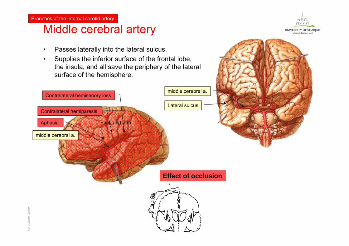
rMiddle cerebral artery• Passes laterally into the lateral sulcus.• Supplies the inferior surface of the frontal lobe,
the insula, and all save the periphery of the lateral surface of the hemisphere.
Branches of the internal carotid artery
Lateral sulcus
middle cerebral a.
middle cerebral a.
Effect of occlusion
Contralateral hemiparesis
Contralateral hemisenory loss
Face and armAphasia

Dr. Akram JaffarDr.
Akra
m J
affa
rOther branches
• Ophthalmic artery orbit• Anterior choroidal artery choroid
plexus of the lateral ventricle• Posterior communicating artery
posterior cerebral artery
Branches of the internal carotid artery
Ophthalmic a.
Post. communicating a.
Ant. Choroidal a.
Post. cerebral a.
Dr. Akram JaffarDr.
Akra
m J
affa
rVertebral artery
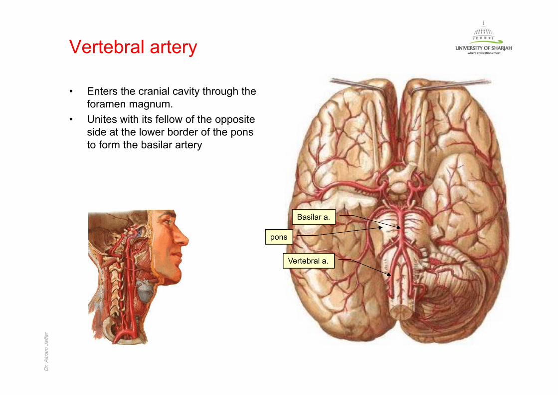
• Enters the cranial cavity through the foramen magnum.
• Unites with its fellow of the opposite side at the lower border of the pons to form the basilar artery
Basilar a.
Vertebral a.
pons
Dr. Akram JaffarDr.
Akra
m J
affa
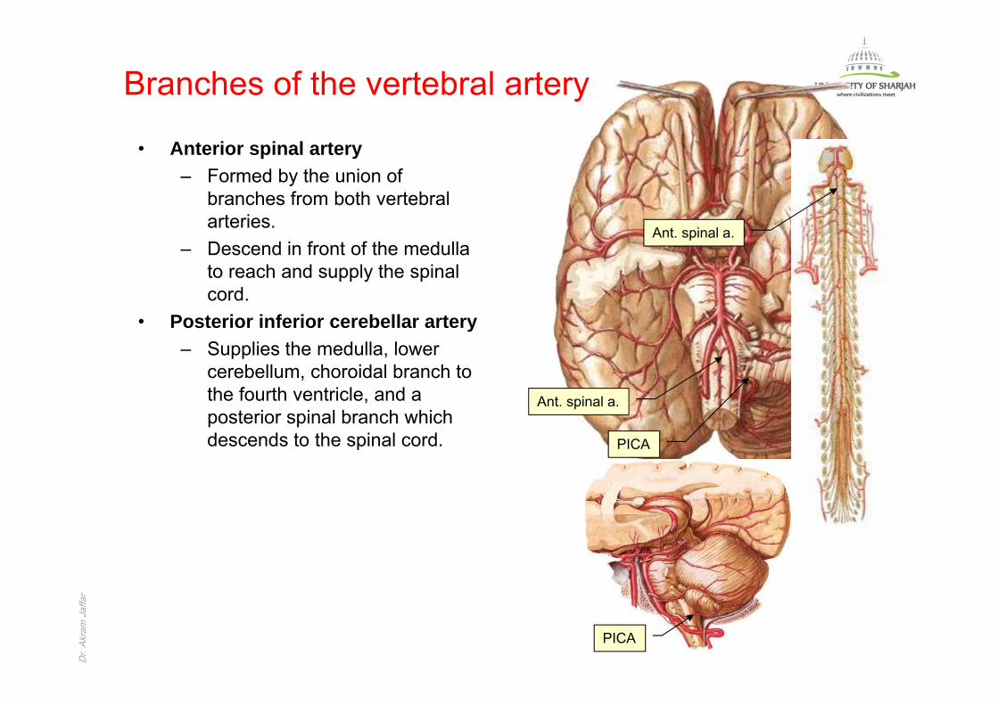
rBranches of the vertebral artery
• Anterior spinal artery– Formed by the union of
branches from both vertebral arteries.
– Descend in front of the medulla to reach and supply the spinal cord.
• Posterior inferior cerebellar artery– Supplies the medulla, lower
cerebellum, choroidal branch to the fourth ventricle, and a posterior spinal branch which descends to the spinal cord.
Ant. spinal a.
Ant. spinal a.
PICA
PICA
Dr. Akram JaffarDr.
Akra
m J
affa
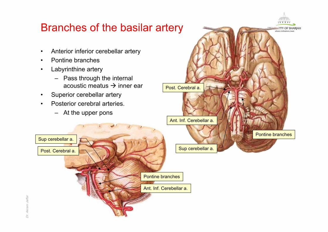
rBranches of the basilar artery
• Anterior inferior cerebellar artery• Pontine branches• Labyrinthine artery
– Pass through the internal acoustic meatus inner ear
• Superior cerebellar artery• Posterior cerebral arteries.
– At the upper pons
Post. Cerebral a.
Ant. Inf. Cerebellar a.
Sup cerebellar a.
Pontine branches
Post. Cerebral a.
Ant. Inf. Cerebellar a.
Sup cerebellar a.
Pontine branches

Dr. Akram JaffarDr.
Akra
m J
affa
rPosterior cerebral artery• Passes backwards around the cerebral
peduncles to reach and supply:– Medial surface of the occipital
lobe.– Inferior surfaces of the occipital
and temporal lobes and the adjacent lateral surface
Branches of the basilar artery
Post. Cerebral a.
Effect of occlusion
Blindness
Impairment of memory
Dr. Akram JaffarDr.
Akra
m J
affa
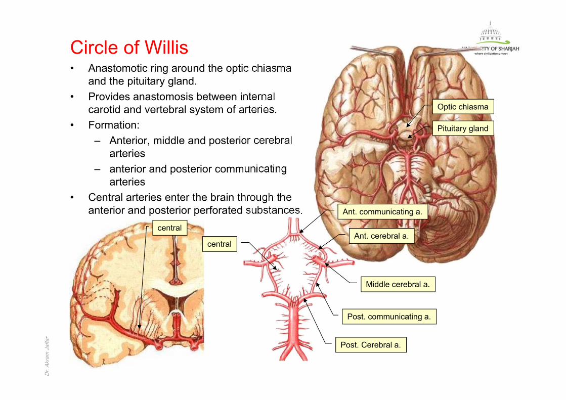
rCircle of Willis• Anastomotic ring around the optic chiasma
and the pituitary gland.• Provides anastomosis between internal
carotid and vertebral system of arteries.• Formation:
– Anterior, middle and posterior cerebral arteries
– anterior and posterior communicating arteries
• Central arteries enter the brain through the anterior and posterior perforated substances.
Post. Cerebral a.
Post. communicating a.
Ant. communicating a.
Ant. cerebral a.
Middle cerebral a.
central
central
Optic chiasma
Pituitary gland
Dr. Akram JaffarDr.
Akra
m J
affa
rVenous drainage of the brain• The veins draining the hemispheres may be divided into superficial and deep
veins.
Dr. Akram JaffarDr.
Akra
m J
affa
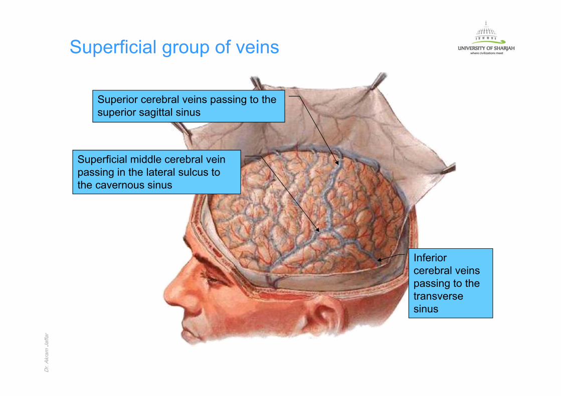
rSuperficial group of veins
Superior cerebral veins passing to the superior sagittal sinus
Superficial middle cerebral vein passing in the lateral sulcus to the cavernous sinus
Inferior cerebral veins passing to the transverse sinus
Dr. Akram JaffarDr.
Akra
m J
affa
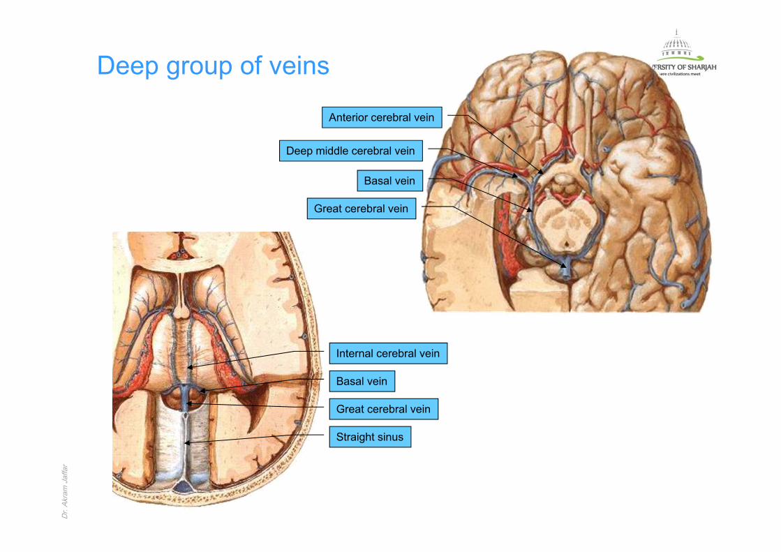
rDeep group of veins
Anterior cerebral vein
Deep middle cerebral vein
Basal vein
Great cerebral vein
Great cerebral vein
Basal vein
Straight sinus
Internal cerebral vein

Dr. Akram JaffarDr.
Akra
m J
affa
rSectional anatomy of the brian
Dr. Akram JaffarDr.
Akra
m J
affa
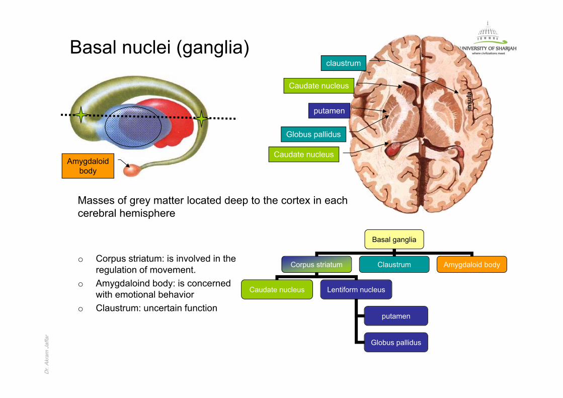
rBasal nuclei (ganglia)
Basal ganglia
Corpus striatum Claustrum Amygdaloid body
Caudate nucleus Lentiform nucleus
putamen
Globus pallidus
claustrum
Caudate nucleus
putamen
Globus pallidus
Amygdaloidbody
Caudate nucleus
Masses of grey matter located deep to the cortex in each cerebral hemisphere
insu
la
o Corpus striatum: is involved in the regulation of movement.
o Amygdaloind body: is concerned with emotional behavior
o Claustrum: uncertain function
Dr. Akram JaffarDr.
Akra
m J
affa
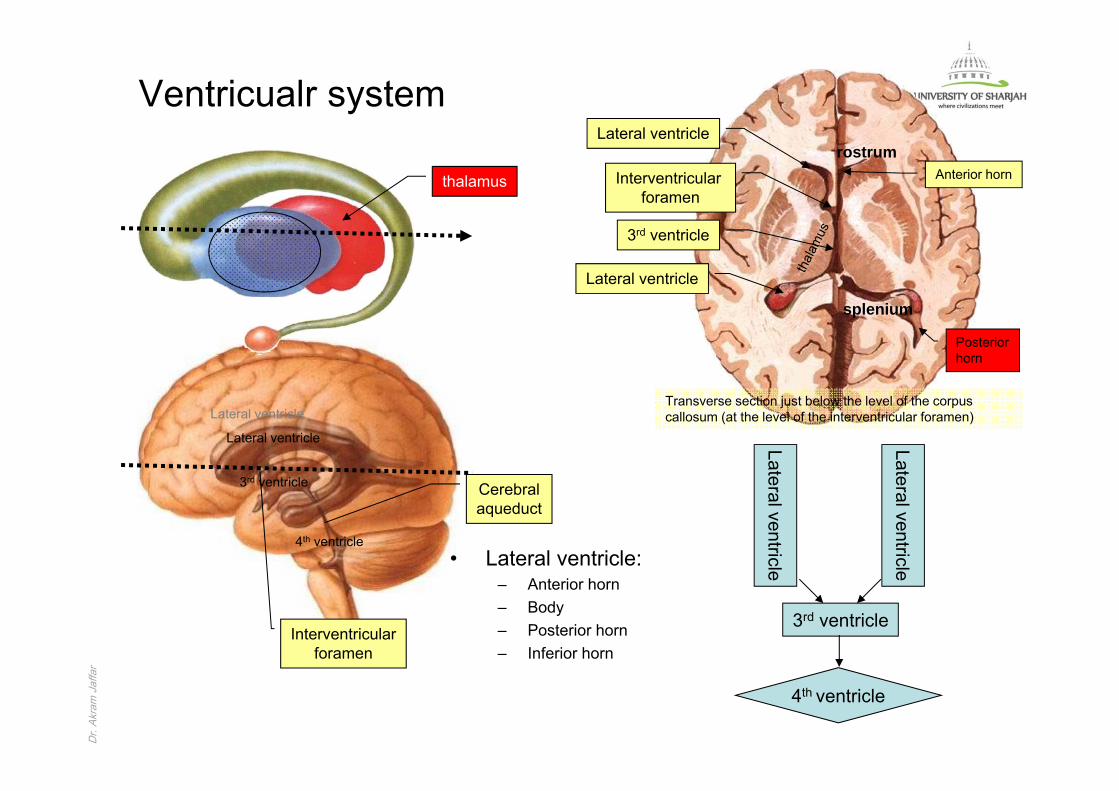
rVentricualr system
Transverse section just below the level of the corpus callosum (at the level of the interventricular foramen)
Lateral ventricle
Interventricular foramen
3rd ventricle
Lateral ventricle
Lateral ventricle
3rd ventricle
Interventricularforamen
4th ventricle
Cerebralaqueduct
Lateral ventricle
Lateral ventricle
3rd ventricle
4th ventricle
Lateral ventricle
rostrum
splenium
thalamus
• Lateral ventricle:– Anterior horn– Body– Posterior horn– Inferior horn
Posteriorhorn
Anterior horn

Dr. Akram JaffarDr.
Akra
m J
affa
rInternal capsule
putamen
Globuspallidus
claustrum• band of projection fibers, appears
in a horizontal section as V-shaped.
• Connects the cerebral cortex with lower centers.
• Has an anterior limb, apex (genu), and a posterior limb.
genu
Caudatenucleus
Dr. Akram JaffarDr.
Akra
m J
affa
r
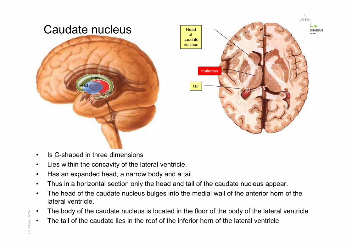
head
body
tail
Caudate nucleus Headof
caudatenucleus
tail
thalamus
• Is C-shaped in three dimensions• Lies within the concavity of the lateral ventricle.• Has an expanded head, a narrow body and a tail. • Thus in a horizontal section only the head and tail of the caudate nucleus appear.• The head of the caudate nucleus bulges into the medial wall of the anterior horn of the
lateral ventricle.• The body of the caudate nucleus is located in the floor of the body of the lateral ventricle • The tail of the caudate lies in the roof of the inferior horn of the lateral ventricle

Dr. Akram JaffarDr.
Akra
m J
affa
rCorpus striatumo The head of the caudate nucleus and
the lentiform nucleus are connected by fibers and grey matter across the anterior limb of the internal capsule resulting in a striated appearance (hence the name corpus striatum).
putamen
Globuspallidus
claustrum
Caudatenucleus
thalamus
genu

Dr. Akram JaffarDr.
Akra
m J
affa
rAxial section of the brain
putamen
Globuspallidus
insula
HeadOf
Caudatenucleus
thalamus
genu
MRI
Dr. Akram JaffarDr.
Akra
m J
affa
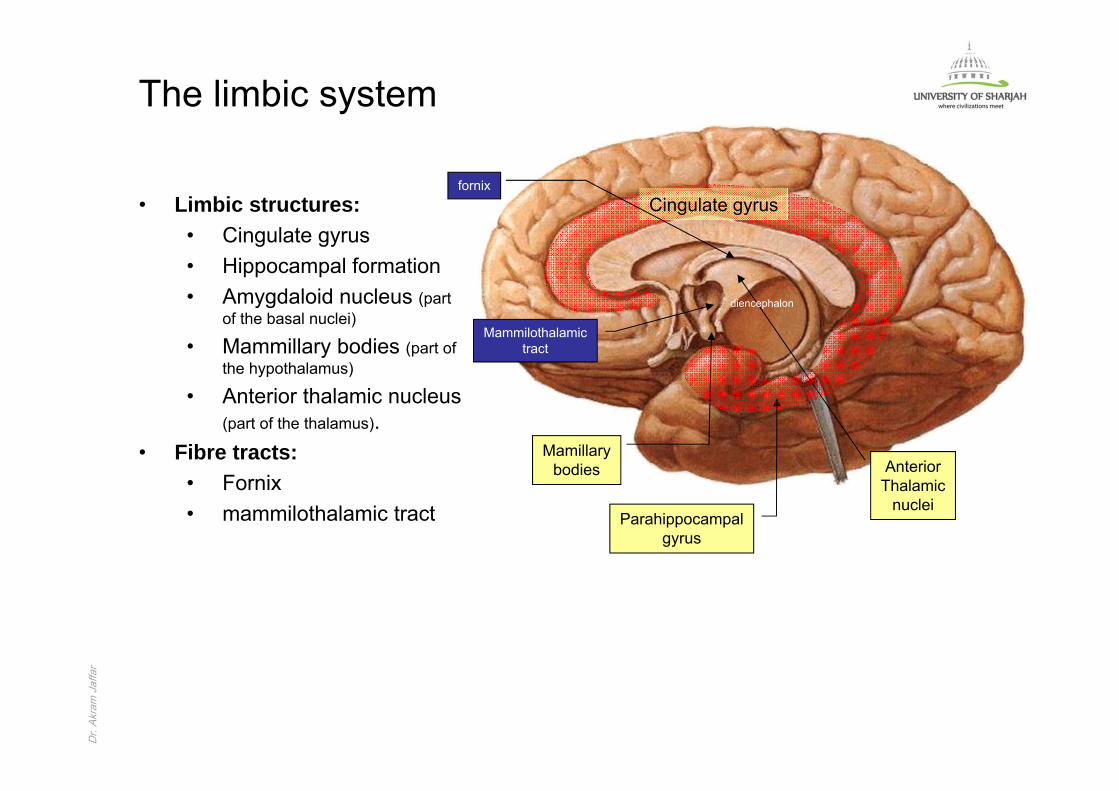
rThe limbic system
Parahippocampalgyrus
Mamillarybodies Anterior
Thalamicnuclei
• Limbic structures:• Cingulate gyrus• Hippocampal formation• Amygdaloid nucleus (part
of the basal nuclei)
• Mammillary bodies (part of the hypothalamus)
• Anterior thalamic nucleus (part of the thalamus).
• Fibre tracts:• Fornix• mammilothalamic tract
Cingulate gyrus
diencephalon
fornix
Mammilothalamictract
Dr. Akram JaffarDr.
Akra
m J
affa
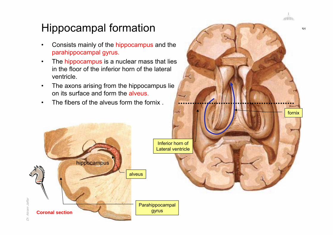
rHippocampal formation• Consists mainly of the hippocampus and the
parahippocampal gyrus.• The hippocampus is a nuclear mass that lies
in the floor of the inferior horn of the lateral ventricle.
• The axons arising from the hippocampus lie on its surface and form the alveus.
• The fibers of the alveus form the fornix .
hippocampus
Coronal section
Inferior horn ofLateral ventricle
alveus
Parahippocampalgyrus
fornix

Dr. Akram JaffarDr.
Akra
m J
affa
rFunctions of the limbic system This system is widely connected, many of its functions are not clearly understood;
nevertheless the limbic system controls:• Emotional behavior such as fear, anger, and the emotions associated with sexual
behavior• Recent memory• Olfaction
Fear
Anger
Emotions
Recent memorySmell
Dr. Akram JaffarDr.
Akra
m J
affa
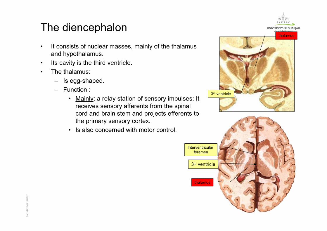
rThe diencephalon • It consists of nuclear masses, mainly of the thalamus
and hypothalamus.• Its cavity is the third ventricle.• The thalamus:
– Is egg-shaped.– Function :
• Mainly: a relay station of sensory impulses: It receives sensory afferents from the spinal cord and brain stem and projects efferents to the primary sensory cortex.
• Is also concerned with motor control.
thalamus
3rd ventricle
thalamus
Interventricular foramen
3rd ventricle
Dr. Akram JaffarDr.
Akra
m J
affa
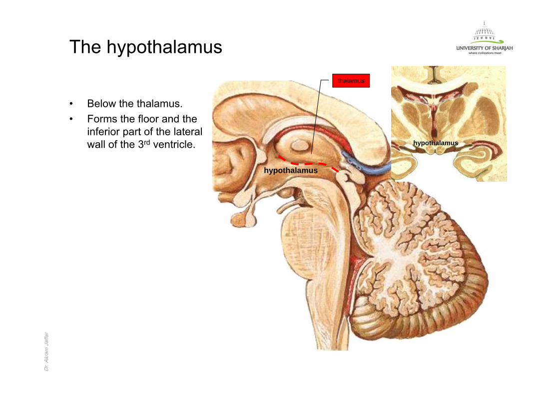
rThe hypothalamus
• Below the thalamus.• Forms the floor and the
inferior part of the lateral wall of the 3rd ventricle.
hypothalamus
hypothalamus
thalamus

Dr. Akram JaffarDr.
Akra
m J
affa
rThe hypothalamus• Behind the optic
chiasma the floor of the 3rd ventricle gives rise to the stalk (infundibulum) of the hypophysis cerebri.
• The mamillary bodieslie behind.
infundibulum
Opticchiasma
Hypophysiscerebri
Mamillary body
Opticchiasma
Hypophysiscerebri
infundibulum
Mamillary body

Dr. Akram JaffarDr.
Akra
m J
affa
rThe pituitary gland (hypophysis cerebri)
• Endocrine gland.• Situated in the hypophyseal fossa of
the body of the sphenoid bone.• Is closely related to the optic chiasma:
– Tumors may produce pressure effects on the adjacent optic chiasma visual defects. Hypophyseal fossa
Opticchiasma
Hypophysiscerebri
Dr. Akram JaffarDr.
Akra
m J
affa
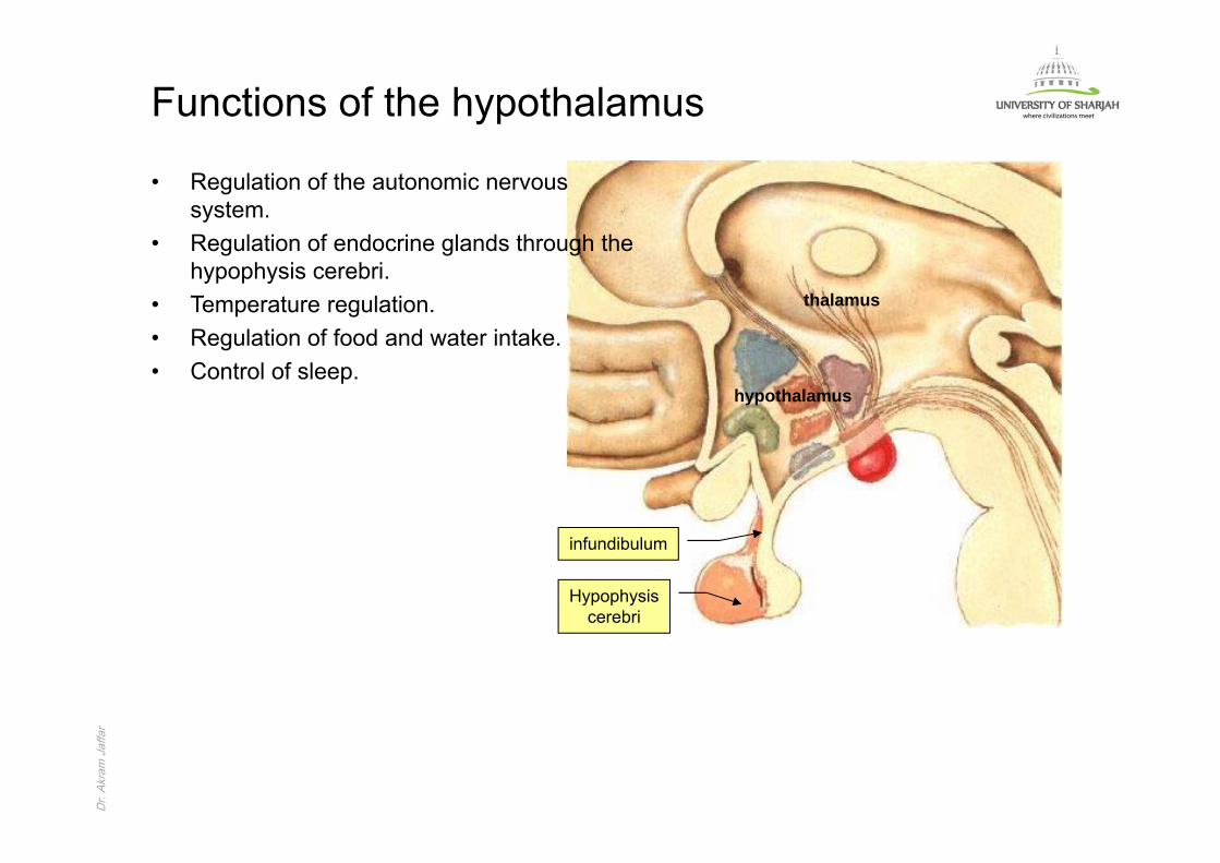
rFunctions of the hypothalamus
• Regulation of the autonomic nervous system.
• Regulation of endocrine glands through the hypophysis cerebri.
• Temperature regulation.• Regulation of food and water intake.• Control of sleep.
hypothalamus
thalamus
Hypophysiscerebri
infundibulum
Dr. Akram JaffarDr.
Akra
m J
affa
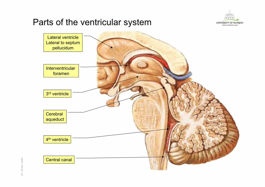
rParts of the ventricular system
Lateral ventricleLateral to septum
pellucidum
Interventricular foramen
3rd ventricle
Cerebralaqueduct
4th ventricle
Central canal

Dr. Akram JaffarDr.
Akra
m J
affa
rThe choroid plexus
• The ventricles are lined with a single epithelial layer called the epindyma.
• In each ventricle the lining of the cavity comes to the surface, i.e. the lining epindyma comes in contact with the pia mater.
• Blood capillaries invaginate at these regions, covered by pia and epindyma constitute the choroids plexus.
• The choroid plexuses secrete the CSF into each ventricle.
Choroid plexusLateral ventricle
Choroid plexus3rd ventricle
Choroid plexusLateral ventricle
Choroid plexus4th ventricle
Dr. Akram JaffarDr.
Akra
m J
affa
rBlood barriers in the CNS
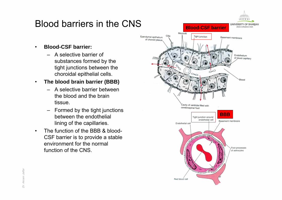
• Blood-CSF barrier:– A selective barrier of
substances formed by the tight junctions between the choroidal epithelial cells.
• The blood brain barrier (BBB)– A selective barrier between
the blood and the brain tissue.
– Formed by the tight junctions between the endothelial lining of the capillaries.
• The function of the BBB & blood-CSF barrier is to provide a stable environment for the normal function of the CNS.
BBB
Blood-CSF barrier
Dr. Akram JaffarDr.
Akra
m J
affa
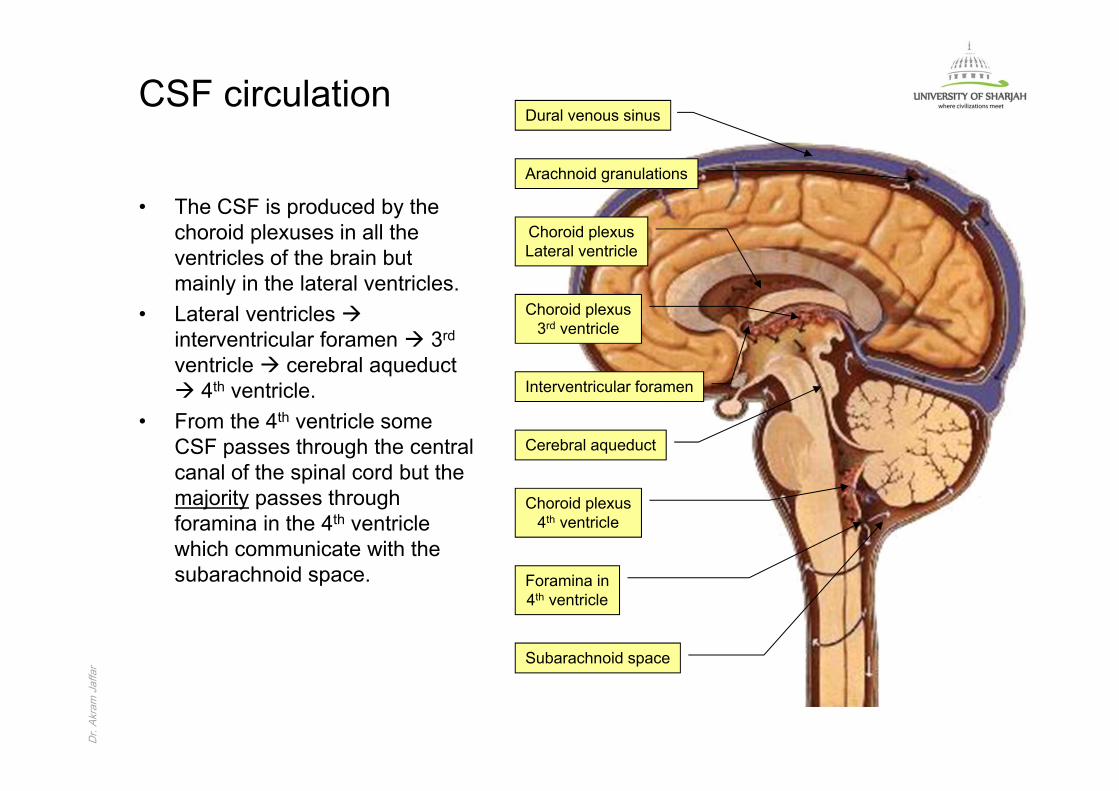
rCSF circulation
Choroid plexusLateral ventricle
Choroid plexus3rd ventricle
Interventricular foramen
Cerebral aqueduct
Choroid plexus4th ventricle
Subarachnoid space
Foramina in4th ventricle
Arachnoid granulations
Dural venous sinus
• The CSF is produced by the choroid plexuses in all the ventricles of the brain but mainly in the lateral ventricles.
• Lateral ventricles interventricular foramen 3rd
ventricle cerebral aqueduct 4th ventricle.
• From the 4th ventricle some CSF passes through the central canal of the spinal cord but the majority passes through foramina in the 4th ventricle which communicate with the subarachnoid space.
Dr. Akram JaffarDr.
Akra
m J
affa
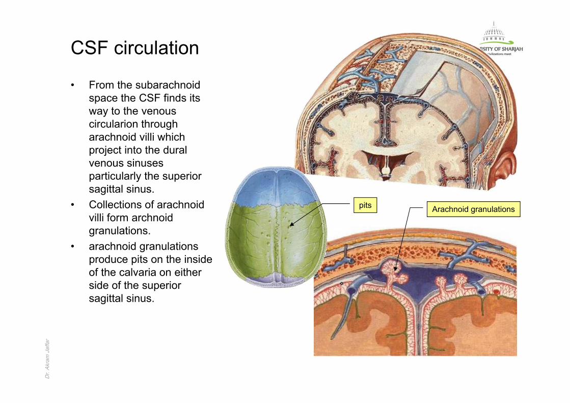
rCSF circulation
• From the subarachnoid space the CSF finds its way to the venous circularion through arachnoid villi which project into the dural venous sinuses particularly the superior sagittal sinus.
• Collections of arachnoid villi form archnoid granulations.
• arachnoid granulations produce pits on the inside of the calvaria on either side of the superior sagittal sinus.
Arachnoid granulationspits