Blood is a complex, specialised tissue containing a number of different cell types suspended in a...
21
is a complex, specialised tissue containing a number of diff cell types suspended in a fluid matrix en whole blood is spun in a centrifuge, the ood cells collect towards the lower half of ube leaving a pale, straw-coloured liquid above ere are between 5 and 10 thousand white blood cells in every mm 3 of blood ere are between 5 and 6.5 million red blood cells in every mm 3 of blood straw-coloured liquid is plasma, a fluid that ains 92% water in which are dissolved various cules such as nutrients, salts, hormones and plasma proteins White blood cells collect at the top of the lower fraction and these include cell types known as granulocytes, lymphocytes and monocytes Red blood cells form the bulk of the lower raction and these cells occupy about 45% of the total volume of the blood Blood is a Tissue Blood is a Tissue
-
Upload
shannon-chloe-powell -
Category
Documents
-
view
223 -
download
5
Transcript of Blood is a complex, specialised tissue containing a number of different cell types suspended in a...
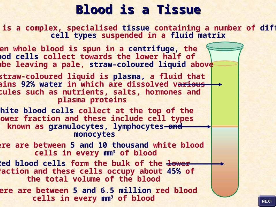
Blood is a complex, specialised tissue containing a number of differentcell types suspended in a fluid matrix
When whole blood is spun in a centrifuge, theblood cells collect towards the lower half of
the tube leaving a pale, straw-coloured liquid above
There are between 5 and 10 thousand white bloodcells in every mm3 of blood
There are between 5 and 6.5 million red bloodcells in every mm3 of blood
The straw-coloured liquid is plasma, a fluid thatcontains 92% water in which are dissolved variousmolecules such as nutrients, salts, hormones and
plasma proteins
White blood cells collect at the top of thelower fraction and these include cell typesknown as granulocytes, lymphocytes and
monocytes
Red blood cells form the bulk of the lowerfraction and these cells occupy about 45% of
the total volume of the blood
Blood is a TissueBlood is a Tissue
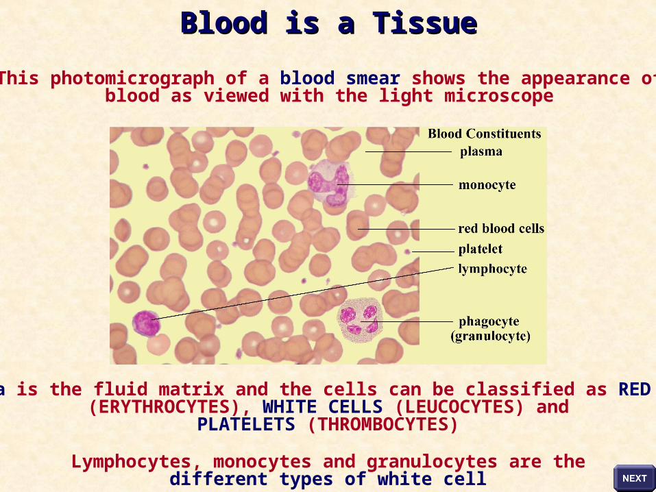
Plasma is the fluid matrix and the cells can be classified as RED CELLS(ERYTHROCYTES), WHITE CELLS (LEUCOCYTES) and
PLATELETS (THROMBOCYTES)
Lymphocytes, monocytes and granulocytes are thedifferent types of white cell
This photomicrograph of a blood smear shows the appearance ofblood as viewed with the light microscope
Blood is a TissueBlood is a Tissue
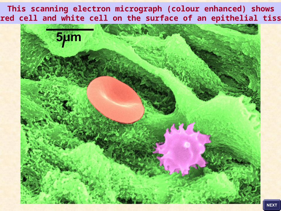
This scanning electron micrograph (colour enhanced) showsa red cell and white cell on the surface of an epithelial tissue
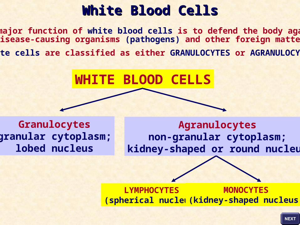
The major function of white blood cells is to defend the body againstdisease-causing organisms (pathogens) and other foreign matter
White cells are classified as either GRANULOCYTES or AGRANULOCYTES
WHITE BLOOD CELLS
Granulocytesgranular cytoplasm;
lobed nucleus
Agranulocytesnon-granular cytoplasm;
kidney-shaped or round nucleus
LYMPHOCYTES(spherical nucleus)
MONOCYTES(kidney-shaped nucleus)
White Blood CellsWhite Blood Cells
GRANULOCYTE
MONOCYTE LYMPHOCYTE
Lobed nucleus
Granular cytoplasm
Kidney-shaped nucleus
Non-granular cytoplasm
Spherical nucleus
occupying alarge volume
of the cell
Non-granular cytoplasm
White Blood CellsWhite Blood Cells
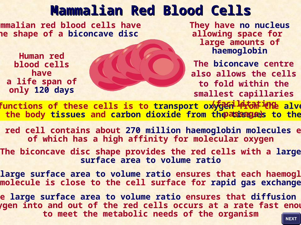
Human red blood cells have
a life span of only 120 days
Mammalian red blood cells havethe shape of a biconcave disc
They have no nucleus allowing space for large amounts of
haemoglobin
The major functions of these cells is to transport oxygen from the alveoli of thelungs to the body tissues and carbon dioxide from the tissues to the alveoli
Each red cell contains about 270 million haemoglobin molecules each of which has a high affinity for molecular oxygen
The biconcave disc shape provides the red cells with a largesurface area to volume ratio
The large surface area to volume ratio ensures that each haemoglobinmolecule is close to the cell surface for rapid gas exchange
The large surface area to volume ratio ensures that diffusion ofoxygen into and out of the red cells occurs at a rate fast enough
to meet the metabolic needs of the organism
The biconcave centre also allows the cells to fold within
the smallest capillaries (facilitating passage)
Mammalian Red Blood CellsMammalian Red Blood Cells

Scanning electron micrograph of mammalian red blood cells showing thebiconcave disc shape that gives these cells a large surface area to volume ratio
Red Blood CellsRed Blood Cells
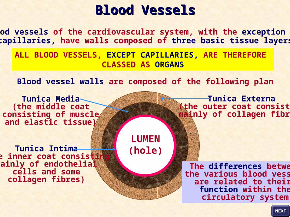
The blood vessels of the cardiovascular system, with the exception of the capillaries, have walls composed of three basic tissue layers
ALL BLOOD VESSELS, EXCEPT CAPILLARIES, ARE THEREFORE CLASSED AS ORGANS
Blood vessel walls are composed of the following plan
LUMEN(hole)
The differences betweenthe various blood vessels
are related to their function within thecirculatory system
Tunica Externa(the outer coat consistingmainly of collagen fibres)
Tunica Media(the middle coat
consisting of muscleand elastic tissue)
Tunica Intima(the inner coat consisting
mainly of endothelialcells and somecollagen fibres)
Blood VesselsBlood Vessels
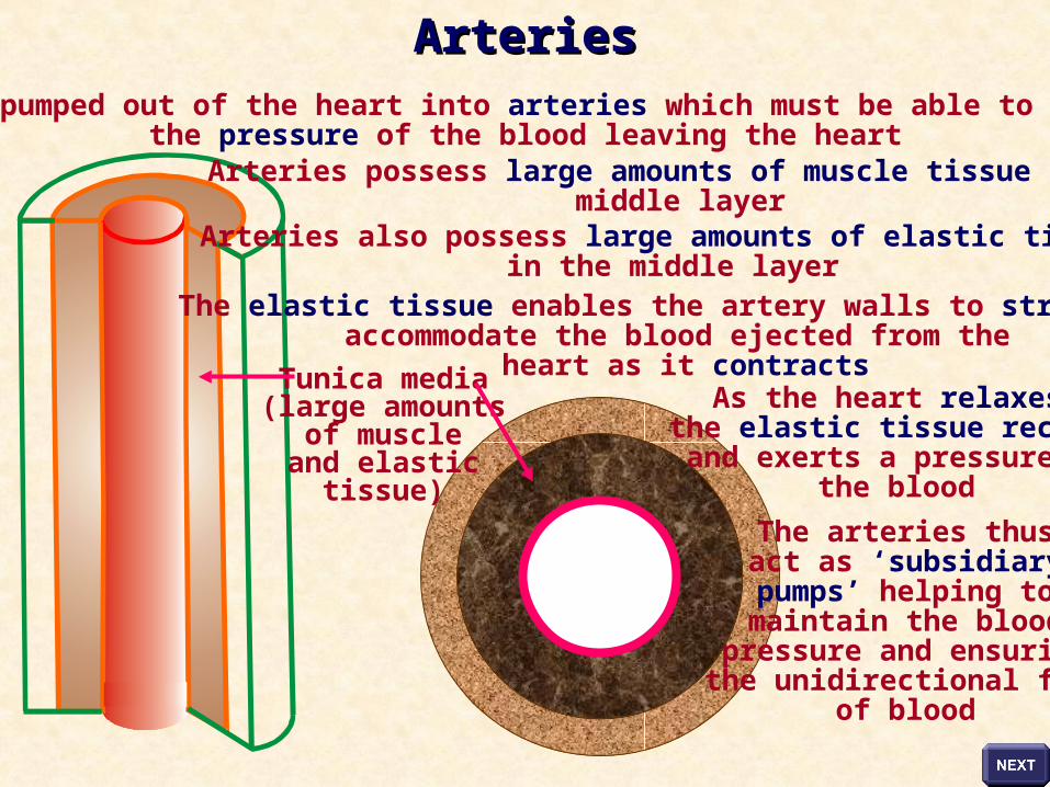
Blood is pumped out of the heart into arteries which must be able to withstandthe pressure of the blood leaving the heart
Arteries possess large amounts of muscle tissue in themiddle layer
Arteries also possess large amounts of elastic tissue in the middle layer
The elastic tissue enables the artery walls to stretch to accommodate the blood ejected from the
heart as it contractsAs the heart relaxes,
the elastic tissue recoilsand exerts a pressure on
the blood
The arteries thusact as ‘subsidiarypumps’ helping tomaintain the blood
pressure and ensuringthe unidirectional flow
of blood
Tunica media(large amounts
of muscleand elastic
tissue)
ArteriesArteries
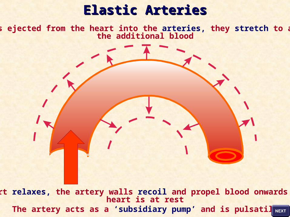
As blood is ejected from the heart into the arteries, they stretch to accommodatethe additional blood
As the heart relaxes, the artery walls recoil and propel blood onwards whilst theheart is at rest
The artery acts as a ‘subsidiary pump’ and is pulsatile
Elastic ArteriesElastic Arteries
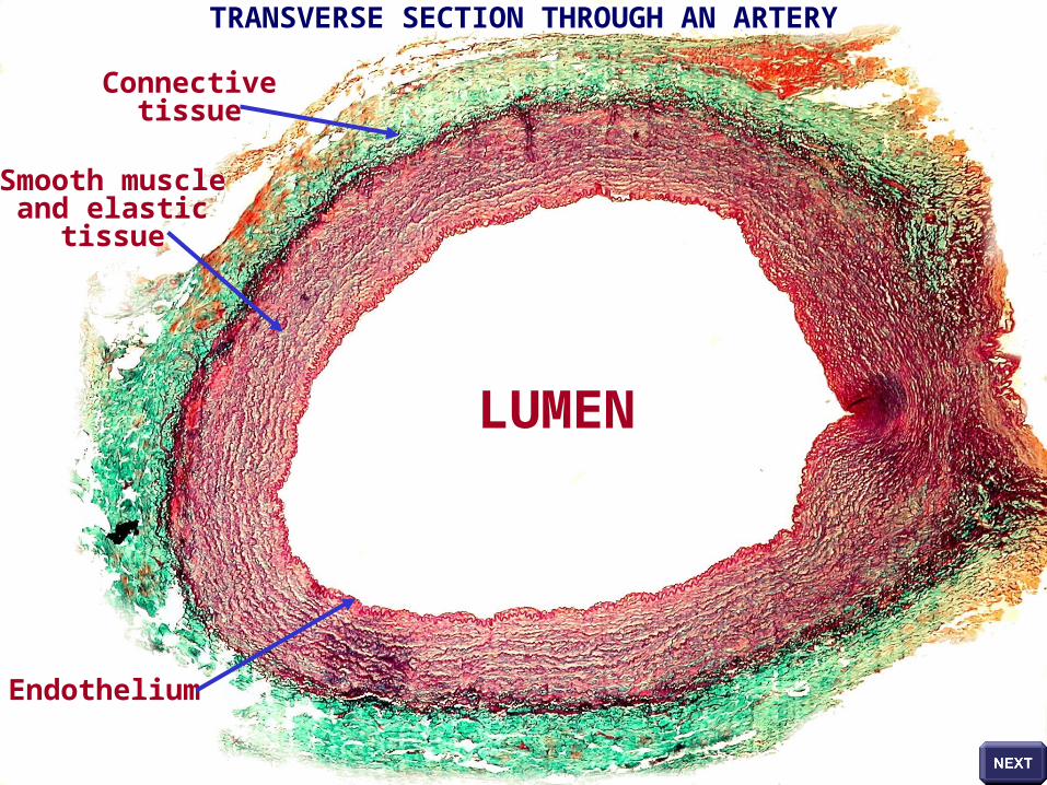
TRANSVERSE SECTION THROUGH AN ARTERY
LUMEN
Connectivetissue
Smooth muscleand elastic
tissue
Endothelium
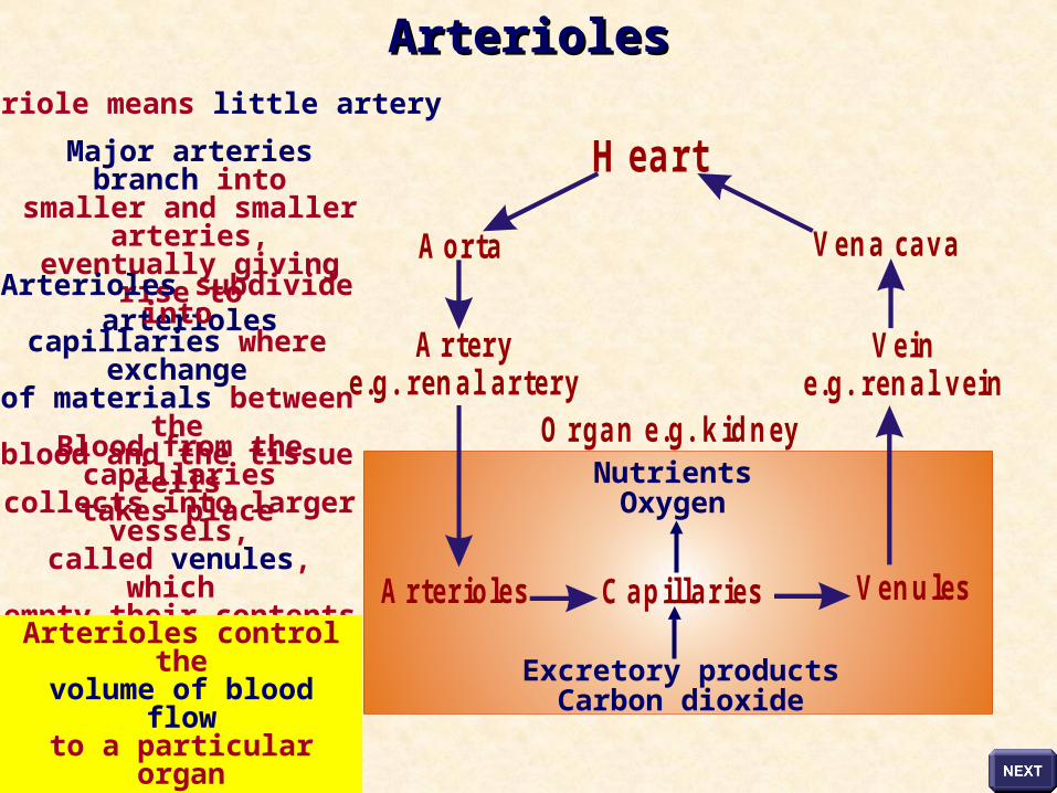
A rter io le s C a p illa r ie s
A rter ye .g . ren a l a r te ry
A o rta
H ea rt
O r g a n e .g . k id n ey
Arteriole means little artery
Major arteries branch intosmaller and smaller
arteries,eventually giving rise to
arteriolesArterioles subdivide into
capillaries where exchangeof materials between theblood and the tissue cells
takes place
NutrientsOxygen
Excretory productsCarbon dioxide
Blood from the capillariescollects into larger vessels,
called venules, which empty their contents into
the thin-walled veins
Arterioles control thevolume of blood flow
to a particularorgan
Ve n u les
Ve ine .g . ren a l v e in
Ve n a ca v a
ArteriolesArterioles
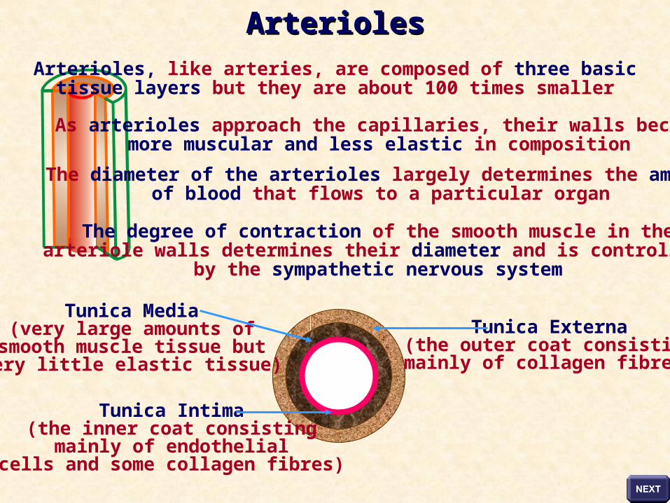
Tunica Externa(the outer coat consistingmainly of collagen fibres)
Tunica Media(very large amounts of
smooth muscle tissue butvery little elastic tissue)
Tunica Intima(the inner coat consisting
mainly of endothelialcells and some collagen fibres)
Arterioles, like arteries, are composed of three basictissue layers but they are about 100 times smaller
As arterioles approach the capillaries, their walls becomemore muscular and less elastic in composition
The diameter of the arterioles largely determines the amountof blood that flows to a particular organ
The degree of contraction of the smooth muscle in thearteriole walls determines their diameter and is controlled
by the sympathetic nervous system
ArteriolesArterioles
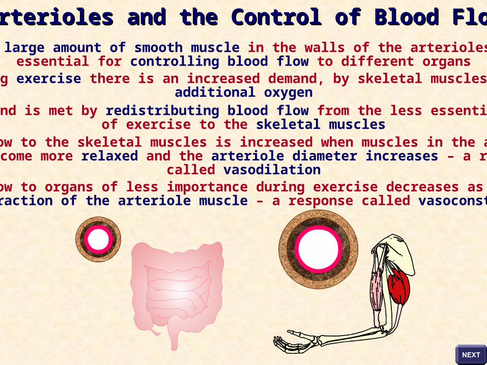
The large amount of smooth muscle in the walls of the arterioles isessential for controlling blood flow to different organs
During exercise there is an increased demand, by skeletal muscles, foradditional oxygen
This demand is met by redistributing blood flow from the less essential organsof exercise to the skeletal muscles
Blood flow to the skeletal muscles is increased when muscles in the arteriolewall become more relaxed and the arteriole diameter increases – a response
called vasodilationBlood flow to organs of less importance during exercise decreases as a result
of contraction of the arteriole muscle – a response called vasoconstriction
Arterioles and the Control of Blood FlowArterioles and the Control of Blood Flow
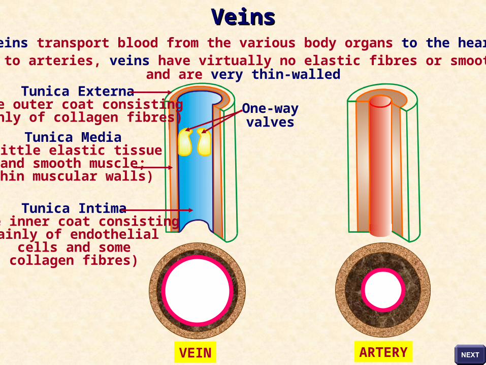
Veins transport blood from the various body organs to the heartCompared to arteries, veins have virtually no elastic fibres or smooth muscle
and are very thin-walled
VEIN ARTERY
Tunica Externa(the outer coat consistingmainly of collagen fibres)
Tunica Media(little elastic tissue
and smooth muscle;thin muscular walls)
Tunica Intima(the inner coat consisting
mainly of endothelialcells and somecollagen fibres)
One-wayvalves
VeinsVeins
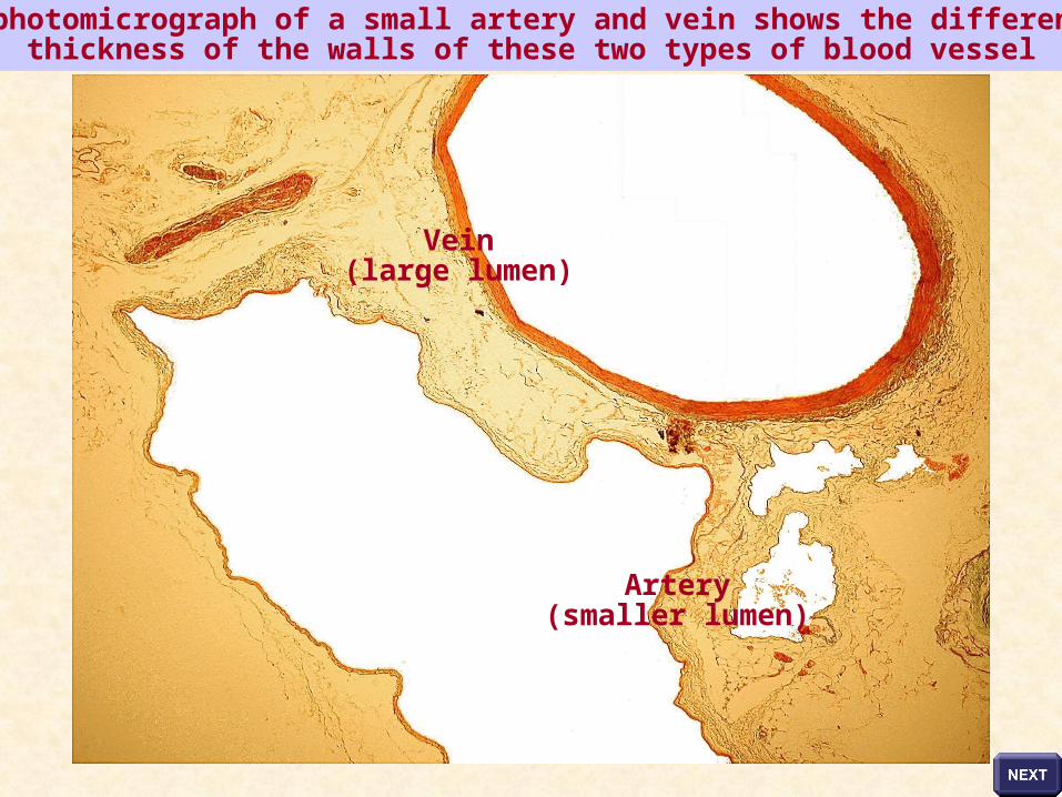
This photomicrograph of a small artery and vein shows the difference inthickness of the walls of these two types of blood vessel
Vein(large lumen)
Artery(smaller lumen)
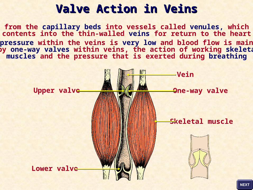
Blood flows from the capillary beds into vessels called venules, which empty theircontents into the thin-walled veins for return to the heart
Blood pressure within the veins is very low and blood flow is maintained by one-way valves within veins, the action of working skeletal
muscles and the pressure that is exerted during breathing
Vein
Lower valve
One-way valve
Skeletal muscle
Upper valve
Valve Action in VeinsValve Action in Veins
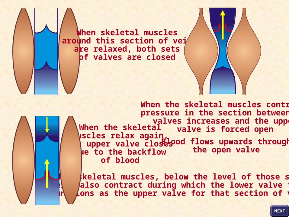
When skeletal musclesaround this section of vein
are relaxed, both setsof valves are closed
When the skeletal muscles contract,pressure in the section between the
valves increases and the uppervalve is forced open
Blood flows upwards throughthe open valve
Other skeletal muscles, below the level of those shownhere, also contract during which the lower valve thenfunctions as the upper valve for that section of vein
When the skeletalmuscles relax again,
the upper valve closesdue to the backflow
of blood
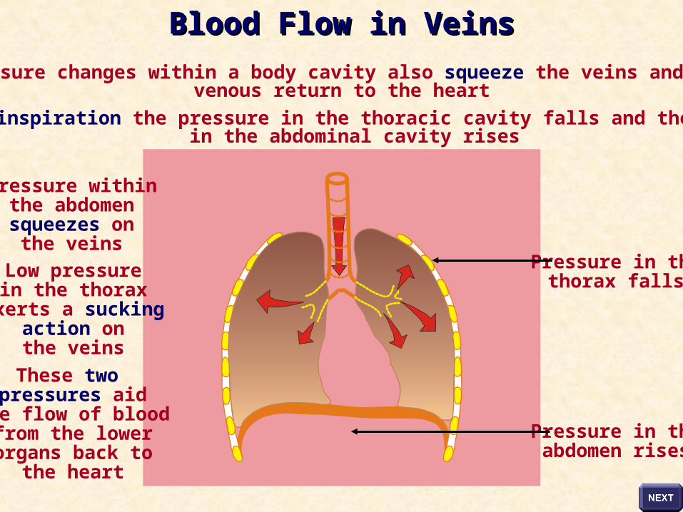
Pressure changes within a body cavity also squeeze the veins and aidvenous return to the heart
During inspiration the pressure in the thoracic cavity falls and the pressurein the abdominal cavity rises
Pressure withinthe abdomensqueezes on
the veins
Low pressurein the thorax
exerts a suckingaction onthe veins
These two pressures aid
the flow of bloodfrom the lowerorgans back to
the heart
Pressure in thethorax falls
Pressure in theabdomen rises
Blood Flow in VeinsBlood Flow in Veins
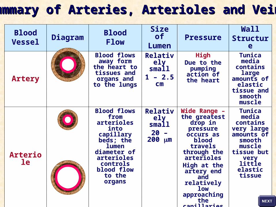
BloodVessel
DiagramBloodFlow
Size ofLumen
PressureWall
Structure
Artery
Blood flows away form the heart to
tissues and organs and to the
lungs
Relatively small
1 – 2.5 cm
HighDue to the
pumping action of the heart
Tunica media contains large
amounts of elastic tissue and smooth
muscle
Arteriole
Blood flows from arterioles into capillary beds;
the lumen diameter of
arterioles controls blood flow to the
organs
Relatively small
20 – 200 m
Wide Range – the greatest drop
in pressure occurs as blood travels through the arteriolesHigh at the
artery end and relatively low
approaching the capillaries
Tunica media contains very large amounts
of smooth muscle tissue but very little elastic tissue
Vein
Blood flows from the capillary beds into venules and then into veins.
Veins carry blood towards the heart
Relatively large
2 - 3 cm
LowOne-way valves, working skeletal
muscles and inspiration aid
the flow of blood in veins
Tunica media contains little elastic tissue and smooth
muscle tissue; thin muscular
wall
Summary of Arteries, Arterioles and VeinsSummary of Arteries, Arterioles and Veins

Acknowledgements
Copyright © 2003 SSER Ltd. and its licensors.All rights reserved. All graphics are for viewing purposes only.


![Crit-Line IV User’s Guides-manuals---hemo...[Na+] affect the micro centrifuge-derived hematocrit values as follows: 1 unit decrease in “spun” hematocrit per 12 mEq/L increases](https://static.fdocuments.in/doc/165x107/5e5b7c0db7d6f71b2568789a/crit-line-iv-useras-guide-s-manuals-hemo-na-affect-the-micro-centrifuge-derived.jpg)