Blood-brain barrier permeability and tPA-mediated …€“br… · · 2012-09-13of human tPA to...
9
Bloodebrain barrier permeability and tPA–mediated neurotoxicity Rami Abu Fanne a,1 , Taher Nassar b,1 , Sergei Yarovoi b , Anwar Rayan c , Itschak Lamensdorf d , Michael Karakoveski e , Polianski Vadim e , Mahmud Jammal a , Douglas B. Cines b , Abd Al-Roof Higazi a, b, * a Department of Clinical Biochemistry, Hebrew University - Hadassah Medical Center, Jerusalem, Israel b Department of Pathology and Laboratory Medicine, University of Pennsylvania, 513A Stellar-Chance, 422 Curie Boulevard, Philadelphia, PA 19104, USA c QRC-Qasemi Research Center, Baka El-Garbiah, Israel d Thrombotech Ltd, Ness Ziona, Israel e PharmaSeed Ltd, Ness Ziona, Israel article info Article history: Received 19 August 2009 Received in revised form 26 November 2009 Accepted 18 December 2009 Keywords: Stroke Tissue type plasminogen activator (tPA) Bloodebrain barrier abstract Tissue type plasminogen activator (tPA) can induce neuronal apoptosis, disrupt the bloodebrain barrier (BBB), and promote dilation of the cerebral vasculature. The timing, sequence and contributions of these and other deleterious effects of tPA and their contribution to post-ischemic brain damage after stroke, have not been fully elucidated. To dissociate the effects of tPA on BBB permeability, cerebral vasodilation and protease-dependent pathways, we developed several tPA mutants and PAI-1 derived peptides constructed by computerized homology modeling of tPA. Our data show that intravenous administration of human tPA to rats increases BBB permeability through a non-catalytic process that is associated with reversible neurotoxicity, brain damage, mortality and contributes significantly to its brief therapeutic window. Furthermore, our data show that inhibiting the effect of tPA on BBB function without affecting its catalytic activity, improves outcome and significantly extends its therapeutic window in mechanical as well as in thromboembolic models of stroke. Ó 2009 Elsevier Ltd. All rights reserved. 1. Introduction Endogenous and exogenous tPA have the potential to aggravate ischemic brain damage after vascular occlusion (Nagai et al., 1999; Wang et al., 1998) or traumatic brain injury (TBI) (Mori et al., 2001) by increasing infarct size and cerebral edema. The mechanism by which tPA causes these neurotoxic effects has not been fully elucidated. tPA promotes the activation of metalloproteases that lead to matrix degradation and disrupts the bloodebrain barrier (BBB) (Yepes et al., 2003). tPA also activates glutamate receptors, which mobilize intracellular calcium and mediate apoptosis of cortical cells (Choi, 1988; Nicole et al., 2001), and augments dilation of the cerebral vasculature in non-ischemic areas, thereby decreasing blood flow to the penumbra (Armstead et al., 2006; Nassar et al., 2004). The specific contribution of each of these components to the neurotoxicity of tPA is not clear since in most experimental models many of these pathways are activated concurrently. The primary goal of this study is to elucidate the molecular bases of tPA mediated opening of the BBB and its contribution to post-ischemic brain damage. Brain parenchyma is exposed to tPA released from endogenous sources after injury (Armstead et al., 2005; Yepes, 2000) and from exogenous sources when tPA is used to treat ischemic stroke. The importance of this distinction is highlighted by recent findings that intraventricular, but not intravenous (IV), administration of cata- lytically active murine tPA increases BBB permeability in mice (Su et al., 2008). This finding helps to explain the deleterious effect of endogenous tPA in stroke (Wang et al., 1998) and TBI (Mori et al., 2001). It also suggests that the neurotoxicity of recombinant tPA (rtPA) given IV to treat stroke may not derive primarily from its ability to further enhance BBB permeability. Identifying the mechanism by which tPA permeabilizes the BBB will not only increase our understanding of the mechanism neurotoxicity but might point to ways to mitigate neurological injury. We have previously reported that tPA initiates intracellular signaling in vascular cells through receptor mediated processes that are independent of its catalytic activity (Armstead et al., 2005; Nassar et al., 2004). Signaling is inhibited by a hexapeptide derived from plasminogen activator inhibitor 1 (PAI-1). The peptide inter- acts with the PAI-1 docking site in tPA, which lies outside of its * Corresponding author at: Department of Pathology and Laboratory Medicine, University of Pennsylvania, 513A Stellar-Chance, 422 Curie Boulevard, Philadelphia, PA 19104, USA. Tel.: þ1 215 662 3966; fax: þ1 215 673 2012. E-mail address: [email protected] (A. Al-Roof Higazi). 1 These authors have equally contributed to the article. Contents lists available at ScienceDirect Neuropharmacology journal homepage: www.elsevier.com/locate/neuropharm 0028-3908/$ e see front matter Ó 2009 Elsevier Ltd. All rights reserved. doi:10.1016/j.neuropharm.2009.12.017 Neuropharmacology 58 (2010) 972e980
Transcript of Blood-brain barrier permeability and tPA-mediated …€“br… · · 2012-09-13of human tPA to...

lable at ScienceDirect
Neuropharmacology 58 (2010) 972e980
Contents lists avai
Neuropharmacology
journal homepage: www.elsevier .com/locate/neuropharm
Bloodebrain barrier permeability and tPA–mediated neurotoxicity
Rami Abu Fanne a,1, Taher Nassar b,1, Sergei Yarovoi b, Anwar Rayan c, Itschak Lamensdorf d,Michael Karakoveski e, Polianski Vadim e, Mahmud Jammal a,Douglas B. Cines b, Abd Al-Roof Higazi a,b,*aDepartment of Clinical Biochemistry, Hebrew University - Hadassah Medical Center, Jerusalem, IsraelbDepartment of Pathology and Laboratory Medicine, University of Pennsylvania, 513A Stellar-Chance, 422 Curie Boulevard, Philadelphia, PA 19104, USAcQRC-Qasemi Research Center, Baka El-Garbiah, Israeld Thrombotech Ltd, Ness Ziona, Israele PharmaSeed Ltd, Ness Ziona, Israel
a r t i c l e i n f o
Article history:Received 19 August 2009Received in revised form26 November 2009Accepted 18 December 2009
Keywords:StrokeTissue type plasminogen activator (tPA)Bloodebrain barrier
* Corresponding author at: Department of PatholoUniversity of Pennsylvania, 513A Stellar-Chance, 422 CPA 19104, USA. Tel.: þ1 215 662 3966; fax: þ1 215 67
E-mail address: [email protected] (A. A1 These authors have equally contributed to the ar
0028-3908/$ e see front matter � 2009 Elsevier Ltd.doi:10.1016/j.neuropharm.2009.12.017
a b s t r a c t
Tissue type plasminogen activator (tPA) can induce neuronal apoptosis, disrupt the bloodebrain barrier(BBB), and promote dilation of the cerebral vasculature. The timing, sequence and contributions of theseand other deleterious effects of tPA and their contribution to post-ischemic brain damage after stroke,have not been fully elucidated. To dissociate the effects of tPA on BBB permeability, cerebral vasodilationand protease-dependent pathways, we developed several tPA mutants and PAI-1 derived peptidesconstructed by computerized homology modeling of tPA. Our data show that intravenous administrationof human tPA to rats increases BBB permeability through a non-catalytic process that is associated withreversible neurotoxicity, brain damage, mortality and contributes significantly to its brief therapeuticwindow. Furthermore, our data show that inhibiting the effect of tPA on BBB function without affectingits catalytic activity, improves outcome and significantly extends its therapeutic window in mechanicalas well as in thromboembolic models of stroke.
� 2009 Elsevier Ltd. All rights reserved.
1. Introduction
Endogenous and exogenous tPA have the potential to aggravateischemic brain damage after vascular occlusion (Nagai et al., 1999;Wang et al., 1998) or traumatic brain injury (TBI) (Mori et al., 2001)by increasing infarct size and cerebral edema. The mechanism bywhich tPA causes these neurotoxic effects has not been fullyelucidated. tPA promotes the activation of metalloproteases thatlead to matrix degradation and disrupts the bloodebrain barrier(BBB) (Yepes et al., 2003). tPA also activates glutamate receptors,which mobilize intracellular calcium and mediate apoptosisof cortical cells (Choi, 1988; Nicole et al., 2001), and augmentsdilation of the cerebral vasculature in non-ischemic areas, therebydecreasing blood flow to the penumbra (Armstead et al., 2006;Nassar et al., 2004). The specific contribution of each of thesecomponents to the neurotoxicity of tPA is not clear since in mostexperimental models many of these pathways are activated
gy and Laboratory Medicine,urie Boulevard, Philadelphia,3 2012.l-Roof Higazi).ticle.
All rights reserved.
concurrently. The primary goal of this study is to elucidate themolecular bases of tPA mediated opening of the BBB and itscontribution to post-ischemic brain damage.
Brain parenchyma is exposed to tPA released from endogenoussources after injury (Armstead et al., 2005; Yepes, 2000) and fromexogenous sources when tPA is used to treat ischemic stroke. Theimportance of this distinction is highlighted by recent findings thatintraventricular, but not intravenous (IV), administration of cata-lytically active murine tPA increases BBB permeability in mice(Su et al., 2008). This finding helps to explain the deleterious effectof endogenous tPA in stroke (Wang et al., 1998) and TBI (Mori et al.,2001). It also suggests that the neurotoxicity of recombinant tPA(rtPA) given IV to treat stroke may not derive primarily from itsability to further enhance BBB permeability. Identifying themechanism by which tPA permeabilizes the BBB will not onlyincrease our understanding of the mechanism neurotoxicity butmight point to ways to mitigate neurological injury.
We have previously reported that tPA initiates intracellularsignaling in vascular cells through receptor mediated processesthat are independent of its catalytic activity (Armstead et al., 2005;Nassar et al., 2004). Signaling is inhibited by a hexapeptide derivedfrom plasminogen activator inhibitor 1 (PAI-1). The peptide inter-acts with the PAI-1 docking site in tPA, which lies outside of its

R.A. Fanne et al. / Neuropharmacology 58 (2010) 972e980 973
catalytic center and therefore does not affect its capacity to activateits physiological substrate plasminogen to inhibit fibrinolysis(Armstead et al., 2006; Nassar et al., 2004). This peptide providesneuroprotection against endogenous as will exogenous tPA inmodels of stroke and TBI (Armstead et al., 2006), data that has beenrecently reproduced by others (Tan et al., 2009).
If tPA permeabilizes the BBB from within the CNS througha process dependent on its catalytic activity (Yepes et al., 2003), itwould be difficult to envision how this deleterious effect could bevitiated without compromising its fibrinolytic activity. On otherhand, these observations were made with recombinant mouse tPA(Yepes et al., 2003), which differs from human tPAwithin the PAI-1docking (human “aa 295e99” AKHRR; mouse VKNKR), which isessential for intracellular signaling (Armstead et al., 2006; Nassaret al., 2004). This suggests that effect of intravenous human tPA onBBB function may differ.
To pursue this possibility, we used human recombinant tPA toelucidate the molecular bases of BBB permeability and its contri-bution to post-ischemic brain damage. We tested the hypothesisthat intravenous human tPA may increase BBB permeabilitythrough non-catalytic processes, which would afford the oppor-tunity to inhibit this receptor mediated process without compro-mising fibrinolytic (catalytic) activity. Our findings suggest a novelapproach to preserving BBB function that improves the outcome ofstroke in animals treated with tPA and increases its therapeuticwindow.
2. Materials and methods
2.1. PAI-1 derived peptides
Peptides were synthesized as previously described by Peptisyntha (Brussels,Belgium and San Diego, CA). rtPA: (Actilase) was purchased from Boehringer(Ingelheim, Germany). Catalytically inactive rtPA (tPA-S481A): cDNA encoding mousewild type (WT-tPA) and tPA-S481Awere expressed in Drosophila S2 cells and purifiedusing antibody-affinity chromatography. The proteins migrate as single bands at Mrw55 kDa on SDS-PAGE and their plasminogen activator activity was analyzed usingthe plasmin chromogenic substrate Spectrazyme PL (gift of American Diagnostica),as described (Higazi et al., 1995).
2.2. Anesthesia
All experimental protocols involving the use of vertebrate animals wereapproved by the Israeli Board for Animal Experiments. SpragueeDawley rats (HarlanLaboratories, Jerusalem, Israel) were anesthetized with an intraperitoneal injectionof ketamine (75 mg/mL) and xylazine (5 mg/mL) (Kepro, Holland) prior to study.
2.3. Transient occlusion of the middle cerebral artery
Transient occlusion of the middle cerebral artery (MCA) was performed exactlyas described previously (Armstead et al., 2006). Briefly, the left common carotidartery (CCA) was exposed and permanently ligated. An incisionwas made in the CCAto insert a 4e0monofilament nylon suture. The monofilament was inserted throughthe CCA into the lumen of the ICA. The monofilament was advanced into the circle ofWillis, effectively occluding the middle carotid artery . Two to four hours after MCAocclusion, the monofilament was removed.
2.4. Thromboembolic stroke
Focal ischemia was induced by injecting microemboli as described (Armsteadet al., 2006). Donor rats were anesthetized as above and their femoral arteries werecatheterized. Arterial blood (0.1 mL) was obtained and incubated at room temper-ature for 2 h to induce clot formation. The clot was fragmented by passage througha 26-gauge needle. Recipient ratswere anesthetizedwith 1.5% isoflurane tomaintainspontaneous respiration. The bifurcations of the right CCA, ICA and ECA wereexposed. The occipital artery branches of the ECA were then isolated. The ICA wasisolated and from the adjacent vagus nerve, and the pterygopalatine artery wasligated close to its origin. Next, a 4e0 silk suture was tied loosely around themobilized ECA stump, and a PE-50 polyethylene tube was connected to a 21-guideneedle, positioned within the ECA to a location 1 or 2 mm distal to the bifurcation ofthe CCA and then fixed. The microclots were injected gently into the ICA overa period of 10 s. The PE-50 tube was removed and the ECA ligated. Rectal temper-atures were maintained at 37 �C with a heating lamp.
2.5. Treatment
Treatments were administered by intravenous (IV) infusion. After the inductionof the ischemic or thrombotic stroke, animals were randomly divided into severalcohorts (n ¼ 12e16 animals per group as indicated in figure legends). Animals ineach cohort were injected with either: 1) saline 2) saline containing rtPA (6 mg/kg),or 3) saline containing rtPA together PAI-1 derived peptide 6-aa or 18-aa Tyr(1 mg/kg each) or 4) 6-aa or 18-aa Tyr peptides alone. Fifty percent of the dose wasgiven as a bolus injection and the remainder was infused over 60 min. Tubes con-taining each form of treatment were coded and handed to the surgeon who wasblinded to nature of the intervention. Statistical analysis was performed by a thirdparty who was also blinded to the experimental conditions.
2.6. Neuro-score assessment
Two assessments of neurological function (neuro-scores, NSS) were performed24 h post induction of ischemia on a total of 12 and 18 animals, respectively, asdescribed (Chen et al., 2001).
2.7. Assessment of BBB integrity
BBB permeability was measured using Evans blue (Adelson et al., 1998) orradiolabeled fibrinogen as described (Higazi et al., 1998). Anesthetized rats wereinjected IV with 100 mL of saline or saline containing either WT-rtPA or catalyticallyinactive rtPA-Ser481Ala (1 mg/kg each) alone or together PAI-1 or the PA-1 derivedpeptides (1 mg/kg). Five minutes later, the rats were given an IV injection of 2%Evans blue (EB) or radiolabeled fibrinogen (0.5 mg/kg) in saline. One hour after thissecond injection, organs were cleared of blood by transcardiac perfusion. The brainswere removed, weighed, homogenized in N, N dimethylformamide and centrifuged.Extruded dye was quantified by absorbance at 620 nm or by quantifying radioac-tivity, as described (Higazi et al., 1998). Data are expressed as absorbance or cpm pergram of tissue.
2.8. Plasminogen activator activity
Plasminogen activator activity was determined as previously reported (Higaziet al., 2005) by adding WT tPA, WT uPA or PA variants (20 nM each) to a reactionmixture containing Glu-plasminogen (100 nM) and the plasmin chromogenicsubstrate Spectrazyme PL (500 mM) in phosphate-buffered saline (PBS) at 37 �C. Thelight absorbance at 405 nm was measured continuously over time. The initialvelocity of plasmin generation was calculated from the change in the rate of A405
over different time intervals and the results were expressed as the change in opticaldensity (DOD) per minute.
2.9. Assessment of infarction and hemorrhage
Twenty-four hours after induction of stroke, ratswere euthanized using CO2. Thebrains were rapidly removed and sectioned coronally into 2 mm segments. All sliceswere examined under a surgical microscope to evaluate for hemorrhagic trans-formation. The slices were then incubated in 2% 2,3,5-triphenyltetrazolium chloride(TTC) solution for 30 min at 37 �C and fixed overnight at room temperature in 4%buffered formaldehyde solution (Gadot, Haifa, Israel). Each TTC-stained brain slicewas then photographed with a digital camera (Olympus C-4000, Japan).
2.10. Measurement of infarct size
The infarcted area in each section was measured using an image J analysissystem. The total infarct volume was determined by integrating the areas from allsections and the results were expressed as a percentage of the total volume of theipsilateral hemisphere, as described (Tejima et al., 2001).
2.11. Mortality
The animals were included in the study once treatment had been initiated.Death was recorded only in treated animals.
2.12. Modeling of tPA
The resolution coordinates of the serine protease domain at 3.25 Å, ID code 1PDA(Renatus et al., 1995) defined by X-ray, and the coordinates of the kringle-2 domain,ID code 1PK2 (Byeon and Llinás, 1991) defined by NMR, were downloaded from theProtein Data Bank (Abola et al., 1997) and used to model the 3D structure of tPA. Thedynamics simulation module in Discovery Studio 2.0 was used for moleculardynamics (MD) simulations. All calculations were made using a dual Intel PentiumIV 3.0 GHz processor.
The initial structure was built through the following steps: 1. Assembly of serineprotease with kringle-2: Cys-Ser-Thr-Cys segment was found in more than 50 PDBs.Their coordinates were extracted and used to build dozens of optional models.Hydrogens were added and charges were assigned in pH ¼ 7.0. Structures were
R.A. Fanne et al. / Neuropharmacology 58 (2010) 972e980974
minimized by fixing the main chain coordinates except the two residues (Ser andThr) from the connecting loop with 1000 steps of steep descent and 5000 steps ofconjugate gradients. 2. Homology modeling of Kringle-1: the sequence identitybetween kringle-1 and kringle-2 is 53%. 1PK2 (the NMR structure of kringle-2) wasused as a template for homology modeling of kringle-1 in Discovery Studio 2.0.Restraints for backbone atoms of identical residues (kringle-1/kringle-2) wereapplied. One thousand steps of steep descent and five thousand of conjugategradient minimizationwere performed at first for hydrogens followed by side chainsand later for the whole model of kringle-1. 3. Assembly with kringle-1: the lowestenergy structure obtained previously was used to connect with the lowest energymodel of kringle-1.
The time step of 1 fs was used to integrate the equations of motion. The structurewas minimized by fixing the main chain coordinates of the whole protein with theexception of the two connecting loops with 1000 steps of steep descent and 5000steps of conjugate gradients. The structure was heated to 1000 K and run for 1 ns ofproduction simulation at constant pressure (1 atm). The structure was then mini-mized with the combination of 1000 steps of steep descent and 5000 steps ofconjugate gradients. This was repeated 100 times, generating 100 frames. The lowestenergy structure obtained byMD (see Fig.1) was used for docking. This model shows
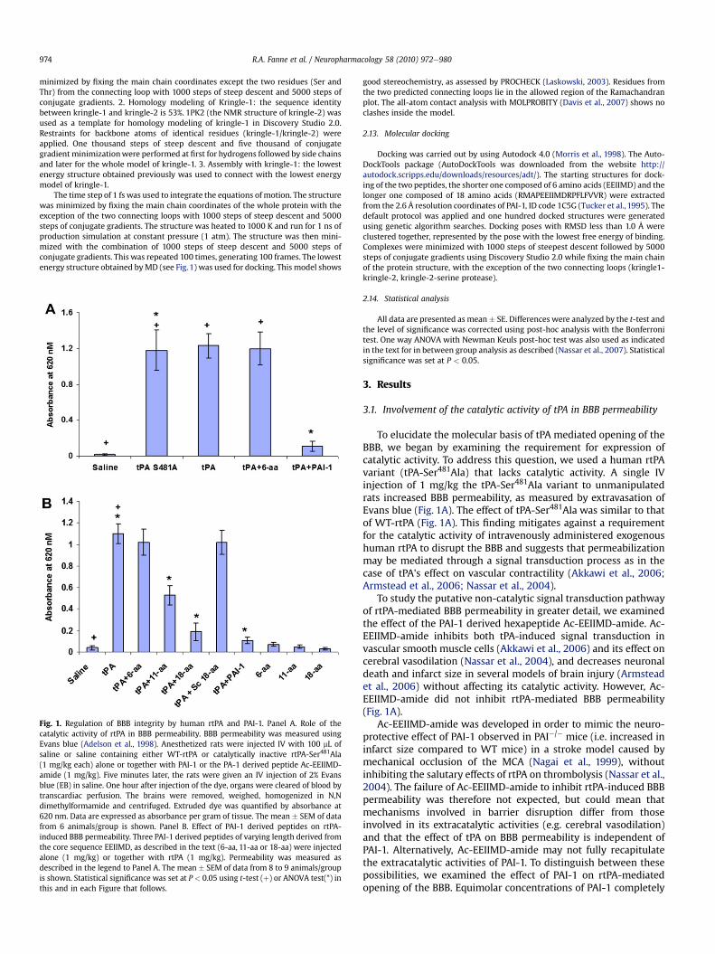
Fig. 1. Regulation of BBB integrity by human rtPA and PAI-1. Panel A. Role of thecatalytic activity of rtPA in BBB permeability. BBB permeability was measured usingEvans blue (Adelson et al., 1998). Anesthetized rats were injected IV with 100 mL ofsaline or saline containing either WT-rtPA or catalytically inactive rtPA-Ser481Ala(1 mg/kg each) alone or together with PAI-1 or the PA-1 derived peptide Ac-EEIIMD-amide (1 mg/kg). Five minutes later, the rats were given an IV injection of 2% Evansblue (EB) in saline. One hour after injection of the dye, organs were cleared of blood bytranscardiac perfusion. The brains were removed, weighed, homogenized in N,Ndimethylformamide and centrifuged. Extruded dye was quantified by absorbance at620 nm. Data are expressed as absorbance per gram of tissue. The mean � SEM of datafrom 6 animals/group is shown. Panel B. Effect of PAI-1 derived peptides on rtPA-induced BBB permeability. Three PAI-1 derived peptides of varying length derived fromthe core sequence EEIIMD, as described in the text (6-aa, 11-aa or 18-aa) were injectedalone (1 mg/kg) or together with rtPA (1 mg/kg). Permeability was measured asdescribed in the legend to Panel A. The mean � SEM of data from 8 to 9 animals/groupis shown. Statistical significance was set at P < 0.05 using t-test (þ) or ANOVA test(*) inthis and in each Figure that follows.
good stereochemistry, as assessed by PROCHECK (Laskowski, 2003). Residues fromthe two predicted connecting loops lie in the allowed region of the Ramachandranplot. The all-atom contact analysis with MOLPROBITY (Davis et al., 2007) shows noclashes inside the model.
2.13. Molecular docking
Docking was carried out by using Autodock 4.0 (Morris et al., 1998). The Auto-DockTools package (AutoDockTools was downloaded from the website http://autodock.scripps.edu/downloads/resources/adt/). The starting structures for dock-ing of the two peptides, the shorter one composed of 6 amino acids (EEIIMD) and thelonger one composed of 18 amino acids (RMAPEEIIMDRPFLFVVR) were extractedfrom the 2.6 Å resolution coordinates of PAI-1, ID code 1C5G (Tucker et al., 1995). Thedefault protocol was applied and one hundred docked structures were generatedusing genetic algorithm searches. Docking poses with RMSD less than 1.0 Å wereclustered together, represented by the pose with the lowest free energy of binding.Complexes were minimized with 1000 steps of steepest descent followed by 5000steps of conjugate gradients using Discovery Studio 2.0 while fixing the main chainof the protein structure, with the exception of the two connecting loops (kringle1-kringle-2, kringle-2-serine protease).
2.14. Statistical analysis
All data are presented as mean� SE. Differences were analyzed by the t-test andthe level of significance was corrected using post-hoc analysis with the Bonferronitest. One way ANOVA with Newman Keuls post-hoc test was also used as indicatedin the text for in between group analysis as described (Nassar et al., 2007). Statisticalsignificance was set at P < 0.05.
3. Results
3.1. Involvement of the catalytic activity of tPA in BBB permeability
To elucidate the molecular basis of tPA mediated opening of theBBB, we began by examining the requirement for expression ofcatalytic activity. To address this question, we used a human rtPAvariant (tPA-Ser481Ala) that lacks catalytic activity. A single IVinjection of 1 mg/kg the tPA-Ser481Ala variant to unmanipulatedrats increased BBB permeability, as measured by extravasation ofEvans blue (Fig. 1A). The effect of tPA-Ser481Ala was similar to thatof WT-rtPA (Fig. 1A). This finding mitigates against a requirementfor the catalytic activity of intravenously administered exogenoushuman rtPA to disrupt the BBB and suggests that permeabilizationmay be mediated through a signal transduction process as in thecase of tPA's effect on vascular contractility (Akkawi et al., 2006;Armstead et al., 2006; Nassar et al., 2004).
To study the putative non-catalytic signal transduction pathwayof rtPA-mediated BBB permeability in greater detail, we examinedthe effect of the PAI-1 derived hexapeptide Ac-EEIIMD-amide. Ac-EEIIMD-amide inhibits both tPA-induced signal transduction invascular smooth muscle cells (Akkawi et al., 2006) and its effect oncerebral vasodilation (Nassar et al., 2004), and decreases neuronaldeath and infarct size in several models of brain injury (Armsteadet al., 2006) without affecting its catalytic activity. However, Ac-EEIIMD-amide did not inhibit rtPA-mediated BBB permeability(Fig. 1A).
Ac-EEIIMD-amide was developed in order to mimic the neuro-protective effect of PAI-1 observed in PAI�/� mice (i.e. increased ininfarct size compared to WT mice) in a stroke model caused bymechanical occlusion of the MCA (Nagai et al., 1999), withoutinhibiting the salutary effects of rtPA on thrombolysis (Nassar et al.,2004). The failure of Ac-EEIIMD-amide to inhibit rtPA-induced BBBpermeability was therefore not expected, but could mean thatmechanisms involved in barrier disruption differ from thoseinvolved in its extracatalytic activities (e.g. cerebral vasodilation)and that the effect of tPA on BBB permeability is independent ofPAI-1. Alternatively, Ac-EEIIMD-amide may not fully recapitulatethe extracatalytic activities of PAI-1. To distinguish between thesepossibilities, we examined the effect of PAI-1 on rtPA-mediatedopening of the BBB. Equimolar concentrations of PAI-1 completely
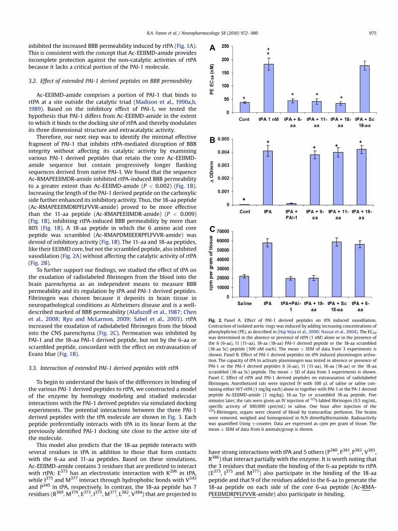
Fig. 2. Panel A. Effect of PAI-1 derived peptides on tPA induced vasodilation.Contraction of isolated aortic rings was induced by adding increasing concentrations ofphenylephrine (PE), as described in (Haj-Yejia et al., 2000; Nassar et al., 2004). The EC50was determined in the absence or presence of rtPA (1 nM) alone or in the presence ofthe 6 (6-aa), 11 (11-aa), 18-aa (18-aa) PAI-1 derived peptide or the 18-aa scrambled(18-aa Sc) peptide (100 nM each). The mean � SEM of data from 3 experiments isshown. Panel B. Effect of PAI-1 derived peptides on tPA induced plasminogen activa-tion. The capacity of tPA to activate plasminogen was tested in absence or presence ofPAI-1 or the PAI-1 derived peptides 6 (6-aa), 11 (11-aa), 18-aa (18-aa) or the 18-aascrambled (18-aa Sc) peptide. The mean � SD of data from 3 experiments is shown.Panel C. Effect of rtPA and PAI-1 derived peptides on extravasation of radiolabeledfibrinogen. Anesthetized rats were injected IV with 100 mL of saline or saline con-taining either WT-rtPA (1 mg/kg each) alone or together with PAI-1 or the PA-1 derivedpeptide Ac-EEIIMD-amide (1 mg/kg), 18-aa Tyr or scrambled 18-aa peptide. Fiveminutes later, the rats were given an IV injection of 125I-labled fibrinogen (0.5 mg/mL,specific activity of 300,000 cpm/mL) in saline. One hour after injection of the125I-fibrinogen, organs were cleared of blood by transcardiac perfusion. The brainswere removed, weighed and homogenized in N,N dimethylformamide. Radioactivitywas quantified Using g-counter. Data are expressed as cpm per gram of tissue. Themean � SEM of data from 6 animals/group is shown.
R.A. Fanne et al. / Neuropharmacology 58 (2010) 972e980 975
inhibited the increased BBB permeability induced by rtPA (Fig. 1A).This is consistent with the concept that Ac-EEIIMD-amide providesincomplete protection against the non-catalytic activities of rtPAbecause it lacks a critical portion of the PAI-1 molecule.
3.2. Effect of extended PAI-1 derived peptides on BBB permeability
Ac-EEIIMD-amide comprises a portion of PAI-1 that binds tortPA at a site outside the catalytic triad (Madison et al., 1990a,b,1989). Based on the inhibitory effect of PAI-1, we tested thehypothesis that PAI-1 differs from Ac-EEIIMD-amide in the extentto which it binds to the docking site of rtPA and thereby modulatesits three dimensional structure and extracatalytic activity.
Therefore, our next step was to identify the minimal effectivefragment of PAI-1 that inhibits rtPA-mediated disruption of BBBintegrity without affecting its catalytic activity by examiningvarious PAI-1 derived peptides that retain the core Ac-EEIIMD-amide sequence but contain progressively longer flankingsequences derived from native PAI-1. We found that the sequenceAc-RMAPEEIIMDR-amide inhibited rtPA-induced BBB permeabilityto a greater extent than Ac-EEIIMD-amide (P < 0.002) (Fig. 1B).Increasing the length of the PAI-1 derived peptide on the carboxylicside further enhanced its inhibitory activity. Thus, the 18-aa peptide(Ac-RMAPEEIIMDRPFLFVVR-amide) proved to be more effectivethan the 11-aa peptide (Ac-RMAPEEIIMDR-amide) (P < 0.009)(Fig. 1B), inhibiting rtPA-induced BBB permeability by more than80% (Fig. 1B). A 18-aa peptide in which the 6 amino acid corepeptide was scrambled (Ac-RMAPDMIIEERPFLFVVR-amide) wasdevoid of inhibitory activity (Fig. 1B). The 11-aa and 18-aa peptides,like their EEIIMD core, but not the scrambled peptide, also inhibitedvasodilation (Fig. 2A) without affecting the catalytic activity of rtPA(Fig. 2B).
To further support our findings, we studied the effect of tPA onthe exudation of radiolabeled fibrinogen from the blood into thebrain parenchyma as an independent means to measure BBBpermeability and its regulation by tPA and PAI-1 derived peptides.Fibrinogen was chosen because it deposits in brain tissue inneuropathological conditions as Alzheimers disease and is a well-described marked of BBB permeability (Alafuzoff et al., 1987; Chenet al., 2008; Ryu and McLarnon, 2009; Sabel et al., 2003). rtPAincreased the exudation of radiolabeled fibrinogen from the bloodinto the CNS parenchyma (Fig. 2C). Permeation was inhibited byPAI-1 and the 18-aa PAI-1 derived peptide, but not by the 6-aa orscrambled peptide, concordant with the effect on extravasation ofEvans blue (Fig. 1B).
3.3. Interaction of extended PAI-1 derived peptides with rtPA
To begin to understand the basis of the differences in binding ofthe various PAI-1 derived peptides to rtPA, we constructed a modelof the enzyme by homology modeling and studied molecularinteractions with the PAI-1 derived peptides via simulated dockingexperiments. The potential interactions between the three PAI-1derived peptides with the tPA molecule are shown in Fig. 3. Eachpeptide preferentially interacts with tPA in its linear form at thepreviously identified PAI-1 docking site close to the active site ofthe molecule.
This model also predicts that the 18-aa peptide interacts withseveral residues in tPA in addition to those that form contactswith the 6-aa and 11-aa peptides. Based on these simulations,Ac-EEIIMD-amide contains 3 residues that are predicted to interactwith rtPA: E373 has an electrostatic interaction with K296 in tPA,while I375 and M377 interact through hydrophobic bonds with V343
and P345 in tPA, respectively. In contrast, the 18-aa peptide has 7residues (R369, M370, E373, I375, M377, L382, V384) that are projected to
have strong interactions with tPA and 5 others (P380, F381, F383, V385,R386) that interact partially with the enzyme. It is worth noting thatthe 3 residues that mediate the binding of the 6-aa peptide to rtPA(E373, I375 and M377) also participate in the binding of the 18-aapeptide and that 9 of the residues added to the 6-aa to generate the18-aa peptide on each side of the core 6-aa peptide (Ac-RMA-PEEIIMDRPFLFVVR-amide) also participate in binding.

Fig. 3. Representation of energetically favorable rtPA/PAI-1 derived peptideepeptide complexes. The peptides adopt an extended conformation upon binding to the previouslydescribed PAI-docking site in tPA. Most of the hydrophobic residues of the 18-aa peptide are buried within the hydrophobic surface regions of the enzyme.
R.A. Fanne et al. / Neuropharmacology 58 (2010) 972e980976
To assess the validity of the model, we compared the capacity ofthe 6-aa and 18-aa peptides to compete with the binding of PAI-1 tortPA. The EC50 of the 18-aa peptide was approximately 3-fold lowerthan that of the 6-aa peptide (Fig. 4A), supporting the contentionthat the binding of the longer peptide simulates the binding ofPAI-1 more closely than does EEIIMD.
Fig. 4. Panel A: Inhibition of the binding of PAI-1 to rtPA by PAI-1 derived peptides. Thebinding of PAI-1 (1 nM) to immobilized rtPA was measured in the absence (cont.) orpresence of increasing concentrations of each of the 3 PAI-1 derived peptides. Equi-molar concentrations of rtPA (tPA) served as the positive control to determine 100%inhibition. One of 3 such experiments with equivalent results is shown. Panel B.Energetically favorable conformation of the complex between rtPA (in blue) and the18-aa peptide (in red) showing that the aromatic rings of Phe383 (in white) are directedoutward toward the solvent.
3.4. Enhancing the bioavailability of the 18-aa peptide
The 18-aa peptide is relatively insoluble in an aqueous envi-ronment. The lyophilized peptide required resuspension in 50%ethanol at 37 �C and it had to be vortexed for maximal solubility(0.2 mg/mL). This limitation has the potential to compromise itsbioavailability and efficacy at high concentrations (Fig. 4A).
We attributed the limited solubility of the original 18-aa peptideto the prevalence of hydrophobic amino acid residues on the car-boxy-terminal side of the elongated peptide (PFLFVV). Therefore,we modified the composition of the 18-aa peptide to enhance itssolubility in aqueous solutions, a process that would facilitateattempts to maximize dosing in vitro and in vivo and improve itsdelivery to ischemic tissue. We chose to enhance solubility bysubstituting a hydrophilic amino acid based on the model thatpredicts the aromatic ring of Phe383 is directed outward toward thesolvent (Fig. 4B). We reasoned that substituting Tyr for Phe at thisposition would maintain the aromatic ring but improve solubilityby adding a hydroxyl group without affecting significantly itsinteraction with tPA. As predicted, Ac-RMAPEEIIMDRPFLYVVR-amide was more soluble in an aqueous environment than theoriginal sequence; specifically, at room temperature,18-aa-Tyr383 issoluble in 50% ethanol at concentrations up to 5 mg/mL, with noadditional steps required. Substitution of Tyr383 for Phe383
improved the capacity of the 18-aa peptide to compete with thebinding of PA-1 to rtPA, reducing the EC50 from 272 nM to 42 nM(Fig. 4A) and its capacity to inhibit rtPA-induced BBB permeabilitywas similar to that of PAI-1 itself (data not shown). Moreover, theoriginal 18-aa- Phe383 and the mutated 18-aa-Tyr383 peptides, liketheir 6-aa core (EEIIMD), inhibited rtPA-induced vasodilationwithout inhibiting the catalytic activity of rtPA. Therefore, westudied the in vivo activity of 18-aa-Tyr383 in two models of stroke.
3.5. Effect of PAI-1 derived peptides and BBB permeabilityon neurological sequelae of ischemic stroke
Studied together, the 6-aa, 18-aa wild-type and 18-aa-Tyr383
peptides can be used to dissociate the effect of tPA on BBBpermeability from its effect on cerebral vasodilation. This enabledus to evaluate the role of BBB disruption in accentuating post-ischemic brain damage induced by rtPA. In order to examine this

R.A. Fanne et al. / Neuropharmacology 58 (2010) 972e980 977
question, we compared the effect of 6-aa and 18-aa-Tyr383 peptideson neurological recovery in rats treated with rtPA after induction ofstroke by mechanical occlusion of the MCA (tMAO).
Injection of rtPA (6 mg/kg) 1 h after the initiation of MCAocclusion and withdrawal of the filament increased infarct size byapproximately 56% (P < 0.0007) (Fig. 5A). At this time, the increase
Fig. 5. Inhibition of post-ischemic rtPA-induced brain damage by PA-1 derived peptides. Thethread was withdrawn 1 h later. One (Panel 5A), two (Panel 5B) or three (Panel 5C) hours apeptides 6-aa or 18-aa Tyr was injected IV (1 mg/kg each). Controls were injected with salinin Methods. The mean � SEM of data from 15 animals/group is shown. The appearance of
in infarct size was almost entirely prevented (P < 0.001) in animalsthat had been given either the 6-aa or 18-aa-Tyr383 peptidetogether with rtPA (Fig. 5A). By 2 h after withdrawal of the filament,animals injected with rtPA alone showed an increase in infarct sizeof approximately 62% (P< 0.024) (Fig. 5B). At this time point, infarctsize was significantly smaller in animals given the 18-aa-Tyr383
MCA of rats was occluded with an intraluminal filament as described in Methods. Thefter withdrawal of the thread, rtPA (6 mg/kg) alone or together with the PAI-1 derivede. Twenty-four hours later, brain infarct size and volumes were measured, as describedrepresentative sections is shown in Panel 5D.

Fig. 7. Time course of BBB permeability post ischemia. BBB permeability was measuredin untreated animals using Evans blue as described in the legend to Fig. 1, before and1, 2 and 3 h after induction of brain ischemia by mechanical occlusion. In second set ofanimals, three hours after induction of brain ischemia, BBB permeability was measuredafter rtPA was given alone or together with the 6- or 18-aa-Tyr PAI-1 derived peptides.The mean � SEM of data from 12 animals/group is shown.
R.A. Fanne et al. / Neuropharmacology 58 (2010) 972e980978
peptide together with rtPA compared with those receiving the 6-aapeptidewith rtPA (P< 0.004) or rtPA alone. By 3 h after reperfusion,only 18-aa-Tyr383 maintained its capacity to inhibit the extension ofinfarct size in rtPA-treated animals (Fig. 5C and D).
Administration of rtPA 3 h after withdrawal of the filamentresulted in increased mortality (Fig. 6A) and aggravated theneurological disability, as reflected in the neurological severityscore (NSS) (Fig. 6B). Co-injection of 18-aa-Tyr383, but not the 6-aapeptide, decreased the mortality associated with rtPA from28.6 � 4.2% to 15.4 � 2.9% (P < 0.05) (Fig. 6A). Furthermore,18-aa-Tyr383 significantly (P < 0.04) improved the NSS in animalsgiven rtPA comparedwith animals treated with rtPA alone (Fig. 6B).Thus, there was a close correlation between the effect of rtPA andthe PAI-1 derived peptides on infarct volume and the improvementin neurological function in all settings tested.
We then tested the hypothesis that the prolonged neuro-protection provided by 18-aa-Tyr383 stems from its capacity toneutralize the disruption of BBB permeability by rtPA. To addressthis question, we analyzed the relationship between the timecourse of BBB permeabilization after tMCAO and loss of neuro-protection by the 6-aa peptide. The time course of BBB perme-ability post tMCAO is shown in Fig. 7. Permeability was maximal1 h after withdrawal of the filament. Barrier integrity was signif-icantly (P < 0.007), but not totally, restored by 3 h. Administrationof rtPA 3 h after the filament was withdrawn re-opened the BBB.This late effect of rtPA on BBB permeability was almost completely(96.7%) inhibited by 18-aa-Tyr383 but not by the 6-aa peptide(Fig. 7).
Fig. 6. Effect of PAI-derived peptides on mortality and neurological recovery. Panel A.Inhibition of post ischemia rtPA-induced mortality. Animals were treated as describedin the legend to Fig. 5. Death was recorded. Animals were included in the study if theysurvived up to the initiation of treatment with saline (Cont) or with rtPA or rtPAtogether with the 6- or 18-aa-Tyr PAI-1 derived peptides. The number of deaths in eachgroup was recorded. The mean from 16 animals/group is shown. Panel B: Preservationof neurological function. Animals were treated as described in the legend to Fig. 5. Theneurological score was measured 24 h after induction of ischemia in animals treatedwith saline (Cont), rtPA or rtPA together with 18-aa-Tyr, as described in Methods. Themean � SEM of data from 12 animals/group is shown.
3.6. Effect of extending the duration of BBB integrityon neurological sequelae
Lastly, we addressed the implications of extending the durationof BBB integrity. To do so, we asked whether blocking rtPA-mediated BBB permeability would extend its therapeutic windowin a thromboembolic model of stroke (MCAO) in which the netbeneficial effect of rtPA stemming from its fibrinolytic activity isevaluated in the context of its deleterious effects. Three hours afterembolic stroke was established, administration of rtPA togetherwith 18-aa-Tyr383, but not with the 6-aa peptide, improved theoutcome compared with animals treated with rtPA alone, as evi-denced by a reduction in stroke volume (Fig. 8A and B) andpreservation of neurological function (Fig. 8C). Furthermore,co-injection of rtPA and 18-aa-Tyr383 3 h after embolic strokereduced rtPA-induced mortality and ICH by 66.7% and 53.64%,respectively, compared with animals treated with rtPA alone(P < 0.05 for each).
4. Discussion
The data presented in this paper support the contention that theneurotoxicity of tPA is multifactorial and time-dependent. The 6-aaPAI-1 derivedpeptide antagonist that inhibits rtPA-induced cerebralvasodilationwithout affecting BBB permeability, reduces infarct sizeat 1 h but not at 3 h after brain ischemia induced by mechanicalocclusion of the MCA. This finding indicates that cerebral vasoac-tivity plays an important role in the deleterious effect of rtPA duringthe first hour after the onset of cerebral ischemia. Over the ensuingseveral hours, the activity of the 6-aa peptide is not sufficient toreduce infarct size or to preserve motor function, suggesting thatother mechanisms are operative and dominant by this time.
Three hours after induction of brain ischemia by mechanicalvascular occlusion, the BBB remains disrupted in animals treatedwith rtPA. The 6-aa peptide fails to block this effect, limit infarctsize or preserve motor function, whereas the 18-aa peptide thatblocks rtPA-induced BBB permeabilization in addition to prevent-ing cerebral vasodilation, is neuroprotective at both time points.This finding suggests that permeabilization of the BBB contributesto the delayed and protracted neurotoxicity of rtPA and thatapproaches that inhibit its effect on BBB permeability may extendits therapeutic window. This concept is supported by the results

Fig. 8. Neurological outcome 4 h after thromboembolic stroke in rats treated with rtPA and PAI-1 derived peptides. Four hours after induction of thromboembolic stroke, rats weregiven saline (Cont), rtPA (6 mg/kg) alone or together with 18-aa-Tyr or 6-aa PAI-1 derive peptides (1 mg/kg each). Twenty-four hours later, infarct volume (Panels 8A and 8B) andassessment of neurological function, expressed as the NSS (Panel 8C) were determined as described in Methods. The mean � SEM of data from 12 animals/group is shown.
R.A. Fanne et al. / Neuropharmacology 58 (2010) 972e980 979
using the peptide 18-aa-Try in a thromboembolic model of stroke.The benefit seen 3 h post brain ischemia in animals that had beengiven 18-aa-Tyr may reflect a combined effect of mitigating the lossof BBB integrity and inhibiting specific signal transduction mech-anisms, such as preventing untoward vasodilation (Armstead et al.,2006).
Our data together with those of Su et al. (2008), suggest that tPAand rtPA-mediated permeabilization of the BBB is species specific.Mouse tPA appears to disrupt the BBB only from within the CNS,whereas human rtPA induces BBB permeabilizationwhen its is givenintravenously as well. Moreover, induction of BBB permeability bymouse, but not human rtPA, requires catalytic activity (Su et al.,2008). The fortuitous finding that the effect of human rtPA on BBBintegrity and cerebral vasodilation does not require its catalyticactivity and the difference in amino acid composition of the regionresponsible for this signal-transduction effect (Armstead et al., 2006),provide an opportunity to develop agents that isolate its beneficialeffect on fibrinolysis, prolong its therapeutic window and have thepotential to improve the management of patients with stroke.
5. Funding sources
This work was supported by Grants HLHL077760, NS53410,HD5735501 and HL076406 from the National Institutes of Healthand 930/04 from the Israel Science Foundation.
6. Disclosures
I.L. works at Thrombotech Ltd and M.K., and P.V., work atPharmaSeed Ltd5, Ness Ziona, Israel.
7. Contributions
A.A.H. designed the research; T.N., S.Y., A.R., M.K., P.V., R.A.F., andM.J. performed the research and collected data; A.A.H., I.L., and D.B.C analyzed the data; A.A.H., and D.B.C. wrote the paper.

R.A. Fanne et al. / Neuropharmacology 58 (2010) 972e980980
References
Abola, E.E., Sussman, J.L., Prilusky, J., Manning, N.O., 1997. Protein data bank archivesof three-dimensional macromolecular structures. Methods Enzymol. 227,556e571.
Adelson, P.F., Whalen, M.J., Kochanek, P.M., et al., 1998. Bloodebrain barrierpermeability and acute inflammation in two models of traumatic brain injury inthe immature rat: a preliminary report. Acta Neurochir. Suppl. 71, 104e106.
Akkawi, S., Nassar, T., Tarshis, M., Cines, B.C., Higazi, A.A.-R., 2006. LRP and avB3mediate tPA-activation of smooth muscle cells. Am. J. Physiol. Heart Circ.Physiol. 291, H1351eH1359.
Alafuzoff, I., Adolfsson, R., Grundke-Iqbal, I., Winblad, B., 1987. Bloodebrain barrierin Alzheimer dementia and in non-demented elderly. An immunocytochemicalstudy. Acta Neuropathol. 73, 160e166.
Armstead, W., Cines, D., Higazi, A.A.-R., 2005. Plasminogen activators contribute toage dependent impairment of NMDA cerebrovasodilation after brain injury.Develop. Brain Res. 156, 139e146.
Armstead, W.M., Nassar, T., Akkawi, S., Smith, D.H., Chen, X.H., Cines, D.B.,Higazi, A.A.-R., 2006. Neutralizing the neurotoxic effects of exogenous andendogenous tPA. Nat. Neurosci. 9, 1150e1155.
Byeon, I.J., Llinás, M., 1991. Solution structure of the tissue-type plasminogen acti-vator kringle 2 domain complexed to 6-aminohexanoic acid an antifibrinolyticdrug. J. Mol. Biol. 222, 1035e1051.
Chen, J., Sanberg, P.R., Li, Y., Wang, L., Lu, M., Willing, A.E., Sanchez-Ramos, J.,Chopp, M., 2001. Intravenous administration of human umbilical cord bloodreduces rehavioral deficits after stroke in rats. Stroke 32, 2682e2688.
Chen, X., Gawryluk, J., Wagener, J., Ghribi, O., Geiger, J., 2008. Caffeine blocksdisruption of bloodebrain barrier in a rabbit model of Alzheimer's disease.J. Neuroinflammation 5, 12. PMID: 18387175.
Choi, D., 1988. Glutamate toxicity and diseases of the nervous system. Neuron 1,623e634.
Davis, I.W., Leaver-Fay, A., Chen, V.B., Block, J.N., Kapral, G.J., Wang, X., Murray, L.W.,Arendall, W.B., Snoeyink, J., Richardson, J.S., Richardson, D.C., 2007. MolProbity:all-atom contacts and structure validation for proteins and nucleic acids.Nucleic Acids Res. 35, W375eW383.
Haj-Yehia, A., Nassar, T., Sachais, B.S., Kuo, A., Bdeir, K., Al-Mehdi, A.B., Mazar, A.,Cines, D.B., Higazi, A.A.-R., 2000. Urokinase-derived peptides regular vascularsmooth muscle cell contraction in vitro and in vivo. FASEB J. 14, 1411e1422.
Higazi, A.A.-R., Ajawi, F., Akkawi, S., Hess, E., Kuo, A., Cines, B.C., 2005. Regulation ofthe single-chain urokinaseeurokinase receptor complex activity by plasmin-ogen and fibrin: novel mechanism of fibrin specificity. Blood 105, 1021e1028.
Higazi, A.A.-R., Barghouti, I.I., Abu-Much, R., 1995. Identification of an inhibitor oftissue-type plasminogen activator-mediated fibrinolysis in human neutrophils.J. Biol. Chem. 270, 9472e9477.
Higazi, A.A.-R., Bdeir, K., Hiss, E., Arad, S., Kuo, A., Barghouti, I., Cines, D.B., 1998. Lysisof plasma clots by urokinase-soluble urokinase receptor complexes. Blood 92,2075e2083.
Laskowski, R.A., 2003. Structural quality assurance. Methods Biochem. Anal. 44,273e303.
Madison, E., Goldsmith, E., Gething, M., Sambrook, J., Gerard, R., 1990a. Restorationof serine protease-inhibitor interaction by protein engineering. J. Biol. Chem.265, 21423e21426.
Madison, E.L., Goldsmith, E.J., Gerard, R.D., Gething, M.J.H., Sambrook, J.F., Bassel-Duby, R.S., 1990b. Amino acid residues that affect interaction of tissue plas-minogen activator with plasminogen activator inhibitor 1. Proc. Natl. Acad. Sci.U S A 87, 3530e3534.
Madison, E.L., Goldsmith, E.J., Gerard, R., Gething, M.J., Sambrook, J., 1989. Serpin-resistant mutants of human tissue-type plasminogen activator. Nature 339,721e725.
Mori, T., Wang, X., Kline, A.E., Siao, C.J., Dixon, C.E., Tsirka, S.E., Lo, E.H., 2001.Reduced cortical injury and edema in tissue plasminogen activator knockoutmice after brain trauma. Neuroreport 12, 4117e4120.
Morris, G.M., Goodsell, D.S., Halliday, R.S., Huey, H., Hart, W.E., Belew, R.K., Olson, J.A.,1998. Automated docking using a Lamarckian genetic algorithm and an empiricalbinding free energy function. J. Comput. Chem. 19, 1639e1662.
Nagai, N., De Mol, M., Lijnen, H.R., Carmeliet, P., Collen, D., 1999. Role of plasminogensystem components in focal cerebral ischemic infarction. Circulation 99,2440e2444.
Nassar, H., Lavi, E., Akkawi, S., Bdeir, K., Heyman, N.S., Raghunath, O.N.,Tomaszewski, J., Higazi, A.A.-R., 2007. Alpha-defensin: link between inflam-mation and atherosclerosis. Atherosclerosis 194, 452e457.
Nassar, T., Akkawi, S., Shina, A., Haj-Yehia, A., Bdeir, K., Tarshis, M., Heyman, S.,Higazi, A.A.-R., 2004. The in vitro and in vivo effect of tPA and PAI-1 on bloodvessel tone. Blood 103, 897e902.
Nicole, O., Docagne, F., Ali, C., Margaill, I., Carmeliet, P., MacKenzie, E.T., Vivien, D.,Buisson, A., 2001. The proteolytic activity of tissue-plasminogen activatorenhances NMDA receptor-mediated signaling. Nat. Med. 7, 59e64.
Renatus, M., Engh, R.A., Stubbs, M.T., Huber, R., Fischer, S., Kohnert, U., Bode, W.,1995. Lysine 156 promotes the anomalous proenzyme activity of tPA: X-raycrystal structure of single-chain human tPA. EMBO J. 16, 4797e4805.
Ryu, J., McLarnon, J., 2009. A leaky bloodebrain barrier, fibrinogen infiltration andmicroglial reactivity in inflamed Alzheimer's disease brain. J. Cell Mol. Med.PMID: 18657226.
Sabel, M., Rommel, F., Kondakci, M., Gorol, M., Willers, R., Bilzer, T., 2003. Locore-gional opening of the rodent bloodebrain barrier for paclitaxel using Nd:YAGlaser-induced thermo therapy: a new concept of adjuvant glioma therapy?Lasers Surg. Med. 33, 75e80.
Su, J.E., Fredriksson, L., Geyer, M., Folestad, E., Cale, J., Andrae, J., Gao, Y.,Pietras, K., Mann, K., Yepes, M., Strickland, K.D., Betsholtz, C., Eriksson, U.,Lawrence, A.D., 2008. Activation of PDGF-CC by tissue plasminogen activatorimpairs bloodebrain barrier integrity during ischemic stroke. Nat. Med. 14,731e737.
Tan, Z., Li, X., Kelly, K., Rosen, C., Huber, J., 2009. Plasminogen activator inhibitortype 1 derived peptide, EEIIMD, diminishes cortical infarct but fails to improveneurological function in aged rats following middle cerebral artery occlusion.Brain Res. 1281, 84e90.
Tejima, E., Katayama, Y., Suzuki, Y., Kano, T., . Lo, E.H., 2001. Hemorrhagic trans-formation after fibrinolysis with tissue plasminogen activator: evaluation ofrole of hypertension with rat thromboembolic stroke model. Stroke 32,1336e1340.
Tucker, H.M., Mottonen, J., Goldsmith, E.J., Gerard, R.D., 1995. Engineering of plas-minogen activator inhibitor-1 to reduce the rate of latency transition. Nat.Struct. Biol. 2, 442e445.
Wang, Y., Tsirka, S., Strickland, S., Stieg, P., Soriano, S., Lipton, S., 1998. Tissue plas-minogen activator (tPA) increases neuronal damage after focal cerebralischemia in wild-type and tPA-deficient mice. Nat. Med. 4, 228e231.
Yepes, M., 2000. Neuroserpin reduces cerebral infarct volume and protects neuronsfrom ischemia-induced apoptosis. Blood 96, 569e576.
Yepes, M., Sandkvist, M., Moore, E.G., Bugge, T.H., Strickland, D.S., Lawrence, D.A., 2003. Tissue-type plasminogen activator induces opening of thebloodebrain barrier via the LDL receptor-related protein. J. Clin. Invest. 112,1533e1540.