
BIPOLAR DISORDER: MICRORNA PROFILING AND...
163
i BIPOLAR DISORDER: MICRORNA PROFILING AND GENETICS COMPARISON WITH SCHIZOPHRENIA IN A MALAYSIAN POPULATION LIM CHOR HONG DISSERTATION SUBMITTED IN FULFILMENT OF THE REQUIREMENT FOR THE DEGREE OF MASTER OF MEDICAL SCIENCES FACULTY OF MEDICINE UNIVERSITY OF MALAYA KUALA LUMPUR 2016
-
Upload
truongduong -
Category
Documents
-
view
212 -
download
0
Transcript of BIPOLAR DISORDER: MICRORNA PROFILING AND...

i
BIPOLAR DISORDER: MICRORNA PROFILING AND
GENETICS COMPARISON WITH SCHIZOPHRENIA IN
A MALAYSIAN POPULATION
LIM CHOR HONG
DISSERTATION SUBMITTED IN FULFILMENT OF
THE REQUIREMENT FOR THE DEGREE OF MASTER
OF MEDICAL SCIENCES
FACULTY OF MEDICINE UNIVERSITY OF MALAYA
KUALA LUMPUR
2016

ii
UNIVERSITY OF MALAYA
ORIGINAL LITERARY WORK DECLARATION
Name of Candidate: Lim Chor Hong (I.C/Passport No: )
Registration/Matric No: MGN 130003
Name of Degree: Master of Medical Sciences
Title of Project Paper/Research Report/Dissertation/Thesis (“this Work”):
Bipolar Disorder: MicroRNA profiling and genetics comparison with Schizophrenia
in a Malaysian population
Field of Study: Pharmacogenomics
I do solemnly and sincerely declare that:
(1) I am the sole author/writer of this Work; (2) This Work is original; (3) Any use of any work in which copyright exists was done by way of fair
dealing and for permitted purposes and any excerpt or extract from, or reference to or reproduction of any copyright work has been disclosed expressly and sufficiently and the title of the Work and its authorship have been acknowledged in this Work;
(4) I do not have any actual knowledge nor do I ought reasonably to know that the making of this work constitutes an infringement of any copyright work;
(5) I hereby assign all and every rights in the copyright to this Work to the University of Malaya (“UM”), who henceforth shall be owner of the copyright in this Work and that any reproduction or use in any form or by any means whatsoever is prohibited without the written consent of UM having been first had and obtained;
(6) I am fully aware that if in the course of making this Work I have infringed any copyright whether intentionally or otherwise, I may be subject to legal action or any other action as may be determined by UM.
Candidate’s Signature Date:
Subscribed and solemnly declared before,
Witness’s Signature Date:
Name:
Designation:

iii
ABSTRACT
Recent studies have shown that bipolar disorder (BPD) and schizophrenia (SZ) share
some common genetic risk factors. This study aimed to examine the association
between candidate single nucleotide polymorphisms (SNPs) identified from genome-
wide association studies (GWAS) and risk of BPD and SZ. A total of 715 patients (244
BPD and 471 SZ) and 593 controls were genotyped using the Sequenom MassARRAY
platform. We showed a positive association between LMAN2L (rs6746896) and risk of
both BPD and SZ in a pooled population (P-value=0.001 and 0.009, respectively).
Following stratification by ethnicity, variants of the ANK3 gene (rs1938516 and
rs10994336) were found to be associated with BPD in Malays (P-value=0.001 and
0.006, respectively). Furthermore, an association exists between another variant of
LMAN2L (rs2271893) and SZ in the Malay and Indian ethnic groups (P-value=0.003
and 0.002, respectively). Gene-gene interaction analysis revealed a significant
interaction between the ANK3 and LMAN2L genes (empirical P=0.0107). Significant
differences were shown between patients and controls for two haplotype frequencies of
LMAN2L: GA (P=0.015 and P=0.010, for BPD and SZ, respectively) and GG (P=0.013
for BPD). Our study showed a significant association between LMAN2L and risk of
both BPD and SZ. Although major progress has been achieved in terms of research and
development, there stills exists gap in the knowledge of molecular mechanisms
underlying bipolar disorder (BPD) and action pathway of atypical antipsychotics.
MicroRNAs (miRNAs) are small non-coding RNA molecules that regulate gene
expression, including genes involved in neuronal function and plasticity. This study
aimed to examine the changes in miRNA expression in the blood of 14 bipolar mania
patients following 12 weeks of treatment with asenapine and risperidone using miRNA
microarray. A total of 24 miRNAs were differentially expressed after treatment in
asenapine group, 22 of which were significantly up-regulated and the other two were

iv
significantly down-regulated. However, all three differentially expressed miRNAs in the
risperidone group were down-regulated. MiRNA target gene prediction and gene
ontology analysis revealed significant enrichment for pathways associated with immune
system response and regulation of programmed cell death and transcription. Our results
show that miRNAs were involved in the pathway mechanism of both antipsychotics.

v
ABSTRAK
Kajian menunjukkan bahawa gangguan bipolar dan skizofrenia berkongsi factor beriski
yang sama. Kajian pada kali ini bertujuan untuk mengkaji pertalian antara Single
nucleotide polymorphisms (SNP) terpilih daripada kajian GWAS yang terdahulu
dengan risiko gangguan bipolar dan skizofrenia. Sejumlah 715 pesakit ( 244 pesakit
bipolar dan 471 skizofrenia) degan 593 subjek control menjalani genotyped
menggunakan platform Sequenom MassARRAY. Kami menunjukan terdapat pertalian
positif antara LMAN2L (rs6746896) dengan risiko mendapat gangguan bipolar dan
skizofrenia di populasi berkongsi ( p = 0.001 dan 0.009). Berikut perpecahan kepada
kaum, SNP dari ANK3 gen ( rs1938516 dan rs10994336) telah didapati pertalian
dengan bipolar di kaum Melayu ( p = 0.001 dan 0.006). Selain itu, terdapat juga petalian
antara rs2271893 dari LMAN2L gen dengan risiko skizofrenia di kaum Melayu dan
India ( p =0.003 dan 0.002). Interaksi antara gen menunjukkan terdapat pertalian antara
ANK3 dengan LMAN2L gen ( p=0.0107). Perbezaan yang penting juga terlihat dalam
dua frekuensi haplotype antara pesakit dan control bagi LMAN2L gene, iaitu GA (
p=0.015 dan 0.010 bagi Bipolar dan Skizofrenia) dan GG ( p =0.013 bagi BPD). Kajian
kami menunjukkan terdapat pertalian yang penting antara LMAN2L dengan risiko
bipolar dan skizofrenia.Walaupun perkembangan dalam kajian terhadap antipsychotic
telah bermaju, namun masih terdapat perkosongan dalam perfahaman mekanisme
molekul bipolar dengan antipsychotic. MicroRNA ( miRNA) adalah rna molekul yang
kecil dan mampu mengawal ekpressi gen, termasuk gen yang terlibat dalam fungsi
neuron yang mempunyai kepentingan bagi fungsi otak dan kesihatan mental. Kajian ini
bertujuan untuk mengkaji perubahan dalam expressi miRNA di darah 14 pesakit bipolar
setelah 12 minggu dirawat dengan asenapine dan risperidone menggunakan miRNA
microarray. Terdapat 24 miRNA yang berbeza expres setelah rawatan di kumpulan
asenapine, di mana 22 miRNA di atas express dan 2 lagi di bawah express. Manakala

vi
hanya 3 miRNA yang terdapat di kumpulan risperidone, dan semuanya telah di bawah
express. Kajian target miRNA menunjukkan terdapat 35 gen yang bertalian dengan
bipolar di kumpulan asenapine, manakala terdapat 6 gen yang bertalian dengan bipolar
di kumpulan risperidone. Kajian kami menunjukkan bahawa miRNA berkenanan
terlibat dengan tindakan mekanisme kedua-dua antipsychotic berkenaan.
.

ACKNOWLEDGEMENTS
First and foremost, I would like to express my foremost gratitude to both my
supervisors, Prof Datin Dr Zahurin Mohamed and Prof Dr Nor Zuraida Zainal for their
continuous guidance to allows me to finish my research project.
Next, I would like to thanks Prof Gavin Reynolds, the visiting professor in the
department, who is also the project advisor for my project. He always advised me on
improving my research design. I would also like to thanks Dr Shamsul Mohd Zain, for
his efforts in assists me in analyzing the data.
My sincere gratitude also goes to Assoc. Prof Datin Dr Sharmilla Kanagasundram, all
the medical officers and staff nurse of Psychological Medicine Department in providing
any kinds of assistance in helping me to finish my research project.
I would like to acknowledge the financial assistance and technical support provided by
University Malaya, Department of Pharmacology and also all the members of
Pharmacogenomics Laboratory. Last but not least, I would like to thanks my parents for
their continuous support in pursing my study.

VIII
Table of Contents
ABSTRACT ........................................................................................................................... iii
ABSTRAK ............................................................................................................................... v
ACKNOWLEDGEMENTS ................................................................................................. VII
LIST OF FIGURES ...................................................................................................... XI
LIST OF TABLES ....................................................................................................... XII
LIST OF SYMBOLS AND ABBREVIATIONS ......................................................... XIV
LIST OF APPENDICES .............................................................................................. XVII
CHAPTER ONE: INTRODUCTION ............................................................................ 1
CHAPTER TWO: GENETIC ASSOCIATION OF LMAN2L GENE IN BIPOLAR DISORDER AND SCHIZOPHRENIA AND ITS INTERACTION WITH ANK3 GENE POLYMORPHISM........................................................................................................... 3
2.1 INTRODUCTION .................................................................................................... 4
2.1.1 Objectives ............................................................................................................... 6
2.1.2 Justification of Study ............................................................................................. 6
2.1.3 Research Hypothesis .............................................................................................. 7
2.2 LITERATURE REVIEW ......................................................................................... 8
2.2.1 Bipolar Disorder .................................................................................................... 8
2.2.2 Genetics of Bipolar Disorder ................................................................................10
2.2.3 Schizophrenia ........................................................................................................ 11
2.2.4 Genetics of Schizophrenia .....................................................................................12
2.2.5 Overlap in susceptibility genes between Schizophrenia and Bipolar Disorder ...12
2.3 MATERIALS AND METHODS .............................................................................14
2.3.1 Materials ...............................................................................................................14
2.3.1.1 Blood collection ..................................................................................................14
2.3.1.2 Buccal Swab .......................................................................................................14
2.3.1.3 DNA Extraction ..................................................................................................14
2.3.1.4 Agarose Gel Electrophoresis ..............................................................................14
2.3.1.5 DNA measurement .............................................................................................14
2.3.1.6 Real time PCR ....................................................................................................14
2.3.1.7 Sequenom MassArray ........................................................................................14
2.3.1.8 Instruments ........................................................................................................15
2.3.2 Methods .................................................................................................................15
2.3.2.1 Subject recruitment ...........................................................................................15
2.3.2.2 DNA Extraction ..................................................................................................16
2.3.2.3 Agarose Gel Electrophoresis ..............................................................................17
2.3.2.4 Genotyping .........................................................................................................17
2.3.2.5 Statistical analyses .............................................................................................18

IX
2.3.2.6 List of SNPs Studied.........................................................................................20
2.4 RESULTS .................................................................................................................21
2.4.1 Demographic Data ................................................................................................21
2.4.2 Single Nucleotide Polymorphisms: LMAN2L ......................................................22
2.4.3 Single Nucleotide Polymorphisms: ANK3 ............................................................27
2.4.4 Single Nucleotide Polymorphism: KCTD12 .........................................................31
2.4.5 Single Nucleotide Polymorphism: SP4 ................................................................34
2.4.6 Single Nucleotide Polymorphism: NAPG gene ....................................................35
2.4.7 Single Nucleotide Polymorphism: NRG1 gene .....................................................36
2.4.8 Single Nucleotide Polymorphism: Other candidate genes ...................................37
2.4.9 Overall Results ......................................................................................................37
2.4.10 Gene-gene Interaction between LMAN2L and ANK3 genes .............................38
2.5 DISCUSSIONS ........................................................................................................39
2.5.1 Single Nucleotide Polymorphisms: LMAN2L ......................................................39
2.5.2 Single Nucleotide Polymorphisms: ANK3 ............................................................40
2.5.3 Single Nucleotide Polymorphisms: KTCD12 .......................................................41
2.5.4 Single Nucleotide Polymorphisms: Other candidate genes ..................................42
2.5.5 Gene-gene interaction ...........................................................................................42
2.5.6 Justification of Study ............................................................................................43
2.5.7 Genetic Overlapping of Schizophrenia and BPD .................................................43
2.5.8 Strength and limitation of the Study ..................................................................44
2.6 CONCLUSION ........................................................................................................45
CHAPTER THREE: DIFFERENTIAL MICRORNA EXPRESSION IN BIPOLAR DISORDER I MANIC PATIENT FOLLOWING TREATMENT WITH ASENAPINE AND RISPERIDONE ....................................................................................................46
3.1 INTRODUCTION ...................................................................................................47
3.1.1 Objectives ..............................................................................................................50
3.1.2 Justification of Study .....................................................................................50
3.1.3 Research Hypothesis......................................................................................51
3.2 LITERATURE REVIEW ........................................................................................52
3.2.1 Antipsychotics .......................................................................................................52
3.2.2 MicroRNA Production ..........................................................................................54
3.3 MATERIALS AND METHOD ................................................................................59
3.3.1 Materials ...............................................................................................................59
3.3.2 Instruments ...........................................................................................................59
3.3.3 Subjects .................................................................................................................60
3.3.3.1 Recruitment .........................................................................................................60
3.3.3.2 Measurement .......................................................................................................60
3.3.4 RNA extraction .....................................................................................................61

X
3.3.5 Quantitative microRNA expression analysis ........................................................62
3.3.6 Statistical analysis and target prediction .............................................................66
3.4 RESULTS .................................................................................................................67
3.4.1 Demographic data of the patient ..........................................................................67
3.4.2 YMRS Score ..........................................................................................................68
3.4.3 Analysis of miRNA expression changes following treatment with asenapine and risperidone .....................................................................................................................69
3.4.4 MicroRNA target prediction and pathway analysis .............................................71
3.5 DISCUSSION ..........................................................................................................79
3.6 CONCLUSION ........................................................................................................88
CHAPTER FOUR: GENERAL CONCLUSION ........................................................89
REFERENCES ..............................................................................................................91
Appendix A List of Publications and Papers Presented ................................................ 109
Appendix B Certificate of Conference Attended........................................................... 112
Appendix C Ethics Committee Approval Letter ........................................................... 114
Appendix D YMRS Assessment Forms ........................................................................ 116
Appendix E Patient Consent Form ................................................................................. 119
Appendix F RNA Integrity Report with Bioanalyzer ...................................................... 122
Appendix G Gel Image of DNA Sample ........................................................................ 126
Appendix H Predicted Target Gene for MiRNA Identified in Asenapine Group .............. 127
Appendix I Predicted Target Gene of MiRNA Identified in Risperidone Group .............. 144

XI
LIST OF FIGURES Figure 2.4 1 Haplotype view for LMAN2L gene rs2271893 and rs6746896 in BPD .... 23
Figure 2.4 2 Haplotype view for LMAN2L gene rs2271893 and rs6746896 in SZ ....... 23
Figure 3.2 1 Production of miRNA……………………………………………..…….. 56
Figure 3.3.1 Overview procedure of Flashtag Biotin RNA Labelling………………….64
Figure 3.3.2 Enzyme Linked Oligosorbent Assay with negative and positive control………………………………………………………………………………..…65 Figure 3.4.1 Mean YMRS scores during twelve weeks of treatment…………………..68
Figure 3.4.2 Top ten enriched items in three category for gene ontology in asenapine
group……………………………………………………………………………………73
Figure 3.4.3 Top ten enriched items in three category for gene ontology in risperidone
group……………………………………………………………………………………74

XII
LIST OF TABLES Table 2.1.1 Symptoms of Bipolar Disorders .................................................................. 9
Table 2.4.1 Demographic Table of BD and SZ patients with healthy controls .............. 21
Table 2.4.2 Genotype Distribution of LMAN2L gene in BPD, SZ and Controls…...….24
Table 2.4.3 Odds ratio of SNPs of LMAN2L gene in BPD and SZ in pooled
Population………………………………………………………………… 25
Table 2.4.4 Odds ratio of SNPs of LMAN2L gene in BPD and SZ according to
Ethnicity………………………………………………………………….. .26
Table 2.4.5 Genotype Distribution of ANK3 gene in BPD, SZ and Controls…………..28
Table 2.4.6 Odds ratio of SNPs of ANK3 gene in BPD and SZ in pooled population…29
Table 2.4.7 Odds ratio of SNPs of ANK3 gene in BPD and SZ according to
Ethnicity………………………………………………………………........30
Table 2.4.8 Genotype distribution of rs2073831 of KCTD12………………………….32
Table 2.4.9 Odds ratio of rs2073831 of KCTD12 in BPD and SZ in pooled
Population……………………………………………………………….…32
Table 2.4.10 Odds ratio of rs2073831 of KCTD12 in BPD and SZ according to
Ethnicity………………………………………………………………….33
Table 2.4.11 Odds ratio of SNPs of SP4 gene in BPD and SZ in BPD and SZ…….….34
Table 2.4.12 Odds ratio of SNPs of NAPG gene in BPD and SZ………………….......35
Table 2.4.13 Odds ratio of SNPs of NRG1 gene in BPD and SZ……………………...36
Table 2.4.14 Best fitted gene-gene interaction model………………………………….38
Table 3.4.1 Demographic data of patient………………………………………………67
Table 3.4.2 Differential expression of miRNA in asenapine group…………..…….….70
Table 3.4.3 Differential expression of miRNA in risperidone group……………...…...71
Table 3.4.4 Target gene prediction for selected miRNA in asenapine group……….….75
Table 3.4.5 Target gene prediction for miRNA in risperidone group……………..……76

XIII
Table 3.4.6 Six target genes regulated by miR-146b that were associated with
BPD ………………………………………………………………………………........76
Table 3.4.7 35 target genes that were associated with BPD in asenapine
group............................................................................................................................... 77
Table 3.4.8. Summary of the target gene for miRNA in asenapine group……………...78

XIV
LIST OF SYMBOLS AND ABBREVIATIONS % Percentage
ºC
Degree Celsius
±
plus minus
μg microgram
μL microliter
AD alzheimer disease
ANK3 Ankyrin-3
ANOVA Analysis of variance
ATP Atypical Antipsychotics
BDNF Brain-derived neurotrophic factor
BPD Bipolar Disorder
CACNA1C calcium channel, voltage-dependent, L type, alpha 1C subunit
CACNB2 calcium channel, voltage-dependent, beta 2 subunit
cDNA Complementary Deoxyribonucleic acid
CI Confidence Interval
ct comparative threshold
DAVID Database for Annotation, Visualization and Integrated Discovery
DNA Deoxyribonucleic acid
EDTA Ethylenediaminetetraacetic acid
g Gravity force
GABA gamma-aminobutyric acid
GMDR Generalized Multifactor Dimensionality Reduction
GWAS Genome Wide Association Studies

XV
HWE Hardy–Weinberg equilibrium
IPA Ingenuity Pathway Analysis
KCTD12 Potassium channel tetramerization domain containing 12
LMAN2L lectin, mannose-binding 2-like
LD Linkage disequilibrium
MAF Minor Allele Frequency
MARK1 MAP/microtubule affinity-regulating kinase 1
MeCP2 methyl CpG binding protein 2
miRNA microRNA
ml Millilitre
NAPG N-Ethylmaleimide-Sensitive Factor Attachment Protein, Gamma
NPAS4 neuronal PAS domain protein 4
NRG1 Neuregulin 1
OR Odds ratio
PDE10A phosphodiesterase 10A
PER1 period circadian protein homolog 1
PTSD Post-traumatic stress disorder
RGS4 Regulator of G protein signaling 4
RNA ribonucleic acid
SD Standard Deviation
SNP Single nucleotide Polymorphisms
SP4 SP4 transcription factor
SP8 specificity protein 8
ST8SIA2 ST8alpha-N-acetyl-neuraminide alpha-2,8-sialyltransferase 2

XVI
SZ Schizophrenia
TAE Tris-acetate-EDTA
TBE Tris/Borate/EDTA
TRANK1 tetratricopeptide repeat and ankyrin repeat containing 1
UMMC University Malaya Medical Centre
UV Ultra violet
YMRS Young Mania Rating Scale

XVII
LIST OF APPENDICES Appendix A List of Publications and Papers Presented...…………………………..109
Appendix B Certificate of Conference Attended…………………...………………111
Appendix C Ethics Committee Approval Letter……………...…………………….113
Appendix D YMRS Assessment Forms………………………………………..….. 115
Appendix E Patient Consent Form………………………..…………….………….118
Appendix F RNA Integrity Report with Bioanalyzer………………………………121
Appendix G Gel Image of DNA Sample……...……………………………………125
Appendix H Predicted Target Gene for MiRNA Identified in Asenapine Group.....126
Appendix I Predicted Target Gene of MiRNA Identified in Risperidone Group….143

1
CHAPTER ONE: INTRODUCTION Bipolar Disorder is a chronic affective disorder with significant social burden. Although
the exact causes of BPD are unknown, genetics factors have been proposed to be one of
the risk factors. This has been well established through twin and family studies. Despite
the fact that development of drugs for the treatment for BPD has achieved major
advancement, such as the atypical antipsychotic agents, which have long been the first-
line treatment for acute mania, there stills exists variability in terms of treatment
response and the existence of various adverse effects. These include correct diagnosis
and medications, adequate dose given, optimum duration and good compliance. Genetic
variability in different individuals has been suggested as the possible causes of the
variability in occurrence of treatment response and occurrence of adverse effects.
Single nucleotide polymorphism (SNP) have been the subject of research since
the past decades, as variation in DNA sequence could bring about varying degree of
susceptibility to the diseases, as well as in treatment response. The fact is, BPD, like
other psychiatry illness, is a complex disorder that can be influenced by multiple gene
interactions, and also by environmental factors. More recently, microRNA (miRNA) has
gained important insights in molecular research of BPD, most probably due to its ability
to regulate post translational gene expression.
With the involvement of advance genetic research tools, the molecular
mechanisms underlying BPD, as well as treatment mechanism have begun to unfold.
This is rather important, as brain disorders, like BPD, may involve changes in the brain
long before the first symptoms emerged. With early detection and early intervention,
this can bring about better treatment outcome.

2
Objectives:
The aims of this study are:
1. To examine the association of SNP of the candidate genes with susceptibility to
BPD and SZ in a multi-ethnic Malaysian population.
2. To study the genetic overlap between BPD and SZ in a multi-ethnic Malaysian
population.
3. To investigate the gene-gene interaction of the candidate SNPs.
4. To examine the changes in the expression of miRNA in the patients of BPD
mania before and after the treatment of atypical antipsychotics.
5. To examine the changes in the scores of YMRS in the patients of BPD mania
before and after the treatment of atypical antipsychotics.
Research Hypothesis
1. There is an association of SNP of the candidate genes with susceptibility to BPD
and SZ in multi-ethnic Malaysian population.
2. There is genetic overlap between BPD and SZ in multi-ethnic Malaysian
population.
3. There is gene-gene interaction between the candidate SNPs.
4. There are changes in the expression of miRNA in the patients of BPD mania
before and after the treatment of atypical antipsychotics.
5. There are changes in the scores of YMRS in the patients of BPD mania before
and after the treatment of atypical antipsychotics.

3
CHAPTER TWO: GENETIC ASSOCIATION OF LMAN2L
GENE IN BIPOLAR DISORDER AND SCHIZOPHRENIA
AND ITS INTERACTION WITH ANK3 GENE
POLYMORPHISM

4
2.1 INTRODUCTION
Bipolar Disorder (BPD) is a severe mental disease with profound social and economic
impacts. BPD accounted for 7% of disability-adjusted life years worldwide, as indicated
by the Global Burden of Diseases 2010 report (Whiteford et al. 2013). It is a condition
where the patients have excessive mood swings between depression and mania, in
addition to changes in daily activities.
The possible causes of BPD are still under study, but among those, the changes in
the brain structure and genetics factors seems to be important factors. BPD tends to run
in families. Person with a family history of BPD are at a higher risk of having BPD.
Besides that, studies also suggests that BPD share some common genetic roots with
another psychiatric illness, schizophrenia (SZ). (Berrettini 2000, Lichtenstein et al.
2009)
Every individual carries different genetic material. This contributes to genetic
variations among individual and allows one to differentiate from the other. There are a
few types of genetic variations in human, such as single nucleotide polymorphism
(SNP), structural alterations, and chromosome alterations. Among these, SNP are the
most extensively studied. SNP is a single alteration in the DNA sequence. Since DNA
sequence are translated into RNA, and followed by protein synthesis, any alteration in
DNA sequence will result in subsequent alteration in the protein synthesis, and hence
alteration in function of the body. SNP are preferably studied compared to other
variation due to its occurrence at significant frequency (>1%) in human population, and
complete SNP study are attainable through high-throughput genotyping (Wang et al.
1998). Although not all SNP will cause diseases, there are some SNP, however that are
associated with certain diseases. This is known via studies done through case-control

5
studies in which comparison is made of the SNP genotyping between cases and healthy
control of particular disease.
As psychiatric illness is a complex disease that results from the interaction of
environment factors and genetic factors, searching for the exact patho-physiology of
BPD seems far beyond reach. The environmental factors include early parental,
particularly maternal loss, which increases the risk for BPD in the offspring (Tsuchiya et
al. 2005) and also exposure to childhood traumatic event (Etain et al. 2008). However,
through identifying SNP that are associated with susceptibility to the disease, a few
things can be achieved. First, the mechanisms and molecular pathway underlying the
disease can be further studied through identification of the gene involved. Second, a
particular genotyping test can be developed to help in improved diagnosis of the
disease, and also personalized treatment with better treatment outcome and lesser side
effects.
Considering the possible genetic overlap between BPD and SZ (Skibinska et al.
2004, Cordeiro et al. 2005, Green et al. 2005), we set out to examine the association
between variants within these genes and in these two disorders. We hypothesized the
root genetic cause of BPD and SZ by highlighting genetic variants that overlap in both
disorders, as well as the genetic variants that are distinctly different between the two
conditions. The BPD and SZ patients in our study were of three major ethnic groups in
Malaysia: Malays, Chinese, and Indians; each of which are presumably from a different
genetic pool. Hence, this sample provided a good opportunity to study ethnic
differences in susceptibility to BPD and SZ as well as the association with gene
polymorphisms.

6
2.1.1 Objectives
The aims of this study are:
1. To examine the association of SNP of the candidate genes with susceptibility to BPD
in a multi-ethnic Malaysian population.
2. To examine the association of SNP of the candidate genes with susceptibility to SZ in
a multi-ethnic Malaysian population.
3. To study the genetic overlap between BPD and SZ in a multi-ethnic Malaysian
population.
4. To investigate the gene-gene interaction of the candidate SNPs.
2.1.2 Justification of Study
BPD affects 2.4 percent of the world's population, with the highest rates of prevalence
in the United States and lowest in India (Merikangas et al. 2011). In Malaysia, there is
still lack of research data regarding the genetic susceptibility of BPD in the Malaysian
population. According to best estimates, about 1% of the Malaysian population suffers
from BPD, that is, there are about 250,000 people with BPD in Malaysia. Thus, genetic
study on BPD is necessary to provide more genetic information on BPD especially in
the Malaysian population. Besides, this study also aims to recognize susceptible genes
for BPD and also SZ. It is hoped that the result of this study can provide more clues on
the genetic overlapping of both BPD and SZ, and further the molecular mechanisms
underlying both diseases.

7
2.1.3 Research Hypothesis
The null hypothesis
1. There is no association of SNP of the candidate genes with susceptibility to BPD in
multi-ethnic Malaysian population.
2. There is no association of SNP of the candidate genes with susceptibility to SZ in
multi-ethnic Malaysian population.
3. There is no genetic overlapping between BPD and SZ in multi-ethnic Malaysian
population.
4. There is no gene-gene interaction between the candidate SNPs.
The research hypothesis
1. There is no association of SNP of the candidate genes with susceptibility to BPD in
multi-ethnic Malaysian population.
2. There is no association of SNP of the candidate genes with susceptibility to SZ in
multi-ethnic Malaysian population.
3. There is no genetic overlap between BPD and SZ in multi-ethnic Malaysian
population.
4. There is no gene-gene interaction between the candidate SNPs.

8
2.2 LITERATURE REVIEW
2.2.1 Bipolar Disorder
BPD refers to a group of affective disorders, in which patients experience two different
episodes of mood changes, namely the episode of mania/hypomania and the episode of
depression, with the episodes alternating between each other (Phillips and Kupfer
2013). According to Diagnostic and Statistical Manual of Mental Disorders fourth
edition (DSM-IV), there are four main subtypes of BPD:
i. BPD type I (episodes of depression and at least one episode of full-blown mania)
ii. BPD type II (several prolong episodes of depression and at least one hypomanic
episode but no manic episodes)
iii. cyclothymic disorder (many periods of hypomanic and depressive symptoms, in
which the depressive symptoms do not meet the criteria for depressive episodes)
iv. BPD not otherwise specified (depressive and hypomanic-like symptoms and
episodes that might alternate rapidly, but do not meet the full diagnostic criteria for
any of the above mentioned illnesses).
BPD I and II have high prevalence of being wrongly diagnosed, especially in their early
stages (Hirschfeld et al. 2003). The main reason being the difficulty to differentiate BPD
patients experiencing depressive phrase from Major Depression, especially those with
no history of mania or hypomania (Hirschfeld et al. 2003). Increasing evidence have
shown that patients diagnosed with Major Depression may actually have a
misdiagnosed BPD (Goldberg et al. 2001).

9
Table 2.1.1 Symptoms of Bipolar Disorder
(National Institute of Mental Health, United States)
Symptoms of mania or a manic
episode include:
Symptoms of depression or a
depressive episode include:
Mood Changes
A long period of feeling "high," or an
overly happy or outgoing mood.
Extreme irritability
Behavioral Changes
Talking very fast, jumping from one
idea to another, having racing thoughts
Being easily distracted
Increasing activities, such as taking on
new projects
Being overly restless
Sleeping little or not being tired
Having an unrealistic belief in one's
abilities
Behaving impulsively and engaging in
pleasurable, high-risk behaviors
Mood Changes
An overly long period of feeling sad or
hopelessness.
Loss of interest in activities once
enjoyed, including sex.
Behavioral Changes
Feeling tired or "slowed down"
Having problems concentrating,
remembering, and making decisions
Being restless or irritable
Changing eating, sleeping, or other habits
Thinking of death or suicide, or
attempting suicide.

10
2.2.2 Genetics of Bipolar Disorder
Research from twins, family and adoption studies have established powerful evidence
for considerable contribution of genetic factor to the risk of BPD. (Kato et al. 2005)
There have been reports that the approximate lifetime risk of having BPD in the family
of BPD patient are: monozygotic co-twin 40-70%; first degree relative 5-10%; and
unrelated person 0.5-1.5% (Craddock and Jones 1999). Over the years, numerous
studies have reported various linkage loci and candidate genes in association with BPD.
These results, however, are not consistent and the rate of successful replication in
different sample sets is low. (Kato 2007) Thus, there is yet to discover any well-
established genetic risk factor for BPD.
Among the reported candidate genes, the following have been studied in more
than two population: DRD2, DRD3, DRD4, HTT, DRD1, MAOA, DAT1, TPH1,
HTR2A, and IMPA2 (Leszczyńska-Rodziewicz et al. 2005, Lai et al. 2005, Keikhaee et
al. 2005). The promising candidate genes that are gaining support from various studies
include Val66Met polymorphism of BDNF (Kunugi et al. 2004, Green et al. 2006,
Harrisberger et al. 2015). In terms of the pathophysiology of BPD, a number of theories
have been proposed. Considering that BPD is a mood disorder, sleep disturbance and
circadian rhythms disrupts had been proposed to play a role in BPD (Harvey 2008).
Numerous studies have yielded some candidate genes, which include TIMELESS
(Mansour et al. 2006) and PERIOD3 (Nievergelt et al. 2006). Besides, a theory that
have gaining much attention is the one related to mitochondrial dysfunction (Kato
2011), based on the observation that the occurrence of mood disorders were statistically
higher in maternal relatives of children with mitochondrial diseases, as compared to
their paternal relatives (Boles et al. 2005, Burnett et al. 2005). Studies on this

11
hypothesis have alluded to the involvement of some candidate genes in BPD among the
Japanese population, such as NDUFV2 (Washizuka et al. 2006).
2.2.3 Schizophrenia
SZ is a mental disorder that causes huge human suffering and abundant financial and
societal burden (Mueser and McGurk 2004). The diagnosis criteria of SZ as according
to DSM-IV includes:
Delusions
Hallucinations
Disorganized speech
Grossly disorganized or catatonic behaviour
Social /occupational dysfunction
There should be two or more symptoms, with each present for a significant portion of
time during a one month period.
Clinical management of SZ includes primarily the use of antipsychotics. The use of first
generation antipsychotics have been associated with profound extrapyramidal (EPS)
side effects (Pierre 2005). This led to the development of atypical antipsychotics, with
much less EPS. However, the atypical antipsychotics was later found to be associated
with some metabolic side effects (Patel et al. 2009), and the high variability in treatment
outcome have suggested the role of genetic factor in treatment response to
antipsychotics (Reynolds et al. 2002).

12
2.2.4 Genetics of Schizophrenia
Studies of identical twins and meta-analysis have revealed that heritability of
susceptibility to SZ can achieve as high as 80% (Cardno and Gottesman 2000).
Research on genetic risk factor of SZ have been carried out extensively, but to date,
there is still lack of linkage between the DNA variants of proposed candidate genes
with specific protein of biological process alterations. In spite of these challenges,
several positions of the chromosome have given consistent results for the linkage with
SZ. Some of these studies, which incorporate the linkage analysis and association
studies in the same family set have yielded several candidate genes, such as DTNBP1,
NRG1, G72/G30, TRAR4 (Chumakov et al. 2002, Stefansson et al. 2002, van denOord
et al. 2003, Duan et al. 2004). Furthermore, numerous studies combining associations of
candidate genes with their functional roles have also yielded several promising
candidate genes, which include COMT, RGS4, PPP3CC, and AKT1 (Egan et al. 2001,
Mirnics et al. 2001). Results of the potential candidate genes as discussed above have
yielded an important theory for the pathophysiology of SZ, which is through the
glutamate neurotransmitter dysfunction (Javitt 2010).
2.2.5 Overlap in susceptibility genes between Schizophrenia and Bipolar Disorder
In considering a possible association of BPD with genetic variants, their interplay with
schizophrenia (SZ) cannot be overlooked. Although the two disorders may present
distinct symptoms, growing evidence has shown that they share some common genetic
variations (Berrettini 2000, Maier et al. 2005, Maier et al. 2006, Lichtenstein et al.
2009). Some of the candidate gene for SZ have been replicated in BPD, and have shown
some promising results. These include NRG1, DISC1, DAOA (G72) and BDNF

13
(Skibinska et al. 2004, Cordeiro et al. 2005, Green et al. 2005). Furthermore,
antipsychotics, which are initially being used for the treatment of SZ, have been
increasingly important as the first line treatment for BPD (Derry and Moore 2007).
Whereas different theory for the pathophysiology of BPD and SZ have been proposed,
and absence of any overlap between these two, studies on the promising common
candidate genes may shed light on the molecular mechanisms underlying both diseases.
The molecular aetiology of BPD has yet to be fully understood, although one hypothesis
that is gaining much support is the involvement of ion channelopathies (Ferreira et al.
2008). Genome wide association studies (GWAS) have identified susceptibility genes
associated with BPD, a few of which are in a class of genes related to the structure and
regulation of ion channel (Ferreira et al. 2008, Group 2011). The notion of ion channel
involvement is further supported by findings of increased intracellular calcium in the
lymphocytes of BPD patients (Brotman et al. 1986, Goodnick 2000) and the
effectiveness of calcium channel blockers in the treatment of BPD (Hough et al. 1999).
These findings add more weight to the evidence for cellular calcium imbalance and
calcium ion channelopathy in the aetiology of BPD.

14
2.3 MATERIALS AND METHODS
2.3.1 Materials
2.3.1.1 Blood collection Purple top vacutainer blood collection tube with EDTA, syringe and needle, tourniquet,
alcohol swab, cotton wool, plasters, disposable examination gloves were used.
2.3.1.2 Buccal Swab DNA buccal swab with cotton head and plastic handle, 1.5 ml microcentrifuge tubes,
scissors, disposable examination gloves.
2.3.1.3 DNA Extraction QIAamp DNA Blood Mini Kit, (QIAGEN, Hilden, Germany), GeneAll Exgene Tissue
DNA Extraction Kit (for buccal swab), 1.5 ml microcentrifuge tubes
2.3.1.4 Agarose Gel Electrophoresis 10X TBE buffer (Biorad); 6X Loading Dye Solution (Fermentas); GelRed (Biorad);
100bp DNA ladder (Fermentas); PCR agarose powder (Biorad)
2.3.1.5 DNA measurement Ultrapure water
2.3.1.6 Real time PCR PCR strip tubes with cap, TaqMan Genotyping Master Mix, TaqMan SNP Genotyping
Assays.
2.3.1.7 Sequenom MassArray 96-well plate and plate seal film (for quality control process). Subsequent experiments
were carried out by the Centre for Genomic Sciences, Hong Kong University.

15
2.3.1.8 Instruments BIO-RAD UV Transluminator; Gel Electrophoresis System with Power Pack; Vortex
machine, Microcentrifuge; Microwave; Freezer; Refrigerator, Weighing Machine, Ice-
maker Machine , micropipettes (20μL,200μL and 1000μL); Step One Plus Real-
Time PCR (Applied Biosystems), Thermoblock, Thermo Scientific Nanodrop 2000c
Spectrophotometers. ; Milli-Q® Water Purification Systems.
2.3.2 Methods
2.3.2.1 Subject recruitment A case control study was conducted at the University Malaya Medical Centre (UMMC).
A total of 244 unrelated BPD patients and 471 SZ patients were recruited, together with
593 healthy controls. The patients were from the psychiatry outpatient clinic of UMMC,
whereas healthy controls were recruited from healthy volunteers from the University of
Malaya, as well as from blood donors at UMMC. All patients were diagnosed through
consensus by at least two experienced psychiatrists, according to the criteria for BPD
and SZ, as set out by the Diagnostic and Statistical Manual of Mental Disorders, Fourth
Edition. Subjects were excluded from the study if they had a history of substance abuse
or alcoholism, or other chronic diseases, including hypertension and diabetes. Most of
the BPD patients (90%) were BPD Type I patients and all were in euthymic state during
the period of sample collection. The controls were interviewed by a psychiatrist to
confirm that they were free from any mental disorders and had no family history of
mental illness (at least for one generation). Ethnicity of the cases and controls was
confirmed by the absence of mixed marriage for at least three generations through self-
report by the subjects. Subjects provided written informed consent after they were given
a full explanation of the research outline. The study protocol was reviewed and
approved by the Medical Ethics Committee of UMMC.
16
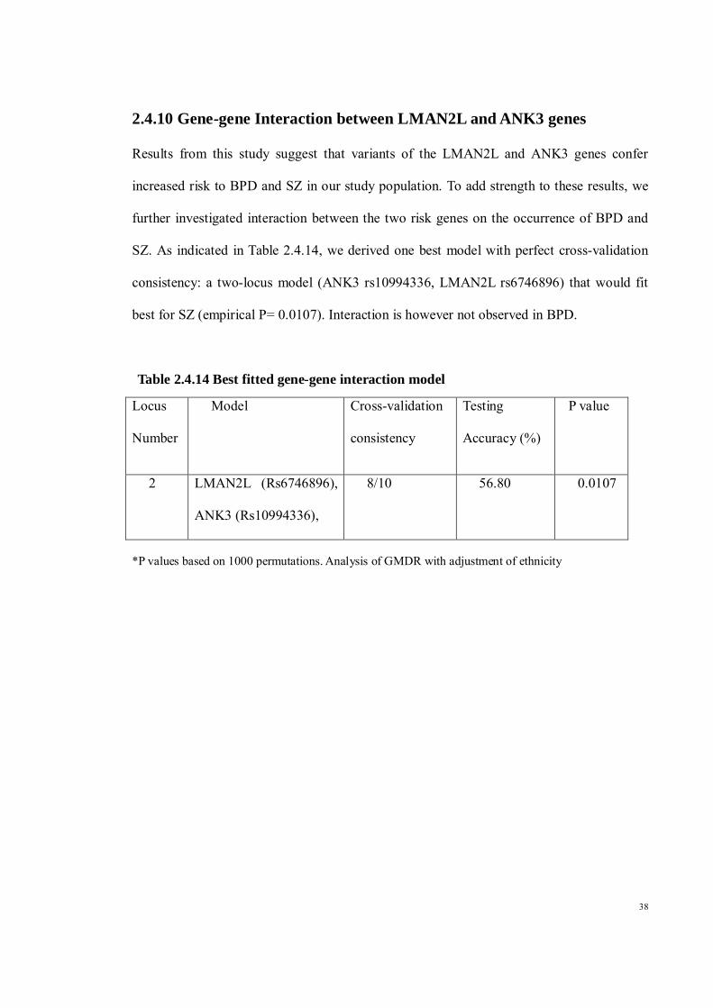
2.3.2.2 DNA Extraction Blood samples collected were centrifuged at 3000 rpm for 10 minutes. The blood
samples were then separated into plasma and buffy coat layer. The buffy coat layers
were then used for DNA extraction using QIAamp DNA Blood Mini Kit, (QIAGEN,
Hilden, Germany). A volume of 20 μl QIAGEN Protease (or proteinase K) was pipetted
into the bottom of a 1.5 ml microcentrifuge tube. Then, 200 μl volume of sample was
added to the microcentrifuge tube. Buffer AL (200 μl) was then added to the sample.
Next, the sample was mixed by pulse-vortexing for 15 s. Following this, the sample was
incubated at 56°C for 10 min. The 1.5 ml microcentrifuge tube was briefly centrifuged
to remove drops from the inside of the lid.
Next, 200 μl ethanol (96–100%) was added to the sample, and mixed again by pulse-
vortexing for 15 s. After mixing, the 1.5 ml microcentrifuge tube was briefly
centrifuged to remove drops from the inside of the lid. The cap was closed, and
centrifuged at 6000 x g (8000 rpm) for 1 min. The QIAamp Mini spin column was
placed in a clean 2 ml collection tube, and the tube containing the filtrate was discarded.
Next, the QIAamp Mini spin column was carefully opened and 500 μl Buffer AW1 was
added without wetting the rim. The cap was closed and centrifuged at 6000 x g (8000
rpm) for 1 min. The QIAamp Mini spin column was put in a clean 2 ml collection tube,
and the collection tube containing the filtrate were discarded. Next, the QIAamp Mini
spin column was carefully opened and 500 μl Buffer AW2 was added without wetting
the rim. The cap was closed and centrifuged at full speed (20,000 x g; 14,000 rpm) for 3
min. The QIAamp Mini spin column was carefully opened and 200 μl Buffer AE or
distilled water was added. It was then incubated at room temperature (15–25°C) for 1
min, and then centrifuged at 6000 x g (8000 rpm) for 1 min., Elution of DNA in Buffer
AE and stored at –20°C were recommended for long-term storage of DNA. The quality
of DNA was checked using Nanodrop 2000 c (Thermo Scientific)

17
2.3.2.3 Agarose Gel Electrophoresis Gel electrophoresis was carried out to determine the quality of DNA and also the
absence of other impurities. First, 1% of agarose was prepared by adding 1 g of
powdered agarose and 6 ul GelRed into 100 ml 1X TAE buffer. Then, the agarose gel
was cooked in a microwave on high power for 40-60 sec. The gel was then allowed to
cool down. In the meantime, the casting tray and comb were assembled. The mixture
was quickly poured onto the casting tray and left to solidify. When the gel was
solidified, the gel tray was moved into the gel tank filled with TAE buffer enough to
cover the top of the gel. A volume of 1ul of the genomic DNA was mixed with 1.5ul of
loading dye and then loaded into the well. The loading dye was first prepared by
diluting a volume of 1 ul of GelRed into 599ul of 6x loading buffer. The sample was run
together with a 100bp ladder at 110 V for 30 minutes. After that, the sample was viewed
under ultraviolet (UV) light using BIO-RAD UV Transluminator.
2.3.2.4 Genotyping Genomic DNA was extracted from peripheral white blood cells using the QIAamp DNA
Mini Kit (Qiagen, Hilden, Germany). LMAN2L gene variants (rs6746896 and
rs2271893), a BTF3L1/KCTD12 gene variant (rs2073831), and ANK3 gene variants
(rs10994336 and rs1938526) were genotyped at the University of Hong Kong Genome
Research Centre using the Sequenom MassARRAY technology platform with iPLEX
Gold chemistry (Sequenom, San Diego, CA, USA) according to the manufacturer's
protocols. Briefly, the MassARRAY Assay Design software package (v4.0) was used to
design the specific assays with proximal SNP filtering. The quality of PCR fragment
amplification and extension primer specificity was checked prior to running the
reaction.
Residual nucleotides were dephosphorylated prior to the iPLEX Gold reaction.
Following a single-base extension, reaction products were desalted with SpectroClean

18
resin (Sequenom), and 10 nL was spotted onto the SpectroCHIP using the MassARRAY
Nanodispenser. A MassARRAY Analyzer Compact MALDI-TOF mass spectrometer
was used to determine product mass. For proper data acquisition and analysis,
MassARRAY Typer 4.0 software was used. Genotypes were called after cluster analysis
by using the default setting of the Gaussian mixture model. The clusters were inspected
to ensure a clear cluster separation with good signal-to-noise cut-off. A manual review
was done to further clarify uncertain genotype calls. An assay with a call rate of less
than 80% within the same SpectroChip was considered to have failed. A blank and five
duplicates were introduced as quality controls. A SpectroChip with a call rate of more
than 25% in the blank control or concordance of less than 99.5% in the duplicate
checks, along with a call rate of more than 10% in the blank check, was considered to
have failed and would need to be repeated. Any significant result will be validated using
real time PCR.
2.3.2.5 Statistical analyses The genotype distribution was assessed for Hardy-Weinberg equilibrium (HWE) using a
chi squared test. A P-value of more than 0.05 indicates agreement with HWE.
Determination of allele associations was performed by using logistic regression. In
order to avoid false discoveries due to population difference, we performed the
association analysis for each SNP marker separately by ethnicity. Linkage
disequilibrium (LD) and haplotype analysis were performed with the Haploview 4.2
program. Pairwise LD was used to investigate the inter-marker relationship through D′
values in case and control subjects. A permutation test with 5000 replications was used
to obtain empirical levels of Significance. To investigate the influence of gene-gene
interaction on BPD and SZ, the Generalized Multifactor Dimensionality Reduction
(GMDR) method (Lou, Chen et al. 2007) was employed. All possible interactions were

19
tested by using 10-fold cross-validation with an exhaustive search that considers all
possible variable combinations. GMDR provides a cross-validation consistency score,
which is a measure of the degree of consistency with which the selected interaction is
identified as the best model among all possibilities considered. The testing-balanced
accuracy generated is a measure of the degree to which the interaction accurately
predicts case-control status. Testing accuracy is a measure of the strength of gene-gene
interaction with a power of 80% with accuracy of 0.58-0.60, given a sample of
500(Chen, Xu et al. 2011). Ethnicity was used as the covariate in the gene-gene
interaction analysis. Power analysis was carried out by assuming a gene-only effect. A
calculated sample size of 244 bipolar cases and 593 controls was used to provide a
desired power of 80% at a P-value of 0.05 with the following assumptions: the allele
frequency range was 0.20-0.80, the baseline risk for the Malaysian population was 0.16,
and the minimum detectable odds ratio (OR) was 1.5. As for SZ, a sample size of 471
SZ cases and 593 controls would provide 80% power at a P-value of 0.05 with the
following assumptions: the allele frequency range was 0.31-0.41, the baseline risk for
the Malaysian population was 0.15, and the minimum detectable OR was 1.5.

20
2.3.2.6 List of SNPs Studied
No. Gene
RS Number Location in Chromosome
SNP1 SP8 Rs2709736 7p21.2
SNP2 ST8SIA2 Rs8040009 15q26
SNP3 CACNB2 Rs11013860 10p12
SNP4 KCTD12 Rs 2073831 13q22.3
SNP5 ANK3 Rs1938526 10q21 SNP6 Rs 10994336 SNP7 Chromosome 3 Rs 11720452 3q21-q25 SNP8 TRANK1 Rs 9834970 3p22.2
SNP9 LMAN2L Rs 6746896, 2q11.2 SNP10 Rs 2271893 2q11.2 SNP11 SP4 Rs12673091
7p15.3
SNP12 Rs3735440 SNP13 NF1A Rs41350144 1p31.3-p31.2 SNP14 SCN8A Rs 303810 12q13 SNP15
NAPG Rs 473938
18p11.22
SNP16 Rs229079 SNP17 Rs 495484
SNP18 ODZ4 Rs 12576775 11q14.1 SNP19 RGS4 Rs951436 1q23.3 SNP20 CACNA1C Rs1006737 12p13.3 SNP21 PDE10A Rs 1039002 2p16.3 SNP22 MARK1 Rs12563333 1q41 SNP23 COMT/Val158Met Rs4680 22q11.21
SNP24 NRG1 Rs35753505 8p12 SNP25 Rs1081062

21
2.4 RESULTS
2.4.1 Demographic Data Table 2.4.1 shows the demographic data of the patients and controls. The 244 BPD
patients consisted of 73 Malays, 98 Chinese, and 73 Indians, while the 471 SZ patients
were made up of 132 Malays, 222 Chinese, and 117 Indians. Out of the 593 controls,
173 were Malays, 286 Chinese, and 134 Indians. There is a significant difference in the
mean age between BPD patients and healthy controls (P= 0.027). Likelihood tests
indicated a significant effect of ethnicity, but no significant effect of age and gender.
None of the genotypes for the tested SNPs deviated from Hardy–Weinberg equilibrium
for BPD patients, SZ patients, and controls.
Table 2.4.1 Demographic Table of BD and SZ patients with healthy controls
BPD
( N=244)
Schizophrenia
(N=471)
Controls
(N=593)
Age (mean± SD ) 42.95 ± 11.975 40.49 ± 12.082 41.09 ± 10.255
Gender
Male 128 (53%) 270 (57%) 375 (63%)
Female
116 (48%) 201 (43%) 218 (37%)
Ethnicity
Malay 73 (30%) 132 (28%) 173 (29%)
Chinese 98 (40%) 222 (47%) 286 (48%)
Indian 73 (30%) 117 (25%) 134 (23%)

22
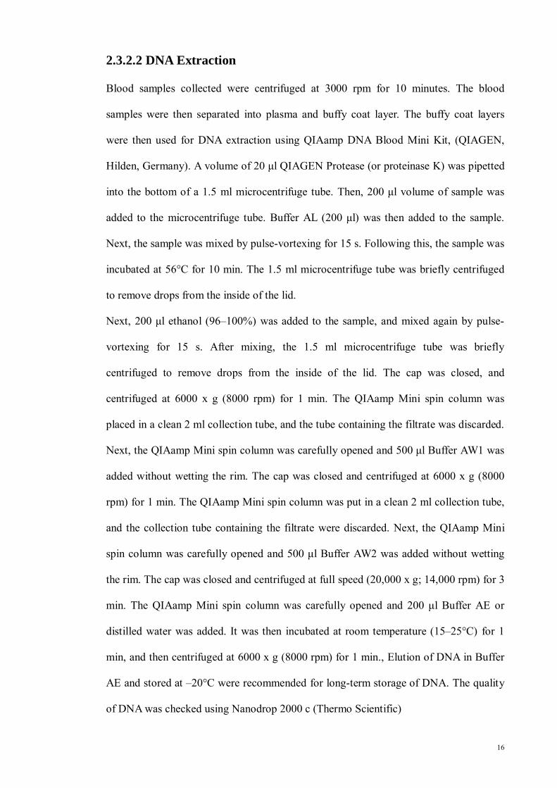
2.4.2 Single Nucleotide Polymorphisms: LMAN2L LMAN2L rs6746896 shows significant differences in allelic distribution between both
BPD and SZ patients and controls for the pooled subjects (P =0.009 and 0.001,
respectively). The LMAN2L rs2271893 C allele frequency was significantly higher in
SZ patients compared with healthy controls, a condition observed in Malays and Indians
(P = 0.002 and P = 0.003, respectively).
After Bonferroni correction, LMAN2L rs6746896 remained significant for both BPD
and SZ (P = 0.009 for BPD and 0.001 for SZ), ANK3 rs1938526 and rs10994336 for
BPD in Malays (P= 0.001 and 0.006, respectively), and LMAN2L rs2271893 for SZ in
Malays (P = 0.003) and Indians (P= 0.002).
When we examined the haplotypes for the LMAN2L gene, the GA, AG, and GG
showed frequencies of 68%, 17% and 15%, respectively. A significant difference exists
between BPD and SZ patients and controls for haplotype frequencies of GA (P = 0.015
and 0.010 for BPD and SZ, respectively), even after the permutation test correction with
5000 permutations. There also exists a significant difference between BPD patients and
controls for haplotypes frequencies of GG (P = 0.013).

23
Figure 2.4 1 Haplotype view for LMAN2L gene rs2271893 and rs6746896 in BPD
Figure 2.4 2 Haplotype view for LMAN2L gene rs2271893 and rs6746896 in SZ

24
Table 2.4.2 Genotype Distribution of LMAN2L gene in BPD, SZ and Controls
All ethnicities Malay Chinese Indian
Controls Patients BPD
SZ
Controls Patients BPD
SZ
Controls Patients BPD
SZ
Controls Patients BPD
SZ
LMAN2L Rs6746896 AA AG GG A G
270 247 76 787 399
137 85 22 359 129
259 170 42 688 254
73 72 28 218 128
32 31 10 95 51
69 53 9 191 71
111 133 42 355 217
53 37 8 143 53
105 87 30 297 147
86 42 6 214 54
51 17 5 119 27
80 30 6 190 42
Rs2271893 GG AG AA G A
417 153 23 987 199
173 63 8 409 79
350 109 12 809 133
132 34 8 298 50
64 5 7 133 19
113 13 6 239 25
186 83 17 455 117
85 6 6 176 18
130 81 12 341 105
90 36 7 216 50
57 8 6 122 20
100 15 5 215 25
25
Table 2.4.3 Odds ratio of SNPs of LMAN2L gene in BPD and SZ in pooled population
ALL ETHNICITY
LMAN2L BPD SZ Rs6746896 0R (CI) P-value OR (CI) P-value A 1 – 1 –
G 1.36 (1.08–1.71) 0.009 1.34 (1.12–1.61) 0.001
AA 1 – 1 –
AG 0.59 (0.35–1.00) 0.048 1.35 (1.04–1.75) 0.024
GG 0.88 (0.51–1.51) 0.634 1.55 (1.03–2.32) 0.036
Rs2271893 G 1 – 1 –
A 1.12 (0.85–1.48) 0.429 1.24 (0.99–1.56) 0.06
GG 1 – 1 –
GA 0.69 (0.29–1.62) 0.388 1.22 (0.92–1.62) 0.164
AA 0.73 (0.30–1.78) 0.482 1.73 (0.87–3.42) 0.117

26
Table 2.4.4 Odds ratio of SNPs of LMAN2L gene in BPD and SZ according to ethnicity
Malay Chinese Indian
LMAN2L BPD SZ BPD SZ BPD SZ
Rs6746896 OR (CI) P value OR (CI) P
value OR (CI) P value OR (CI) P
value OR (CI) P value OR (CI) P
value A 1 – 1 – 1 – 1 – 1 – 1 –
G 1.09 (0.74–1.60) 0.678 1.50 (1.07–
2.10) 0.02 1.63 (1.14–2.34) 0.007 1.21 (0.94–
1.55) 0.148 1.21 (0.73–2.00) 0.471 1.59 (0.98–
2.58) 0.061
AA 1 – 1 – 1 – 1 – 1 – 1 –
AG 1.05 (0.58–1.91)
0.876 1.30 (0.80–2.12)
0.295 1.80 (1.09–2.96)
0.021 1.45 (0.99–2.13)
0.058 1.49 (0.75–2.95)
0.251 1.39 (0.78–2.45)
0.261
GG 1.29 (0.56–3.00) 0.549 2.53 (1.13–
5.63) 0.023 2.42 (1.05–5.57) 0.037 1.28 (0.74–
2.19) 0.379 0.91 (0.23–3.54) 0.89 5.71 (0.67–
9.96) 0.112
Rs2271893 G 1 – 1 – 1 – 1 – 1 – 1 –
A 0.83 (0.49–1.41) 0.49 2.63 (1.40–
4.97) 0.003 1.15 (0.77–1.71) 0.495 0.86 (0.64–
1.15) 0.297 1.31 (0.73–2.30) 0.363 2.76 (1.46–
5.20) 0.002
GG 1 – 1 – 1 – 1 – 1 – 1 –
GA 0.77 (0.40–1.50) 0.444 2.28 (1.14–
4.58) 0.02 1.00 (0.60–1.67) 1 0.72 (0.49–
1.06) 0.091 1.42 (0.69–2.92) 0.34 2.51 (1.28–
4.95) 0.008
AA 1.02 (0.19–5.49) 0.983 1.30 (0.00–
2.13) 0.999 1.75 (0.49–6.20) 0.39 0.99 (0.46–
2.16) 0.997 0.96 (0.15–6.06) 0.961 1.74 (0.00–
2.15) 0.999

27
2.4.3 Single Nucleotide Polymorphisms: ANK3 The ANK3 rs1938526 and rs10994336 were significantly different between BPD and SZ
patients and controls of Malay ethnicity (P = 0.001 and 0.012, for BPD and SZ,
respectively). ANK3 rs1938526 was associated with BPD in Indians (P = 0.015).
In the Malay ethnic subgroup, the ANK3 rs1938526 C allele and rs10994336 T allele were
found to be associated with a relatively reduced risk of BPD and SZ (ANK3 rs1938526:
OR0.471, 95% CI 0.30–0.74, P = 0.001 and OR 0.60, 95% CI 0.41–0.89, P = 0.012, for
BPD and SZ, respectively; ANK3 rs10994336: OR0.49, 95% CI 0.30–0.81, P = 0.006 and
OR 0.62, 95% CI 0.40–0.95,P = 0.029, for BPD and SZ, respectively). The LD of the
ANK3 gene was not significantly different between BPD and SZ patients and controls.

28
Table2.4.5 Genotype Distribution of ANK3 gene in BPD, SZ and Controls
All ethnicities Malay Chinese Indian Controls Patients
BPD
SZ
Controls Patients BPD
SZ
Controls Patients BPD
SZ
Controls Patients BPD
SZ
ANK3 Rs1938526 TT TC CC T C
357 202 34 916 270
141 87 16 369 119
263 174 34 700 242
111 57 5 279 67
34 30 9 98 48
69 53 10 191 73
167 96 23 430 142
52 40 6 144 52
116 88 16 320 120
79 48 7 206 62
50 15 8 115 31
77 32 7 186 46
Rs10994336 CC TC TT C T
411 164 18 986 200
168 70 6 406 82
325 130 16 780 162
128 39 7 295 53
41 26 6 108 34
87 38 7 212 52
194 81 10 469 101
63 28 7 154 42
152 66 5 370 76
85 44 5 214 54
51 16 6 118 28
86 26 4 198 34

29
Table 2.4.6 Odds ratio of SNPs of ANK3 gene in BPD and SZ in pooled population
ANK3 ALL ETHINICITY BPD
SZ
Rs1938526 OR ( CI) P value OR (CI) P value T 1 – 1 – C 0.92 (0.72–1.17) 0.483 0.86 (0.71–1.04) 0.123 TT 1 – 1 – TC 1.23 (0.65–2.32) 0.518 0.87 (0.67–1.12) 0.273
CC 1.11 (0.58–2.13) 0.76 0.79 (0.48–1.30) 0.347 Rs10994336 C 1 – 1 – T 1.01 (0.76–1.33) 0.976 0.98 (0.78–1.23) 0.84 CC 1 – 1 – CT 0.94 (0.67–1.31) 0.698 1.02 (0.78–1.34) 0.878 TT 1.11 (0.43–2.87) 0.825 0.85 (0.43–1.68) 0.643

30
Table 2.4.7 Odds ratio of SNPs of ANK3 gene in BPD and SZ according to ethnicity
MALAY CHINESE INDIAN
ANK3 BPD SZ BPD SZ BPD SZ
Rs1938526 OR ( CI) P value OR ( CI) P
value OR ( CI) P value OR ( CI) P
value OR ( CI) P value OR ( CI) P
value
T 1 – 1 – 1 – 1 – 1 – 1 –
C 0.47 (0.30–0.74) 0.001 0.60 (0.41–
0.89) 0.012 0.92 (0.64–1.32) 0.646 0.87 (0.66–
1.15) 0.334 2.03 (1.15–3.58) 0.015 1.21 (0.79–
1.83) 0.384
TT 1 – 1 – 1 – 1 – 1 – 1 –
TC 0.59 (0.33–1.08) 0.085 0.71 (0.44–
1.15) 0.166 0.72 (0.44–1.18) 0.193 0.76 (0.52–
1.11) 0.149 1.94 (0.99–3.83) 0.054 1.36 (0.77–
2.37) 0.289
CC 0.13 (0.04–0.46) 0.001 0.21 (0.06–
0.72) 0.013 1.14 (0.43–2.98) 0.795 1.00 (0.50–
1.99) 0.999 5.52 (0.63–7.52) 0.122 1.01 (0.33–
3.08) 0.984
Rs10994336 C 1 – 1 – 1 – 1 – 1 – 1 –
T 0.49 (0.30–0.81) 0.006 0.62 (0.40–
0.95) 0.029 1.19 (0.76–1.86) 0.439 1.05 (0.76–
1.46) 0.776 1.81 (1.01–3.26) 0.047 1.45 (0.91–
2.32) 0.116
CC 1 – 1 – 1 – 1 – 1 – 1 –
CT 0.49 (0.27–0.89)
0.02 0.71 (0.42–1.20)
0.2 0.95 (0.57–1.60)
0.852 0.96 (0.65–1.42)
0.829 1.78 (0.89–3.54)
0.102 1.68 (0.94–3.00)
0.082
TT 0.24 (0.05–1.16) 0.076 0.27 (0.07–
1.09) 0.066 2.95 (0.37–3.57) 0.309 1.48 (0.49–
4.44) 0.488 4.29 (0.47–5.96) 0.199 1.19 (0.30–
4.70) 0.804

31
2.4.4 Single Nucleotide Polymorphism: KCTD12 KCTD12 rs2073831 was genotyped in this study. All the genotype distribution are in
Hardy-Weinberg Equilibrium. No allelic associations were observed with BPD or SZ in the
pooled population. However, after stratification by ethnicity, there is a significant
association between the allele T with SZ in both Malays and Chinese (P = 0.024 and 0.025,
respectively).

32
Table 2.4.8 Genotype distribution of rs2073831 of KCTD12
All ethnicities Malay Chinese Indian
Control Patient BPD
SZ
Control Patient BPD
SZ
Control Patient BPD
SZ
Controls Patient BPD
SZ
KCTD12 Rs2073831 CC TC TT C T
226 280 87 732 454
95 109 40 299 189
191 216 64 598 344
72 82 20 226 122
44 6 27 94 60
41 66 25 148 116
99 133 52 331 237
83 5 8 171 21
92 107 24 291 155
55 65 15 175 95
8 57 6 73 69
58 43 15 159
Table 2.4.9 Odds ratio of rs2073831 of KCTD12 in BPD and SZ in pooled population
ALL ETHNICITY KCTD12 BPD SZ Rs2073831 0R (CI) P-value OR (CI) P-value C 1 – 1 –
T 0.99 (0.80–1.23) 0.94 1.07 (0.90–1.28) 0.445
CC 1 – 1 –
CT 1.05 (0.76–1.47) 0.752 1.05 (0.81–1.37) 0.695
TT 0.91 (0.58–1.43) 0.683 1.12 (0.77–1.62) 0.57

33
Table 2.4.10 Odds ratio of rs2073831 of KCTD12 in BPD and SZ according to ethnicity
Malay Chinese Indian
KCTD12 BPD SZ BPD SZ BPD SZ
Rs2073831 OR (CI) P value OR (CI) P
value OR (CI) P value OR (CI) P
value OR (CI) P value OR (CI) P
value C 1 – 1 – 1 – 1 – 1 – 1 –
T 0.90 (0.40–1.35) 0.602 0.68 (0.49–
0.95) 0.025 0.92 (0.07–1.26) 0.612 1.35 (1.04–
1.74) 0.024 1.16 (0.74–1.80) 0.524 1.18 (0.81–
1.70) 0.387
CC 1 – 1 – 1 – 1 – 1 – 1 –
CT 1.13 (0.60–2.10) 0.86 0.69 (0.41–
1.14) 0.147 1.08 (0.63–1.84) 0.783 1.15 (0.78–
1.69) 0.492 1.13 (0.60–2.10) 0.711 1.69 (0.98–
2.93) 0.061
TT 1.47 (0.50–4.28) 0.668 0.41 (0.20–
0.84) 0.015 0.74 (0.39–1.41) 0.362 1.99 (1.13–
3.51) 0.018 1.47 (0.50–4.28) 0.483 1.16 (0.50–
2.67) 0.727

34
2.4.5 Single Nucleotide Polymorphism: SP4 Two SNPs of the SP4 gene (transcription factor gene) were genotyped in this study,
rs12673091 and rs3735440. Both of the SNP are intron variants, located on chromosome 7.
Table 2.4.11 shows the association test between cases and controls in BPD and SZ. All the
genotype distributions are in Hardy-Weinberg Equilibrium. No allelic association with BPD
and SZ were observed in the pooled population, or after stratification by ethnicity.
Table 2.4.11 Odds ratio of SNPs of SP4 gene in BPD and SZ in BPD and SZ
SNP BPD SZ
Rs12673091 OR ( CI) P value OR (CI) P value
Overall 0.563 (0.432-0.734) 0.06 1.159 (0.974-1.379) 0.096
Malay 0.455 (0.274-0.754) 0.08 1.060 (0.769-1.462) 0.722
Chinese 0.633 (0.372-1.075) 0.09 1.246 (0.957-1.621) 0.103
Indian 0.723 (0.471-1.111) 0.139 1.079 (0.777-2.554) 0.595
Rs 3735440
Overall 0.841 (0.639-1.108) 0.219 0.878 (0.695-1.109) 0.275
Malay 0.779 (0.482-1.258) 0.307 0.826 (0.536-1.272) 0.386
Chinese 1.828 (0.969-3.448) 0.062 0.957 (0.642-1.427) 0.829
Indian 0.655 (0.413-1.039) 0.072 0.886 (0.585-1.343) 0.569

35
2.4.6 Single Nucleotide Polymorphism: NAPG gene Three SNPs of the NAPG gene (N-ethylmaleimide-sensitive factor attachment protein,
gamma) were genotyped in this study, rs473938, rs229079 and rs495484. All of the SNP
are intron variants, located on chromosome 18. Table 2.4.12 shows the association test
between cases and controls in BPD and SZ. All the genotype distributions are in Hardy-
Weinberg Equilibrium. No allelic association with BPD and SZ were observed in the
pooled population, or after stratification by ethnicity.
Table 2.4.12 Odds ratio of SNPs of NAPG gene in BPD and SZ
SNP BPD SZ
OR ( CI) P value OR (CI) P value
Rs473938 Overall 1.148 (0.835-1.410) 0.187 0.924 (0.784-1.089) 0.347
Malay 1.394 (0.966-2.012) 0.076 0.991 (0.730-1.346) 0.954
Chinese 0.893 (0.631-1.264) 0.522 0.9 (0.693-1.169) 0.431
Indian 1.008 (0.663-1.532) 0.971 0.741 (0.530-1.035) 0.078
Rs229079 Overall 1.067 (0.840-1.356) 0.595 1.119 (0.921-1.360) 0.257
Malay 1.037 (0.667-1.612) 0.872 0.887 (0.626-1.286) 0.554
Chinese 1.053 (0.729-1.522) 0.783 1.209 (0.905-1.617) 0.199
Indian 1.087 (0.689-1.717) 0.719 1.283 (0.855-1.924) 0.228
Rs495484 Overall 0.993 (0.803-1.229) 0.951 0.974 (0.820-1.157) 0.765
Malay 1.039 (0.694-1.556) 0.853 0.806 (0.583-1.116) 0.194
Chinese 0.821 (0.593-1.137) 0.236 1.037 (0.800-1.345) 0.782
Indian 1.155 (0.763-1.747) 0.495 0.934 (0.665-1.311) 0.693

36
2.4.7 Single Nucleotide Polymorphism: NRG1 gene Two SNPs of the NRG1 gene (neuregulin 1) were genotyped in this study, rs35753503 and
rs1081062. Both of the SNP are intron variants, located on chromosome 8. Table 2.4.13
shows the association test between cases and controls in BPD and SZ. All the genotype
distributions are in Hardy-Weinberg Equilibrium. No allelic association with BPD and SZ
were observed in the pooled population, or after stratification by ethnicity.
Table 2.4.13 Odds ratio of SNPs of NRG1 gene in BPD and SZ
SNP BPD SZ
Rs35753503 OR ( CI) P value OR (CI) P value
Overall 1.136 (0.924-1.397) 0.226 0.878 (0.695-1.109) 0.275
Malay 1.305 (0.902-1.888) 0.158 0.826 (0.536-1.272) 0.386
Chinese 1.016 (0.735-1.404) 0.925 0.927 (0.642-1.427) 0.829
Indian 0.998 (0.660-1.511) 0.994 0.886 (0.585-1.343) 0.569
Rs1081062
Overall 1.031 (0.750-1.418) 0.851 0.986 (0.800-1.214) 0.893
Malay 0.911 (0.516-1.611) 0.749 1.162 (0.727-1.857) 0.530
Chinese 1.061 (0.648-1.738) 0.814 1.021 (0.706-1.478) 0.911
Indian 1.732 (0.608-2.107) 0.696 0.866 (0.610-1.229) 0.421

37
2.4.8 Single Nucleotide Polymorphism: Other candidate genes For the SNP of CACNA1C, PDE10A, MARK1, COMT/Val158Me, NRG1, there is no
allelic association with BPD and SZ observed in the pooled population, or after
stratification by ethnicity. The genotype distribution of the SNP of SP8, ST8SIA2,
CACNB2, TRANK1, and NF1A are deviated from Hardy Weinberg equilibrium. For the
SNP of SCN8A, ODZ4 and RGS4, the association test do not proceed as more than 30% of
the samples failed to be genotyped.
2.4.9 Overall Results Overall, rs1938526 and rs10994336 of the ANK3 gene and rs6746896 of the LMAN2L
gene were found to be significantly associated with BPD and SZ. It is also important to note
that the homozygous mutant genotype of the LMAN2L rs6746896 confers greater risk of
both disorders, while the ANK3 rs1938526 confers lower risk, indicating stronger effects of
risk alleles in their homozygous form.
38
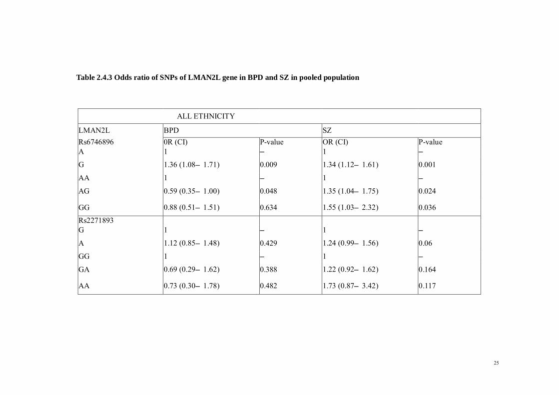
2.4.10 Gene-gene Interaction between LMAN2L and ANK3 genes Results from this study suggest that variants of the LMAN2L and ANK3 genes confer
increased risk to BPD and SZ in our study population. To add strength to these results, we
further investigated interaction between the two risk genes on the occurrence of BPD and
SZ. As indicated in Table 2.4.14, we derived one best model with perfect cross-validation
consistency: a two-locus model (ANK3 rs10994336, LMAN2L rs6746896) that would fit
best for SZ (empirical P= 0.0107). Interaction is however not observed in BPD.
Table 2.4.14 Best fitted gene-gene interaction model
*P values based on 1000 permutations. Analysis of GMDR with adjustment of ethnicity
Locus
Number
Model Cross-validation
consistency
Testing
Accuracy (%)
P value
2 LMAN2L (Rs6746896),
ANK3 (Rs10994336),
8/10 56.80 0.0107

39
2.5 DISCUSSIONS In this study, we investigated the association of gene variants involved in ion channel
transport and ER transport with BPD and SZ.
2.5.1 Single Nucleotide Polymorphisms: LMAN2L We showed a significant association between LMAN2L rs6746896 and both BPD and SZ in
the pooled population. However, following ethnic stratification, this association was
modified, with a strong and significant association that was apparent in the Chinese for
BPD and in the Malays for SZ. Our pooled result on BPD is consistent with two previous
GWAS: one by (Chen et al. 2013) on the European and Asian ancestry samples and another
by the Psychiatric GWAS Consortium BPD Working Group (Group 2011). However, to
date, no study has investigated the association between LMAN2L rs6746896 and SZ; ours
is the first to report a significant positive association. LMAN2L rs2271893 was also found
to be associated with SZ but not with BPD in Malays and Indians.
These findings are partially consistent with those of Andreassen et al. (2013) in that the
authors reported an overlap of rs2271893 with BPD and SZ, whereas we found an
association with SZ but not BPD. Our finding is inconsistent, however, with the significant
association with BPD reported by (Chen et al. 2013). Nevertheless, our results suggest that
the A allele of rs2271893 confers an over 2.5-fold increased risk of SZ in ethnic Malays and
Indians. The GG haplotype frequency of the LMAN2L gene was found to be significantly
different between patients and controls in both BPD and SZ. Our study provides an early
report of such a haplotype finding and needs to be confirmed by other studies.
The role of the lectin, mannose-binding 2-like (LMAN2L) gene, also known as the VIPL
(VIP36-Like) gene, as an ER export receptor has been suggested in a study by (Neve et al.

40
2003). Studies have demonstrated that LMAN2L may act as a regulator for the ERGIC-53
(LMAN 1) gene. Mutation of the latter gene has been associated with a genetic bleeding
disorder with a combined deficiency of coagulation factors FV and FVIII (F5F8D)
(Nicholas et al. 1999). However, the functional role of LMAN2L in the pathophysiology of
BPD has yet to be discovered. Nonetheless, two large-scale GWAS have shown that this
gene is associated with BPD (Group 2011, Chen et al. 2013).
2.5.2 Single Nucleotide Polymorphisms: ANK3 The variants ANK3 rs1938516 and rs10994336 were not significantly associated with the
occurrence of BPD and SZ in the pooled subjects. However, after ethnic stratification,
significant differences were observed between BPD patients and controls and between SZ
patients and controls among ethnic Malays. The rs1938526 variant was also significantly
associated with BPD in Indians. The association between ANK3 gene variants and BPD has
gained support from various studies (Ferreira et al. 2008, Schulze et al. 2009, Paez-
Gonzalez et al. 2011, Tesli et al. 2011, Dedman et al. 2012) while other studies have failed
to report any significant association (Gella et al. 2011, Lett et al. 2011, Takata et al. 2011,
Gonzalez et al. 2013, Kondo et al. 2013), suggesting that ethnicity contributes to the risk of
these disorders. Among supporting studies were those by (Takata et al. 2011), who showed
an association in the East Asian population, including Han Chinese, Japanese, and Koreans,
and by (Gella et al. 2011) and (Kondo et al. 2013), who reported an association of
rs1938526 and rs10994336 with SZ. These studies are in contrast, however, with the work
of Tesli et al. (2011), who reported no association. We failed to show any association
between rs1938526 and rs10994336 with SZ and BPD in our Chinese subgroup.

41
Ankyrin-G protein (ANK3) is an adaptor protein expressed in the axonal initial segment
and the nodes of Ranvier in the central and peripheral nervous systems (Bennett and Baines
2001). It has been shown to regulate the assembly of voltage-gated sodium channels. (Zhou
et al. 1999) reported that Purkinje cells with knockout ANK3 failed to initiate action
potential to support rapid and repetitive firing. Furthermore, a study with mouse brain
samples treated with lithium, one of the drugs commonly used to treat BPD, found that
ANK3 and subunits of the calcium channel were down-regulated (Baum et al. 2008).
2.5.3 Single Nucleotide Polymorphisms: KTCD12 Variant BTF3L1/KTCD12 rs2073831 was found to be associated with SZ but not with BPD
in the Malays and Chinese. This result is in contrast with the significant positive finding in
the GWAS of the Han Chinese BPD (Yuan et al. 2012). There are, however, no reports of
association of rs2073831 and risk of SZ.
An ion channel gene, potassium channel tetramerization domain containing 12 (KCTD12),
is involved in potassium ion transport. KCNC2, another potassium channel, was previously
identified in the Wellcome Trust Case Control Consortium Study as being associated with
BPD (Baum, Akula et al. 2008). The role of potassium channel mutations has also been
found to be particularly prominent in human channelopathies (Ryan and Ptácek 2010). In
addition, an association between altered gene function related to potassium ion transport
and BPD was reported in a study by (Goldstein et al. 2009). Sodium- and potassium-
activated adenosine triphosphatases (Na+, K+–ATPase), major plasma membrane
transporters for sodium and potassium, were found to be associated with BPD (Goldstein et

42
al. 2009). These studies have clearly suggested a role for potassium ion transport in the
etiology of BPD.
2.5.4 Single Nucleotide Polymorphisms: Other candidate genes The present study also investigated a number of SNP in various candidate genes, which
includes CACNA1C, PDE10A, MARK1, COMT/Val158Me, NRG1, SP8, ST8SIA2,
CACNB2, TRANK1, NF1A, SCN8A, ODZ4 and RGS4. None of the SNP of these
candidate genes was significantly associated with susceptibility to BPD and SZ. These
genes and their related SNP were chosen to be candidate genes in this study because before
this current study were conducted, a literature search was carried out and a number of
research have shown that these candidate genes were significantly associated with
susceptibility to BPD and SZ in other populations. Therefore these SNP were chosen as a
prescreening test for susceptibility to BPD and SZ in Malaysia population. The non-
significant result may be due to different genetic make-up between the Malaysian
population and other populations being studied.
2.5.5 Gene-gene interaction The result from gene-gene interaction analysis suggests that the ANK3 and LMAN2L genes
interact with each other in the pathophysiology of BPD and SZ. Surprisingly, a literature
search failed to provide any reports that link these two genes. However, gene ontology
enrichment analysis (Huang et al. 2009) identified protein localization as a common shared
biological pathway.

43
2.5.6 Justification of Study There were more Chinese participants in both of our BPD and SZ samples than other
ethnicities, which could be due to the catchment area having a higher proportion of Chinese
patients in UMMC as compared with other races. However, sampling was done without
bias or preference for any ethnic group. A contributing factor in the failure to replicate a
positive association with either BPD or SZ, as reported in other studies, in our Chinese
ethnic group may be the differences in genetic variability between Southeast Asian Chinese,
Chinese from Shanghai, and Beijing Han Chinese (Chen et al. 2009). Allele frequency
differences between each population may also be a factor. The smaller sample size in our
study could also explain the failure to replicate findings. Nonetheless, overall, our result is
consistent with previous studies in the Asian population.
2.5.7 Genetic Overlapping of Schizophrenia and BPD Genetic association studies have provided clues on possible risk genes associated with
psychiatric disorders including BPD and SZ (Berrettini 2000). These genetic variations are
responsible for the pathophysiology of the disorders mainly by altering the protein
functions, thereby regulating brain activity (Meyer-Lindenberg et al. 2006, Bigos et
al.2010). Expression studies have shown that the activities of the BPD/SZ genes were
altered following treatments (Pandey et al. 2008, Rueckert et al. 2013). These observations
have strongly hinted that the genetic factor may play a significant role in etiology of these
two disorders.

44
2.5.8 Strength and limitation of the Study A limitation of our study is the small sample size compared with earlier GWAS samples,
which could have contributed to the small effect size of the associations. Taking into
consideration that BPD and SZ develop as a result of the interplay of genetic and
environmental factors, the lack of environmental background information of the subjects
such as exposure to nicotine, advanced paternal age and stressful or traumatic events could
be a further limitation of the study (de Leon and Diaz 2005, Goldberg and Garno 2005,
Torrey et al. 2009). One of the objectives of the study is to examine the gene-gene
interaction of the candidate SNPs. In order to achieve this objective, we employed the
GMDR method. The limitation was that only one to several SNPs per candidate genes were
chosen. These numbers of SNPs were not sufficient to capture most genetic information of
the candidate genes. Future studies should elaborate on the findings by including more
functional SNPs in the studied candidate genes.
On the other hand, its strength lies in the genotyping of polymorphisms across the BPD and
SZ population simultaneously, which enabled us to study common genetic variants
involved in the pathophysiology of both BPD and SZ. The results of our study favor the
hypothesis that both BPD and SZ share some common genetic risk factors. Moreover,
comparison of ethnicities in relation to BPD and SZ allowed us to investigate how ethnicity
modifies the association between gene variants and BPD and SZ.

45
2.6 CONCLUSION
To our knowledge, this study is the first to report an association between LMAN2L
rs6746896 and SZ. Further evidence is needed to confirm this interaction in a larger
population. We found a positive association between LMAN2L (rs6746896) with both BPD
and SZ. We also showed that variants of the ANK3 gene (rs1938516 and rs10994336) are
associated with BPD in Malays and that LMAN2L rs2271893 is associated with SZ in
Malays and Indians. The results of our study favour the hypothesis that both BPD and SZ
share some common genetic risk factors. In addition to our result, functional study of the
role of candidate gene in animal model and in-vitro study could provide more insights of
the molecular mechanisms of BPD. As BPD and SZ are complex diseases with interaction
of huge amount of genetic factor, searching for the causative risk factor may be difficult.
Nevertheless, with the recent advancement of high throughput technology, and introduction
of other aspect of genetic variation in human diseases, such as microRNA, understanding of
pathophysiology of BPD can be achievable.

46
CHAPTER THREE: DIFFERENTIAL MICRORNA
EXPRESSION IN BIPOLAR DISORDER I MANIC PATIENT
FOLLOWING TREATMENT WITH ASENAPINE AND
RISPERIDONE

47
3.1 INTRODUCTION
Mood stabilizers and atypical antipsychotic agent have long been the first-line treatment for
acute mania with or without psychotic features. The atypical antipsychotic have several
advantages in that they do not induce depressive episodes compared to mono-therapy with
mood stabilizers, while several studies have revealed that some atypical antipsychotic
shows improvement in depressive symptoms in manic patients (Szegedi et al. 2011). Over
the years, numerous reviews and studies have shown that these atypical antipsychotics were
significantly more effective than placebo in the treatment of acute mania, although these
studies do not usually seek to align the efficacy of these individual atypical antipsychotics
(Scherk et al. 2007, Correll et al. 2010, Yildiz et al. 2011).
Although major progress have been achieved in terms of research and development,
there stills exists gap in the knowledge of molecular mechanisms underlying bipolar mania
and action pathways of atypical antipsychotic are still waiting to be filled. Asenapine is a
relatively new atypical antipsychotic in the market, and its molecular structure was
described as a tetracyclic antipsychotic (Shahid et al. 2009). Being administered as a
sublingual preparation, which enhances its direct absorption, this increases asenapine
bioavailability compared to other atypical antipsychotic that are administered orally and
thus possess much lower bioavailability due to hepatic metabolism (Reynolds 2011). This
makes asenapine rather unique than its fellow atypical antipsychotic.
MicroRNAs (miRNA) are small non-coding RNA molecules that regulate gene
expression, including genes involved in neuronal function and plasticity that have relevance
for brain function and mental health (Chan and Kocerha 2012). In several studies, miRNA

48
have been shown to be altered in BPD patient and other psychiatric disorder (Kim et al.
2010, Moreau et al. 2011). There are vast evidence of miRNAs’ regulatory roles in a
number of central nervous system processes, including neurogenesis and synaptic plasticity
(Kolshus et al. 2014).
Being a complex disorder, the polygenic characteristics of BPD have invited a large
number of studies carried out in various sample sizes and population subjects, and results in
a list of candidate genes, each acts in small effects contributing to the complexity of the
disorder. The search for novel intervention targets for pharmacology interventions is further
made difficult by its interaction and overlapping with genetics of other psychiatric illness
(Maier et al. 2005). Given that miRNA levels are frequently altered in psychiatric disorders
and also findings that they have been able to be influenced by psychotropic medications
(Dinan 2010), the ability of a single miRNA to target whole network of proteins may make
it fruitful for psychiatric disorders studies where it is the sum of small effects of many
genes that is thought to underlie habitability.
In contrast to postmortem studies and other invasive techniques, uses of peripheral
blood as a research subject in psychiatric disorders provides advantages of easy access and
of being relatively non-invasive. In addition to that, stability of miRNA in human blood is
assured (Turchinovich et al. 2011). While peripheral blood provides an easy assessable and
non-invasive way for biochemical investigation of BPD, there have been concerns as to
whether the peripheral changes reflects the exact alterations in the brain function. In
respond to this, interaction between the central nervous system (CNS) and immune system,
particularly lymphocytes have been proposed. In view of this, there have been correlation
and similarity in pattern between the receptor expression, altered metabolism and cellular

49
function of the cells in CNS with lymphocytes (Gladkevich et al. 2004). Moreover, miRNA
that are specific to brain disorders are detectable in peripheral blood (Lai et al. 2011,
Leidinger et al. 2013).
The majority of clinical studies of miRNAs in psychiatric disorders are focused on
schizophrenia. Being a new player in the field of atypical antipsychotic, there is a relatively
lack of understanding of asenapine in terms of its molecular mechanism. This study aims to
examine the changes in microRNA expression in bipolar mania patient following 12 weeks
of treatment with asenapine and in comparison with risperidone. Risperidone have been
among the first-line treatment in acute mania. In a recent up to dated meta-analysis, the
author proposed that risperidone were among the most effective atypical antipsychotic in
the treatment of acute mania (Cipriani et al. 2011). It is hoped that the results of this study
were to provide more insights into the molecular mechanism underlying the action of the
two atypical antipsychotics, as well as the biological pathophysiology underlying BPD
mania.

50
3.1.1 Objectives
The aims of this study are:
1. To examine the changes in the expression of miRNA in the patients of BPD mania before
and after the treatment of asenapine and risperidone.
2. To examine the changes in the scores of YMRS in the patients of BPD mania before and
after the treatment of asenapine and risperidone.
3. To compare the changes in the expression of miRNA in the patients of BPD mania before
and after the treatment between asenapine and risperidone.
4. To compare the changes in the scores of YMRS in the patients of BPD mania before and
after the treatment between asenapine and risperidone.
3.1.2 Justification of Study
BPD, like any other psychiatric illness, is a complex diseases influenced by genetic factors
that interact with environmental factors. There is no single gene that is responsible for the
illness. Each genetic factor act in a very small effects, and the sum and interaction of
hundreds of these genes play a role in the pathogenesis of the diseases. The ability of a
single miRNA to target whole network of proteins make it an important subject of research
for psychiatric disorders where it is the sum of many genes of small effects that are thought
to underlie the pathophysiology of the disorder.

51
3.1.3 Research Hypothesis
The null hypothesis
1. There are no changes in the expression of miRNA in the patients of BPD mania before
and after the treatment of asenapine and risperidone.
2. There are no changes in the scores of YMRS in the patients of BPD mania before and
after the treatment of asenapine and risperidone.
3. There are no differences in the expression of miRNA in the patients of BPD mania before
and after the treatment between asenapine and risperidone.
The research hypothesis
1. There are changes in the expression of miRNA in the patients of BPD mania before and
after the treatment of asenapine and risperidone.
2. There are changes in the scores of YMRS in the patients of BPD mania before and after
the treatment of asenapine and risperidone.
3. There are differences in the expression of miRNA in the patients of BPD mania before
and after the treatment between asenapine and risperidone.

52
3.2 LITERATURE REVIEW
3.2.1 Antipsychotics
Antipsychotic drugs are widely used as pharmacotherapy prescribed to schizophrenia and
related disorders. However, their use in the treatment of mania in BPD have increased over
the past decade. This have now led to antipsychotic drugs being the first line treatment for
bipolar mania. The neuronal mechanisms of mania remains ambiguous, but antipsychotic
mechanisms observed in schizophrenia may provide clues on this matter. As occupancy of
dopamine D2 receptor in the striatum correlates best with relief of positive symptoms of
schizophrenia (Agid et al. 2007), it is likely that the similar mechanisms underlying the
anti-manic effects, given the efficacy of antipsychotic in alleviation of manic symptoms in
BPD. In common to other antipsychotic, asenapine have high affinity for dopamine D2
receptor. In fact, asenapine have affinity for D2 receptor that is higher than other
antipsychotic except aripiprazole. (Shahid et al. 2009) Other than its affinity for D2
receptor, asenapine have been shown to have broad activities at 5-HT2 receptor. Its
antagonism at 5-HT2B, 5-HT2C, 5-HT5A, 5-HT6 and 5-HT7 receptors is apparent at
affinities at or greater than that for the D2 receptor; with activity at the 5-HT2C site that is
particularly high (Shahid et al. 2009).
One of the side effects of atypical antipsychotic that is of most concern is weight
gain. Affinity of antipsychotic at 5-HT2C has been associated with weight gain in patients
(Reynolds et al. 2002). However, it is remarkable that even though asenapine possesses
relatively high affinities at 5-HT2C and H1 receptors, similar to that been observed for
clozapine and olanzapine, which is known for their weight gain induction, yet it avoids

53
inducing the profound weight gain associated with treatment using asenapine (McIntyre et
al. 2009).
Another undesirable side effects associated with antipsychotic is
hyperprolactinaemia. As discussed above, most antipsychotic were designed to be
dopamine D2 antagonism which correlates with symptoms relief. But at the same time, this
dopamine D2 antagonism by antipsychotic have introduced an unpleasant consequence,
which is disinhibition of the release of prolactin via their action at receptors in the pituitary
gland (Kapur et al. 2002). In view of this, the antipsychotic used in the treatment of BPD
that were frequently reported to be associated with hyperprolactinaemia is risperidone
(Kearns et al. 2000). This can result in galactorrhoea and gynaecomastia, and may
contribute to sexual dysfunction (Haddad and Wieck 2004).
The mechanism that underlies prolactin secretion is the synergy interaction between
dopamine D2 antagonism and serotonin receptors, particularly 5-HT2A and 5-HT2C
receptors. It has been reported that these receptors have opposing effects to those of D2
receptors (Bagdy 1996). In addition to this, 5-HT2C agonists, are specifically effective in
blocking prolactin release and this action is lacking in risperidone (Scheepers et al. 2001).In
this aspect, even though asenapine has a high affinity for D2 receptor, it also possesses a
strong affinity for 5-HT2C receptor, and in this way, the unwanted side effect is somewhat
prevented (Shahid et al. 2009).

54
3.2.2 MicroRNA Production
MiRNA are non-protein coding endogenous RNA molecules, consisting of approximately
21 to 23 bases. MiRNA production starts with the transcription of miRNA genes by RNA
polymerase Pol II promoters, and followed by splicing and polyadenylation. The product of
the transcription is known as primary miRNA (pri-miRNA). Next, the pri-miRNA is
cleaved in the nucleus by an enzyme complex that contains Drosha and Pasha. Following
this, the cleavage product is known as precursor miRNA (pre miRNA). Pre miRNA is
transported out to the cytoplasm from the nucleus by exportin 5 and further cleaved by an
enzyme complex which consists of Dicer and Loquacious. After that, the miRNA duplex is
then unwound, where it binds to a target mRNA, and the passenger strand is destroyed.
(Kosik 2006)
Due to their abilities in regulating post-transcriptional gene expression, miRNAs
have promptly gained important insights in molecular research since its discovery. Until
now, there are over 2000 mature human miRNA sequences that have been reported
(Friedman et al. 2009), and the numbers are increasing. A miRNA have the ability to target
and regulate hundreds of mRNA, and reciprocally, a single mRNA can be regulated by
multiple miRNAs. As reported, nearly 50 % of mammalian mRNA are potential targets for
miRNAs (Friedman et al. 2009).
Generally, miRNAs act to suppress the expression of their target gene. However,
the corresponding relationship between miRNA levels and their target genes are being
extensively studied (Pasquinelli 2012). MiRNA are expressed not only during development
stage, but also in adulthood. Their expression pattern is rather unique, in that some of the

55
miRNA are being expressed in specific organs (Sempere et al. 2004), or even specific
region within an organs such as the brain (Olsen et al. 2009). Functional studies reveal that
miRNAs play vital roles in the control of cellular processes which include neurogenesis,
apoptosis, cell fate decision and synaptic plasticity (Kloosterman and Plasterk 2006, Bredy
et al. 2011). Likewise, Dicer, one of the key enzymes in the production of miRNA, have
been shown to be necessary for maintenance of neuron survival in the mature brain and also
precise development of embryonic brain in zebra fish model (O'Connor et al. 2012).

56
Figure 3.2 1 Production of miRNA
Adapted from Kosik KS. The neuronal microRNA system. Nat Rev Neurosci. 2006 Dec; 7(12):911–20

57
The ability of synapses to response to activity in their neuronal circuits by making
adaptive changes is known as synaptic plasticity. These adaptive changes include
modification of the strength or efficacy of synaptic transmission, modification of dendritic
size and density and receptor expression. Synaptic plasticity also plays an important roles in
the early development of neural circuity (Martin et al. 2000). Given that these modification
are crucial parts of the mechanisms underlying higher brain function such as memory and
learning, dysregulation of synaptic plasticity are postulated to be involved in the
pathogenesis of neuropsychiatric disorders.
Certain miRNA, such as miR-132 and miR-134 have been identified to be
associated with altered dendritic spine density and volume (Schratt et al. 2006, Edbauer et
al. 2010). The target genes of these miRNA are deeply associated with synaptic function
and regulation. These includes genes that encodes brain-derived neurotrophic factor
(BDNF), cAMP response element-binding protein (CREB), and LIM domain kinase-1
(LimK-1). BDNF are widely expressed throughout the brain where it promotes neuronal
survival and maturation, synaptic plasticity and synaptic function; CREB is a transcription
factor involved in synaptic plasticity and memory formation; while LimK-1 is an enzyme
that regulates dendrite size. Furthermore, BDNF can reverse the suppression action of
miRNA towards LimK-1 (Schratt et al. 2006).
In addition, miRNA also acts as a vital regulators of postnatal neurogenesis - the
growth and development of neuron. One relevant example of this is miR-124, which has
been shown to command adult neurogenesis in mice (Åkerblom et al. 2012). In relation to
psychiatry, a study has shown that antidepressant therapies stimulate neurogenesis, while
stress and depression inhibit neurogenesis (Baudry et al. 2011). Given their role in

58
neurogenesis, the study of miRNA is of relevance in further understanding the molecular
mechanisms of psychiatric disorders.
The interaction of miRNA and antipsychotic treatment has also gained the
attention from researchers, as shown by various studies that examine the miRNA changes in
postmortem studies of individuals receiving antipsychotic treatment. In a studies using
animal model, haloperidol was found to upregulate the level of a set of miRNAs (Perkins et
al. 2007), and these results have supported the results of another similar study using cortex
of SZ patient (Beveridge et al. 2010). Among those miRNA that have shown alteration in
expression, miR-219 is of particular interest. Animal studies have shown that a reduction in
miR-219 mediates schizophrenia-like effects similar to those demonstrated by NMDA (N-
Methyl-D-aspartate) antagonists such as hyperlocomotion and stereotypy (Kocerha et al.
2009). On the other hand, this condition can be reversed via pretreatment of the animal with
antipsychotics. Furthermore, miR-219 has been reported to be upregulated in the
dorsolateral prefrontal cortex (DLPFC) of schizophrenic patients of the previously
mentioned study.
Besides antipsychotics, studies also revealed the roles of miRNA in mood
stabilizer treatment in BPD patients. Preclinical studies have shown the effects of mood
stabilizers (lithium and valproate) on miRNA levels in rats, with up regulation of several
miRNAs and down regulation of miR-144 in response to mood stabilizer treatment (Zhou et
al. 2009). Another such studies with lithium using lymphoblastoid cell lines have revealed
that three miRNAs were upregulated (miR-221, miR-152 and miR-15a) while miR-494 was
shown to be down regulated (Chen et al. 2009).

59
3.3 MATERIALS AND METHOD
3.3.1 Materials
3.3.1.1 Blood collection
Tempus RNA tube (Applied Biosystems), syringe and needle, tourniquet, alcohol swab,
cotton wool, plasters, disposable examination gloves.
3.3.1.2 RNA Extraction
Tempus RNA Extraction Kit (Applied Biosystems), 1.5 ml microcentrifuge tube
3.3.1.3 RNA measurement
Nanodrop (Thermo Scientific), Bioanlyzer
3.3.1.4 Real time PCR
PCR strip tubes with cap, Custom-made Taqman miRNA expression assay (Applied
Biosystems), TaqMan MicroRNA Reverse Transcription Kit (Applied Biosystems).
3.3.1.5 MicroRNA Microarray
GeneChip® miRNA 4.0 Array, Flashtag™ Bundle, Enzyme Linked Oligosorbent Assay
3.3.2 Instruments
BIO-RAD UV Transluminator; Gel Electrophoresis System with Power Pack; Vortex
machine, Microcentrifuge; Microwave; Freezer; Refrigerator, Weighing Machine, Ice-
maker Machine , micropipettes (20μL,200μL and 1000μL); Step One Plus Real-Time
PCR (Applied Biosystems), Thermoblock, Thermo Scientific Nanodrop 2000c
Spectrophotometers. ; Milli-Q® Water Purification Systems.

60
3.3.3 Subjects
3.3.3.1 Recruitment The subjects for this study were recruited from University Malaya Medical Centre inpatient
psychiatric ward. The inclusion criteria for recruitment includes patients who have been
diagnosed with BPD I, currently in manic phrase, according to DSM V by psychiatrists,
aged 18 to 65, and is either treatment-naive or have defaulted medication for the past three
months prior to admission. The exclusion criteria were as follows: a history of substance
abuse and psychiatric disorders other than BPD, currently in pregnancy or actively seeking
pregnancy and severe mania who needed ECT. The patients were recruited into the study if
the patient and relatives have consented and were then divided into two groups, by
randomization. One groups of patients were prescribed asenapine, while the other group
were given risperidone. Both groups have also been prescribed sodium valproate as mood
stabilizer. Each patients was commenced on the dose optimum for the patient’s condition.
The patient’s progress was monitored by the psychiatrists. The dose of antipsychotics was
escalated depending on the patient’s mental state. The dose for sodium valproate was
started and maintained on 20 mg/kg body weight of the patient. This study is approved by
Medical Ethics Committee of UMMC, and all the subjects provided written consent to be
enrolled into this study.
3.3.3.2 Measurement The patients were assessed on their sociodemographic profiles and Young Mania Rating
Scale (YMRS) to monitor the improvement of the patient’s condition at baseline (WO),
week 1, week 4 and after 12 weeks of treatment with asenapine or risperidone. Blood were
taken and put into Tempus RNA blood tube (Applied Biosystems) and the miRNA
expression were analyzed and compared using GeneChip miRNA 4.0 array (Affymetrix).
The details of the demography of subjects were listed in Table 3.4.1. Patients were

61
discharged based on clinical improvement and YMRS scored showed more than 50%
reduction. The rater was not blinded to the randomized drug.
3.3.4 RNA extraction
The blood sample in the Tempus tube was poured into a clean 50-mL tube. A volume of
3ml of 1xPBS were pipetted into the tube to bring the total volume to 12ml. The tube was
then vortexed for 30 seconds to ensure proper mixing of the contents. Next, the mixture was
centrifuged at 40C at 3000 x g for 30 minutes. The supernatant was carefully poured off and
the tube were left inverted on absorbent paper for 1 to 2 minutes. Then, the remaining drops
of liquid off the rim of the tube were blotted with clean absorbent paper. A volume of 400μl
of RNA purification suspension solution was pipetted into the tube, and the tube was briefly
vortexed to suspend the RNA pellet. Suspended RNA was then ready for the next step.
First, the RNA purification filter was inserted into a waste collection tube. The filtration
membrane was pre-wet with RNA Purification Wash Solution 1. Next, the suspended RNA
was pipetted into the purification filter and centrifuged at 16,000 x g for 30 seconds. Then,
the purification filter was removed and re-inserted into the waste tube after the liquid waste
collected in the waste tube was discarded. The steps was repeated again with RNA
Purification Wash Solution 2. After the liquid waste collected in the waste tube was
discarded and the purification filter re-inserted into the waste tube, the tube was centrifuged
at 16,000 x g for 30 seconds. Following that, the purification filter was transferred into a
new collection tube to collect the eluate. Then, the nucleic acid purification elution solution
was pipetted into the purification filter and the cap was closed and incubated at 700C for 2
minutes followed by centrifuged at 16,000 x g for 30 seconds. The collected RNA eluate
was pipetted back into the purification filter and was then centrifuged at maximum speed
for 2 minutes. The purification filter was discarded, and approximately 90μl of the RNA

62
eluate was transferred to a new, labeled collection tube. The RNA was then stored at -80 0C
for long-term storage.
3.3.5 Quantitative microRNA expression analysis
RNA integrity and concentration were assessed using NanoDrop spectrophotometer
(Thermo Scientific) and Bioanalyzer (Agilent). To ensure that the quality of the result of
microarray is not affected, only samples with RNA integrity number equal or exceeds 8 are
accepted for subsequent analysis. The RNA was labelled using the FlashTag Biotin RNA
Labeling Kit. Prior to hybridize to GeneChip miRNA 4.0 microarrays (Affymetrix Inc.)
containing probes for a total of 2025 human miRNAs, the Biotin-labeled RNA was checked
for its quality using the Enzyme Linked Oligosorbent Assay (ELOSA). A positive ELOSA
result confirms that the biotin-labeling process of RNA was properly carried out. The
sample labeling, microarray hybridization and washing were performed based on the
manufacturer's standard protocols. Briefly, total RNA were tailed with Poly A and then
labeled with Biotin. Afterwards, the labeled RNAs were hybridized onto the GeneChip
miRNA 4.0 microarrays at 48°C for 16 hours. Having washed and stained, the arrays were
scanned by the Affymetrix Scanner 3000 (Affymetrix). The scanned images were analyzed
using Expression Console software (version1.3.1, Affymetrix).
Selected candidate miRNA with significant difference were validated using real time PCR
method. Custom-made TaqMan miRNA expression assay (Applied Biosystems) were used
for the quantification of expression level of selected miRNA candidate. All experiments
were ran in triplicate and RNU 48 were selected to act as the endogenous control. The total
RNA were transcripted into cDNA using the TaqMan MicroRNA Reverse Transcription Kit
(Applied Biosystems). Briefly, total RNA were diluted to suitable concentration as the

63
volume of RNA needed for the following reaction is 5µl per reaction. Next, 5µl of RNA
were mixed together with 3µl of primer and 7µl of master mix which consists of 100 mM
deoxynucleotide triphosphates (dNTP) (with deoxythymidine triphosphate, 0.15
μl/reaction), Multiscribe™ Reverse Transcriptase 50 U/μl (1 μl/reaction), 10 x Reverse
Transcriptase Buffer (1.5 μl/reaction), RNase Inhibitor 20 U/μl (0.19 μl/reaction) and
nuclease-free water (4.16 μl/reactions). This brings up to total volume of the mixture to
15µl. The mixture were then incubated for 30 min at 16 °C and followed by 30 min of
incubation at 42 °C. The reaction was terminated at 85 °C for 5 min before being held at
4 °C until required for qPCR.
After that, a PCR master mix was prepared for each miRNA gene assay. Each 20 μl reaction
contained 1 μl 20X TaqMan miRNA assay mix, 10 μl TaqMan 2X Fast Universal PCR
Master Mix with No Amperase UNG, 7.67 μl of nuclease-free water, and 1.33 μl of the
reverse transcription product. Each reaction was performed in triplicates. For negative
control of the reverse transcription template, nuclease-free water was used. PCR reactions
were placed in the StepOne Plus machine, and the reaction was performed at 95 °C for 20 s,
followed by 40 cycles of denaturing at 95 °C for 1 s, and then annealing and extension at
60 °C for 20 s. The results were analyzed with StepOne Plus software (version 2.3).
Relative gene expression levels were calculated using the comparative threshold (ct)
method, commonly known as the delta-delta ct method.
Delta Ct = Ct value of a miRNA in a sample - Ct value of the endogenous control in the
Same sample
Delta-delta ct = Mean value of delta Ct of treated group - Mean value of delta Ct of
Untreated group (control group).
Fold change = 2delta-delta ct

64
Note: The Ct or cycle threshold value is the cycle number at which the fluorescence
generated within a reaction crosses the fluorescence threshold, a fluorescent signal that is
significantly above the background fluorescence.
Figure 3.3.1 Overview procedure of Flashtag Biotin RNA Labelling

65
Figure 3.3.2 Enzyme Linked Oligosorbent Assay with negative and positive control.
(This serves as a quality control to ensure that the microRNA have been labeled prior to
microarray scanning in order to be detected.)

66
3.3.6 Statistical analysis and target prediction
The statistical analysis relative to miRNA expression were performed using the Affymetrix
Transcriptome Analysis Console (TAC) Software. The regulatory targets of differentially
expressed miRNAs were identified using Ingenuity Pathway Analysis's (IPA) miRNA
Target Filter function, which queries TarBase and miRecords for experimentally validated
miRNA targets and TargetScan for predicted miRNA targets. In addition, the Ingenuity
Knowledge Base contains information on miRNA targets from the peer-reviewed literature.
The list of experimentally observed and predicted miRNA targets was then submitted to
DAVID (Database for Annotation, Visualization and Integrated Discovery) to identify gene
ontology annotation analysis and biological pathway.
The changes in YMRS score during week 0, week 4 and week 12 was examined using one-
way ANOVA. The difference between YMRS score between the two groups were compared
using t test.

67
3.4 RESULTS
3.4.1 Demographic data of the patient There are no significant differences between the two treatment groups in terms of gender
and age (asenapine group = 28.64, risperidone group = 29.83). Initially, there were a total of
fourteen patients recruited, seven on each group. However, during the assessment, four
patients dropped out, two were due to loss of follow up and withdrawal from the study
with the patients’ consent, while another two patients was due to lack of efficacy ( one in
each group). Hence, at week twelfth, there were a total of ten patients who completed the
study.
Table 3.4.1 Demographic data of patient
Asenapine Risperidone
Age 28.64 29.83
Gender
Male 2 2
Female 5 5
Ethnicity
Malay 2 2
Chinese
Indian
5
-
3
2

68
3.4.2 YMRS Score For the four patients who dropped out, last observation carried forward method (LOCF)
were used for the calculation of mean YMRS scores. At week twelfth, the mean change
from the baseline in total YMRS scores was -28.20 with the asenapine group and -28.42
with the risperidone group. Statistical analysis indicates no significant difference between
asenapine and risperidone. In terms of measurement of the efficacy of antipsychotics, the
percentage of YMRS responders is calculated. The percentage of YMRS responders is
defined as 50% reduction from baseline in YMRS total score at the endpoint of study
(Mclntyre et al 2009). In the present study, the percentage of asenapine and risperidone
patients meeting the criteria for YMRS response was not significantly different at any
assessment during the study period (all p > 0.05). The rates of response at twelfth week
were 90% with asenapine and risperidone, respectively.
Figure 3.4.1 Mean YMRS scores during twelve weeks of treatment
0
5
10
15
20
25
30
35
week 0 week 1 week 4 week 12
Mean YMRS Scores
asenapine risperidone

69
3.4.3 Analysis of miRNA expression changes following treatment with asenapine and risperidone By using the Transcriptome Analysis Console (TAC) Software, the miRNA expression
profiling were analyzed using one-way ANOVA test and the p-value was adjusted for
multiple testing with the Benjamini Hochberg test. At the fourth week of the study, there
were no statistically significant difference in expression of miRNA compared to baseline,
for both groups. However, during the twelfth week of the study, differentially expressed
miRNA that were statistically significant were identified. There were 24 such miRNA in
asenapine group while only three in risperidone group.
In Table 3.4.2, out of 24 miRNA, 22 miRNA were upregulated with the fold change ranging
from 2.05 to 8.56, while two miRNA were down regulated with the fold change ranging
from -2.36 to -2.44. On the other hand, in Table 3.4.3, all the three miRNA were down
regulated with the fold change ranging from -2.41 to 4.2. Of these, none of the miRNA that
was identified overlaps between asenapine and risperidone treatment group. Each miRNA
identified were unique to respective group.

70
Table 3.4.2 Differential expressed miRNA in asenapine group
Transcript ID(Array Design) Fold Change ANOVA p-value
hsa-miR-18a-5p 8.56 0.010761
hsa-miR-19b-3p 8.07 0.013057 hsa-miR-145-5p 7.82 0.029543
hsa-miR-27a-3p 6.54 0.000161
hsa-miR-148b-3p 5.53 0.005188 hsa-miR-210-3p 5.3 0.005157
hsa-miR-17-3p 5.19 0.018034
hsa-miR-30b-5p 5.11 0.015608
hsa-miR-378g 5.11 0.047909 hsa-miR-106b-5p 3.93 0.00445 hsa-miR-505-3p 3.71 0.031197 hsa-miR-339-5p 3.55 0.002185 hsa-miR-194-5p 3.16 0.021328
hsa-miR-6068 3.05 0.047453
hsa-miR-20b-5p 2.95 0.043146 hsa-miR-106a-5p 2.58 0.006898 hsa-miR-20a-5p 2.54 0.002247 hsa-miR-17-5p 2.43 0.011219
hsa-miR-15a-5p 2.18 0.002422 hsa-miR-1231 2.11 0.005526 hsa-miR-4516 2.08 0.044268 SNORD121B 2.05 0.012617 hsa-miR-92b-5p -2.36 0.04547 hsa-miR-1343-5p -2.44 0.019721

71
Table 3.4.3 Differential expressed miRNA in risperidone group
3.4.4 MicroRNA target prediction and pathway analysis
In order to further understand the role of miRNA and their possible gene target, we have
performed a bioinformatics analysis using IPA software. In asenapine treatment group,
initial analysis show that there are about 3000 target genes predicted. When the confidence
level were set to experimentally observed, only those gene that are mentioned in previous
studies in the database will be shown. This have filtered the target genes to 444. From this
444 target genes, we then cross-referenced with a database which includes all the
susceptibility genes associated with BPD mentioned in previous publications, and this
resulted in 35 genes that was reported to be associated with BPD. Out of 35 genes
associated with BPD, 13 are regulated by one miRNA, which is miR-15a. Summary of the
findings were listed in Table 3.4.9
Transcript ID(Array Design) Fold Change ANOVA p-value
hsa-miR-664b-5p -2.41 0.035348 hsa-miR-6778-5p -3.83 0.047124 hsa-miR-146b-5p -4.2 0.005919

72
While initial analysis of risperidone treatment group show that there are about 300 target
genes predicted, when the confidence level were set to experimentally observed, those gene
that are mentioned in previous studies in the database will be shown. This have filtered
down the target genes to 77. From these 77 target genes, we further cross-referenced with a
database which includes all the susceptibility genes associated with BPD mentioned in
previous publications, and this resulted in 6 genes that was reported to be associated with
BPD. All these six target genes are regulated by one miRNA, which is miR-146b.
The target gene were submitted to DAVID for gene ontology annotation at three levels,
which is biological processes, cellular component and molecular function. The top ten of
the most enriched items for each category are shown in Figure 3.4.2 and Figure 3.4.3
In the asenapine group, the enriched gene list was mainly focused on regulation of cell
death and regulation of transcription. However in the risperidone group, the list was
enriched on response of immune system and body defense mechanism.

73
Figure 3.4.2 Top ten enriched items in three category for gene ontology in the asenapine
group

74
Figure 3.4.3 Top ten enriched item in three category for gene ontology in risperidone group

75
Table 3.4.4 Target gene prediction for selected miRNA in the asenapine group
Hsa-miR-145-5p MYC , MMP1 ,CCNA2 , DFFA , MDM2 ,CDK4 ,EIF4E , MAPK7 , DDR1, MUC1 ,F11R ,AHNAK ,UNG ,
PADI1 , CES1 ,SPTB, PPP3CA , RASA1 , KLF5 , IRS1 , TPM3 , IGF1R , FSCN1 ,CLINT1, NDUFA4, USP46,
EIF4EBP2, RTKN , KLF4
Hsa-miR-15a-5p MAP2K1 ,MAP2K4 , PDCD6IP , CDC25A , TNFSF9 , ITGA2 .FGF2 , FGF7 ,CCND1 , FGFR1 ,CLDN12 , LAMC1 , HSPA1A/HSPA1B, DNAJB4 , SLC12A2 , PPP2R5C , BCL2 ,VEGFA ,CCNE1 ,CHEK1 ,CDK6 ,CCND3 ,RTN4 ,WNT3A ,EIF4E, BDNF ,E2F3 ,LAMTOR3 ,WEE1, KCNN4 ,SPTLC1, SLC7A1 ,ZYX ,NOTCH2 ,IGF2R ,NAPG ,HMGA1 ,RECK ,TXN2 ,UCP2KIF23 ,CDC14B ,UBE4A , KPNA3 ,PAFAH1B2 , ARHGDIA , ANLN , PSAT1 , PNP , TPM3 , ATF6 ,VTI1B , ECHDC1
hsa-miR-19b-3p HIPK3 , PTEN , CCND1 , BMPR2 , ERBB4 , BCL2L11 , ESR1 , THBS1 , CTGF , MYLIP
hsa-miR-210-3p ISCU, FGFRL1 ,EFNA3 ,E2F3 ,ACVR1B , SDHD
hsa-miR-27a-3p GCA , ST14, ZBTB10 , FOXO1, CYP1B1 , RUNX1 , SMAD5 , PPARG , FBXW7 , MMP13, SMAD4 , RXRA , ADORA2B, BBC3 , PHB , THRB
hsa-miR-30b-5p PPP3CA , NT5E , GNAI2 , MAP4K4 , BCL6 , RUNX2 , ATP2A2 ,AP2A1 , MAT2A , ACVR1 , PGM1 , UBE2I , SLC38A1, NEUROD1 , NAPG , RAD23B , PTPRK , PAFAH1B2, PRPF40A
hsa-miR-339-5p HOXA 1, HOXA 11, BCL 6, GRM3

76
Table 3.4.5 Target gene prediction for miRNA in risperidone group
Table 3.4.6 Six target gene associated with bipolar disorder in risperidone group
Hsa-miR-146b-5p BRCA1 ,C8A ,CAM ,CCL8 ,CCNA2 ,CCR3, CD1D ,CD40 ,CDKN3,CFH ,CHUK ,COL13A1 ,CRP , CXCL8 ,CXCR4,DMBT1 ,FADD ,IFNA1/IFNA13 ,IFNB1 , IL10 ,IL12RB2 ,IL1F10 ,IL1R1 ,IL1RAP ,IL1, RAPL2 ,IL1RL2 ,IL36A ,IL36B, IL36 , IL36RN ,IL37 , IRAK1 , IRAK2
NFIX nuclear factor I/X (CCAAT-binding transcription factor)
TIMELESS timeless circadian clock
POLE2 polymerase (DNA directed), epsilon 2, accessory subunit
IL10 interleukin 10
MMP16 matrix metallopeptidase 16 (membrane-inserted)
TLR4 toll-like receptor 4

77
Table 3.4.7 35 target genes that were associated with bipolar disorder in asenapine
group
BDNF brain-derived neurotrophic factor GRM3 glutamate receptor, metabotropic 3 NAPG N-ethylmaleimide-sensitive factor attachment protein, gamma NPTX1 neuronal pentraxin I CACNA2D1 calcium channel, voltage-dependent, alpha 2/delta subunit 1 ERBB4 erb-b2 receptor tyrosine kinase 4 ATP2A2 ATPase, Ca++ transporting, cardiac muscle, slow twitch 2 BCL2 B-cell CLL/lymphoma 2 CREB1 cAMP responsive element binding protein 1 NDUFA4 NDUFA4, mitochondrial complex associated JAK1 Janus kinase 1 FBXO28 F-box protein 28 CRIM1 cysteine rich transmembrane BMP regulator 1 (chordin-like) NT5DC1 5’-nucleotidase domain containing 1 MBNL1 muscleblind-like splicing regulator 1 NR4A2 nuclear receptor subfamily 4, group A, member 2 NFIA nuclear factor I/A CCKBR cholecystokinin B receptor ADORA2B adenosine A2b receptor HARS histidyl-tRNA synthetase MMP3 matrix metallopeptidase 3 HSP90B1 heat shock protein 90kDa beta (Grp94), member 1 SPTLC1 serine palmitoyltransferase, long chain base subunit 1 USP46 ubiquitin specific peptidase 46 FGFRL1 fibroblast growth factor receptor-like 1 ESR1 estrogen receptor 1 EGFR epidermal growth factor receptor GNAI2 guanine nucleotide binding protein (G protein), CRHBP corticotropin releasing hormone binding protein RTN4 reticulon 4 HSF2 heat shock transcription factor 2 WIPF1 WAS/WASL interacting protein family, member 1 CEP72 centrosomal protein 72kDa PPP2R5C protein phosphatase 2, regulatory subunit B’, gamma BCL2L11 BCL2-like 11 (apoptosis facilitator)

78
Table 3.4.8 Summary of the target gene for miRNA in asenapine group
MicroRNA Target Gene miR-15a BDNF, NAPG, CACNA2D1, BCL2,
NTSDC1, NF1A, HARS, HSP90B1, SPTLC1, FGFRL1, EGFR, CRHBP, RTN4, W1PF1,
miR-106b BCL2, CREB1, JAK1, CR1M1, MMP3, ESR1, BCL2L11,
miR-30b NAPG, ATP2A2, MBNL1, GNA12,CEP72,
miR-145 NDUFA4, FBXO28, USP46,
miR18a, miR-19b, miR-148b, miR-27a, miR-339, miR-210
GRM3, NPTX1, ERBB4, NR4A2, CCKBR, ADORA2B, HSF2, PPP2R5C

79
3.5 DISCUSSION
The development of atypical antipsychotics has provided patients with drugs which are
better in terms of much less side effects, and the uses of atypical antipsychotics have
consistently emerged as the first line treatment for bipolar mania. Despite this, there are still
significant numbers of patients who are impaired. In general, the current pharmacology of
the atypical antipsychotics were concentrated on several specific pathways, which include
dopamine receptors system, 5-HT receptors and GABA system. The molecular mechanisms
of the pharmacology of these atypical antipsychotics remain elusive, making it difficult to
personalize the treatment for better therapeutic effects. Given the heterogeneity of
psychiatric disorders, in which there is no single gene that exerts a huge impact on the
disorders, and the pathophysiology of psychiatric disorders mainly involves dynamic
interactions of several complex pathways, such as synaptic plasticity and neurogenesis, the
emergence of research on miRNA have claimed its importance in this particular issues.
MiRNA, which is a non-coding RNA, have the potential to regulate the expression of
several hundred target genes, opens the possibility to be manipulated as the novel target for
antipsychotic actions. Increasing amount of findings have shown that miRNA are involved
in the control of cellular processes including neurogenesis, synaptic plasticity, apoptosis
and cell fate decision (Magill et al. 2010, Bredy et al. 2011).
Several studies have shown that miRNA can act as diverse pharmacology target
sites for certain psychotropic drugs. For example, miRNA-504 have been shown to increase
dopamine D1 receptor expression, while the expression can be reversed by miR-504
inhibition (Huang and Li 2009). Perkin at el (2007) examined the differential expression of
miRNA in 179 rats treated with haloperidol with untreated control, and have found that

80
miR-199a, miR-128a and miR-128b were elevated in haloperidol treated rats (Perkins et al.
2007). Albeit the findings that miRNA expression can be altered by antipsychotics and
other psychotropic drugs, there is hardly any evidence that these miRNA changes were
associated with symptoms improvement in bipolar mania.
This present study were intended to examine the miRNA that are associated with
or involved in the pathway mechanism of action for both atypical antipsychotics, namely,
asenapine and risperidone. In terms of efficacy of the atypical antipsychotics in treating the
acute mania symptoms, the YMRS scores do not show any significant differences between
the two treatment groups. Both treatment group achieved 90% responder at the twelfth
weeks of study. While the atypical antipsychotics do not differentiate much in terms of
efficacy and symptoms relief, other aspects relating to the treatment such as antipsychotics-
induced weight gain would be more prominent in differentiating and classifying these
atypical antipsychotics. (Reynolds 2011). But this is not within the scope of our study.
In the miRNA expression profiling, we have identified 22 miRNA that was
significantly differentially expressed after treatment with asenapine, while only 3 such
miRNA that was identified in the treatment group of risperidone. In order to further
understand the role of miRNA identified in the molecular mechanisms of action of both
atypical antipsychotics, a thorough literature search have been performed. In treatment
group of asenapine, out of 22 miRNAs, seven miRNAs were found to be related to neuronal
disorders.

81
MiR-19b
miR-19b was found to be associated with three neurological disorders, where it was
upregulated in the serum of Autism Spectrum Disorder patient (Mundalil Vasu et al. 2014)
and the serum and amygdala of animal model of PTSD (post-traumatic stress disorder)
(Balakathiresan et al. 2014) ; while down-regulated in idiopathic Parkinson’s Disease
(Botta-Orfila et al. 2014). In the study of PTSD, the author showed that miR-19b was
enriched in glial cells and was able to inhibit aberrant glial expression of neuronal proteins.
The author suggested that miR-19b was able to cause neurodegenerative disease by
inducing dopaminergic neuronal apoptosis. Besides, miR-19b was also shown to be down-
regulated in animal model of aging, and their down-regulation was associated with
increased transcription of several target genes identified to be associated with aging in a
previous study (Hackl et al. 2010).
MiR-145
In a study to examine the neuronal apoptosis in Alzheimer diseases, MiR-145 was found to
be upregulated in Alzheimer disease (Li et al. 2014). The author hypothesized that the role
of miR-145 in Alzheimer disease is dependent on a tumor suppressor gene, p53, based on
the observation that previous studies have shown that increased p53 reactivity has been
associated with apoptosis in Alzheimer disease, and its inhibition has been shown to protect
neurons from amyloid-induced cell death. It has therefore been postulated that up-
regulation of miR-145 is associated with increased neuron apoptosis through its interaction
with p53. Besides, miR-145 was found to be down-regulated in the brain of rats exposed to
prenatal stress (Zucchi et al. 2013). Apart from this, miR-145 was identified to be expressed
in the primary neurons of dorsal root ganglion and it was down-regulated following nerve

82
injury (Zhang et al. 2011). The author hypothesized that miR-145 may act as a repressor of
neurite growth during the regeneration of primary neurons after injury, as it was shown that
miR-145 can inhibit neurite growth in normal neuron in an in- vitro study. The down
regulation of miR-145 may thus facilitate peripheral nerve regeneration in an injured
neuron.
MiR-15a
MiR-15a was associated with neuronal maturation through its link with brain-derived
neurotrophic factor (BDNF) (Gao et al. 2015). BDNF is a potent neurotrophic factor for
neuronal maturation and is well known for its role in the pathology of BPD and other
related neuropsychiatry disorders (Post 2007). Studies have shown that BDNF expression is
regulated by another gene, MeCP2 (Zhou et al. 2006). MeCP2 gene regulates the
expression of BDNF through its interaction with miR-15a. Overexpression of miR-15a was
able to inhibit dendritic morphogenesis in immature neurons while reduction of miR-15a
has the converse effect. MiR-15a was also able to regulate the expression level of BDNF,
where exogenous BDNF was able to rescue the neuronal maturation deficits resulting from
overexpression of miR-15a. Furthermore, inhibition of miR-15a could reverse the effect of
neuronal maturation deficits in MeCP2-deficient adult-born new neurons. All of these have
supported the hypothesis that miR-15a plays a role in neuronal development through its
interaction with BDNF and MeCP2.
MiR-30b-5p
MiR-30b was associated with schizophrenia, a psychiatric disorder that have been shown to
share common genetic roots with BPD. In a study of the relationship between estrogen
signaling pathways and schizophrenia, miR-30b expression was significantly reduced in the

83
cerebral cortex of females but not male subjects with schizophrenia (Mellios et al. 2012).
Estrogen signaling pathways was known to affect cortical function and metabolism and are
thought to play a role in the pathophysiology of schizophrenia, where it exert neuro-
protective effects in female subjects at risk (Cyr et al. 2002). It is then postulated that miR-
30b in schizophrenia could be influenced by gender and potentially regulated by estrogen
signaling. Reduced miR-30b expression causes the neuro-protective effects in females to be
lost, thereby increasing the female subjects at risk of schizophrenia. Of note, miR-30b is
predicted with high probability to target at least 20 genes implicated in schizophrenia,
including metabotropic glutamate receptors GRM3 and GRM567.
MiR-210-3p
Overexpression of miR-210 was found to be able to induce angiogenesis and neurogenesis
in the normal adult mouse brain, and both processes are crucial for brain tissue repair and
remodeling after brain injury (Zeng et al. 2014). Other than the validated gene targets
ISCU1/2 and COX10, which have important roles in mitochondrial respiration and
function, miR-210 is also predicted to target several neuro-protective proteins, such as
BDNF, SYNGAP1 and IGF-1R (Li et al. 2014). Of note, mitochondrial dysfunction were
proposed to be one of the pathology of BPD by several studies (Stork and Renshaw 2005,
Clay et al. 2011).
MiR-339-5p
MiR-339-5p was associated with the pathology of Alzheimer diseases, where its levels were
found to be significantly reduced in brain specimens isolated from Alzheimer Disease (AD)
patients as compared with age-matched controls (Long et al. 2014). The author showed that
miR-339 plays a significant role in the regulation of an enzyme, β-site APP-cleaving

84
enzyme 1 (BACE1). Since BACE 1 was involved in the rate-limiting step of production of
amyloid-β (Aβ) peptide, and excess accumulation of the peptide is recognized as one of the
main contributor to the occurrence of AD, thus miR-339 is directly involved in the
development of AD. Delivery of the miR-339 mimic significantly inhibited expression of
BACE1 protein in human glioblastoma cells and human primary brain cultures. In contrast,
delivery of target protectors designed against the miR-339-5p BACE1 3'-UTR target sites
in primary human brain cultures significantly elevated BACE1 expression.
MiR-146b
MiR-146b was linked to BPD through its interaction with BDNF val 66 met polymorphism,
a factor which is associated with BPD and shown by numerous studies (Lohoff et al. 2005).
In a study using rats with induced BDNF val 66 met polymorphism, miR-146b was down-
regulated, which subsequently increased Per1 and Npas4 mRNA levels in the hippocampus
of mice (Hsu et al. 2015). Per1 is a transcriptional repressor controlling circadian rhythm
(Takahashi et al. 2008). Per1 expression is also altered in postmortem brains of
schizophrenia patients (Aston et al. 2004), while its homolog PER3 is considered a
candidate gene for risk of BPD and schizophrenia (Nievergelt et al. 2006). Npas4 is
homologous to Npas2, which is another core component of circadian regulation (DeBruyne
et al. 2007). Circadian rhythm disruption were hypothesized as one of the factor in the
pathology of BPD (Harvey 2008).
In the gene ontology and biological function analysis for the asenapine group,
the biological process were mainly enriched on regulation of programmed cell death and
transcription. This is in concordance with studies carried out in genetic model organisms,
where it have been proposed that programmed cell death is one of the important aspects

85
during the neuronal development, supported by findings that regulation of cell death are
observed during the early development of neural tube (Deshmukh and Johnson 1997, Buss
and Oppenheim 2004, Kristiansen and Ham 2014). Bai and colleagues (Bai et al. 2006)
examined the effect of haloperidol and other antipsychotics on the regulation of expression
of neurotrophin factors, which are capable of mediating programmed cell death. They
reported that the expression of the neurotrophin factors gene was influenced by
antipsychotics. A similar result were also shown by another relevant study (Noh, Kang et al.
2000).
In the risperidone group, the gene ontology analysis showed that biological
processes were enriched on immune response and body defense. Although BPD is a brain
disorder, and it would seem that there is no direct link between brain disorder and immune
systems response, this have been answered by several studies regarding this matter. In a
meta-analysis covering 30 years of study, it was shown that psychological stress is
associated with vulnerability of the immune system (Segerstrom and Miller 2004). Besides,
accumulating evidence have shown the association between dysregulated immune system
with the etiology of psychiatric disorders (Gibney and Drexhage 2013, Jones and Thomsen
2013). For instance, a proportion of schizophrenia patients were observed to have disrupted
cytokines level (Miller et al. 2011). In addition, several studies have shown that
antipsychotics were capable of altering the levels of cytokines, an important component of
immune response (Himmerich et al. 2011, Al-Amin et al. 2013).
By cross-referencing the predicted target genes of the miRNA that were
differentially expressed with a genetic database for BPD (Chang et al. 2013), we managed
to find 35 genes that were previously reported to be associated with BPD in the asenapine

86
group, while there are 6 such genes in the risperidone group. Out of the 35 target genes in
the asenapine group, 14 of such genes is being regulated by miR-15a. In accordance with a
previous study who reported the role of miR-15a in neuronal maturation (Gao et al. 2015),
we therefore hypothesized the possibility of miR-15a as a biomarker in treatment response
in BPD.
Our study of the micro profiling were conducted in peripheral blood. Since
psychiatric disorders involves the mechanisms in the brain, there exists concern as to
whether miRNA changes in the blood would reflect the real mechanisms taking places in
the brain. This concern could have been answered by several findings that studied the
miRNA across brain and blood samples, and examine the expression pattern in these
samples. Liu and colleagues (Liu et al. 2010) examined the miRNA expression profiling in
ischemic strokes, brain hemorrhage and kainate-induced seizures rats using brain and blood
samples. The author reported that expression pattern of several miRNA in the blood
samples correlates with that of the brain samples. A related studies have also reported
similar miRNA changes in the serum of subjects exposed to brain injury (Jickling et al.
2014). Besides, peripheral blood miRNA have been shown to be potential biomarkers in
various human diseases and pathology (Gupta et al. 2010, Leidinger et al. 2013).
Furthermore, the miRNA changes observed may possibly be due to pathway interaction
between the central nervous system and immunological system, which leads to changes that
can be observed in the lymphocytes of peripheral blood (Gladkevich et al. 2004). These
observation, however still warrants validation from in-vitro and animal model studies.
There were more Chinese participants in the samples than other ethnicities,
which could be due to the catchment area (Klang Valley) having a higher proportion of

87
Chinese patients attending clinics and wards at the University Malaya Medical Centre as
compared with the other races.
Limitation of the study
In this study, we have shown that miRNA plays a role in the mechanism of action of
atypical antipsychotics, and these miRNA regulates several target genes, including those in
the neuronal functions and in the signaling pathway. However, due to the small sample size
of our study, further validation in larger sample sizes and in other psychiatric disorders such
as schizophrenia and major depression are needed to evaluate the specificity of the result.
Secondly, the identified target genes would be needed to examine for its expression to
verify the interaction of miRNA and mRNA.
In summary, our study have shown that 22 miRNAs were upregulated with the
fold change ranging from 2.05 to 8.56 , while 2 miRNAs were downregulated with the fold
change ranging from -2.36 to -2.44 . On the other hand, there were only 3 statistically
significant miRNAs that were identified for the treatment group with risperidone. All the 3
miRNAs were downregulated with the fold change ranging from -2.41 to 4.2. Out of the
miRNAs that were identified, 7 miRNAs were found to be related to neuronal disorders
which includes miR-19b, miR-145, miR-15a, miR-30b, miR210, miR339 and miR-146b.

88
3.6 CONCLUSION
In this present study, we have shown that certain miRNAs may involve in the mechanisms
of action of antipsychotics. The miRNA that were being identified were further analyzed
for target prediction. Furthermore, some of the target genes have been shown to be involved
in or being reported previously to be associated with BPD and other related neurological
disorder. Our results support the hypothesis that miRNA have the potential to act as novel
target sites for antipsychotics drugs, due to its ability to target several hundred genes, and
also due to the heterogeneity of BPD. Of note, the target gene expression would be needed
to replicate in larger sample size to determine the specificity of the result. Although the
research on miRNA in psychiatric, and in particular, BPD, is still in its infancy, it has been
shown to have promising results and huge potential as the new research area in developing
personalized medicine in psychiatry.
Future studies would require the expression of predicted target genes being studied in larger
sample size to further validate the functional role of specific miRNA. Besides, replication
in other related psychiatric disorders such as schizophrenia and major depression would be
needed to determine the specificity of the functional role of the miRNA, since overlapping
of genetic roots have been hypothesized in these disorders.

89
CHAPTER FOUR: GENERAL CONCLUSION
Bipolar Disorder is a complex disease which is a result of the interaction of both genetic
and environmental factors. The study was designed to examine the role of genetic
component in the pathophysiology of BPD. It was divided into two parts; first, to examine
the association of SNP of the candidate genes with susceptibility to BPD, and second, to
examine the changes in the expression of miRNA in the patients of BPD mania before and
after the treatment of atypical antipsychotics.
In the first study, we have managed to show the association of several gene with
susceptibility to BPD, and these includes LMAN2L and ANK3. Besides, our result also
support the hypothesis that BPD is in fact sharing some common genetic root cause with
another psychiatric illness, SZ. The LMAN2L gene deserved much attention on its
association with BPD and SZ in the pooled population. Further functional study would be
required to unfold its roles in the pathophysiology of both disorders.
In the second study, the changes in the expression of miRNA observed in the BPD patients
following treatment have shown that certain miRNA are involved in the mechanisms of
action of the antipsychotics. Furthermore, target gene prediction also revealed that parts of
the genes regulated by miRNA were associated with BPD.
The two parts of the study have enabled us to look into more details at the underlying
molecular mechanisms of BPD. Interestingly, the candidate genes in part one of the study
(ANK3,CACNA1C) were found to be potential target genes for miRNA in part two of the
study (miRNA-27a). This has further strengthened the findings that those genes were

90
involved in the molecular pathway of BPD. Functional study using animal model in future
studies would be able to provide a clearer picture of the functional role of these candidate
genes.
Although genetic risk alleles have been identified for BPD, these have not been translated
into clinical applications. This suggests that there are complex pathways underlying this
disease, and larger sample size are required to replicate the findings due to genetic
variability among individuals. In addition, the risk alleles were required to be associated
specific treatment response. On the other hand, the introduction of miRNA, which is
capable of controlling the expression of various genes, have shed a new angle into
molecular research of BPD. However, the knowledge of miRNA were still in its infancy,
and there is still a large amount of work to be done to uncover its function.
The discovery of risk allele and predicted target gene associated with BPD have been
generated from a large numbers of studies. It is therefore the pathway that links all the
proposed risk genes that needs to be identified, rather than single gene product. Besides,
integration of genetic data generated with other biological information, such as
neuroimaging would be needed to produce a clearer picture of the pathway underlying the
disease. Any models developed from these data, be it cell model or animal model, would be
required to be validated experimentally to provide a powerful tool for the understanding of
BPD.

91
REFERENCES Agid, O., D. Mamo, N. Ginovart, I. Vitcu, A. A. Wilson, R. B. Zipursky and S. Kapur
(2007). "Striatal vs extrastriatal dopamine D2 receptors in antipsychotic response--a double-blind PET study in schizophrenia." Neuropsychopharmacology 32(6): 1209-1215.
Al-Amin, M. M., M. M. Nasir Uddin and H. Mahmud Reza (2013). "Effects of
antipsychotics on the inflammatory response system of patients with schizophrenia in peripheral blood mononuclear cell cultures." Clin Psychopharmacol Neurosci 11(3): 144-151.
Andreassen, O. A., W. K. Thompson, A. J. Schork, S. Ripke, M. Mattingsdal, J. R. Kelsoe,
K. S. Kendler, M. C. O'Donovan, D. Rujescu, T. Werge, P. Sklar, J. C. Roddey, C. H. Chen, L. McEvoy, R. S. Desikan, S. Djurovic, A. M. Dale, P. G. C. (PGC) and B. D. a. S. W. Groups (2013). "Improved detection of common variants associated with schizophrenia and bipolar disorder using pleiotropy-informed conditional false discovery rate." PLoS Genet 9(4): e1003455.
Aston, C., L. Jiang and B. P. Sokolov (2004). "Microarray analysis of postmortem temporal
cortex from patients with schizophrenia." J Neurosci Res 77(6): 858-866. Bagdy, G. (1996). "Role of the hypothalamic paraventricular nucleus in 5-HT1A, 5-HT2A
and 5-HT2C receptor-mediated oxytocin, prolactin and ACTH/corticosterone responses." Behav Brain Res 73(1-2): 277-280.
Bai, O., H. Xu and X. M. Li (2006). "Haloperidol and atypical antipsychotics share a same
action of decreasing P75(NTR) mRNA levels in PC12 cells." Life Sci 79(6): 570-574.
Balakathiresan, N. S., R. Chandran, M. Bhomia, M. Jia, H. Li and R. K. Maheshwari
(2014). "Serum and amygdala microRNA signatures of posttraumatic stress: fear correlation and biomarker potential." J Psychiatr Res 57: 65-73.
Baudry, A., S. Mouillet-Richard, J. M. Launay and O. Kellermann (2011). "New views on
antidepressant action." Curr Opin Neurobiol 21(6): 858-865.

92
Baum, A. E., N. Akula, M. Cabanero, I. Cardona, W. Corona, B. Klemens, T. G. Schulze, S. Cichon, M. Rietschel, M. M. Nöthen, A. Georgi, J. Schumacher, M. Schwarz, R. Abou Jamra, S. Höfels, P. Propping, J. Satagopan, S. D. Detera-Wadleigh, J. Hardy and F. J. McMahon (2008). "A genome-wide association study implicates diacylglycerol kinase eta (DGKH) and several other genes in the etiology of bipolar disorder." Mol Psychiatry 13(2): 197-207.
Bennett, V. and A. J. Baines (2001). "Spectrin and ankyrin-based pathways: metazoan
inventions for integrating cells into tissues." Physiol Rev 81(3): 1353-1392. Berrettini, W. H. (2000). "Susceptibility loci for bipolar disorder: overlap with inherited
vulnerability to schizophrenia." Biol Psychiatry 47(3): 245-251. Beveridge, N. J., E. Gardiner, A. P. Carroll, P. A. Tooney and M. J. Cairns (2010).
"Schizophrenia is associated with an increase in cortical microRNA biogenesis." Mol Psychiatry 15(12): 1176-1189.
Bigos, K. L., V. S. Mattay, J. H. Callicott, R. E. Straub, R. Vakkalanka, B. Kolachana, T. M.
Hyde, B. K. Lipska, J. E. Kleinman and D. R. Weinberger (2010). "Genetic variation in CACNA1C affects brain circuitries related to mental illness." Arch Gen Psychiatry 67(9): 939-945.
Bipolar Disorder in Adults. (n.d.). Retrieved June 2, 2015, from
http://www.nimh.nih.gov/health/publications/bipolar-disorder-in adults/index.shtml?rf
Boles, R. G., B. B. Burnett, K. Gleditsch, S. Wong, A. Guedalia, A. Kaariainen, J. Eloed, A.
Stern and V. Brumm (2005). "A high predisposition to depression and anxiety in mothers and other matrilineal relatives of children with presumed maternally inherited mitochondrial disorders." Am J Med Genet B Neuropsychiatr Genet 137B(1): 20-24.
Botta-Orfila, T., X. Morató, Y. Compta, J. J. Lozano, N. Falgàs, F. Valldeoriola, C. Pont-
Sunyer, D. Vilas, L. Mengual, M. Fernández, J. L. Molinuevo, A. Antonell, M. J. Martí, R. Fernández-Santiago and M. Ezquerra (2014). "Identification of blood serum micro-RNAs associated with idiopathic and LRRK2 Parkinson's disease." J Neurosci Res 92(8): 1071-1077.
Bredy, T. W., Q. Lin, W. Wei, D. Baker-Andresen and J. S. Mattick (2011). "MicroRNA
regulation of neural plasticity and memory." Neurobiol Learn Mem 96(1): 89-94.

93
Brotman, A. W., A. M. Farhadi and A. J. Gelenberg (1986). "Verapamil treatment of acute
mania." J Clin Psychiatry 47(3): 136-138. Burnett, B. B., A. Gardner and R. G. Boles (2005). "Mitochondrial inheritance in
depression, dysmotility and migraine?" J Affect Disord 88(1): 109-116. Buss, R. R. and R. W. Oppenheim (2004). "Role of programmed cell death in normal
neuronal development and function." Anat Sci Int 79(4): 191-197. Cardno, A. G. and I. I. Gottesman (2000). "Twin studies of schizophrenia: from bow-and-
arrow concordances to star wars Mx and functional genomics." Am J Med Genet 97(1): 12-17.
Chan, A. W. and J. Kocerha (2012). "The Path to microRNA Therapeutics in Psychiatric
and Neurodegenerative Disorders." Front Genet 3: 82. Chang, S. H., L. Gao, Z. Li, W. N. Zhang, Y. Du and J. Wang (2013). "BDgene: a genetic
database for bipolar disorder and its overlap with schizophrenia and major depressive disorder." Biol Psychiatry 74(10): 727-733.
Chen, D. T., X. Jiang, N. Akula, Y. Y. Shugart, J. R. Wendland, C. J. Steele, L. Kassem, J. H. Park, N. Chatterjee, S. Jamain, A. Cheng, M. Leboyer, P. Muglia, T. G. Schulze, S. Cichon, M. M. Nöthen, M. Rietschel, F. J. McMahon, A. Farmer, P. McGuffin, I. Craig, C. Lewis, G. Hosang, S. Cohen-Woods, J. B. Vincent, J. L. Kennedy, J. Strauss and BiGS (2013). "Genome-wide association study meta-analysis of European and Asian-ancestry samples identifies three novel loci associated with bipolar disorder." Mol Psychiatry 18(2): 195-205.
Chen, G. B., Y. Xu, H. M. Xu, M. D. Li, J. Zhu and X. Y. Lou (2011). "Practical and
theoretical considerations in study design for detecting gene-gene interactions using MDR and GMDR approaches." PLoS One 6(2): e16981.
Chen, H., N. Wang, M. Burmeister and M. G. McInnis (2009). "MicroRNA expression
changes in lymphoblastoid cell lines in response to lithium treatment." Int J Neuropsychopharmacol 12(7): 975-981.
Chen, J., H. Zheng, J. X. Bei, L. Sun, W. H. Jia, T. Li, F. Zhang, M. Seielstad, Y. X. Zeng,
X. Zhang and J. Liu (2009). "Genetic structure of the Han Chinese population revealed by genome-wide SNP variation." Am J Hum Genet 85(6): 775-785.

94
Chumakov, I., M. Blumenfeld, O. Guerassimenko, L. Cavarec, M. Palicio, H. Abderrahim, L. Bougueleret, C. Barry, H. Tanaka, P. La Rosa, A. Puech, N. Tahri, A. Cohen-Akenine, S. Delabrosse, S. Lissarrague, F. P. Picard, K. Maurice, L. Essioux, P. Millasseau, P. Grel, V. Debailleul, A. M. Simon, D. Caterina, I. Dufaure, K. Malekzadeh, M. Belova, J. J. Luan, M. Bouillot, J. L. Sambucy, G. Primas, M. Saumier, N. Boubkiri, S. Martin-Saumier, M. Nasroune, H. Peixoto, A. Delaye, V. Pinchot, M. Bastucci, S. Guillou, M. Chevillon, R. Sainz-Fuertes, S. Meguenni, J. Aurich-Costa, D. Cherif, A. Gimalac, C. Van Duijn, D. Gauvreau, G. Ouellette, I. Fortier, J. Raelson, T. Sherbatich, N. Riazanskaia, E. Rogaev, P. Raeymaekers, J. Aerssens, F. Konings, W. Luyten, F. Macciardi, P. C. Sham, R. E. Straub, D. R. Weinberger, N. Cohen, D. Cohen, G. Ouelette and J. Realson (2002). "Genetic and physiological data implicating the new human gene G72 and the gene for D-amino acid oxidase in schizophrenia." Proc Natl Acad Sci U S A 99(21): 13675-13680.
Cipriani, A., C. Barbui, G. Salanti, J. Rendell, R. Brown, S. Stockton, M. Purgato, L. M.
Spineli, G. M. Goodwin and J. R. Geddes (2011). "Comparative efficacy and acceptability of antimanic drugs in acute mania: a multiple-treatments meta-analysis." Lancet 378(9799): 1306-1315.
.Clay, H. B., S. Sillivan and C. Konradi (2011). "Mitochondrial dysfunction and pathology
in bipolar disorder and schizophrenia." Int J Dev Neurosci 29(3): 311-324. Cordeiro, Q., M. E. Talkowski, K. V. Chowdari, J. Wood, V. Nimgaonkar and H. Vallada
(2005). "Association and linkage analysis of RGS4 polymorphisms with schizophrenia and bipolar disorder in Brazil." Genes Brain Behav 4(1): 45-50.
Correll, C. U., E. M. Sheridan and M. P. DelBello (2010). "Antipsychotic and mood
stabilizer efficacy and tolerability in pediatric and adult patients with bipolar I mania: a comparative analysis of acute, randomized, placebo-controlled trials." Bipolar Disord 12(2): 116-141.
Craddock, N. and I. Jones (1999). "Genetics of bipolar disorder." J Med Genet 36(8): 585-
594. Cyr, M., F. Calon, M. Morissette and T. Di Paolo (2002). "Estrogenic modulation of brain
activity: implications for schizophrenia and Parkinson's disease." J Psychiatry Neurosci 27(1): 12-27.
de Leon, J. and F. J. Diaz (2005). "A meta-analysis of worldwide studies demonstrates an
association between schizophrenia and tobacco smoking behaviors." Schizophr Res 76(2-3): 135-157.

95
DeBruyne, J. P., D. R. Weaver and S. M. Reppert (2007). "CLOCK and NPAS2 have overlapping roles in the suprachiasmatic circadian clock." Nat Neurosci 10(5): 543-545.
Dedman, A., A. McQuillin, R. Kandaswamy, S. Sharp, A. Anjorin and H. Gurling (2012).
"Sequencing of the ANKYRIN 3 gene (ANK3) encoding ankyrin G in bipolar disorder reveals a non-conservative amino acid change in a short isoform of ankyrin G." Am J Med Genet B Neuropsychiatr Genet 159B(3): 328-335.
Derry, S. and R. A. Moore (2007). "Atypical antipsychotics in bipolar disorder: systematic
review of randomised trials." BMC Psychiatry 7: 40. Deshmukh, M. and E. M. Johnson (1997). "Programmed cell death in neurons: focus on the
pathway of nerve growth factor deprivation-induced death of sympathetic neurons." Mol Pharmacol 51(6): 897-906.
Dinan, T. G. (2010). "MicroRNAs as a target for novel antipsychotics: a systematic review
of an emerging field." Int J Neuropsychopharmacol 13(3): 395-404. Duan, J., M. Martinez, A. R. Sanders, C. Hou, N. Saitou, T. Kitano, B. J. Mowry, R. R.
Crowe, J. M. Silverman, D. F. Levinson and P. V. Gejman (2004). "Polymorphisms in the trace amine receptor 4 (TRAR4) gene on chromosome 6q23.2 are associated with susceptibility to schizophrenia." Am J Hum Genet 75(4): 624-638.
Edbauer, D., J. R. Neilson, K. A. Foster, C. F. Wang, D. P. Seeburg, M. N. Batterton, T. Tada, B. M. Dolan, P. A. Sharp and M. Sheng (2010). "Regulation of synaptic structure and function by FMRP-associated microRNAs miR-125b and miR-132." Neuron 65(3): 373-384.
Egan, M. F., T. E. Goldberg, B. S. Kolachana, J. H. Callicott, C. M. Mazzanti, R. E. Straub,
D. Goldman and D. R. Weinberger (2001). "Effect of COMT Val108/158 Met genotype on frontal lobe function and risk for schizophrenia." Proc Natl Acad Sci U S A 98(12): 6917-6922.
Etain, B., Henry, C., Bellivier, F., Mathieu, F., & Leboyer, M. (2008). Beyond genetics:
childhood affective trauma in bipolar disorder. Bipolar Disord, 10(8), 867-876. doi:10.1111/j.1399-5618.2008.00635.x
Ferreira, M. A., M. C. O'Donovan, Y. A. Meng, I. R. Jones, D. M. Ruderfer, L. Jones, J.
Fan, G. Kirov, R. H. Perlis, E. K. Green, J. W. Smoller, D. Grozeva, J. Stone, I. Nikolov, K. Chambert, M. L. Hamshere, V. L. Nimgaonkar, V. Moskvina, M. E.

96
Thase, S. Caesar, G. S. Sachs, J. Franklin, K. Gordon-Smith, K. G. Ardlie, S. B. Gabriel, C. Fraser, B. Blumenstiel, M. Defelice, G. Breen, M. Gill, D. W. Morris, A. Elkin, W. J. Muir, K. A. McGhee, R. Williamson, D. J. MacIntyre, A. W. MacLean, C. D. St, M. Robinson, M. Van Beck, A. C. Pereira, R. Kandaswamy, A. McQuillin, D. A. Collier, N. J. Bass, A. H. Young, J. Lawrence, I. N. Ferrier, A. Anjorin, A. Farmer, D. Curtis, E. M. Scolnick, P. McGuffin, M. J. Daly, A. P. Corvin, P. A. Holmans, D. H. Blackwood, H. M. Gurling, M. J. Owen, S. M. Purcell, P. Sklar, N. Craddock and W. T. C. C. Consortium (2008). "Collaborative genome-wide association analysis supports a role for ANK3 and CACNA1C in bipolar disorder." Nat Genet 40(9): 1056-1058.
Friedman, L. M., A. A. Dror, E. Mor, T. Tenne, G. Toren, T. Satoh, D. J. Biesemeier, N.
Shomron, D. M. Fekete, E. Hornstein and K. B. Avraham (2009). "MicroRNAs are essential for development and function of inner ear hair cells in vertebrates." Proc Natl Acad Sci U S A 106(19): 7915-7920.
Gao, Y., J. Su, W. Guo, E. D. Polich, D. P. Magyar, Y. Xing, H. Li, R. D. Smrt, Q. Chang
and X. Zhao (2015). "Inhibition of miR-15a Promotes BDNF Expression and Rescues Dendritic Maturation Deficits in MeCP2-Deficient Neurons." Stem Cells 33(5): 1618-1629.
Gella, A., M. Segura, N. Durany, B. Pfuhlmann, G. Stöber and M. Gawlik (2011). "Is
Ankyrin a genetic risk factor for psychiatric phenotypes?" BMC Psychiatry 11: 103. Gibney, S. M. and H. A. Drexhage (2013). "Evidence for a dysregulated immune system in
the etiology of psychiatric disorders." J Neuroimmune Pharmacol 8(4): 900-920. Gladkevich, A., H. F. Kauffman and J. Korf (2004). "Lymphocytes as a neural probe:
potential for studying psychiatric disorders." Prog Neuropsychopharmacol Biol Psychiatry 28(3): 559-576.
Goldberg, J. F. and J. L. Garno (2005). "Development of posttraumatic stress disorder in
adult bipolar patients with histories of severe childhood abuse." J Psychiatr Res 39(6): 595-601.
Goldberg, J. F., M. Harrow and J. E. Whiteside (2001). "Risk for bipolar illness in patients
initially hospitalized for unipolar depression." Am J Psychiatry 158(8): 1265-1270. Goldstein, I., E. Lerer, E. Laiba, J. Mallet, M. Mujaheed, C. Laurent, H. Rosen, R. P.
Ebstein and D. Lichtstein (2009). "Association between sodium- and potassium-

97
activated adenosine triphosphatase alpha isoforms and bipolar disorders." Biol Psychiatry 65(11): 985-991.
Gonzalez, S. D., C. Xu, M. E. Ramirez, J. M. Zavala, R. Armas, S. A. Contreras, J.
Contreras, A. Dassori, R. J. Leach, D. Flores, A. Jerez, H. Raventós, A. Ontiveros, H. Nicolini and M. Escamilla (2013). "Family-based association of an ANK3 haplotype with bipolar disorder in Latino populations." Transl Psychiatry 3: e265.
Goodnick, P. J. (2000). "The use of nimodipine in the treatment of mood disorders." Bipolar
Disord 2(3 Pt 1): 165-173. Green, E. K., R. Raybould, S. Macgregor, K. Gordon-Smith, J. Heron, S. Hyde, D.
Grozeva, M. Hamshere, N. Williams, M. J. Owen, M. C. O'Donovan, L. Jones, I. Jones, G. Kirov and N. Craddock (2005). "Operation of the schizophrenia susceptibility gene, neuregulin 1, across traditional diagnostic boundaries to increase risk for bipolar disorder." Arch Gen Psychiatry 62(6): 642-648.
Green, E. K., R. Raybould, S. Macgregor, S. Hyde, A. H. Young, M. C. O'Donovan, M. J.
Owen, G. Kirov, L. Jones, I. Jones and N. Craddock (2006). "Genetic variation of brain-derived neurotrophic factor (BDNF) in bipolar disorder: case-control study of over 3000 individuals from the UK." Br J Psychiatry 188: 21-25.
Group, P. G. C. B. D. W. (2011). "Large-scale genome-wide association analysis of bipolar
disorder identifies a new susceptibility locus near ODZ4." Nat Genet 43(10): 977-983.
Gupta, S. K., C. Bang and T. Thum (2010). "Circulating microRNAs as biomarkers and
potential paracrine mediators of cardiovascular disease." Circ Cardiovasc Genet 3(5): 484-488.
Hackl, M., S. Brunner, K. Fortschegger, C. Schreiner, L. Micutkova, C. Mück, G. T.
Laschober, G. Lepperdinger, N. Sampson, P. Berger, D. Herndler-Brandstetter, M. Wieser, H. Kühnel, A. Strasser, M. Rinnerthaler, M. Breitenbach, M. Mildner, L. Eckhart, E. Tschachler, A. Trost, J. W. Bauer, C. Papak, Z. Trajanoski, M. Scheideler, R. Grillari-Voglauer, B. Grubeck-Loebenstein, P. Jansen-Dürr and J. Grillari (2010). "miR-17, miR-19b, miR-20a, and miR-106a are down-regulated in human aging." Aging Cell 9(2): 291-296.
Haddad, P. M. and A. Wieck (2004). "Antipsychotic-induced hyperprolactinaemia:
mechanisms, clinical features and management." Drugs 64(20): 2291-2314.

98
Harrisberger, F., R. Smieskova, A. Schmidt, C. Lenz, A. Walter, K. Wittfeld, H. J. Grabe, U.
E. Lang, P. Fusar-Poli and S. Borgwardt (2015). "BDNF Val66Met polymorphism and hippocampal volume in neuropsychiatric disorders: A systematic review and meta-analysis." Neurosci Biobehav Rev.
Harvey, A. G. (2008). "Sleep and circadian rhythms in bipolar disorder: seeking synchrony,
harmony, and regulation." Am J Psychiatry 165(7): 820-829. Himmerich, H., J. Schönherr, S. Fulda, A. J. Sheldrick, K. Bauer and U. Sack (2011).
"Impact of antipsychotics on cytokine production in-vitro." J Psychiatr Res 45(10): 1358-1365.
Hirschfeld, R. M., L. Lewis and L. A. Vornik (2003). "Perceptions and impact of bipolar
disorder: how far have we really come? Results of the national depressive and manic-depressive association 2000 survey of individuals with bipolar disorder." J Clin Psychiatry 64(2): 161-174.
Hough, C., S. J. Lu, C. L. Davis, D. M. Chuang and R. M. Post (1999). "Elevated basal and
thapsigargin-stimulated intracellular calcium of platelets and lymphocytes from bipolar affective disorder patients measured by a fluorometric microassay." Biol Psychiatry 46(2): 247-255.
Hsu, P. K., B. Xu, J. Mukai, M. Karayiorgou and J. A. Gogos (2015). "The BDNF
Val66Met variant affects gene expression through miR-146b." Neurobiol Dis 77: 228-237.
Huang, d. W., B. T. Sherman and R. A. Lempicki (2009). "Bioinformatics enrichment tools:
paths toward the comprehensive functional analysis of large gene lists." Nucleic Acids Res 37(1): 1-13.
Huang, W. and M. D. Li (2009). "Differential allelic expression of dopamine D1 receptor
gene (DRD1) is modulated by microRNA miR-504." Biol Psychiatry 65(8): 702-705.
Javitt, D. C. (2010). "Glutamatergic theories of schizophrenia." Isr J Psychiatry Relat Sci
47(1): 4-16.

99
Jickling, G. C., B. P. Ander, X. Zhan, D. Noblett, B. Stamova and D. Liu (2014). "microRNA expression in peripheral blood cells following acute ischemic stroke and their predicted gene targets." PLoS One 9(6): e99283.
Jones, K. A. and C. Thomsen (2013). "The role of the innate immune system in psychiatric
disorders." Mol Cell Neurosci 53: 52-62. Kapur, S., X. Langlois, P. Vinken, A. A. Megens, R. De Coster and J. S. Andrews (2002).
"The differential effects of atypical antipsychotics on prolactin elevation are explained by their differential blood-brain disposition: a pharmacological analysis in rats." J Pharmacol Exp Ther 302(3): 1129-1134.
Kato, T. (2007). "Molecular genetics of bipolar disorder and depression." Psychiatry Clin
Neurosci 61(1): 3-19. Kato, T. (2011). "Mitochondrial dysfunction and bipolar disorder." Curr Top Behav
Neurosci 5: 187-200. Kato, T., G. Kuratomi and N. Kato (2005). "Genetics of bipolar disorder." Drugs Today
(Barc) 41(5): 335-344. Kearns, A. E., D. C. Goff, D. L. Hayden and G. H. Daniels (2000). "Risperidone-associated
hyperprolactinemia." Endocr Pract 6(6): 425-429. Keikhaee, M. R., Fadai, F., Sargolzaee, M. R., Javanbakht, A., Najmabadi, H., & Ohadi, M.
(2005). Association analysis of the dopamine transporter (DAT1)-67A/T polymorphism in bipolar disorder. Am J Med Genet B Neuropsychiatr Genet, 135B(1), 47-49. doi:10.1002/ajmg.b.30174
Kim, A. H., M. Reimers, B. Maher, V. Williamson, O. McMichael, J. L. McClay, E. J. van
den Oord, B. P. Riley, K. S. Kendler and V. I. Vladimirov (2010). "MicroRNA expression profiling in the prefrontal cortex of individuals affected with schizophrenia and bipolar disorders." Schizophr Res 124(1-3): 183-191.
Kloosterman, W. P. and R. H. Plasterk (2006). "The diverse functions of microRNAs in
animal development and disease." Dev Cell 11(4): 441-450. Kocerha, J., M. A. Faghihi, M. A. Lopez-Toledano, J. Huang, A. J. Ramsey, M. G. Caron,
N. Sales, D. Willoughby, J. Elmen, H. F. Hansen, H. Orum, S. Kauppinen, P. J.

100
Kenny and C. Wahlestedt (2009). "MicroRNA-219 modulates NMDA receptor-mediated neurobehavioral dysfunction." Proc Natl Acad Sci U S A 106(9): 3507-3512.
Kolshus, E., V. S. Dalton, K. M. Ryan and D. M. McLoughlin (2014). "When less is more--
microRNAs and psychiatric disorders." Acta Psychiatr Scand 129(4): 241-256. Kondo, K., M. Ikeda, Y. Kajio, T. Saito, Y. Iwayama, B. Aleksic, K. Yamada, T. Toyota, E.
Hattori, H. Ujike, T. Inada, H. Kunugi, T. Kato, T. Yoshikawa, N. Ozaki and N. Iwata (2013). "Genetic variants on 3q21 and in the Sp8 transcription factor gene (SP8) as susceptibility loci for psychotic disorders: a genetic association study." PLoS One 8(8): e70964.
Kosik, K. S. (2006). "The neuronal microRNA system." Nat Rev Neurosci 7(12): 911-920. Kristiansen, M. and J. Ham (2014). "Programmed cell death during neuronal development:
the sympathetic neuron model." Cell Death Differ 21(7): 1025-1035. Kunugi, H., Y. Iijima, M. Tatsumi, M. Yoshida, R. Hashimoto, T. Kato, K. Sakamoto, T.
Fukunaga, T. Inada, T. Suzuki, N. Iwata, N. Ozaki, K. Yamada and T. Yoshikawa (2004). "No association between the Val66Met polymorphism of the brain-derived neurotrophic factor gene and bipolar disorder in a Japanese population: a multicenter study." Biol Psychiatry 56(5): 376-378.
Lai, C. Y., S. L. Yu, M. H. Hsieh, C. H. Chen, H. Y. Chen, C. C. Wen, Y. H. Huang, P. C.
Hsiao, C. K. Hsiao, C. M. Liu, P. C. Yang, H. G. Hwu and W. J. Chen (2011). "MicroRNA expression aberration as potential peripheral blood biomarkers for schizophrenia." PLoS One 6(6): e21635.
Lai, T. J., Wu, C. Y., Tsai, H. W., Lin, Y. M., & Sun, H. S. (2005). Polymorphism
screening and haplotype analysis of the tryptophan hydroxylase gene (TPH1) and association with bipolar affective disorder in Taiwan. BMC Med Genet, 6, 14. doi:10.1186/1471-2350-6-14
Leidinger, P., C. Backes, S. Deutscher, K. Schmitt, S. C. Mueller, K. Frese, J. Haas, K.
Ruprecht, F. Paul, C. Stähler, C. J. Lang, B. Meder, T. Bartfai, E. Meese and A. Keller (2013). "A blood based 12-miRNA signature of Alzheimer disease patients." Genome Biol 14(7): R78.
Leszczyńska-Rodziewicz, A., Hauser, J., Dmitrzak-Weglarz, M., Skibińka, M., Czerski, P.,
Zakrzewska, A., . . . Rybakowski, J. K. (2005). Lack of association between

101
polymorphisms of dopamine receptors, type D2, and bipolar affective illness in a Polish population. Med Sci Monit, 11(6), CR289-295.
Lett, T. A., C. C. Zai, A. K. Tiwari, S. A. Shaikh, O. Likhodi, J. L. Kennedy and D. J.
Müller (2011). "ANK3, CACNA1C and ZNF804A gene variants in bipolar disorders and psychosis subphenotype." World J Biol Psychiatry 12(5): 392-397.
Li, J. J., G. Dolios, R. Wang and F. F. Liao (2014). "Soluble beta-amyloid peptides, but not
insoluble fibrils, have specific effect on neuronal microRNA expression." PLoS One 9(3): e90770.
Lichtenstein, P., B. H. Yip, C. Björk, Y. Pawitan, T. D. Cannon, P. F. Sullivan and C. M.
Hultman (2009). "Common genetic determinants of schizophrenia and bipolar disorder in Swedish families: a population-based study." Lancet 373(9659): 234-239.
Liu, D. Z., Y. Tian, B. P. Ander, H. Xu, B. S. Stamova, X. Zhan, R. J. Turner, G. Jickling
and F. R. Sharp (2010). "Brain and blood microRNA expression profiling of ischemic stroke, intracerebral hemorrhage, and kainate seizures." J Cereb Blood Flow Metab 30(1): 92-101.
Lohoff, F. W., T. Sander, T. N. Ferraro, J. P. Dahl, J. Gallinat and W. H. Berrettini (2005). "Confirmation of association between the Val66Met polymorphism in the brain-derived neurotrophic factor (BDNF) gene and bipolar I disorder." Am J Med Genet B Neuropsychiatr Genet 139B(1): 51-53.
Long, J. M., B. Ray and D. K. Lahiri (2014). "MicroRNA-339-5p down-regulates protein
expression of β-site amyloid precursor protein-cleaving enzyme 1 (BACE1) in human primary brain cultures and is reduced in brain tissue specimens of Alzheimer disease subjects." J Biol Chem 289(8): 5184-5198.
Lou, X. Y., G. B. Chen, L. Yan, J. Z. Ma, J. Zhu, R. C. Elston and M. D. Li (2007). "A
generalized combinatorial approach for detecting gene-by-gene and gene-by-environment interactions with application to nicotine dependence." Am J Hum Genet 80(6): 1125-1137.
Magill, S. T., X. A. Cambronne, B. W. Luikart, D. T. Lioy, B. H. Leighton, G. L.
Westbrook, G. Mandel and R. H. Goodman (2010). "microRNA-132 regulates dendritic growth and arborization of newborn neurons in the adult hippocampus." Proc Natl Acad Sci U S A 107(47): 20382-20387.

102
Maier, W., B. Höfgen, A. Zobel and M. Rietschel (2005). "Genetic models of schizophrenia and bipolar disorder: overlapping inheritance or discrete genotypes?" Eur Arch Psychiatry Clin Neurosci 255(3): 159-166.
Maier, W., A. Zobel and M. Wagner (2006). "Schizophrenia and bipolar disorder:
differences and overlaps." Curr Opin Psychiatry 19(2): 165-170. Mansour, H. A., J. Wood, T. Logue, K. V. Chowdari, M. Dayal, D. J. Kupfer, T. H. Monk,
B. Devlin and V. L. Nimgaonkar (2006). "Association study of eight circadian genes with bipolar I disorder, schizoaffective disorder and schizophrenia." Genes Brain Behav 5(2): 150-157.
Martin, S. J., P. D. Grimwood and R. G. Morris (2000). "Synaptic plasticity and memory:
an evaluation of the hypothesis." Annu Rev Neurosci 23: 649-711. McIntyre, R. S., M. Cohen, J. Zhao, L. Alphs, T. A. Macek and J. Panagides (2009).
"Asenapine versus olanzapine in acute mania: a double-blind extension study." Bipolar Disord 11(8): 815-826.
Mellios, N., M. Galdzicka, E. Ginns, S. P. Baker, E. Rogaev, J. Xu and S. Akbarian (2012).
"Gender-specific reduction of estrogen-sensitive small RNA, miR-30b, in subjects with schizophrenia." Schizophr Bull 38(3): 433-443.
Merikangas, K. R., R. Jin, J. P. He, R. C. Kessler, S. Lee, N. A. Sampson, M. C. Viana, L.
H. Andrade, C. Hu, E. G. Karam, M. Ladea, M. E. Medina-Mora, Y. Ono, J. Posada-Villa, R. Sagar, J. E. Wells and Z. Zarkov (2011). "Prevalence and correlates of bipolar spectrum disorder in the world mental health survey initiative." Arch Gen Psychiatry 68(3): 241-251.
Meyer-Lindenberg, A., T. Nichols, J. H. Callicott, J. Ding, B. Kolachana, J. Buckholtz, V. S.
Mattay, M. Egan and D. R. Weinberger (2006). "Impact of complex genetic variation in COMT on human brain function." Mol Psychiatry 11(9): 867-877, 797.
Miller, B. J., P. Buckley, W. Seabolt, A. Mellor and B. Kirkpatrick (2011). "Meta-analysis
of cytokine alterations in schizophrenia: clinical status and antipsychotic effects." Biol Psychiatry 70(7): 663-671.
Mirnics, K., F. A. Middleton, G. D. Stanwood, D. A. Lewis and P. Levitt (2001). "Disease-
specific changes in regulator of G-protein signaling 4 (RGS4) expression in schizophrenia." Mol Psychiatry 6(3): 293-301.

103
Moreau, M. P., S. E. Bruse, R. David-Rus, S. Buyske and L. M. Brzustowicz (2011).
"Altered microRNA expression profiles in postmortem brain samples from individuals with schizophrenia and bipolar disorder." Biol Psychiatry 69(2): 188-193.
Mueser, K. T. and S. R. McGurk (2004). "Schizophrenia." Lancet 363(9426): 2063-2072. Mundalil Vasu, M., A. Anitha, I. Thanseem, K. Suzuki, K. Yamada, T. Takahashi, T.
Wakuda, K. Iwata, M. Tsujii, T. Sugiyama and N. Mori (2014). "Serum microRNA profiles in children with autism." Mol Autism 5: 40.
Neve, E. P., K. Svensson, J. Fuxe and R. F. Pettersson (2003). "VIPL, a VIP36-like
membrane protein with a putative function in the export of glycoproteins from the endoplasmic reticulum." Exp Cell Res 288(1): 70-83.
Nichols, W. C., V. H. Terry, M. A. Wheatley, A. Yang, A. Zivelin, N. Ciavarella, C.
Stefanile, T. Matsushita, H. Saito, N. B. de Bosch, A. Ruiz-Saez, A. Torres, A. R. Thompson, D. I. Feinstein, G. C. White, C. Negrier, C. Vinciguerra, M. Aktan, R. J. Kaufman, D. Ginsburg and U. Seligsohn (1999). "ERGIC-53 gene structure and mutation analysis in 19 combined factors V and VIII deficiency families." Blood 93(7): 2261-2266.
Nievergelt, C. M., D. F. Kripke, T. B. Barrett, E. Burg, R. A. Remick, A. D. Sadovnick, S.
L. McElroy, P. E. Keck, N. J. Schork and J. R. Kelsoe (2006). "Suggestive evidence for association of the circadian genes PERIOD3 and ARNTL with bipolar disorder." Am J Med Genet B Neuropsychiatr Genet 141B(3): 234-241.
Noh, J. S., H. J. Kang, E. Y. Kim, S. Sohn, Y. K. Chung, S. U. Kim and B. J. Gwag (2000).
"Haloperidol-induced neuronal apoptosis: role of p38 and c-Jun-NH(2)-terminal protein kinase." J Neurochem 75(6): 2327-2334.
O'Connor, R. M., T. G. Dinan and J. F. Cryan (2012). "Little things on which happiness
depends: microRNAs as novel therapeutic targets for the treatment of anxiety and depression." Mol Psychiatry 17(4): 359-376.
Olsen, L., M. Klausen, L. Helboe, F. C. Nielsen and T. Werge (2009). "MicroRNAs show
mutually exclusive expression patterns in the brain of adult male rats." PLoS One 4(10): e7225.

104
Paez-Gonzalez, P., K. Abdi, D. Luciano, Y. Liu, M. Soriano-Navarro, E. Rawlins, V. Bennett, J. M. Garcia-Verdugo and C. T. Kuo (2011). "Ank3-dependent SVZ niche assembly is required for the continued production of new neurons." Neuron 71(1): 61-75.
Pandey, G. N., H. S. Rizavi, Y. Dwivedi and M. N. Pavuluri (2008). "Brain-derived
neurotrophic factor gene expression in pediatric bipolar disorder: effects of treatment and clinical response." J Am Acad Child Adolesc Psychiatry 47(9): 1077-1085.
Pasquinelli, A. E. (2012). "MicroRNAs and their targets: recognition, regulation and an
emerging reciprocal relationship." Nat Rev Genet 13(4): 271-282. Patel, J. K., P. F. Buckley, S. Woolson, R. M. Hamer, J. P. McEvoy, D. O. Perkins, J. A.
Lieberman and C. Investigators (2009). "Metabolic profiles of second-generation antipsychotics in early psychosis: findings from the CAFE study." Schizophr Res 111(1-3): 9-16.
Perkins, D. O., C. D. Jeffries, L. F. Jarskog, J. M. Thomson, K. Woods, M. A. Newman, J.
S. Parker, J. Jin and S. M. Hammond (2007). "microRNA expression in the prefrontal cortex of individuals with schizophrenia and schizoaffective disorder." Genome Biol 8(2): R27.
Phillips, M. L. and D. J. Kupfer (2013). "Bipolar disorder diagnosis: challenges and future
directions." Lancet 381(9878): 1663-1671. Pierre, J. M. (2005). "Extrapyramidal symptoms with atypical antipsychotics : incidence,
prevention and management." Drug Saf 28(3): 191-208. Post, R. M. (2007). "Role of BDNF in bipolar and unipolar disorder: clinical and theoretical
implications." J Psychiatr Res 41(12): 979-990. Reynolds, G. P. (2011). "Receptor mechanisms of antipsychotic drug action in bipolar
disorder - focus on asenapine." Ther Adv Psychopharmacol 1(6): 197-204. Reynolds, G. P., Z. J. Zhang and X. B. Zhang (2002). "Association of antipsychotic drug-
induced weight gain with a 5-HT2C receptor gene polymorphism." Lancet 359(9323): 2086-2087.

105
Rueckert, E. H., D. Barker, D. Ruderfer, S. E. Bergen, C. O'Dushlaine, C. J. Luce, S. D. Sheridan, K. M. Theriault, K. Chambert, J. Moran, S. M. Purcell, J. M. Madison, S. J. Haggarty and P. Sklar (2013). "Cis-acting regulation of brain-specific ANK3 gene expression by a genetic variant associated with bipolar disorder." Mol Psychiatry 18(8): 922-929.
Ryan, D. P. and L. J. Ptácek (2010). "Episodic neurological channelopathies." Neuron
68(2): 282-292. Scheepers, F. E., C. C. Gespen de Wied and R. S. Kahn (2001). "The effect of olanzapine
treatment on m-chlorophenylpiperazine-induced hormone release in schizophrenia." J Clin Psychopharmacol 21(6): 575-582.
Scherk, H., F. G. Pajonk and S. Leucht (2007). "Second-generation antipsychotic agents in
the treatment of acute mania: a systematic review and meta-analysis of randomized controlled trials." Arch Gen Psychiatry 64(4): 442-455.
Schratt, G. M., F. Tuebing, E. A. Nigh, C. G. Kane, M. E. Sabatini, M. Kiebler and M. E.
Greenberg (2006). "A brain-specific microRNA regulates dendritic spine development." Nature 439(7074): 283-289.
Schulze, T. G., S. D. Detera-Wadleigh, N. Akula, A. Gupta, L. Kassem, J. Steele, J. Pearl, J.
Strohmaier, R. Breuer, M. Schwarz, P. Propping, M. M. Nöthen, S. Cichon, J. Schumacher, M. Rietschel, F. J. McMahon and N. G. I. B. D. Consortium (2009). "Two variants in Ankyrin 3 (ANK3) are independent genetic risk factors for bipolar disorder." Mol Psychiatry 14(5): 487-491.
Segerstrom, S. C. and G. E. Miller (2004). "Psychological stress and the human immune
system: a meta-analytic study of 30 years of inquiry." Psychol Bull 130(4): 601-630. Sempere, L. F., S. Freemantle, I. Pitha-Rowe, E. Moss, E. Dmitrovsky and V. Ambros
(2004). "Expression profiling of mammalian microRNAs uncovers a subset of brain-expressed microRNAs with possible roles in murine and human neuronal differentiation." Genome Biol 5(3): R13.
Shahid, M., G. B. Walker, S. H. Zorn and E. H. Wong (2009). "Asenapine: a novel
psychopharmacologic agent with a unique human receptor signature." J Psychopharmacol 23(1): 65-73.

106
Skibinska, M., J. Hauser, P. M. Czerski, A. Leszczynska-Rodziewicz, M. Kosmowska, P.
Kapelski, A. Slopien, M. Zakrzewska and J. K. Rybakowski (2004). "Association analysis of brain-derived neurotrophic factor (BDNF) gene Val66Met polymorphism in schizophrenia and bipolar affective disorder." World J Biol Psychiatry 5(4): 215-220.
Stefansson, H., E. Sigurdsson, V. Steinthorsdottir, S. Bjornsdottir, T. Sigmundsson, S.
Ghosh, J. Brynjolfsson, S. Gunnarsdottir, O. Ivarsson, T. T. Chou, O. Hjaltason, B. Birgisdottir, H. Jonsson, V. G. Gudnadottir, E. Gudmundsdottir, A. Bjornsson, B. Ingvarsson, A. Ingason, S. Sigfusson, H. Hardardottir, R. P. Harvey, D. Lai, M. Zhou, D. Brunner, V. Mutel, A. Gonzalo, G. Lemke, J. Sainz, G. Johannesson, T. Andresson, D. Gudbjartsson, A. Manolescu, M. L. Frigge, M. E. Gurney, A. Kong, J. R. Gulcher, H. Petursson and K. Stefansson (2002). "Neuregulin 1 and susceptibility to schizophrenia." Am J Hum Genet 71(4): 877-892.
Stork, C. and P. F. Renshaw (2005). "Mitochondrial dysfunction in bipolar disorder:
evidence from magnetic resonance spectroscopy research." Mol Psychiatry 10(10): 900-919.
Szegedi, A., J. Zhao, A. van Willigenburg, K. R. Nations, M. Mackle and J. Panagides
(2011). "Effects of asenapine on depressive symptoms in patients with bipolar I disorder experiencing acute manic or mixed episodes: a post hoc analysis of two 3-week clinical trials." BMC Psychiatry 11: 101.
Takahashi, J. S., H. K. Hong, C. H. Ko and E. L. McDearmon (2008). "The genetics of
mammalian circadian order and disorder: implications for physiology and disease." Nat Rev Genet 9(10): 764-775.
Takata, A., S. H. Kim, N. Ozaki, N. Iwata, H. Kunugi, T. Inada, H. Ujike, K. Nakamura, N.
Mori, Y. M. Ahn, E. J. Joo, J. Y. Song, S. Kanba, T. Yoshikawa, Y. S. Kim and T. Kato (2011). "Association of ANK3 with bipolar disorder confirmed in East Asia." Am J Med Genet B Neuropsychiatr Genet 156B(3): 312-315.
Tesli, M., P. Koefoed, L. Athanasiu, M. Mattingsdal, O. Gustafsson, I. Agartz, L. M. Rimol,
A. Brown, K. V. Wirgenes, L. L. Smorr, A. K. Kähler, T. Werge, O. Mors, E. Mellerup, E. G. Jönsson, I. Melle, G. Morken, S. Djurovic and O. A. Andreassen (2011). "Association analysis of ANK3 gene variants in nordic bipolar disorder and schizophrenia case-control samples." Am J Med Genet B Neuropsychiatr Genet 156B(8): 969-974.

107
Torrey, E. F., S. Buka, T. D. Cannon, J. M. Goldstein, L. J. Seidman, T. Liu, T. Hadley, I. M. Rosso, C. Bearden and R. H. Yolken (2009). "Paternal age as a risk factor for schizophrenia: how important is it?" Schizophr Res 114(1-3): 1-5.
Turchinovich, A., L. Weiz, A. Langheinz and B. Burwinkel (2011). "Characterization of
extracellular circulating microRNA." Nucleic Acids Res 39(16): 7223-7233. Tsuchiya, K. J., Agerbo, E., & Mortensen, P. B. (2005). Parental death and bipolar disorder:
a robust association was found in early maternal suicide. J Affect Disord, 86(2-3), 151-159. doi:10.1016/j.jad.2005.01.006
van den Oord, E. J., P. F. Sullivan, Y. Jiang, D. Walsh, F. A. O'Neill, K. S. Kendler and B. P.
Riley (2003). "Identification of a high-risk haplotype for the dystrobrevin binding protein 1 (DTNBP1) gene in the Irish study of high-density schizophrenia families." Mol Psychiatry 8(5): 499-510.
Wang, D. G., J. B. Fan, C. J. Siao, A. Berno, P. Young, R. Sapolsky, G. Ghandour, N.
Perkins, E. Winchester, J. Spencer, L. Kruglyak, L. Stein, L. Hsie, T. Topaloglou, E. Hubbell, E. Robinson, M. Mittmann, M. S. Morris, N. Shen, D. Kilburn, J. Rioux, C. Nusbaum, S. Rozen, T. J. Hudson, R. Lipshutz, M. Chee and E. S. Lander (1998). "Large-scale identification, mapping, and genotyping of single-nucleotide polymorphisms in the human genome." Science 280(5366): 1077-1082.
Washizuka, S., M. Kametani, T. Sasaki, M. Tochigi, T. Umekage, K. Kohda and T. Kato
(2006). "Association of mitochondrial complex I subunit gene NDUFV2 at 18p11 with schizophrenia in the Japanese population." Am J Med Genet B Neuropsychiatr Genet 141B(3): 301-304.
Whiteford, H. A., L. Degenhardt, J. Rehm, A. J. Baxter, A. J. Ferrari, H. E. Erskine, F. J.
Charlson, R. E. Norman, A. D. Flaxman, N. Johns, R. Burstein, C. J. Murray and T. Vos (2013). "Global burden of disease attributable to mental and substance use disorders: findings from the Global Burden of Disease Study 2010." Lancet 382(9904): 1575-1586.
Yildiz, A., E. Vieta, S. Leucht and R. J. Baldessarini (2011). "Efficacy of antimanic
treatments: meta-analysis of randomized, controlled trials." Neuropsychopharmacology 36(2): 375-389.

108
Yuan, A., Z. Yi, Q. Wang, J. Sun, Z. Li, Y. Du, C. Zhang, T. Yu, J. Fan, H. Li and S. Yu (2012). "ANK3 as a risk gene for schizophrenia: new data in Han Chinese and meta analysis." Am J Med Genet B Neuropsychiatr Genet 159B(8): 997-1005.
Zeng, L., X. He, Y. Wang, Y. Tang, C. Zheng, H. Cai, J. Liu, Y. Fu and G. Y. Yang (2014).
"MicroRNA-210 overexpression induces angiogenesis and neurogenesis in the normal adult mouse brain." Gene Ther 21(1): 37-43.
Zhang, H. Y., S. J. Zheng, J. H. Zhao, W. Zhao, L. F. Zheng, D. Zhao, J. M. Li, X. F. Zhang,
Z. B. Chen and X. N. Yi (2011). "MicroRNAs 144, 145, and 214 are down-regulated in primary neurons responding to sciatic nerve transection." Brain Res 1383: 62-70.
Zhou, L., A. Messing and S. Y. Chiu (1999). "Determinants of excitability at transition
zones in Kv1.1-deficient myelinated nerves." J Neurosci 19(14): 5768-5781. Zhou, R., P. Yuan, Y. Wang, J. G. Hunsberger, A. Elkahloun, Y. Wei, P. Damschroder-
Williams, J. Du, G. Chen and H. K. Manji (2009). "Evidence for selective microRNAs and their effectors as common long-term targets for the actions of mood stabilizers." Neuropsychopharmacology 34(6): 1395-1405.
Zhou, Z., E. J. Hong, S. Cohen, W. N. Zhao, H. Y. Ho, L. Schmidt, W. G. Chen, Y. Lin, E.
Savner, E. C. Griffith, L. Hu, J. A. Steen, C. J. Weitz and M. E. Greenberg (2006). "Brain-specific phosphorylation of MeCP2 regulates activity-dependent Bdnf transcription, dendritic growth, and spine maturation." Neuron 52(2): 255-269.
Zucchi, F. C., Y. Yao, I. D. Ward, Y. Ilnytskyy, D. M. Olson, K. Benzies, I. Kovalchuk, O.
Kovalchuk and G. A. Metz (2013). "Maternal stress induces epigenetic signatures of psychiatric and neurological diseases in the offspring." PLoS One 8(2): e56967.
Zkerblom, M., R. Sachdeva, I. Barde, S. Verp, B. Gentner, D. Trono and J. Jakobsson
(2012). "MicroRNA-124 is a subventricular zone neuronal fate determinant." J Neurosci 32(26): 8879-8889.

109
Appendix A List of Publications and Papers Presented

110

111

112
Appendix B Certificate of Conference Attended

113

114
Appendix C Ethics Committee Approval Letter

115

116
Appendix D YMRS Assessment Forms

117

118

119
Appendix E Patient Consent Form CONSENT BY SUBJECT FOR CLINICAL RESEARCH
CONSENT BY CONTROL SUBJECT R.N.
FOR CLINICAL RESEARCH Name
I, ……………………………………………………………… Identity Card No……….…………………………………
(Name of Subject)
of ……………………………………………………………………………………………………………………………………...
(Address)
hereby agree to take part in the clinical research (clinical study/questionnaire study/drug trial) specified below:
Title of Study: Effect of Asenapine and Risperidone medication on the gene expression level of bipolar disorder patient
the nature and purpose of which has been explained to me by Dr. ..………….………………………….………………
(Name & Designation of Doctor) and interpreted by ………………………..……………………………….…..………… (Name & Designation of Interpreter)……………………………………….. to the best of his/her ability in …………………….…………… language/dialect.
I have been told about the nature of the clinical research in terms of methodology, possible adverse effects and complications (as per patient information sheet). After knowing and understanding all the possible advantages and disadvantages of this clinical research, I voluntarily consent of my own free will to participate in the clinical research specified above.
I understand that I can withdraw from this clinical research at any time without assigning any reason whatsoever and in such a situation shall not be denied the benefits of usual treatment by the attending doctors.
Date: ……………...……….. Signature or Thumbprint …………….……………………………
(Subject)
IN THE PRESENCE OF
Name ………………………………………..….……..…) Identity Card No. ………………………….…….…… ) Signature (Witness for Signature of Subject)
Designation ……………………………….……………)
I confirm that I have explained to the patient the nature and purpose of the above-mentioned clinical research.
Date ……………………………. Signature …………………………………………………… (Attending Doctor)

120

121

122
Appendix F RNA Integrity Report with Bioanalyzer

123

124

125

126
Appendix G Gel Image of DNA Sample

127
Appendix H Predicted Target Gene for MiRNA Identified in Asenapine Group ID Confidence Symbol
hsa-miR-145-5p Experimentally Observed ACBD3
hsa-miR-145-5p Experimentally Observed AHNAK
hsa-miR-145-5p Experimentally Observed CCNA2
hsa-miR-145-5p Experimentally Observed CDK4
hsa-miR-145-5p Experimentally Observed CES1
hsa-miR-145-5p Experimentally Observed DDR1
hsa-miR-145-5p Experimentally Observed DFFA
hsa-miR-145-5p Experimentally Observed DTD1
hsa-miR-145-5p Experimentally Observed EIF4E
hsa-miR-145-5p Experimentally Observed F11R
hsa-miR-145-5p Experimentally Observed KRT7
hsa-miR-145-5p Experimentally Observed MAPK7
hsa-miR-145-5p Experimentally Observed MDM2
hsa-miR-145-5p Experimentally Observed MMP1
hsa-miR-145-5p Experimentally Observed MUC1
hsa-miR-145-5p Experimentally Observed MYC
hsa-miR-145-5p Experimentally Observed PADI1
hsa-miR-145-5p Experimentally Observed ROBO4
hsa-miR-145-5p Experimentally Observed SPTB
hsa-miR-145-5p Experimentally Observed UNG
hsa-miR-148b-3p Experimentally Observed DNMT3B
hsa-miR-15a-5p Experimentally Observed ABCF2
hsa-miR-15a-5p Experimentally Observed ABHD10
hsa-miR-15a-5p Experimentally Observed ACP2
hsa-miR-15a-5p Experimentally Observed ADSS
hsa-miR-15a-5p Experimentally Observed ANAPC16

128
hsa-miR-15a-5p Experimentally Observed ASXL2
hsa-miR-15a-5p Experimentally Observed BMI1
hsa-miR-15a-5p Experimentally Observed C17orf80
hsa-miR-15a-5p Experimentally Observed C1orf56
hsa-miR-15a-5p Experimentally Observed C2orf43
hsa-miR-15a-5p Experimentally Observed C2orf74
hsa-miR-15a-5p Experimentally Observed C4orf27
hsa-miR-15a-5p Experimentally Observed CACNA2D1
hsa-miR-15a-5p Experimentally Observed CADM1
hsa-miR-15a-5p Experimentally Observed CARD8
hsa-miR-15a-5p Experimentally Observed CCNF
hsa-miR-15a-5p Experimentally Observed CDK5RAP1
hsa-miR-15a-5p Experimentally Observed CENPJ
hsa-miR-15a-5p Experimentally Observed CEP63
hsa-miR-15a-5p Experimentally Observed CFL2
hsa-miR-15a-5p Experimentally Observed CREBL2
hsa-miR-15a-5p Experimentally Observed CRHBP
hsa-miR-15a-5p Experimentally Observed CSHL1
hsa-miR-15a-5p Experimentally Observed DTD1
hsa-miR-15a-5p Experimentally Observed EGFR
hsa-miR-15a-5p Experimentally Observed F2
hsa-miR-15a-5p Experimentally Observed FAM69A
hsa-miR-15a-5p Experimentally Observed GFM1
hsa-miR-15a-5p Experimentally Observed GFPT1
hsa-miR-15a-5p Experimentally Observed GNL3L
hsa-miR-15a-5p Experimentally Observed GOLGA5
hsa-miR-15a-5p Experimentally Observed GOLPH3L
hsa-miR-15a-5p Experimentally Observed GPAM

129
hsa-miR-15a-5p Experimentally Observed GSTM4
hsa-miR-15a-5p Experimentally Observed GTF2H1
hsa-miR-15a-5p Experimentally Observed H3F3A/H3F3B
hsa-miR-15a-5p Experimentally Observed HACE1
hsa-miR-15a-5p Experimentally Observed HARS
hsa-miR-15a-5p Experimentally Observed HDHD2
hsa-miR-15a-5p Experimentally Observed HMOX1
hsa-miR-15a-5p Experimentally Observed HRSP12
hsa-miR-15a-5p Experimentally Observed HSP90B1
hsa-miR-15a-5p Experimentally Observed HYAL3
hsa-miR-15a-5p Experimentally Observed IPO4
hsa-miR-15a-5p Experimentally Observed JUN
hsa-miR-15a-5p Experimentally Observed LAMTOR5
hsa-miR-15a-5p Experimentally Observed MAPK3
hsa-miR-15a-5p Experimentally Observed MCL1
hsa-miR-15a-5p Experimentally Observed mir-9
hsa-miR-15a-5p Experimentally Observed MLLT1
hsa-miR-15a-5p Experimentally Observed MRPL20
hsa-miR-15a-5p Experimentally Observed MSH2
hsa-miR-15a-5p Experimentally Observed NFIA
hsa-miR-15a-5p Experimentally Observed NIPAL2
hsa-miR-15a-5p Experimentally Observed NPR3
hsa-miR-15a-5p Experimentally Observed NT5DC1
hsa-miR-15a-5p Experimentally Observed OMA1
hsa-miR-15a-5p Experimentally Observed OSGEPL1
hsa-miR-15a-5p Experimentally Observed PANX1
hsa-miR-15a-5p Experimentally Observed PHKB
hsa-miR-15a-5p Experimentally Observed PHLDB2

130
hsa-miR-15a-5p Experimentally Observed PLK1
hsa-miR-15a-5p Experimentally Observed PMS1
hsa-miR-15a-5p Experimentally Observed PNN
hsa-miR-15a-5p Experimentally Observed PRIM1
hsa-miR-15a-5p Experimentally Observed PRIMPOL
hsa-miR-15a-5p Experimentally Observed PTGS2
hsa-miR-15a-5p Experimentally Observed PWWP2A
hsa-miR-15a-5p Experimentally Observed RAB21
hsa-miR-15a-5p Experimentally Observed RAB30
hsa-miR-15a-5p Experimentally Observed RAD51C
hsa-miR-15a-5p Experimentally Observed RARS
hsa-miR-15a-5p Experimentally Observed RFT1
hsa-miR-15a-5p Experimentally Observed RHOT1
hsa-miR-15a-5p Experimentally Observed RNASEL
hsa-miR-15a-5p Experimentally Observed SERPINE2
hsa-miR-15a-5p Experimentally Observed SKAP2
hsa-miR-15a-5p Experimentally Observed SLC16A3
hsa-miR-15a-5p Experimentally Observed SLC35A1
hsa-miR-15a-5p Experimentally Observed SLC35B3
hsa-miR-15a-5p Experimentally Observed SLC38A1
hsa-miR-15a-5p Experimentally Observed SLC38A5
hsa-miR-15a-5p Experimentally Observed SNX15
hsa-miR-15a-5p Experimentally Observed SPI1
hsa-miR-15a-5p Experimentally Observed SQSTM1
hsa-miR-15a-5p Experimentally Observed TIA1
hsa-miR-15a-5p Experimentally Observed TMEM109
hsa-miR-15a-5p Experimentally Observed TMEM251
hsa-miR-15a-5p Experimentally Observed TOMM34

131
hsa-miR-15a-5p Experimentally Observed TPI1
hsa-miR-15a-5p Experimentally Observed TRMT13
hsa-miR-15a-5p Experimentally Observed UBE2S
hsa-miR-15a-5p Experimentally Observed UGDH
hsa-miR-15a-5p Experimentally Observed UGP2
hsa-miR-15a-5p Experimentally Observed UTP15
hsa-miR-15a-5p Experimentally Observed VPS45
hsa-miR-15a-5p Experimentally Observed WIPF1
hsa-miR-15a-5p Experimentally Observed WT1
hsa-miR-15a-5p Experimentally Observed YIF1B
hsa-miR-15a-5p Experimentally Observed ZNF559
hsa-miR-106b-5p Experimentally Observed BCL2
hsa-miR-106b-5p Experimentally Observed HIPK3
hsa-miR-106b-5p Experimentally Observed MEF2D
hsa-miR-106b-5p Experimentally Observed MICA
hsa-miR-106b-5p Experimentally Observed PPARG
hsa-miR-106b-5p Experimentally Observed TP63
hsa-miR-106b-5p Experimentally Observed VIM
hsa-miR-18a-5p Experimentally Observed BCL2L11
hsa-miR-18a-5p Experimentally Observed CDKN1A
hsa-miR-18a-5p Experimentally Observed E2F1
hsa-miR-18a-5p Experimentally Observed E2F2
hsa-miR-18a-5p Experimentally Observed E2F3
hsa-miR-18a-5p Experimentally Observed TP63
hsa-miR-19b-3p Experimentally Observed NR4A2
hsa-miR-210-3p Experimentally Observed CASP8AP2
hsa-miR-210-3p Experimentally Observed HOXA1
hsa-miR-210-3p Experimentally Observed MNT

132
hsa-miR-210-3p Experimentally Observed NPTX1
hsa-miR-210-3p Experimentally Observed PTPN1
hsa-miR-210-3p Experimentally Observed RAD52
hsa-miR-210-3p Experimentally Observed TP53I11
hsa-miR-27a-3p Experimentally Observed BAX
hsa-miR-27a-3p Experimentally Observed CTNNBIP1
hsa-miR-27a-3p Experimentally Observed FADD
hsa-miR-27a-3p Experimentally Observed NOTCH1
hsa-miR-27a-3p Experimentally Observed ODC1
hsa-miR-27a-3p Experimentally Observed PEX7
hsa-miR-27a-3p Experimentally Observed PKMYT1
hsa-miR-27a-3p Experimentally Observed PMAIP1
hsa-miR-27a-3p Experimentally Observed SMAD3
hsa-miR-27a-3p Experimentally Observed SRM
hsa-miR-27a-3p Experimentally Observed WEE1
hsa-miR-30b-5p Experimentally Observed ADPGK
hsa-miR-30b-5p Experimentally Observed ANPEP
hsa-miR-30b-5p Experimentally Observed ATRX
hsa-miR-30b-5p Experimentally Observed C1orf56
hsa-miR-30b-5p Experimentally Observed CDCP1
hsa-miR-30b-5p Experimentally Observed CEP72
hsa-miR-30b-5p Experimentally Observed CTGF
hsa-miR-30b-5p Experimentally Observed F2
hsa-miR-30b-5p Experimentally Observed FRG1
hsa-miR-30b-5p Experimentally Observed FXR2
hsa-miR-30b-5p Experimentally Observed GPD2
hsa-miR-30b-5p Experimentally Observed HNRNPM
hsa-miR-30b-5p Experimentally Observed IFRD1

133
hsa-miR-30b-5p Experimentally Observed ITGA2
hsa-miR-30b-5p Experimentally Observed JUN
hsa-miR-30b-5p Experimentally Observed KDELC2
hsa-miR-30b-5p Experimentally Observed KRT7
hsa-miR-30b-5p Experimentally Observed KRT85
hsa-miR-30b-5p Experimentally Observed LMNB2
hsa-miR-30b-5p Experimentally Observed LTN1
hsa-miR-30b-5p Experimentally Observed MET
hsa-miR-30b-5p Experimentally Observed MLLT1
hsa-miR-30b-5p Experimentally Observed MPDU1
hsa-miR-30b-5p Experimentally Observed MYO10
hsa-miR-30b-5p Experimentally Observed NCL
hsa-miR-30b-5p Experimentally Observed NPR3
hsa-miR-30b-5p Experimentally Observed NT5C3A
hsa-miR-30b-5p Experimentally Observed NUCB1
hsa-miR-30b-5p Experimentally Observed PEX11B
hsa-miR-30b-5p Experimentally Observed PNP
hsa-miR-30b-5p Experimentally Observed PPP2R4
hsa-miR-30b-5p Experimentally Observed PTRH1
hsa-miR-30b-5p Experimentally Observed RBMS1
hsa-miR-30b-5p Experimentally Observed RQCD1
hsa-miR-30b-5p Experimentally Observed SLC12A4
hsa-miR-30b-5p Experimentally Observed SLC4A10
hsa-miR-30b-5p Experimentally Observed SLC7A1
hsa-miR-30b-5p Experimentally Observed SLC9A3R2
hsa-miR-30b-5p Experimentally Observed STRN
hsa-miR-30b-5p Experimentally Observed STX1A
hsa-miR-30b-5p Experimentally Observed STX7

134
hsa-miR-30b-5p Experimentally Observed SYT4
hsa-miR-30b-5p Experimentally Observed THEM4
hsa-miR-30b-5p Experimentally Observed TMCO1
hsa-miR-30b-5p Experimentally Observed TMED10
hsa-miR-30b-5p Experimentally Observed TMED2
hsa-miR-30b-5p Experimentally Observed TMED3
hsa-miR-30b-5p Experimentally Observed TMED7
hsa-miR-30b-5p Experimentally Observed TMEM41B
hsa-miR-30b-5p Experimentally Observed TMEM59
hsa-miR-30b-5p Experimentally Observed TNFAIP2
hsa-miR-30b-5p Experimentally Observed TP53
hsa-miR-30b-5p Experimentally Observed UAP1
hsa-miR-30b-5p Experimentally Observed WDR92
hsa-miR-30b-5p Experimentally Observed WNT5A
hsa-miR-339-5p Experimentally Observed HOXA1
hsa-miR-145-5p Experimentally Observed,High (predicted) ABRACL
hsa-miR-145-5p Experimentally Observed,High (predicted) C11orf58
hsa-miR-145-5p Experimentally Observed,High (predicted) CCDC25
hsa-miR-145-5p Experimentally Observed,High (predicted) CLINT1
hsa-miR-145-5p Experimentally Observed,High (predicted) EIF4EBP2
hsa-miR-145-5p Experimentally Observed,High (predicted) FBXO28
hsa-miR-145-5p Experimentally Observed,High (predicted) FLI1
hsa-miR-145-5p Experimentally Observed,High (predicted) FSCN1
hsa-miR-145-5p Experimentally Observed,High (predicted) IGF1R
hsa-miR-145-5p Experimentally Observed,High (predicted) IRS1
hsa-miR-145-5p Experimentally Observed,High (predicted) KLF4
hsa-miR-145-5p Experimentally Observed,High (predicted) KLF5
hsa-miR-145-5p Experimentally Observed,High (predicted) MDFI

135
hsa-miR-145-5p Experimentally Observed,High (predicted) NDUFA4
hsa-miR-145-5p Experimentally Observed,High (predicted) PPP3CA
hsa-miR-145-5p Experimentally Observed,High (predicted) RASA1
hsa-miR-145-5p Experimentally Observed,High (predicted) RTKN
hsa-miR-145-5p Experimentally Observed,High (predicted) SWAP70
hsa-miR-145-5p Experimentally Observed,High (predicted) TPM3
hsa-miR-145-5p Experimentally Observed,High (predicted) USP46
hsa-miR-145-5p Experimentally Observed,High (predicted) VASN
hsa-miR-148b-3p Experimentally Observed,High (predicted) CCKBR
hsa-miR-148b-3p Experimentally Observed,High (predicted) DNMT1
hsa-miR-148b-3p Experimentally Observed,High (predicted) RPS6KA5
hsa-miR-15a-5p Experimentally Observed,High (predicted) ACTR1A
hsa-miR-15a-5p Experimentally Observed,High (predicted) ANLN
hsa-miR-15a-5p Experimentally Observed,High (predicted) ARHGDIA
hsa-miR-15a-5p Experimentally Observed,High (predicted) ARL2
hsa-miR-15a-5p Experimentally Observed,High (predicted) ATG9A
hsa-miR-15a-5p Experimentally Observed,High (predicted) BCL2
hsa-miR-15a-5p Experimentally Observed,High (predicted) BCL2L2
hsa-miR-15a-5p Experimentally Observed,High (predicted) BDNF
hsa-miR-15a-5p Experimentally Observed,High (predicted) CAPRIN1
hsa-miR-15a-5p Experimentally Observed,High (predicted) CCND1
hsa-miR-15a-5p Experimentally Observed,High (predicted) CCND3
hsa-miR-15a-5p Experimentally Observed,High (predicted) CCNE1
hsa-miR-15a-5p Experimentally Observed,High (predicted) CDC14B
hsa-miR-15a-5p Experimentally Observed,High (predicted) CDC25A
hsa-miR-15a-5p Experimentally Observed,High (predicted) CDK6
hsa-miR-15a-5p Experimentally Observed,High (predicted) CHEK1
hsa-miR-15a-5p Experimentally Observed,High (predicted) CHORDC1

136
hsa-miR-15a-5p Experimentally Observed,High (predicted) CLDN12
hsa-miR-15a-5p Experimentally Observed,High (predicted) DMTF1
hsa-miR-15a-5p Experimentally Observed,High (predicted) DNAJB4
hsa-miR-15a-5p Experimentally Observed,High (predicted) E2F3
hsa-miR-15a-5p Experimentally Observed,High (predicted) EIF4E
hsa-miR-15a-5p Experimentally Observed,High (predicted) FGF2
hsa-miR-15a-5p Experimentally Observed,High (predicted) FGF7
hsa-miR-15a-5p Experimentally Observed, High (predicted) FGFR1
hsa-miR-15a-5p Experimentally Observed,High (predicted) FNDC3B
hsa-miR-15a-5p Experimentally Observed,High (predicted) GALNT7
hsa-miR-15a-5p Experimentally Observed,High (predicted) HMGA1
hsa-miR-15a-5p Experimentally Observed,High (predicted) HSPA1A/HSPA1B
hsa-miR-15a-5p Experimentally Observed,High (predicted) IGF2R
hsa-miR-15a-5p Experimentally Observed,High (predicted) ITGA2
hsa-miR-15a-5p Experimentally Observed,High (predicted) KCNN4
hsa-miR-15a-5p Experimentally Observed,High (predicted) KIF23
hsa-miR-15a-5p Experimentally Observed,High (predicted) KPNA3
hsa-miR-15a-5p Experimentally Observed,High (predicted) LAMC1
hsa-miR-15a-5p Experimentally Observed,High (predicted) LAMTOR3
hsa-miR-15a-5p Experimentally Observed,High (predicted) LUZP1
hsa-miR-15a-5p Experimentally Observed,High (predicted) LYPLA2
hsa-miR-15a-5p Experimentally Observed,High (predicted) MAP2K1
hsa-miR-15a-5p Experimentally Observed,High (predicted) MAP2K4
hsa-miR-15a-5p Experimentally Observed,High (predicted) MYB
hsa-miR-15a-5p Experimentally Observed,High (predicted) NAA15
hsa-miR-15a-5p Experimentally Observed,High (predicted) NAPG
hsa-miR-15a-5p Experimentally Observed,High (predicted) NOTCH2
hsa-miR-15a-5p Experimentally Observed,High (predicted) PAFAH1B2

137
hsa-miR-15a-5p Experimentally Observed,High (predicted) PDCD4
hsa-miR-15a-5p Experimentally Observed,High (predicted) PDCD6IP
hsa-miR-15a-5p Experimentally Observed,High (predicted) PISD
hsa-miR-15a-5p Experimentally Observed,High (predicted) PLAG1
hsa-miR-15a-5p Experimentally Observed,High (predicted) PPP2R5C
hsa-miR-15a-5p Experimentally Observed,High (predicted) PSAT1
hsa-miR-15a-5p Experimentally Observed,High (predicted) PURA
hsa-miR-15a-5p Experimentally Observed,High (predicted) RAB9B
hsa-miR-15a-5p Experimentally Observed,High (predicted) RECK
hsa-miR-15a-5p Experimentally Observed,High (predicted) RTN4
hsa-miR-15a-5p Experimentally Observed,High (predicted) SHOC2
hsa-miR-15a-5p Experimentally Observed,High (predicted) SLC12A2
hsa-miR-15a-5p Experimentally Observed,High (predicted) SLC25A22
hsa-miR-15a-5p Experimentally Observed,High (predicted) SLC7A1
hsa-miR-15a-5p Experimentally Observed,High (predicted) SPTLC1
hsa-miR-15a-5p Experimentally Observed,High (predicted) SRPR
hsa-miR-15a-5p Experimentally Observed,High (predicted) SRPRB
hsa-miR-15a-5p Experimentally Observed,High (predicted) TMEM189-UBE2V1
hsa-miR-15a-5p Experimentally Observed,High (predicted) TNFSF9
hsa-miR-15a-5p Experimentally Observed,High (predicted) TPPP3
hsa-miR-15a-5p Experimentally Observed,High (predicted) TXN2
hsa-miR-15a-5p Experimentally Observed,High (predicted) UBE4A
hsa-miR-15a-5p Experimentally Observed,High (predicted) UCP2
hsa-miR-15a-5p Experimentally Observed,High (predicted) VEGFA
hsa-miR-15a-5p Experimentally Observed,High (predicted) WEE1
hsa-miR-15a-5p Experimentally Observed,High (predicted) WNT3A
hsa-miR-15a-5p Experimentally Observed,High (predicted) ZNF622
hsa-miR-15a-5p Experimentally Observed,High (predicted) ZYX

138
hsa-miR-106b-5p Experimentally Observed,High (predicted) APP
hsa-miR-106b-5p Experimentally Observed,High (predicted) ARID4B
hsa-miR-106b-5p Experimentally Observed,High (predicted) BAMBI
hsa-miR-106b-5p Experimentally Observed,High (predicted) BCL2L11
hsa-miR-106b-5p Experimentally Observed,High (predicted) BMPR2
hsa-miR-106b-5p Experimentally Observed,High (predicted) BNIP2
hsa-miR-106b-5p Experimentally Observed,High (predicted) CCND1
hsa-miR-106b-5p Experimentally Observed,High (predicted) CDKN1A
hsa-miR-106b-5p Experimentally Observed,High (predicted) CREB1
hsa-miR-106b-5p Experimentally Observed,High (predicted) CRIM1
hsa-miR-106b-5p Experimentally Observed,High (predicted) CXCL8
hsa-miR-106b-5p Experimentally Observed,High (predicted) E2F1
hsa-miR-106b-5p Experimentally Observed,High (predicted) E2F2
hsa-miR-106b-5p Experimentally Observed,High (predicted) E2F3
hsa-miR-106b-5p Experimentally Observed,High (predicted) HBP1
hsa-miR-106b-5p Experimentally Observed,High (predicted) ITCH
hsa-miR-106b-5p Experimentally Observed,High (predicted) JAK1
hsa-miR-106b-5p Experimentally Observed,High (predicted) MAP3K12
hsa-miR-106b-5p Experimentally Observed,High (predicted) MYLIP
hsa-miR-106b-5p Experimentally Observed,High (predicted) NCOA3
hsa-miR-106b-5p Experimentally Observed,High (predicted) PAK7
hsa-miR-106b-5p Experimentally Observed,High (predicted) PKD2
hsa-miR-106b-5p Experimentally Observed,High (predicted) PTEN
hsa-miR-106b-5p Experimentally Observed,High (predicted) PURA
hsa-miR-106b-5p Experimentally Observed,High (predicted) RB1
hsa-miR-106b-5p Experimentally Observed,High (predicted) RBL2
hsa-miR-106b-5p Experimentally Observed,High (predicted) RUNX1
hsa-miR-106b-5p Experimentally Observed,High (predicted) S1PR1

139
hsa-miR-106b-5p Experimentally Observed,High (predicted) STAT3
hsa-miR-106b-5p Experimentally Observed,High (predicted) TGFBR2
hsa-miR-106b-5p Experimentally Observed,High (predicted) TUSC2
hsa-miR-106b-5p Experimentally Observed,High (predicted) VEGFA
hsa-miR-106b-5p Experimentally Observed,High (predicted) ZBTB7A
hsa-miR-18a-5p Experimentally Observed,High (predicted) CTGF
hsa-miR-18a-5p Experimentally Observed,High (predicted) ESR1
hsa-miR-18a-5p Experimentally Observed,High (predicted) HSF2
hsa-miR-18a-5p Experimentally Observed,High (predicted) PIAS3
hsa-miR-18a-5p Experimentally Observed,High (predicted) THBS1
hsa-miR-19b-3p Experimentally Observed,High (predicted) BCL2L11
hsa-miR-19b-3p Experimentally Observed,High (predicted) BMPR2
hsa-miR-19b-3p Experimentally Observed,High (predicted) CCND1
hsa-miR-19b-3p Experimentally Observed,High (predicted) CTGF
hsa-miR-19b-3p Experimentally Observed,High (predicted) ERBB4
hsa-miR-19b-3p Experimentally Observed,High (predicted) ESR1
hsa-miR-19b-3p Experimentally Observed,High (predicted) HIPK3
hsa-miR-19b-3p Experimentally Observed,High (predicted) MYLIP
hsa-miR-19b-3p Experimentally Observed,High (predicted) PTEN
hsa-miR-19b-3p Experimentally Observed,High (predicted) THBS1
hsa-miR-210-3p Experimentally Observed,High (predicted) EFNA3
hsa-miR-210-3p Experimentally Observed,High (predicted) FGFRL1
hsa-miR-210-3p Experimentally Observed,High (predicted) ISCU
hsa-miR-27a-3p Experimentally Observed,High (predicted) ADORA2B
hsa-miR-27a-3p Experimentally Observed,High (predicted) BBC3
hsa-miR-27a-3p Experimentally Observed,High (predicted) CYP1B1
hsa-miR-27a-3p Experimentally Observed,High (predicted) FBXW7
hsa-miR-27a-3p Experimentally Observed,High (predicted) FOXO1

140
hsa-miR-27a-3p Experimentally Observed,High (predicted) GCA
hsa-miR-27a-3p Experimentally Observed,High (predicted) MMP13
hsa-miR-27a-3p Experimentally Observed,High (predicted) PHB
hsa-miR-27a-3p Experimentally Observed,High (predicted) PPARG
hsa-miR-27a-3p Experimentally Observed,High (predicted) RUNX1
hsa-miR-27a-3p Experimentally Observed,High (predicted) RXRA
hsa-miR-27a-3p Experimentally Observed,High (predicted) SMAD4
hsa-miR-27a-3p Experimentally Observed,High (predicted) SMAD5
hsa-miR-27a-3p Experimentally Observed,High (predicted) ST14
hsa-miR-27a-3p Experimentally Observed,High (predicted) THRB
hsa-miR-27a-3p Experimentally Observed,High (predicted) ZBTB10
hsa-miR-30b-5p Experimentally Observed,High (predicted) ACVR1
hsa-miR-30b-5p Experimentally Observed,High (predicted) AP2A1
hsa-miR-30b-5p Experimentally Observed,High (predicted) ATP2A2
hsa-miR-30b-5p Experimentally Observed,High (predicted) BCL6
hsa-miR-30b-5p Experimentally Observed,High (predicted) BECN1
hsa-miR-30b-5p Experimentally Observed,High (predicted) CBFB
hsa-miR-30b-5p Experimentally Observed,High (predicted) CHD1
hsa-miR-30b-5p Experimentally Observed,High (predicted) CPNE8
hsa-miR-30b-5p Experimentally Observed,High (predicted) DOCK7
hsa-miR-30b-5p Experimentally Observed,High (predicted) ELMOD2
hsa-miR-30b-5p Experimentally Observed,High (predicted) GALNT1
hsa-miR-30b-5p Experimentally Observed,High (predicted) GALNT7
hsa-miR-30b-5p Experimentally Observed,High (predicted) GNAI2
hsa-miR-30b-5p Experimentally Observed,High (predicted) IDH1
hsa-miR-30b-5p Experimentally Observed,High (predicted) LRRC8C
hsa-miR-30b-5p Experimentally Observed,High (predicted) MAP4K4
hsa-miR-30b-5p Experimentally Observed,High (predicted) MAT2A

141
hsa-miR-30b-5p Experimentally Observed,High (predicted) MBNL1
hsa-miR-30b-5p Experimentally Observed,High (predicted) NAPG
hsa-miR-30b-5p Experimentally Observed,High (predicted) NEUROD1
hsa-miR-30b-5p Experimentally Observed,High (predicted) NT5E
hsa-miR-30b-5p Experimentally Observed,High (predicted) NUFIP2
hsa-miR-30b-5p Experimentally Observed,High (predicted) P4HA2
hsa-miR-30b-5p Experimentally Observed,High (predicted) PAFAH1B2
hsa-miR-30b-5p Experimentally Observed,High (predicted) PGM1
hsa-miR-30b-5p Experimentally Observed,High (predicted) PPP3CA
hsa-miR-30b-5p Experimentally Observed,High (predicted) PRPF40A
hsa-miR-30b-5p Experimentally Observed,High (predicted) PTGFRN
hsa-miR-30b-5p Experimentally Observed,High (predicted) PTPRK
hsa-miR-30b-5p Experimentally Observed,High (predicted) RAB27B
hsa-miR-30b-5p Experimentally Observed,High (predicted) RAD23B
hsa-miR-30b-5p Experimentally Observed,High (predicted) RUNX2
hsa-miR-30b-5p Experimentally Observed,High (predicted) SEC23A
hsa-miR-30b-5p Experimentally Observed,High (predicted) SEC62
hsa-miR-30b-5p Experimentally Observed,High (predicted) SLC38A1
hsa-miR-30b-5p Experimentally Observed,High (predicted) SLC38A2
hsa-miR-30b-5p Experimentally Observed,High (predicted) SLC4A7
hsa-miR-30b-5p Experimentally Observed,High (predicted) SLC7A11
hsa-miR-30b-5p Experimentally Observed,High (predicted) SYPL1
hsa-miR-30b-5p Experimentally Observed,High (predicted) TMEM87A
hsa-miR-30b-5p Experimentally Observed,High (predicted) TNRC6A
hsa-miR-30b-5p Experimentally Observed,High (predicted) TRPS1
hsa-miR-30b-5p Experimentally Observed,High (predicted) UBE2I
hsa-miR-339-5p Experimentally Observed,High (predicted) BCL6
hsa-miR-339-5p Experimentally Observed,High (predicted) HOXA11

142
hsa-miR-145-5p ExperimentallyObserved,High (predicted),Moderate (predicted)
LAMP2
hsa-miR-145-5p Experimentally Observed,Moderate (predicted)
CBFB
hsa-miR-145-5p Experimentally Observed,Moderate (predicted)
PARP8
hsa-miR-148b-3p Experimentally Observed,Moderate (predicted)
NR1I2
hsa-miR-15a-5p Experimentally Observed,Moderate (predicted)
ATF6
hsa-miR-15a-5p Experimentally Observed,Moderate (predicted)
CA12
hsa-miR-15a-5p Experimentally Observed,Moderate (predicted)
CDC14A
hsa-miR-15a-5p Experimentally Observed,Moderate (predicted)
ECHDC1
hsa-miR-15a-5p Experimentally Observed,Moderate (predicted)
FAM122C
hsa-miR-15a-5p Experimentally Observed,Moderate (predicted)
HERC6
hsa-miR-15a-5p Experimentally Observed,Moderate (predicted)
HSDL2
hsa-miR-15a-5p Experimentally Observed,Moderate (predicted)
IFRD1
hsa-miR-15a-5p Experimentally Observed,Moderate (predicted)
PNP
hsa-miR-15a-5p Experimentally Observed,Moderate (predicted)
PPIF
hsa-miR-15a-5p Experimentally Observed,Moderate (predicted)
SEC24A
hsa-miR-15a-5p Experimentally Observed,Moderate (predicted)
TMEM43
hsa-miR-15a-5p Experimentally Observed,Moderate (predicted)
TPM3
hsa-miR-15a-5p Experimentally Observed,Moderate VTI1B

143
(predicted)
hsa-miR-106b-5p Experimentally Observed,Moderate (predicted)
ESR1
hsa-miR-106b-5p Experimentally Observed,Moderate (predicted)
MMP3
hsa-miR-210-3p Experimentally Observed,Moderate (predicted)
ACVR1B
hsa-miR-210-3p Experimentally Observed,Moderate (predicted)
E2F3
hsa-miR-210-3p Experimentally Observed,Moderate (predicted)
SDHD
hsa-miR-30b-5p Experimentally Observed,Moderate (predicted)
NCEH1
hsa-miR-30b-5p Experimentally Observed,Moderate (predicted)
TNFRSF10B
hsa-miR-339-5p Experimentally Observed,Moderate (predicted)
GRM3

144
Appendix I Predicted Target Gene of MiRNA Identified in Risperidone Group ID Confidence Symbol
hsa-miR-146b-5p Experimentally Observed ATOH8
hsa-miR-146b-5p Experimentally Observed BLMH
hsa-miR-146b-5p Experimentally Observed BRCA1
hsa-miR-146b-5p Experimentally Observed C8A
hsa-miR-146b-5p Experimentally Observed CAMP
hsa-miR-146b-5p Experimentally Observed CCL8
hsa-miR-146b-5p Experimentally Observed CCNA2
hsa-miR-146b-5p Experimentally Observed CCR3
hsa-miR-146b-5p Experimentally Observed CD1D
hsa-miR-146b-5p Experimentally Observed CD40
hsa-miR-146b-5p Experimentally Observed CDKN3
hsa-miR-146b-5p Experimentally Observed,Moderate (predicted) CFH
hsa-miR-146b-5p Experimentally Observed CHUK
hsa-miR-146b-5p Experimentally Observed COL13A1
hsa-miR-146b-5p Experimentally Observed CRP
hsa-miR-146b-5p Experimentally Observed CXCL8
hsa-miR-146b-5p Experimentally Observed CXCR4
hsa-miR-146b-5p Experimentally Observed DMBT1
hsa-miR-146b-5p Experimentally Observed FADD
hsa-miR-146b-5p Experimentally Observed IFNA1/IFNA13
hsa-miR-146b-5p Experimentally Observed IFNB1
hsa-miR-146b-5p Experimentally Observed IL10
hsa-miR-146b-5p Experimentally Observed IL12RB2
hsa-miR-146b-5p Experimentally Observed IL1F10
hsa-miR-146b-5p Experimentally Observed IL1R1

145
hsa-miR-146b-5p Experimentally Observed IL1RAP
hsa-miR-146b-5p Experimentally Observed IL1RAPL2
hsa-miR-146b-5p Experimentally Observed IL1RL2
hsa-miR-146b-5p Experimentally Observed IL36A
hsa-miR-146b-5p Experimentally Observed IL36B
hsa-miR-146b-5p Experimentally Observed IL36G
hsa-miR-146b-5p Experimentally Observed IL36RN
hsa-miR-146b-5p Experimentally Observed IL37
hsa-miR-146b-5p Experimentally Observed,High (predicted) IRAK1
hsa-miR-146b-5p Experimentally Observed IRAK2
hsa-miR-146b-5p Experimentally Observed IRF5
hsa-miR-146b-5p Experimentally Observed KIF22
hsa-miR-146b-5p Experimentally Observed LALBA
hsa-miR-146b-5p Experimentally Observed LBP
hsa-miR-146b-5p Experimentally Observed LTB
hsa-miR-146b-5p Experimentally Observed LTF
hsa-miR-146b-5p Experimentally Observed MCM10
hsa-miR-146b-5p Experimentally Observed MCPH1
hsa-miR-146b-5p Experimentally Observed METTL7A
hsa-miR-146b-5p Experimentally Observed,High (predicted) MMP16
hsa-miR-146b-5p Experimentally Observed MR1
hsa-miR-146b-5p Experimentally Observed,High (predicted) NFIX
hsa-miR-146b-5p Experimentally Observed NLGN1
hsa-miR-146b-5p Experimentally Observed NOS2
hsa-miR-146b-5p Experimentally Observed,High (predicted) NOVA1
hsa-miR-146b-5p Experimentally Observed PA2G4
hsa-miR-146b-5p Experimentally Observed,Moderate (predicted) PBLD
hsa-miR-146b-5p Experimentally Observed PDIK1L

146
hsa-miR-146b-5p Experimentally Observed PEX11G
hsa-miR-146b-5p Experimentally Observed PGLYRP1
hsa-miR-146b-5p Experimentally Observed PGLYRP2
hsa-miR-146b-5p Experimentally Observed PLEKHA4
hsa-miR-146b-5p Experimentally Observed POLE2
hsa-miR-146b-5p Experimentally Observed PRR15
hsa-miR-146b-5p Experimentally Observed PTAFR
hsa-miR-146b-5p Experimentally Observed PTGES2
hsa-miR-146b-5p Experimentally Observed RAD54L
hsa-miR-146b-5p Experimentally Observed S100A12
hsa-miR-146b-5p Experimentally Observed SDCBP2
hsa-miR-146b-5p Experimentally Observed SFTPD
hsa-miR-146b-5p Experimentally Observed STAT1
hsa-miR-146b-5p Experimentally Observed,High (predicted) SYT1
hsa-miR-146b-5p Experimentally Observed,High (predicted) TIMELESS
hsa-miR-146b-5p Experimentally Observed TLR1
hsa-miR-146b-5p Experimentally Observed TLR10
hsa-miR-146b-5p Experimentally Observed TLR4
hsa-miR-146b-5p Experimentally Observed TLR9
hsa-miR-146b-5p Experimentally Observed TMSB15A
hsa-miR-146b-5p Experimentally Observed,High (predicted) TRAF6
hsa-miR-146b-5p Experimentally Observed TRIM14
hsa-miR-146b-5p Experimentally Observed,High (predicted) UHRF1
hsa-miR-146b-5p Experimentally Observed VWCE