Biosynthesis of silver and zinc oxide nanoparticles using Pichia fermentans JA2 and their...
9
ORIGINAL ARTICLE Biosynthesis of silver and zinc oxide nanoparticles using Pichia fermentans JA2 and their antimicrobial property Ritika Chauhan • Arpita Reddy • Jayanthi Abraham Received: 31 October 2013 / Accepted: 16 January 2014 Ó The Author(s) 2014. This article is published with open access at Springerlink.com Abstract The development of eco-friendly alternative to chemical synthesis of metal nanoparticles is of great challenge among researchers. The present study aimed to investigate the biological synthesis, characterization, anti- microbial study and synergistic effect of silver and zinc oxide nanoparticles against clinical pathogens using Pichia fermentans JA2. The extracellular biosynthesis of silver and zinc oxide nanoparticles was investigated using Pichia fermentans JA2 isolated from spoiled fruit pulp bought in Vellore local market. The crystalline and stable metallic nanoparticles were characterized evolving several analyti- cal techniques including UV–visible spectrophotometer, X-ray diffraction pattern analysis and FE-scanning electron microscope with EDX-analysis. The biosynthesized metallic nanoparticles were tested for their antimicrobial property against medically important Gram positive, Gram negative and fungal pathogenic microorganisms. Further- more, the biosynthesized nanoparticles were also evaluated for their increased antimicrobial activities with various commercially available antibiotics against clinical patho- gens. The biosynthesized silver nanoparticles inhibited most of the Gram negative clinical pathogens, whereas zinc oxide nanoparticles were able to inhibit only Pseudomonas aeruginosa. The combined effect of standard antibiotic disc and biosynthesized metallic nanoparticles enhanced the inhibitory effect against clinical pathogens. The biological synthesis of silver and zinc oxide nanoparticles is a novel and cost-effective approach over harmful chemical syn- thesis techniques. The metallic nanoparticles synthesized using Pichia fermentans JA2 possess potent inhibitory effect that offers valuable contribution to pharmaceutical associations. Keywords Antimicrobial activity EDX-SEM Metallic nanoparticles Pichia fermentans JA2 Introduction In the modern science of nonmaterial, the interaction between inorganic nanoparticles and biological structures is one of the most emerging and exciting areas of research. The research in nanobiotechnology deals with the devel- opment of eco-friendly processes for the synthesis of stable nanoparticles, possessing well-defined shapes and con- trolled narrow sizes (Kathiresan et al. 2009). Nanoparticles have been abundantly used in nanochemistry to enhance the immobilization and activity of catalysts (Wang 2006) in pharmaceutical nanoengineering for delivery of therapeutic agents (Zhang et al. 2008) in chronic disease diagnostics and in sensors (Hong et al. 2008). Silver nanoparticles (Ag NPs) and zinc oxide nanoparticles (ZnO NPs) have received considerable attention due to their good conduc- tivity, chemical stability, catalytic property, photonics and optoelectronics, unique antibacterial, antifungal and UV filtering properties (Meruvu et al. 2011). ZnO nanomate- rials are also being considered for use in next-generation biological applications including antimicrobial agents, drug delivery and bioimaging probes (Padmavathy and Vijay- araghavan 2008). The ever increasing antibiotic resistance in pathogenic and opportunistic microorganisms encour- ages the scientific community to constantly develop new drugs and antimicrobial agents. In the last decade, several new antibiotics have been introduced by pharmaceutical industries and none of them has improved the activity R. Chauhan A. Reddy J. Abraham (&) Microbial Biotechnology Laboratory, School of Biosciences and Technology, VIT University, Vellore 632014, Tamil Nadu, India e-mail: [email protected] 123 Appl Nanosci DOI 10.1007/s13204-014-0292-7
Transcript of Biosynthesis of silver and zinc oxide nanoparticles using Pichia fermentans JA2 and their...
ORIGINAL ARTICLE
Biosynthesis of silver and zinc oxide nanoparticles using Pichiafermentans JA2 and their antimicrobial property
Ritika Chauhan • Arpita Reddy • Jayanthi Abraham
Received: 31 October 2013 / Accepted: 16 January 2014
� The Author(s) 2014. This article is published with open access at Springerlink.com
Abstract The development of eco-friendly alternative to
chemical synthesis of metal nanoparticles is of great
challenge among researchers. The present study aimed to
investigate the biological synthesis, characterization, anti-
microbial study and synergistic effect of silver and zinc
oxide nanoparticles against clinical pathogens using Pichia
fermentans JA2. The extracellular biosynthesis of silver
and zinc oxide nanoparticles was investigated using Pichia
fermentans JA2 isolated from spoiled fruit pulp bought in
Vellore local market. The crystalline and stable metallic
nanoparticles were characterized evolving several analyti-
cal techniques including UV–visible spectrophotometer,
X-ray diffraction pattern analysis and FE-scanning electron
microscope with EDX-analysis. The biosynthesized
metallic nanoparticles were tested for their antimicrobial
property against medically important Gram positive, Gram
negative and fungal pathogenic microorganisms. Further-
more, the biosynthesized nanoparticles were also evaluated
for their increased antimicrobial activities with various
commercially available antibiotics against clinical patho-
gens. The biosynthesized silver nanoparticles inhibited
most of the Gram negative clinical pathogens, whereas zinc
oxide nanoparticles were able to inhibit only Pseudomonas
aeruginosa. The combined effect of standard antibiotic disc
and biosynthesized metallic nanoparticles enhanced the
inhibitory effect against clinical pathogens. The biological
synthesis of silver and zinc oxide nanoparticles is a novel
and cost-effective approach over harmful chemical syn-
thesis techniques. The metallic nanoparticles synthesized
using Pichia fermentans JA2 possess potent inhibitory
effect that offers valuable contribution to pharmaceutical
associations.
Keywords Antimicrobial activity � EDX-SEM � Metallic
nanoparticles � Pichia fermentans JA2
Introduction
In the modern science of nonmaterial, the interaction
between inorganic nanoparticles and biological structures
is one of the most emerging and exciting areas of research.
The research in nanobiotechnology deals with the devel-
opment of eco-friendly processes for the synthesis of stable
nanoparticles, possessing well-defined shapes and con-
trolled narrow sizes (Kathiresan et al. 2009). Nanoparticles
have been abundantly used in nanochemistry to enhance
the immobilization and activity of catalysts (Wang 2006) in
pharmaceutical nanoengineering for delivery of therapeutic
agents (Zhang et al. 2008) in chronic disease diagnostics
and in sensors (Hong et al. 2008). Silver nanoparticles (Ag
NPs) and zinc oxide nanoparticles (ZnO NPs) have
received considerable attention due to their good conduc-
tivity, chemical stability, catalytic property, photonics and
optoelectronics, unique antibacterial, antifungal and UV
filtering properties (Meruvu et al. 2011). ZnO nanomate-
rials are also being considered for use in next-generation
biological applications including antimicrobial agents, drug
delivery and bioimaging probes (Padmavathy and Vijay-
araghavan 2008). The ever increasing antibiotic resistance
in pathogenic and opportunistic microorganisms encour-
ages the scientific community to constantly develop new
drugs and antimicrobial agents. In the last decade, several
new antibiotics have been introduced by pharmaceutical
industries and none of them has improved the activity
R. Chauhan � A. Reddy � J. Abraham (&)
Microbial Biotechnology Laboratory, School of Biosciences and
Technology, VIT University, Vellore 632014, Tamil Nadu, India
e-mail: [email protected]
123
Appl Nanosci
DOI 10.1007/s13204-014-0292-7
against multi-drug resistant bacteria (Conlon et al. 2004).
Metallic nanoparticles have demonstrated antimicrobial
activities to the development of novel applications in this
field, making them an attractive alternative to antibiotics
(Fidel et al. 2010). With the prevalence and increase of
microorganisms resistant to multiple antibiotics and the
continuing emphasis on health care costs, many researchers
have tried to develop new, effective antimicrobial agents,
free of resistance and cost-effective. To avoid the use of
toxic organic solvents and severe reaction conditions
(temperature, pressure and long refluxing time) for the
preparation of nanomaterials, researchers recently have
been exploring the possibilities of preparing nanomaterials
in aqueous medium with the help of stabilizing or capping
agents (Shervani and Yamamoto 2011). In this work, we
report extracellular biosynthesis of silver (Ag) and zinc
oxide (ZnO) nanoparticles (NPs) from extracellular com-
ponents of Pichia fermentans JA2. The biosynthesized Ag
and ZnO nanoparticles were extensively characterized
through sophisticated analytical instrumentation and their
antimicrobial activity was evaluated against various path-
ogenic bacteria and fungi.
Experimental
Materials and methods
Sample collection
Spoiled fruits were collected from local fruit market in the
month of January 2011 from Vellore, Tamil Nadu, India.
Isolation of Pichia fermentans JA2
The isolation of Pichia fermentans JA2 was carried out by
standard serial dilution method and spread plate was per-
formed on malt yeast peptone glucose agar (yeast extract
0.3 %, malt extract 0.3 %, peptone 0.5 %, glucose 1 % and
agar 1.5 %). The colonies appeared were further purified
by repeatedly streaking on malt yeast peptone glucose agar
(MYPGA).
Molecular identification of isolated yeast
The extraction of genomic DNA of the strain was per-
formed according to the method described by Rainey
et al. (1996). 18S rRNA gene was amplified with the
primers ITS1 (50-TCCGTAGGTGAACCTGCGG-30) and
ITS4 (50-TCCTCCGCTTATTGATATGC-30). The ampli-
fied DNA fragment was separated on 1 % agarose gel.
The purified PCR product was sequenced using the Big-
Dye terminator kit ABI 310 Genetic Analyzer (Applied
Biosystems, USA). The phylogenetic position of the
isolated strain (Pichia fermentans JA2) was assessed by
performing a nucleotide sequence database search using
the BLAST program from NCBI GenBank. Sequence
data of related species were retrieved from NCBI Gen-
Bank. The nucleotide sequencing result was submitted to
the GenBank National Centre for Biotechnology Infor-
mation (NCBI) and accession number obtained is
KC509579.
Microbial synthesis of Ag NPs
The Pichia fermentans JA2 strain was isolated from the
pulp of spoiled fruits. For the synthesis of silver nanopar-
ticles, the active Pichia fermentans JA2 culture was freshly
inoculated on sterile potato dextrose broth and the flasks
were incubated at 28 �C and 200 rpm for 3 days (Gajbhiye
et al. 2009). After the incubation period was complete, the
culture was centrifuged (5,000 9 g) for 30 min and the
supernatant was used for the biosynthesis of Ag NPs.
Deionized water was used as a solvent in the synthesis of
Ag NPs. The collected supernatant (1 %) was added to
conical flask containing 1 mM AgNO3, was further incu-
bated for 96 h at 28 �C and 200 rpm.
Microbial synthesis of ZnO NPs
The Pichia fermentans JA2 was allowed to grow as culture
suspension in yeast peptone glucose medium for 24 h.
After incubation period was complete, the culture was
centrifuged (5,000 9 g) for 30 min and supernatant was
used for the biosynthesis of ZnO NPs. 0.1 g of zinc oxide
was added to 250 ml of Erlenmeyer flasks containing
deionized water followed by 1 % Pichia fermentans JA2
culture supernatant (Prasad and Jha 2009; Kirthi et al.
2011). The flask was further incubated at 37 �C under
agitation (200 rpm) for 24–48 h.
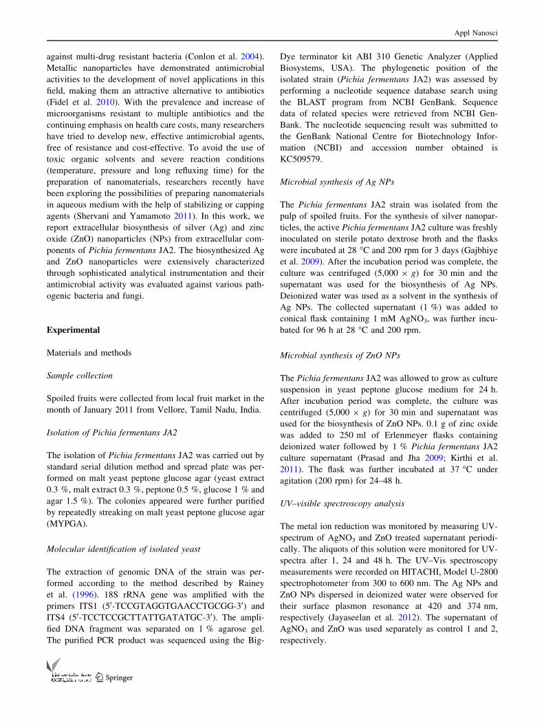
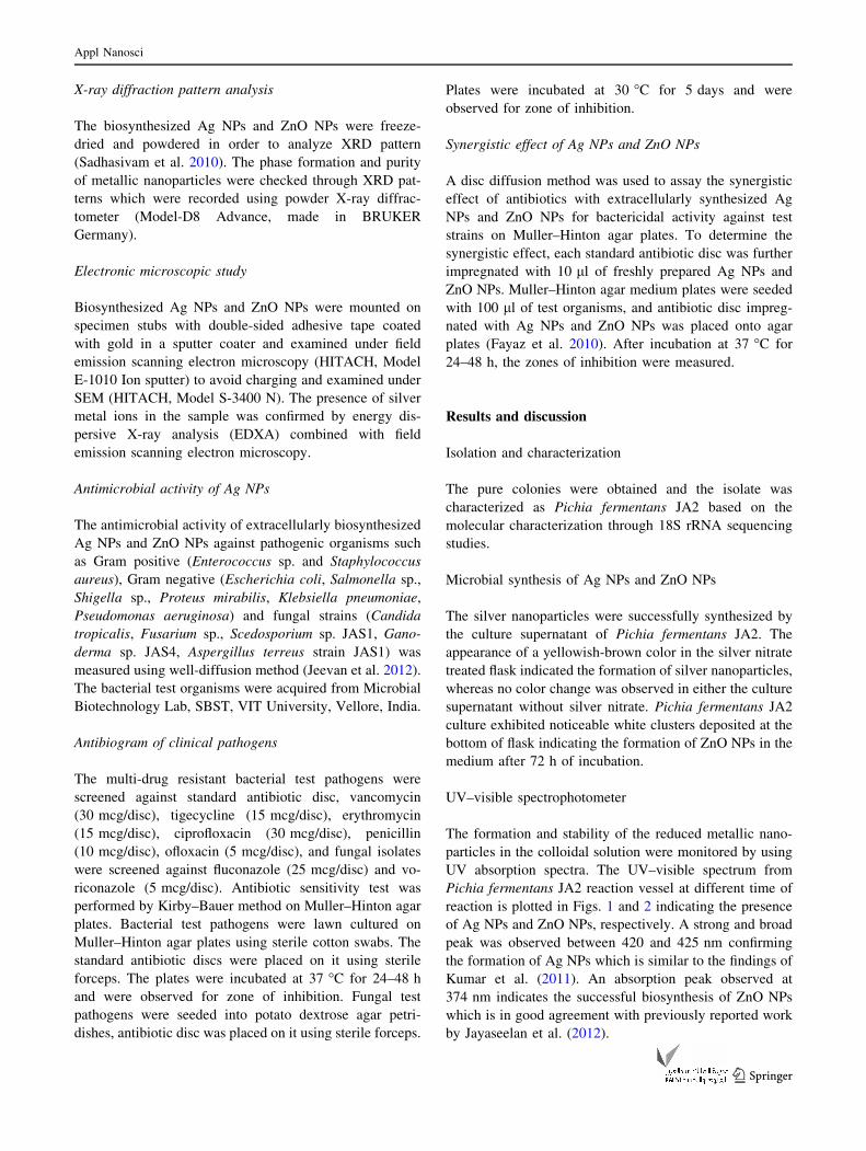
UV–visible spectroscopy analysis
The metal ion reduction was monitored by measuring UV-
spectrum of AgNO3 and ZnO treated supernatant periodi-
cally. The aliquots of this solution were monitored for UV-
spectra after 1, 24 and 48 h. The UV–Vis spectroscopy
measurements were recorded on HITACHI, Model U-2800
spectrophotometer from 300 to 600 nm. The Ag NPs and
ZnO NPs dispersed in deionized water were observed for
their surface plasmon resonance at 420 and 374 nm,
respectively (Jayaseelan et al. 2012). The supernatant of
AgNO3 and ZnO was used separately as control 1 and 2,
respectively.
Appl Nanosci
123
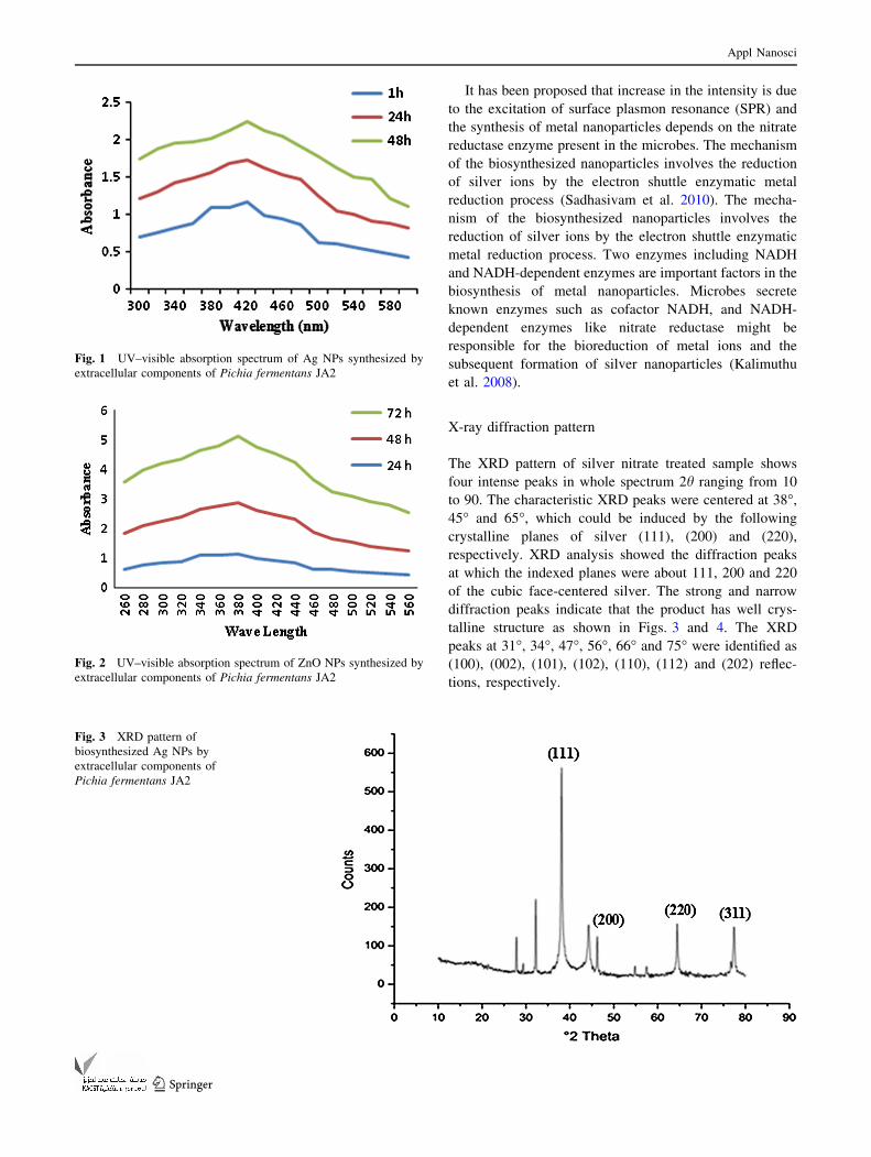
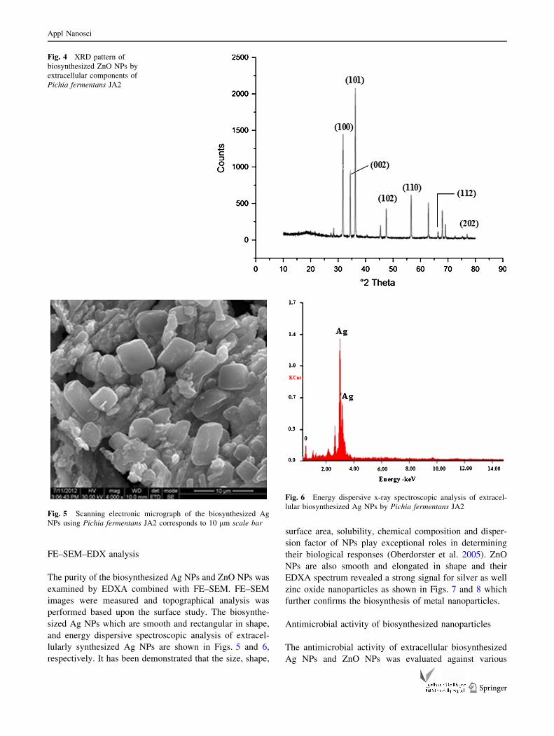
X-ray diffraction pattern analysis
The biosynthesized Ag NPs and ZnO NPs were freeze-
dried and powdered in order to analyze XRD pattern
(Sadhasivam et al. 2010). The phase formation and purity
of metallic nanoparticles were checked through XRD pat-
terns which were recorded using powder X-ray diffrac-
tometer (Model-D8 Advance, made in BRUKER
Germany).
Electronic microscopic study
Biosynthesized Ag NPs and ZnO NPs were mounted on
specimen stubs with double-sided adhesive tape coated
with gold in a sputter coater and examined under field
emission scanning electron microscopy (HITACH, Model
E-1010 Ion sputter) to avoid charging and examined under
SEM (HITACH, Model S-3400 N). The presence of silver
metal ions in the sample was confirmed by energy dis-
persive X-ray analysis (EDXA) combined with field
emission scanning electron microscopy.
Antimicrobial activity of Ag NPs
The antimicrobial activity of extracellularly biosynthesized
Ag NPs and ZnO NPs against pathogenic organisms such
as Gram positive (Enterococcus sp. and Staphylococcus
aureus), Gram negative (Escherichia coli, Salmonella sp.,
Shigella sp., Proteus mirabilis, Klebsiella pneumoniae,
Pseudomonas aeruginosa) and fungal strains (Candida
tropicalis, Fusarium sp., Scedosporium sp. JAS1, Gano-
derma sp. JAS4, Aspergillus terreus strain JAS1) was
measured using well-diffusion method (Jeevan et al. 2012).
The bacterial test organisms were acquired from Microbial
Biotechnology Lab, SBST, VIT University, Vellore, India.
Antibiogram of clinical pathogens
The multi-drug resistant bacterial test pathogens were
screened against standard antibiotic disc, vancomycin
(30 mcg/disc), tigecycline (15 mcg/disc), erythromycin
(15 mcg/disc), ciprofloxacin (30 mcg/disc), penicillin
(10 mcg/disc), ofloxacin (5 mcg/disc), and fungal isolates
were screened against fluconazole (25 mcg/disc) and vo-
riconazole (5 mcg/disc). Antibiotic sensitivity test was
performed by Kirby–Bauer method on Muller–Hinton agar
plates. Bacterial test pathogens were lawn cultured on
Muller–Hinton agar plates using sterile cotton swabs. The
standard antibiotic discs were placed on it using sterile
forceps. The plates were incubated at 37 �C for 24–48 h
and were observed for zone of inhibition. Fungal test
pathogens were seeded into potato dextrose agar petri-
dishes, antibiotic disc was placed on it using sterile forceps.
Plates were incubated at 30 �C for 5 days and were
observed for zone of inhibition.
Synergistic effect of Ag NPs and ZnO NPs
A disc diffusion method was used to assay the synergistic
effect of antibiotics with extracellularly synthesized Ag
NPs and ZnO NPs for bactericidal activity against test
strains on Muller–Hinton agar plates. To determine the
synergistic effect, each standard antibiotic disc was further
impregnated with 10 ll of freshly prepared Ag NPs and
ZnO NPs. Muller–Hinton agar medium plates were seeded
with 100 ll of test organisms, and antibiotic disc impreg-
nated with Ag NPs and ZnO NPs was placed onto agar
plates (Fayaz et al. 2010). After incubation at 37 �C for
24–48 h, the zones of inhibition were measured.
Results and discussion
Isolation and characterization
The pure colonies were obtained and the isolate was
characterized as Pichia fermentans JA2 based on the
molecular characterization through 18S rRNA sequencing
studies.
Microbial synthesis of Ag NPs and ZnO NPs
The silver nanoparticles were successfully synthesized by
the culture supernatant of Pichia fermentans JA2. The
appearance of a yellowish-brown color in the silver nitrate
treated flask indicated the formation of silver nanoparticles,
whereas no color change was observed in either the culture
supernatant without silver nitrate. Pichia fermentans JA2
culture exhibited noticeable white clusters deposited at the
bottom of flask indicating the formation of ZnO NPs in the
medium after 72 h of incubation.
UV–visible spectrophotometer
The formation and stability of the reduced metallic nano-
particles in the colloidal solution were monitored by using
UV absorption spectra. The UV–visible spectrum from
Pichia fermentans JA2 reaction vessel at different time of
reaction is plotted in Figs. 1 and 2 indicating the presence
of Ag NPs and ZnO NPs, respectively. A strong and broad
peak was observed between 420 and 425 nm confirming
the formation of Ag NPs which is similar to the findings of
Kumar et al. (2011). An absorption peak observed at
374 nm indicates the successful biosynthesis of ZnO NPs
which is in good agreement with previously reported work
by Jayaseelan et al. (2012).
Appl Nanosci
123
It has been proposed that increase in the intensity is due
to the excitation of surface plasmon resonance (SPR) and
the synthesis of metal nanoparticles depends on the nitrate
reductase enzyme present in the microbes. The mechanism
of the biosynthesized nanoparticles involves the reduction
of silver ions by the electron shuttle enzymatic metal
reduction process (Sadhasivam et al. 2010). The mecha-
nism of the biosynthesized nanoparticles involves the
reduction of silver ions by the electron shuttle enzymatic
metal reduction process. Two enzymes including NADH
and NADH-dependent enzymes are important factors in the
biosynthesis of metal nanoparticles. Microbes secrete
known enzymes such as cofactor NADH, and NADH-
dependent enzymes like nitrate reductase might be
responsible for the bioreduction of metal ions and the
subsequent formation of silver nanoparticles (Kalimuthu
et al. 2008).
X-ray diffraction pattern
The XRD pattern of silver nitrate treated sample shows
four intense peaks in whole spectrum 2h ranging from 10
to 90. The characteristic XRD peaks were centered at 38�,
45� and 65�, which could be induced by the following
crystalline planes of silver (111), (200) and (220),
respectively. XRD analysis showed the diffraction peaks
at which the indexed planes were about 111, 200 and 220
of the cubic face-centered silver. The strong and narrow
diffraction peaks indicate that the product has well crys-
talline structure as shown in Figs. 3 and 4. The XRD
peaks at 31�, 34�, 47�, 56�, 66� and 75� were identified as
(100), (002), (101), (102), (110), (112) and (202) reflec-
tions, respectively.
Fig. 1 UV–visible absorption spectrum of Ag NPs synthesized by
extracellular components of Pichia fermentans JA2
Fig. 2 UV–visible absorption spectrum of ZnO NPs synthesized by
extracellular components of Pichia fermentans JA2
Fig. 3 XRD pattern of
biosynthesized Ag NPs by
extracellular components of
Pichia fermentans JA2
Appl Nanosci
123
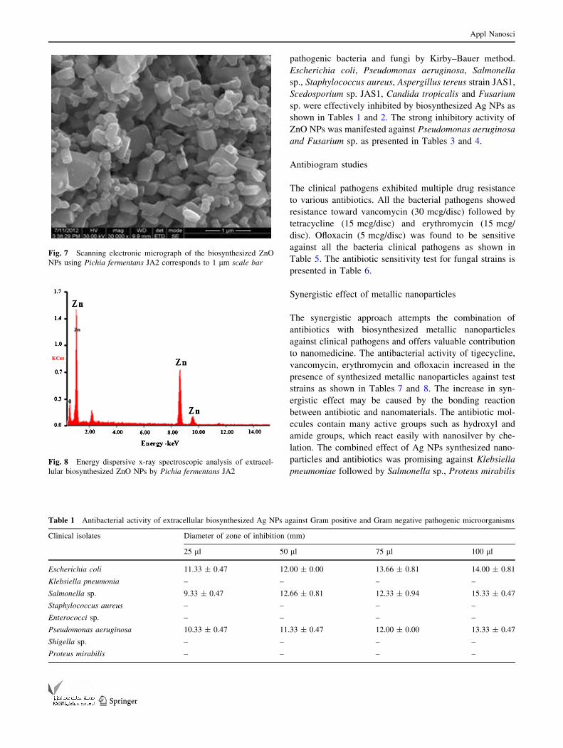
FE–SEM–EDX analysis
The purity of the biosynthesized Ag NPs and ZnO NPs was
examined by EDXA combined with FE–SEM. FE–SEM
images were measured and topographical analysis was
performed based upon the surface study. The biosynthe-
sized Ag NPs which are smooth and rectangular in shape,
and energy dispersive spectroscopic analysis of extracel-
lularly synthesized Ag NPs are shown in Figs. 5 and 6,
respectively. It has been demonstrated that the size, shape,
surface area, solubility, chemical composition and disper-
sion factor of NPs play exceptional roles in determining
their biological responses (Oberdorster et al. 2005). ZnO
NPs are also smooth and elongated in shape and their
EDXA spectrum revealed a strong signal for silver as well
zinc oxide nanoparticles as shown in Figs. 7 and 8 which
further confirms the biosynthesis of metal nanoparticles.
Antimicrobial activity of biosynthesized nanoparticles
The antimicrobial activity of extracellular biosynthesized
Ag NPs and ZnO NPs was evaluated against various
Fig. 4 XRD pattern of
biosynthesized ZnO NPs by
extracellular components of
Pichia fermentans JA2
Fig. 5 Scanning electronic micrograph of the biosynthesized Ag
NPs using Pichia fermentans JA2 corresponds to 10 lm scale bar
Fig. 6 Energy dispersive x-ray spectroscopic analysis of extracel-
lular biosynthesized Ag NPs by Pichia fermentans JA2
Appl Nanosci
123
pathogenic bacteria and fungi by Kirby–Bauer method.
Escherichia coli, Pseudomonas aeruginosa, Salmonella
sp., Staphylococcus aureus, Aspergillus tereus strain JAS1,
Scedosporium sp. JAS1, Candida tropicalis and Fusarium
sp. were effectively inhibited by biosynthesized Ag NPs as
shown in Tables 1 and 2. The strong inhibitory activity of
ZnO NPs was manifested against Pseudomonas aeruginosa
and Fusarium sp. as presented in Tables 3 and 4.
Antibiogram studies
The clinical pathogens exhibited multiple drug resistance
to various antibiotics. All the bacterial pathogens showed
resistance toward vancomycin (30 mcg/disc) followed by
tetracycline (15 mcg/disc) and erythromycin (15 mcg/
disc). Ofloxacin (5 mcg/disc) was found to be sensitive
against all the bacteria clinical pathogens as shown in
Table 5. The antibiotic sensitivity test for fungal strains is
presented in Table 6.
Synergistic effect of metallic nanoparticles
The synergistic approach attempts the combination of
antibiotics with biosynthesized metallic nanoparticles
against clinical pathogens and offers valuable contribution
to nanomedicine. The antibacterial activity of tigecycline,
vancomycin, erythromycin and ofloxacin increased in the
presence of synthesized metallic nanoparticles against test
strains as shown in Tables 7 and 8. The increase in syn-
ergistic effect may be caused by the bonding reaction
between antibiotic and nanomaterials. The antibiotic mol-
ecules contain many active groups such as hydroxyl and
amide groups, which react easily with nanosilver by che-
lation. The combined effect of Ag NPs synthesized nano-
particles and antibiotics was promising against Klebsiella
pneumoniae followed by Salmonella sp., Proteus mirabilis
Fig. 7 Scanning electronic micrograph of the biosynthesized ZnO
NPs using Pichia fermentans JA2 corresponds to 1 lm scale bar
Fig. 8 Energy dispersive x-ray spectroscopic analysis of extracel-
lular biosynthesized ZnO NPs by Pichia fermentans JA2
Table 1 Antibacterial activity of extracellular biosynthesized Ag NPs against Gram positive and Gram negative pathogenic microorganisms
Clinical isolates Diameter of zone of inhibition (mm)
25 ll 50 ll 75 ll 100 ll
Escherichia coli 11.33 ± 0.47 12.00 ± 0.00 13.66 ± 0.81 14.00 ± 0.81
Klebsiella pneumonia – – – –
Salmonella sp. 9.33 ± 0.47 12.66 ± 0.81 12.33 ± 0.94 15.33 ± 0.47
Staphylococcus aureus – – – –
Enterococci sp. – – – –
Pseudomonas aeruginosa 10.33 ± 0.47 11.33 ± 0.47 12.00 ± 0.00 13.33 ± 0.47
Shigella sp. – – – –
Proteus mirabilis – – – –
Appl Nanosci
123
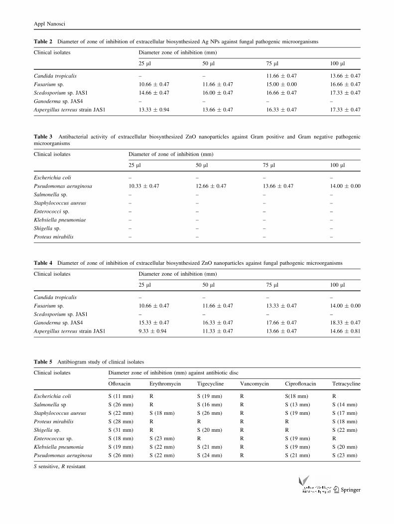
Table 2 Diameter of zone of inhibition of extracellular biosynthesized Ag NPs against fungal pathogenic microorganisms
Clinical isolates Diameter zone of inhibition (mm)
25 ll 50 ll 75 ll 100 ll
Candida tropicalis – – 11.66 ± 0.47 13.66 ± 0.47
Fusarium sp. 10.66 ± 0.47 11.66 ± 0.47 15.00 ± 0.00 16.66 ± 0.47
Scedosporium sp. JAS1 14.66 ± 0.47 16.00 ± 0.47 16.66 ± 0.47 17.33 ± 0.47
Ganoderma sp. JAS4 – – – –
Aspergillus terreus strain JAS1 13.33 ± 0.94 13.66 ± 0.47 16.33 ± 0.47 17.33 ± 0.47
Table 3 Antibacterial activity of extracellular biosynthesized ZnO nanoparticles against Gram positive and Gram negative pathogenic
microorganisms
Clinical isolates Diameter of zone of inhibition (mm)
25 ll 50 ll 75 ll 100 ll
Escherichia coli – – – –
Pseudomonas aeruginosa 10.33 ± 0.47 12.66 ± 0.47 13.66 ± 0.47 14.00 ± 0.00
Salmonella sp. – – – –
Staphylococcus aureus – – – –
Enterococci sp. – – – –
Klebsiella pneumoniae – – – –
Shigella sp. – – – –
Proteus mirabilis – – – –
Table 4 Diameter of zone of inhibition of extracellular biosynthesized ZnO nanoparticles against fungal pathogenic microorganisms
Clinical isolates Diameter zone of inhibition (mm)
25 ll 50 ll 75 ll 100 ll
Candida tropicalis – – – –
Fusarium sp. 10.66 ± 0.47 11.66 ± 0.47 13.33 ± 0.47 14.00 ± 0.00
Scedosporium sp. JAS1 – – – –
Ganoderma sp. JAS4 15.33 ± 0.47 16.33 ± 0.47 17.66 ± 0.47 18.33 ± 0.47
Aspergillus terreus strain JAS1 9.33 ± 0.94 11.33 ± 0.47 13.66 ± 0.47 14.66 ± 0.81
Table 5 Antibiogram study of clinical isolates
Clinical isolates Diameter zone of inhibition (mm) against antibiotic disc
Ofloxacin Erythromycin Tigecycline Vancomycin Ciprofloxacin Tetracycline
Escherichia coli S (11 mm) R S (19 mm) R S(18 mm) R
Salmonella sp S (26 mm) R S (16 mm) R S (13 mm) S (14 mm)
Staphylococcus aureus S (22 mm) S (18 mm) S (26 mm) R S (19 mm) S (17 mm)
Proteus mirabilis S (28 mm) R R R R S (18 mm)
Shigella sp. S (31 mm) R S (20 mm) R R S (22 mm)
Enterococcus sp. S (18 mm) S (23 mm) R R S (19 mm) R
Klebsiella pneumonia S (19 mm) S (22 mm) S (21 mm) R S (19 mm) S (20 mm)
Pseudomonas aeruginosa S (26 mm) S (22 mm) S (24 mm) R S (21 mm) S (23 mm)
S sensitive, R resistant
Appl Nanosci
123

and in case of ZnO NPs, Enterococcus sp., Staphylococcus
aureus, Proteus mirabilis showed effective zone of inhi-
bition. The biosynthetic methods have been recognized as
an alternative to chemical and physical synthesis as bio-
synthetic method is economical, ecofriendly and green low
cost approach. In this study, Ag NPs and ZnO NPs were
synthesized by extracellular components of Pichia fer-
mentans JA2. The biosynthetic route developed in this
study for producing metallic nanoparticles has distinct
advantages over chemical synthetic techniques regarding
biosafety and offers valuable contribution to pharmaceuti-
cal associations.
Table 6 Antibiogram study of
fungal clinical isolates
R resistant, S sensitive
Clinical isolates Diameter of zone of inhibition antibiotic disc (mm)
Fluconazole (25omcg/disc) Voriconazole (5omcg/disc)
Candida tropicalis R S (26)
Fusarium sp. R S (23)
Scedosporium sp. JAS1 R S (29)
Ganoderma sp. JAS4 R S (28)
Aspergillus terreus strain JAS1 R S (32)
Table 7 Diameter of zone of inhibition of combined effect of extracellular biosynthesized Ag NPs with different antibiotics (with and without
antibiotics) against Gram positive and Gram negative bacteria
Antibiotics Diameter zone of inhibition (mm)
Tigecycline Vancomycin Erythromycin Ofloxacin
Microorganisms Ab Ab ? NP % Ab Ab ? NP % Ab Ab ? NP % Ab Ab ? NP %
Escherichia coli R 18 – R 17 – R – – 22 27 22.73
Pseudomonas aeruginosa 17 20 17.65 R 20 – 22 27 22.72 20 21 5.00
Salmonella sp. 10 16 60.00 R – – R – – 25 36 30.56
Staphylococcus aureus 20 21 5.00 R 20 – 30 35 16.66 15 28 86.67
Shigella sp. 12 21 75.00 R 18 – R – – 16 27 68.75
Proteus mirabilis 14 23 64.29 R 18 – R – – 20 25 25.00
Enterococcus sp. R 10 – R 14 – 13 17 30.76 18 20 6.76
Klebsiella pneumoniae 17 20 17.69 R 17 – 28 30 7.142 22 28 27.28
Over all percentile increase % = b - a/a 9 100
Ab antibiotic disc, R resistant, mm millimeters, Ab ? Np antibiotic disc dipped in nanoparticle
Table 8 Diameter of zone of inhibition of combined effect of extracellular biosynthesized ZnO NPs with different antibiotics (with and without
antibiotics) against Gram positive and Gram negative bacteria
Antibiotics Diameter zone of inhibition (mm)
Tigecycline Vancomycin Erythromycin Ofloxacin
Microorganisms Ab Ab ? NP % Ab Ab ? NP % Ab Ab NP % Ab Ab ? NP %
Escherichia coli R 18 – R R R R 18 – 18 22 –
Pseudomonas aeruginosa 25 32 28 R 21 – 12 15 25.00 28 31 10.71
Salmonella sp. 15 24 60.00 R 13 – R 19 – 33 34 3.03
Staphylococcus aureus 12 19 58.34 R 15 – 18 22 22.23 23 27 17.39
Shigella sp. 23 29 8.69 R 27 – R – 31 38 22.58
Proteus mirabilis 18 22 22.23 R 21 – R – 17 28 64.00
Enterococcus sp. R 22 – R 21 – 18 21 16.67 21 27 28.27
Klebsiella pneumoniae 19 21 10.52 R 14 – 13 14 7.69 20 26 30.00
Appl Nanosci
123
Open Access This article is distributed under the terms of the
Creative Commons Attribution License which permits any use, dis-
tribution, and reproduction in any medium, provided the original
author(s) and the source are credited.
References
Conlon JM, Kolodziejek J, Nowotny N (2004) Antimicrobial peptides
from ranid frogs: taxonomic and phylogenetic markers and a
potential source of new therapeutic agents. Biochim Biophys
Acta 1696:1–14
Fayaz AM, Balaji K, Girilal M, Yadav R, Kalaichelvan PT,
Venketesan R (2010) Biogenic synthesis of silver nanoparticles
and their synergistic effect with antibiotics: a study against
Gram-positive and Gram-negative bacteria. Nanomed Nanotech-
nol 6:103–109
Fidel MG, Peggy L, Adriana B, Erasmo O, Nereyda N, Elpidio MS,
Facundo R, Horacio B, Yossef AG (2010) Synthesis, character-
ization, and evaluation of antimicrobial and cytotoxic effect of
silver and titanium nanoparticles. Nanomed Nanotechnol
6:681–688
Gajbhiye M, Kesharwani J, Ingle A, Gade A, Rai M (2009) Fungus-
mediated synthesis of silver nanoparticles and their activity
against pathogenic fungi in combination with fluconazole.
Nanomed Nanotechnol 5:382–386
Hong B, Kai J, Ren Y, Han J, Zou Z, Ahn CH (2008) Highly sensitive
rapid, reliable, and automatic cardiovascular disease diagnosis
with nanoparticle fluorescence enhancer and MEMS. Adv Exp
Med Biol 614:265–273
Jayaseelan C, Rahuman A, Kirthi A, Marimuthu S, Santhoshkumar T,
Bagavan A, Gaurav K, Karthik L, Rao K (2012) Novel microbial
route to synthesize ZnO nanoparticles using Aeromonas hydro-
phila and their activity against pathogenic bacteria and fungi.
Spectrochim Acta A 90:78–84
Jeevan P, Ramya K, Reena A (2012) Extracellular biosynthesis of
silver nanoparticles by culture supernatant of Pseudomonas
aeruginosa Indian. J Biotechnol 11:72–76
Kalimuthu K, Babu RS, Venkataraman D, Bilal M, Gurunathan S
(2008) Biosynthesis of silver nanocrystals by Bacillus licheni-
formis. Colloids Surf B Bioint 65:150–153
Kathiresan K, Manivannan S, Nabeel MA, Dhivya B (2009) Studies
on silver nanoparticles synthesized by a marine fungus, Peni-
cillium fellutanum isolated from coastal mangrove sediment.
Colloids Surf B 71:133–137
Kirthi AV, Rahuman AA, Marimuthu S, Santhoshkumar T, Jayase-
elan C, Kanayairam V (2011) Acaricidal, pediculocidal and
larvicidal activity of synthesized ZnO nanoparticles using wet
chemical route against blood feeding parasites. Parasitol Res
109:461–472
Kumar A, Pandey AK, Singh SS, Shankar R, Dhawan A (2011)
Cellular uptake and mutagenic potential of metal oxide nano-
particles in bacterial cells. Chemosphere 83:1124–1132
Meruvu H, Vangalapati M, Chippada SC, Bammidi SR (2011)
Synthesis and characterization of zinc oxide nanoparticles and its
antimicrobial activity against Bacillus subtilis and Escherichia
coli. J Rasayan Chem 4:217–222
Oberdorster G, Maynard A, Donaldson K, Castranova V (2005)
Principles for characterizing the potential human health effects
from exposure to nanomaterials: elements of a screening
strategy. Part Fibre Toxicol 2:8
Padmavathy N, Vijayaraghavan R (2008) Enhanced bioactivity of
ZnO nanoparticles—an antimicrobial study. Sci Technol Adv
Mater 9:7
Prasad K, Jha AK (2009) ZnO nanoparticles and adsorption study.
Nat Sci 1:129–135
Rainey FA, Rainey NW, Kroppenstedt RM, Stackebrandt E (1996)
The genus Nocardiopsis represents a phylogenetically coherent
taxon and a distinct actinomycete lineage: proposal of Nocar-
diopsiaceae fam. nov. Int J Syst Bacteriol 46:1088–1092
Sadhasivam S, Shanmugam P, Yun K (2010) Biosynthesis of silver
nanoparticles by Streptomyces hygroscopicus and antimicrobial
activity against medically important pathogenic microorganisms.
Colloids Surf B 81:358–362
Shervani Z, Yamamoto Y (2011) Size and morphology controlled
synthesis of gold nanoparticles in green solvent. Mater Lett
65:92–95
Wang P (2006) Nanoscale biocatalyst systems. Curr Opin Biotechnol
17:574–579
Zhang L, Gu FX, Chan JM, Wang AZ, Langer RS, Farokhzad OC
(2008) Nanoparticles in medicine: therapeutic applications and
developments. Clin Pharmacol Ther 83:761–769
Appl Nanosci
123