BIOMECHANICAL CONSIDERATIONS OF PARTIAL …. Functional assessments of the satisfaction of...
41
UNIVERSITY OF MEDICINE AND PHARMACY “GRIGORE T.POPA” IASI PhD ABSTRACT BIOMECHANICAL CONSIDERATIONS OF PARTIAL REMOVABLE DENTURES Scientific coordinator Prof. dr. FORNA NORINA PhD Student CARAS CIBELA 2017
Transcript of BIOMECHANICAL CONSIDERATIONS OF PARTIAL …. Functional assessments of the satisfaction of...

UNIVERSITY OF MEDICINE AND PHARMACY
“GRIGORE T.POPA” IASI
PhD ABSTRACT
BIOMECHANICAL CONSIDERATIONS OF PARTIAL
REMOVABLE DENTURES
Scientific coordinator
Prof. dr. FORNA NORINA
PhD Student
CARAS CIBELA
2017

CONTENTS
CONTENTS ......................................................................................................................... i
LIST OF ABBREVIATIONS ................................................................................................... iii
THE STAGE OF KNOWLEDGE
CHAPTER 1. GENERAL ASPECTS REGARDING THE PARTIAL PARTIAL EDITION…1
1.1. Etiopathogens of the extended partial edentation ............................................................... 1
1.2. Classified partial stretching classes ............................................. ..............................…… 4
1.3. Local, loco-regional and systemic changes .......................................... .........................… 4
1.4. Prevalence of partially movable prosthesis carriers ........................................................... 6
CHAPTER 2. THERAPEUTIC OPTIONS IN A STRONG PARTIAL EDITION .................. 8
2.1.Terapia of partial edentation stretched by conventional acrylic prostheses……………….. 8
2.2. Partial edentation therapy stretched through flexible prostheses ........................................ 11
CHAPTER 3. PARTIAL EDITION TREATMENT THROUGH SCHELET PROTEZS ....... 14
3.1. Design of Components ............................................... ........................................................ 14
3.2. Functional assessments of the satisfaction of prostate-bearing patients ………………… 23
3.3 Impact of wearing prostheses on partially edented prosthetic elements ………………… 24
CHAPTER 4. BIOMECANIC BEHAVIOR OF PROTETIC RESTAURANTS IN
PARTICULAR PARTIAL EDUCATION .............................................................................. 26
4.1. Distribution of forces in the prosthetic field ...................................................................... 26
4.2. Modern computerized evaluation of the effect of mobile prostheses on the alveolar bone
resorption rate ............................................................................................................................ 30
4.3. Data from the literature on the biomechanical behavior of prosthetic restorations in the large
partial edentation ..................................... ................................................................................. 32
PERSONAL PARTY
CHAPTER 5. STUDY ON CHARACTERISTICS OF THE PARTIAL EDITION TARGETS IN
A LOT OF PATIENTS TREATED BY MOBILIZABLE PROTECTION ............................. 36
Introduction................................................................................................................................. 36
Purpose of the study ................................................................................................................... 36
Material and method................................................................................................................... 36
Results ........................................................................................................................................ 37
Discussions ................................................................................................................................. 47
Conclusions ................................................................................................................................. 51

CHAPTER 6. STUDY ON THE PREVALENCE AND DESIGN OF PROTETIC
RESTAURANT PARTIALLY MOBILIZABLE IN REPORT WITH CLINICAL AND
BIOLOGICAL PARAMETERS ......................................................................................... …. 52
Introduction............................................................................................................................... 52
Purpose of the study ................................................................................................................. 52
Material and method.................................................................................................................. 52
Results ....................................................................................................................................... 53
Discussions ................................................................................................................................ 68
Conclusions ............................................................................................................................... 74
CHAPTER 7. BIOMECANICAL CONSIDERATIONS REGARDING THE ACKERS AND T
TYPES OF STERILIZATION OF SCHOLET PARTIALLY AMONGATE PROTEZE ....... 75
Introduction................................................................................................................................ 75
Purpose of the study ................................................................................................................... 75
Material and method................................................................................................................... 75
Results ....................................................................................................................................... 81
Discussions ................................................................................................................................ 90
Conclusions ................................................................................................................................ 95
CHAPTER 8. BIOMECANICAL CONSIDERATIONS REGARDING SPECIAL RESTORATION SYSTEMS IN
IMPROVEMENTS ....................................................................................................................... 96
Introduction............................................................................................................................... 96
Purpose of the study ................................................................................................................. 96
Material and method ................................................................................................................ 96
Results ...................................................................................................................................... 101
Discussions ............................................................................................................................... 111
Conclusions .............................................................................................................................. 116
GENERAL CONCLUSIONS .......................................................................................................... 117
CHAPTER 9. ORIGINAL ELEMENTS AND PERSPECTIVES THAT OPEN THESIS ............................ 118
REFERENCES ............................................................................................................................. 121
ANNEXES
ANNEX 1. FEA images (stress distribution at the Ackers and "T" hooks) - CHAPTER 7
APPENDIX 2. FEA images (stress distribution at the level of special elements) - CHAPTER 8
APPENDIX 3. List of published articles
APPENDIX 4. Copies of published articles

Key words: partial edentation, skeletal prosthesis, special elements of retention, over-
protection, biomechanical behavior, finite element analysis
The doctoral thesis contains:
• Theoretical part organized in 4 chapters (35 pages);
. personal research organized in 4 chapters (78 pages);
• 52 tables and 87 figures (personal part);
• 124 bibliographic references.
Note: This abstract contains bibliographic references, tables and images, respecting the
numbering and content of the PhD thesis.

1
CHAPTER 5. STUDY ON THE CHARACTERISTICS OF THE EXTENDED
PARTIAL EDENTATION FOR A LOT OF PATIENTS TREATED WITH
REMOVABLE DENTURE
5.1.INTRODUCTION
Although the extended or total edentation is considered a normal consequence of the
ageing process, there is a tendency at the global level of the population to maintain a high
number of teeth for a longer period of time. The epidemiologic studies prove that, despite the
decrease of the prevalence of partial and total extended edentation, patients aged over 50
years are significantly affected by the loss of teeth.
5.2. PURPOSE OF THE STUDY
The study has aimed to assess the prevalence and parameters of the types of partial edentation
for a lot of patients who have come for treatment with partial removable denture at the Dental
Education Foundation, ”Grigore T. Popa” University of Medicine and Pharmacy of Iași.
5.3.MATERIAL AND METHOD
The study lot has included a number of 200 patients who have come for treatment
between 2014-2016 at the Dental Education Clinical Foundation of the Faculty of Dental
Medicine, ”Gr. T. Popa” University of Medicine and Pharmacy of Iași.
The investigation has been carried out based on the data recorded in the clinical
charts, of the orthopantomographies and of the specific data recorded in the laboratory charts.
The patients were informed of the use of the clinical and therapeutic data within the
study.
Criteria for the inclusion:
- Clinical charts with complete data regarding the prosthetic treatment;
- Laboratory charts with data which specify the design and elements of preservation,
support and stabilisation of the partial removable denture;
- Completed prosthetic treatment.
Criteria for exclusion:
- Incomplete data in the clinical and laboratory charts;
- Patients who did not take all the treatment steps.
The following parameters of partial edentation have been assessed:
Types of edentation:
- Extended partial edentation;
- Subtotal edentation;
- Total edentation.
Location of the arch:
- Maxillary;
- Mandibular.
Kennedy classification:
Class I – bilateral and biterminaledentation;
Class II – unilateral and uniterminaledentation;
Class III – interbeddededentation;
Class IV – edentation in the frontal area.

2
Edentation extension:
- Number of absent teeth;
- Patients with minimum 20 absent teeth;
- Patients with less than 20 absent teeth.
The data were calculated and expressed in graphics and tables (descriptive statistics) in
Microsoft Excel.
5.4.RESULTS
The prevalence of the types of edentation is presented in figure 5.3.a.
Figure 5.3.a.Prevalence of the types of edentation (study lot)
The prevalence of the types of edentation is presented in figure 5.3.a and the
prevalence of the types of edentation in proportion to their location (maxillary, mandible) is
presented in figure 5.3.b:
0%
10%
20%
30%
40%
50%
60%
70%
80%
90%
100%
EPI ST T
95%
7,50% 10%
0,00%
10,00%
20,00%
30,00%
40,00%
50,00%
60,00%
70,00%
80,00%
90,00%
100,00%
EPI ST T
93,75%
3,75% 2,50%
92,50%
5,00% 2,50%
MX
MD

3
Figure 5.3.b.Prevalence of the types of edentation (maxillary, mandibular arch)
The prevalence of the types of edentation in proportion to sex, age group, class of
edentation:
Figure 5.3.c.Prevalence of the types of edentation (sex)
Figure 5.3.d.Prevalence of the types of edentation (age group)
0,00%
20,00%
40,00%
60,00%
80,00%
100,00%
EPI ST T
96,25%
1,25% 2,50%
96,25%
1,25% 2,50%
M
F
0%
20%
40%
60%
80%
100%
31-40 41-50 51-60 61-70 71-80
100% 100% 100% 100% 100%
0 010%
0 00 0
15%
0
20%
EPI
ST
T

4
Figure 5.4. Prevalence of partial edentation in proportion to the class of edentation
The prevalence of the classes of edentation for each sex is presented in figure 5.6:
- class I edentation is found in similar percentages (66%, respectively 68.4%);
- class II edentation prevails in male patients (84%);
- class III edentation prevails in male patients (71.4%);
- class IV edentation prevails in male patients (42.8%).
Figure 5.6. Prevalence of the classes of edentation in proportion to sex
It is observed that for the age group 31-40 years, class III edentation prevails (37.5%),
for the age group 41-50 years, class I (57.1%) and class IV (50%) edentation prevails, for the
age group 51-60 years, class I edentation (66%) prevails, followed by class IV edentation
(33%), for the age groups 61-70 and 71-80 years, class I edentation is present in a percentage
of 100%.
0%
10%
20%
30%
40%
50%
60%
70%
I II III IV
65%
25%17,50%
27,50%
0%
10%
20%
30%
40%
50%
60%
70%
80%
90%
I II III IV
66%
84%
71,40%
42,80%
68,40%
31,25%
15,60%20,80%
M
F

5
Figure 5.7. Prevalence of the classes of edentation in proportion to the age group
In figure 5.8.e., one can notice that the weight of class I, II, IV edentation is similar at
the level of the two arches (50% vs. 47.5% class I, 16.5% class II, 12.5% vs. 15% class IV)
Figure 5.8. Prevalence of the classes of edentation (maxillary, mandibular arch)
The distribution of the extended partial edentation in proportion to the location and
the class of edentation and the location on the arches (maxillary, mandibular) for male and
female patients is presented in table 5.V and figures 5.9 and 5.10
0%
10%
20%
30%
40%
50%
60%
70%
80%
90%
100%
31-40 41-50 51-60 61-70 71-80
25%
57,10%
66%
100% 100%
12,50% 14,30%20%
60%
40%37,50%42,80%
6,60%0% 0%
25%
50%
33%
0% 0%
I
II
III
IV
0%
10%
20%
30%
40%
50%
I II III IV
50%
16,50%
2,50%
12,50%
47,50%
16,50%10%
15%
MX
MD

6
Figure 5.8. Prevalence of EPE (extended partial edentation) in proportion to the location and
Kennedy class (male sex)
Figure 5.8. Prevalence of EPE (extended partial edentation) in proportion to the location and
Kennedy class (female sex)
Figure 5.11. Average values for the number of absent teeth
0,00%
5,00%
10,00%
15,00%
20,00%
25,00%
30,00%
MX (I) MX (II) MX(III)
MX(IV)
MD (I) MD (II) MD(III)
MD(IV)
27,50%
12,50%
5%7,50%
22,50%
7,50%5%
12,50%
0%
5%
10%
15%
20%
25%
30%
35%
MX (I) MX (II) MX (III) MX (IV) MD (I) MD (II) MD (III) MD(IV)
32%
11%
6% 6%
27%
12%
6% 6%
0
5
10
15
20
LOT MX MD
18,87
9,77 9,1

7
Figure 5.12.Average values for the number of absent teeth (sex)
Figure 5.13. Average values for the number of absent teeth (age groups)
Figure 5.14. Average values for the number of absent teeth (Kennedy class)
5.5.DISCUTII
This study regarding the parameters of the extended partial edentation for a lot of
patients with ages between 30-80 years has been carried out by referring to the definition of
edentation, considered a crippling impairment which influences the quality of life, as well as
the levels of education, economic and social level of the individuals (Basno A. &col.2016).
Under these conditions, local studies regarding the demographic significance of this
phenomenon can be used in order to understand the global significance of this malady
(Locker D.1988).Oral rehabilitation represents an integrative concept which reconstructs each
segment of the dental-maxillary system affected by various forms of edentation, without
eluding the complications induced and without influencing the general status within the
selected therapeutic algorithm (Forna N. 2008, 2011). Taking into consideration the literature
data regarding the prevalence of the extended partial and total edentation in patients over 50
years, oral health suppliers can delay and reduce the extension of the edentation by ensuring
8,5
9
9,5
10
M F
9,9
9,1
0
10
20
30
31-40 41-50 51-60 61-70 71-80
11,315
20,7 22,619
0
2
4
6
8
10
12
14
I II III IV
12,4
8,27,1
9,4
8
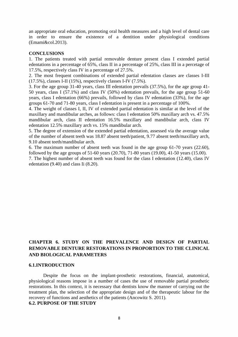
an appropriate oral education, promoting oral health measures and a high level of dental care
in order to ensure the existence of a dentition under physiological conditions
(Emami&col.2013).
CONCLUSIONS
1. The patients treated with partial removable denture present class I extended partial
edentations in a percentage of 65%, class II in a percentage of 25%, class III in a percetage of
17.5%, respectively class IV in a percentage of 27.5%.
2. The most frequent combinations of extended partial edentation classes are classes I-III
(17.5%), classes I-II (15%), respectively classes I-IV (7.5%).
3. For the age group 31-40 years, class III edentation prevails (37.5%), for the age group 41-
50 years, class I (57.1%) and class IV (50%) edentation prevails, for the age group 51-60
years, class I edentation (66%) prevails, followed by class IV edentation (33%), for the age
groups 61-70 and 71-80 years, class I edentation is present in a percentage of 100%.
4. The weight of classes I, II, IV of extended partial edentation is similar at the level of the
maxillary and mandibular arches, as follows: class I edentation 50% maxillary arch vs. 47.5%
mandibular arch, class II edentation 16.5% maxillary and mandibular arch, class IV
edentation 12.5% maxillary arch vs. 15% mandibular arch.
5. The degree of extension of the extended partial edentation, assessed via the average value
of the number of absent teeth was 18.87 absent teeth/patient, 9.77 absent teeth/maxillary arch,
9.10 absent teeth/mandibular arch.
6. The maximum number of absent teeth was found in the age group 61-70 years (22.60),
followed by the age groups of 51-60 years (20.70), 71-80 years (19.00), 41-50 years (15.00).
7. The highest number of absent teeth was found for the class I edentation (12.40), class IV
edentation (9.40) and class Ii (8.20).
CHAPTER 6. STUDY ON THE PREVALENCE AND DESIGN OF PARTIAL
REMOVABLE DENTURE RESTORATIONS IN PROPORTION TO THE CLINICAL
AND BIOLOGICAL PARAMETERS
6.1.INTRODUCTION
Despite the focus on the implant-prosthetic restorations, financial, anatomical,
physiological reasons impose in a number of cases the use of removable partial prosthetic
restorations. In this context, it is necessary that dentists know the manner of carrying out the
treatment plan, the selection of the appropriate design and of the therapeutic labour for the
recovery of functions and aesthetics of the patients (Ancowitz S. 2011).
6.2. PURPOSE OF THE STUDY

9
The study has aimed to assess the prevalence and distribution of removable partial
prosthetic solutions, design and distribution of components in proportion to a series of
clinical and biological parameters.
6.3.MATERIAL AND METHOD
The research was carried out via the analysis of the patients’ charts who have come
for treatment at the Dental Education Clinical Foundation of the Faculty of Dental Medicine,
”Gr. T. Popa” University of Medicine and Pharmacy of Iași. The prevalence and distribution
of the removable partial dentures were investigated, in proportion to the clinical and
biological parameters, at the level of a lot of 200 patients who have come for treatment
between January 2016-January 2017. The design and distribution of the components of the
final removable partial prosthetic restorations (frame-type dentures, elastic dentures) were
assessed in proportion to the clinical and biological parameters at the level of a lot of 125
patients. The investigation has been carried out based on the data recorded in the clinical
charts, of the orthopantomographies and of the specific data recorded in the laboratory charts.
The patients were informed of the use of the clinical and therapeutic data within the study.
The distribution of the removable prosthetic restorations was assessed in proportion to
the following parameters:
- Patients’ sex.
- Age group (31-40 years;41-50 years;51-60 years;61-70 years;71-80 years).
- Arch: Maxillary; Mandibular.
Kennedy classification:
Class I – bilateral and biterminaledentation;
Class II – unilateral and uniterminaledentation;
Class III – interbeddededentation;
Class IV – edentation in the frontal area.
Type of removable partial denture:
- Elastic removable partial denture (ED);
- Frame-type removable partial denture (FD).
Design, main connectors, preservation, support and mobilisation elements:
- Main connectors:
- Palatine plate (PP);
- Palatine plate with reduced width (PPRW);
- U-shaped connector anteriorly and posteriorly opened (CPU);
- Lingual bar (LB);
- Lingual plate (LP).
- preservation, support and mobilisation elements:
Frame-type removable partial dentures:
- Clasps; - Ackers (A);
- T-shaped (T);
- ring-shaped - with or without changes (R);
- Bonwill (B).
Special elements (attachments):
- Staples (overdenture) (SOD);
- Grooves (frame-type dentures) (G);
- Extra-coronation (EC);
- Intra-coronation (IC).
Elastic removable partial dentures:

10
- Elastic clasps (E);
- Pelota (P);
The data were calculated and expressed in graphics and tables (descriptive statistics)
in Microsoft Excel.
6.4.RESULTS
Within the study lot, it was opted for the removable partial denture in 95% of the
cases ( a percentage of 5% of the arches was represented by the total edentation treated with
total removable denture). Frame-type partial removable denture was applied in 30.2% of the
cases, it was opted for elastic dentures in 23.8% of the cases and 46% of the cases were
treated with conventional acrylic partial removable dentures (figure 6.3).
Figure 6.3. Distribution of prosthetic solutions (study lot)
The distribution of partial removable prosthetic solution in proportion to the patients’
sex is presented in figure 6.4.
Figure 6.4. Distribution of partial removable prosthetic solutions (in proportion to sex)
30,20%
23,80%
46,00%Proteza scheletata
Proteza elastica
Proteza acrilica
0%
10%
20%
30%
40%
50%
60%
70%
Proteza scheletata Proteza elastica Proteza acrilica
36%32% 32%
23,70%
15,80%
61%
M
F

11
The frame-type partial removable denture was chosen as a therapeutic solution for the
extended partial edentation in an increased percentage in the age group of 51-60 years (58%),
is similar in the age groups of 41-50 years and 61-70 years (25%) and is absent at the level of
the age group of 71-80 years (figure 6.5).
Figure 6.5. Distribution of partial removable prosthetic solutions (in proportion to age)
At the maxillary level, the distribution of the partial removable prosthetic solutions
was as follows: 27% frame-type denture, 27% elastic denture, 46% acrylic denture. At the
mandibular level, the distribution of the partial removable prosthetic solutions was as
follows: 33% frame-type denture,20% elastic denture, 47% acrylic denture (figure 6.6).
Figure 6.6. Distribution of partial removable prosthetic solutions (in proportion to location)
0%
10%
20%
30%
40%
50%
60%
70%
80%
31-40 41-50 51-60 61-70 71-80
18%
25%
58%
25%
0%
9%
25%22,50%
42%
20%
72%
50%
29,50%33%
80%
Proteza scheletata
Proteza elastica
Proteza acrilica
0%
5%
10%
15%
20%
25%
30%
35%
40%
45%
50%
Proteza scheletata Proteza elastica Proteza acrilica
27% 27%
46%
33%
20%
47%
MX
MD

12
At the level of the class I Kennedy edentation, 57% of the removable prosthetic
solutions are conventional acrylic dentures, 43% are final prosthetic solutions (frame-type,
elastic). In the class II Kennedy edentation, elastic dentures (47%) prevail, followed by the
frame-type dentures (33%). In the class IV Kennedy edentation, acrylic dentures (45%)
prevail, followed by elastic dentures (36%), a reduced percentage (19%) being represented by
frame-type dentures (figure 6.7).
Figure 6.7. Distribution of partial removable prosthetic solutions (in proportion to the Kennedy class)
The data regarding the design and the distribution of the components of the frame-type
partial removable dentures are presented in the following figures. Amongst the patients with
frame-type partial removable dentures, mixed denture was applied in 39% of the cases,
hybrid denture in 48% of the cases, overdenture in 13% of the cases (figure 6.8).
Figure 6.8.Distribution of frame-type prosthetic solutions
In the case of patients with frame-type prosthetic solutions, for male patients, the
mixed denture was applied in 43% of the cases, hybrid denture in 43% of the cases,
respectively overdenture in 14% of the cases. For female patients, mixed denture was applied
in 33% of the cases, hybrid denture in 56% of the cases, respectively overdenture in 11% of
the cases (figure 6.9).
0,00%
10,00%
20,00%
30,00%
40,00%
50,00%
60,00%
Clasa I Clasa II Clasa III Clasa IV
30,70%33%
50%
19%
12,30%
47%
0%
36%
57%
20%
50%45%
Proteza scheletata
Proteza elastica
Proteza acrilica
48,00%
39,00%
13%
Protezare hibrida
Protezare mixta
Supraprotezare

13
Figure 6.9. Distribution of frame-type prosthetic solutions (in proportion to sex)
The frame-type partial removable denture was chosen as a therapeutic solution for the
extended partial edentation in an increased percentage in the age group of 51-60 years (58%),
is similar in the age groups of 41-50 years and 61-70 years (25%) and is absent at the level of
the age group of 71-80 years. Regarding the types of frame-type denture, the weight between
the hybrid denture and the mixed one is similar in the age group of 61-70 years (33%), and
the hybrid denture prevails in the age groups of 41-50 years (66%) and 51-60 years (54%)
(figure 6.10).
Figure 6.10. Distribution of frame-type partial removable prosthetic solutions (in proportion to age)
Regarding the frame-type prosthetic solutions, at the maxillary level, hybrid denture
prevails (50%), followed by the mixed denture (40%), and the mandibular level the hybrid
denture prevails (46%), followed by the mixed denture (38%) (figure 6.11).
0,00%
10,00%
20,00%
30,00%
40,00%
50,00%
60,00%
Protezare hibrida Protezare mixta Supraprotezare
43,00% 43%
14,00%
56%
33%
11%
M
F
0%
10%
20%
30%
40%
50%
60%
70%
80%
90%
100%
31-40 41-50 51-60 61-70 71-80
0%
66%
54%
33%
0%
100%
33% 33% 33%
0%0% 0%
13%
33%
0%
Protezare hibrida
Protezare mixta
Supraprotezare

14
Figure 6.11.Distribution of frame-type partial removable prosthetic solutions
(in proportion to location)
The distribution of the partial removable prosthetic solution in patients with extended
partial edentation, in proportion to the Kennedy edentation class is presented in figure 6.11.
In the class I Kennedy edentation, the hybrid denture (58.7%) prevails, followed by the
mixed denture (33%). In class II and III Kennedy edentations, the distribution of hybrid and
mixed denture is similar (50%). In class IV Kennedy edentation, the mixed denture (66%)
prevails (figure 6.12).
Figure 6.12. Distribution of frame-type partial removable prosthetic solutions
(in proportion to the Kennedy class)
For the maxillary frame-type partial removable dentures, the distribution was the
following (in descending order): palatine plate (50%), palatine plate with reduced width
(20%), U-shaped palatine connector (anteriorly or posteriorly opened) (30%), palatine bar
0%
5%
10%
15%
20%
25%
30%
35%
40%
45%
50%
Protezare hibrida Protezare mixta Supraprotezare
50%
40%
10%
46%
38%
16%
MX
MD
0,00%
10,00%
20,00%
30,00%
40,00%
50,00%
60,00%
70,00%
Clasa I Clasa II Clasa III Clasa IV
58,70%
50% 50%
33%33%
50% 50%
66%
8,30%
0% 0% 0%
Protezare hibrida
Protezare mixta
Supraprotezare

15
(10%). For the mandibular frame-type partial removable dentures, we have observed the
lingual bar (73%) and the lingual plate (27%) (figures 6.13.a-c).
Figure 6.13.a. Distribution of main connectors. Maxillary frame-type denture
Figure 6.13.b. Distribution of main connectors. Mandibular frame-type denture
Figure 6.13.c. Distribution of main connectors. Classes of edentation.
In figures 13.a-c the distribution of the preservation, support and stabilization
elements is presented for the frame-type partial removable dentures (clasps, special
elements).
0%
10%
20%
30%
40%
50%
Placutapalatina
PP latimeredusa
Conectorpalatin in "U"
Bara palatina
50%
20%
30%
10%
0%
20%
40%
60%
80%
Bara linguala Placa linguala
73%
27%

16
Figure 6.14.a. Distribution of preservation, support and stabilization elements (frequency)
Figure 6.14.b. Distribution of preservation, support and stabilization elements (frame-type
dentures)
Figure 6.14.c.Distribution per type of clasps (in proportion to frequency)
40%
60%Crosete
Elemente speciale
42%
58%Crosete
Elemente speciale
0,00%
5,00%
10,00%
15,00%
20,00%
25,00%
30,00%
35,00%
40,00%
45,00%
50,00%
Ackers "T" Inelar Bonwill Continuu
47,20%
30,80%
8,80% 8,80%4,40%

17
Figure 6.14.d. Distribution per classes of clasps (frame-type dentures)
Figure 6.14.e. Distribution of types of special elements (frame-type dentures)
6.5.DISCUSSIONS
Partial removable denture, with dental support or with support on implants, is a
widely used therapeutic solution in the rehabilitationof extended partial edentation due to the
favourable cost-efficiency ratio both for the dentist and the patient (Rehman P. &col.2013).A
reduced number of studies correlate the types of extended partial edentation with the
parameters regarding the patient (sex, age) and with the types of partial removable prosthetic
restorations chosen as treatment solutions (Walid&col.2010, Pellizzer&col.2012).Ankowitz
S. (2011) draws attention to the fact that both the design of the partial removable denture and
the materials used contribute to the long-term success of prosthetic therapy via partial non-
removable restorations. The creation of an optimal design of the partial removable denture is
dependent on the clinical knowledge and experience, appropriate assessment of the patient,
appropriate therapeutic planning, technical expertise and knowledge of the laboratory
procedures and material properties (Farias-Neto A.2012).
6.6.CONCLUSIONS
79%
21%
Culise extracoronare
Capse
38,50%
38,50%
23%
Circumferentiale
Subecuatoriale
Combinate

18
1. Frame-type (30%) and elastic (24%) partial removable prosthetic solutions prevails in
the lot of patients with extended partial edentation.
2. Male patients with extended partial edentation are treated more frequently via frame-
type partial removable denture (36%) comparatively with female patients (23.7%).
3. Patients with extended partial edentation for age groups of 41-50 years and 61-70
years present a similar pattern of treatment via frame-type partial removable denture
(25%); frame-type denture prevails in the age group of 51-60 years (58%). The
highest frequency of the patients treated with elastic dentures was found in the age
group of 60-61 years (42%).
4. Frame-type partial removable denture was found in 27% of the maxillary extended
partial edentations, respectively 33% of the mandibular ones, and the prosthetic
treatment with elastic partial removable dentures was found in 27% of the maxillary
extended partial edentations, respectively 20% of the mandibular extended partial
edentations.
5. Frame-type partial removable denture is frequently found in class I and II Kennedy
edentations (30.7%, respectively 33%) and less frequently in class IV edentations
(19%) and elastic partial removable denture is chosen as a therapeutic solution more
frequently in class II edentations (47%), respectively class IV (36%).
6. Hybrid denture is the most frequent found therapeutic solution (48% of the frame-type
dentures) in patients treated with frame-type partial removable dentures. The
prevalence of hybrid and mixed frame-type prosthetic solutions is similar at the level
of maxillary arches (50%, 40%) and mandibular arches (46%, 38%). In class I
edentations, the hybrid frame-type denture prevails (58.7%), in class IV edentations,
the mixed frame-type denture prevails (66%).
7. The most frequently used main connectors are the palatine plate (50%), used
especially in classes I and IV, followed by the U-shaped palatine connector for
maxillary frame-type dentures, respectively the lingual bar (73%) for mandibular
frame-type dentures. The most frequently used cast clasps are Ackers clasps (47.2%),
frequent in class I maxillary and mandibular edentations, T-shaped clasp (30.8%), in
class I mandibular edentations. In a smaller proportion, the ring-shaped clasps (class
II) are used, the Bonwill clasps (class III maxillary extended) and continuous clasps
(class I maxillary extended). Special elements are represented by extracoronary
grooves (79%), used especially in class I and class IV extended edentations and
staples, used in overdentures.
CHAPTER 7.Biomechanical considerations regarding the Types of Ackers
and T-shaped clasps in the stabilisation of the frame-type partial non-
removable denture
7.1. INTRODUCTION
Mandibular frame-type partial non-removable dentures in class I Kennedy edentations
present a complex biomechanical behaviour due to the support structures present, gum, teeth,
each with different biomechanical properties (Archangelo&col.2012). Both the physician and
the technician must understand the action of the forces which appear and the biomechanical
response of the structures concerned but also of the materials used in making the prosthetic
structures (Bural 2015). Thus, the FEA studies have a high practical applicability in order to

19
understand the biomechanical behaviour of the preservation, support and stabilisation
elements.
7.2. PURPOSE OF THE STUDY
In this chapter, the following objectives are aimed at:
1. Moulding the structure and imposing conditions for the analysis of the finite element;
2. The results of the analysis in the finite element: assessment of the stress at the level of the
Ackers clasp and the T-shaped clasp, upon a physiological burdening of the non-
removable denture;
3. Assessment of the stress at the level of the tooth on which the clasp shall be attached.
7.3.MATERIAL AND METHOD
7.3.1. Moulding the structure and imposing conditions for the analysis of the finite element;
In order to assess the stability, the sustainability in time and the mechanical stress
specific to each type of clasp chosen for the fixation of the mandibular frame-type non-
removable denture, under the conditions of various mechanical forces, two cases are taken
into discussion for the situation in which a class I Kennedy edentation is present and the
fixation of the denture is achieved at the level of the 34, 44 premolars:
a. fixation of the non-removable denture with the help of two Ackers clasps;
b. fixation of the non-removable denture with the help of two T-shaped
clasps.
The assessment of the distribution of stress at the level of the clasps is aimed at,
taking into consideration the elements of mandibular biomechanics, areas of physiological
burdening of the premolars, canine teeth and incisors, the characteristics of the materials of
the non-removable denture and of the tissues around the denture.
In order to assess the distribution of stress at the level of the clasps, tridimensional
models of the study elements were made with the help of the design program Autodesk
Inventor Professional 2017 and the simulation was carried out in the simulation section of the
above-mentioned program.
Two mandibular frame-type non-removable dentures were designed on a functional
class I Kennedy edentation model, each made of two mixed saddles, 6 artificial teeth, main
connector lingual bar and two clasps placed at the level of 34, 44 premolars.
For each type of clasp studied, an individual frame was designed. In figures 7.2.1.and
7.2.1. the model for the metal frame and for the final frame-type partial denture are
illustrated. The Ackers and T-shaped clasps designed are illustrated in figures 7.2.3 and 7.2.4.

20
Figure 7.2.2.Frame-type non-removable denture with T-shaped clasps
Figure 7.2.3.Detail for the frame-type non-removable denture with preservation, support and
stabilisation elements for T-shaped clasps (on level 4.4)
Figure 7.2.4. Detail for the frame-type non-removable denture with preservation, support and
stabilisation elements for Ackers clasps (on level 4.4)
In figure 7.2.6, the complete assembly of the biomechanical simulation is illustrated,
the mandible, the gum, class I Kennedy edentation, central incisors, lateral incisors, canine

21
teeth and premolars 1 and the frame-type partial non-removable denture retained with the T-
shaped clasps.
Figure 7.2.6. Assembly designed frame-type mandible-gum-denture
With the generation of the mesh, the model was divided into a large number of
elements and knots. For the two models to have the same density of the mesh, the conditions
of absolute mesh size and absolute mesh dimension are imposed, to which the value of 1mm
is also imposed, as the analysis with a finite elements is the more precise, the larger the
number of elements and knots is (Bhat&col.2014). This was also achieved for the purpose of
a more accurate analysis and the results of the analysis for the two models must be
comparable. In figure 7.2.7, there is an example of the result of meshing for the non-
removable denture retained with T-shaped clasps.
Figure 7.2.7.a-b Assembly of the frame-type mandible-gum-denture after the meshing

22
In figure 7.2.8, types of support selected for the simulation of both types of dentures
are illustrated. In Autodesk Simulation Mechanical 2014, these types of support are indicated
with circles (figure 34).
Figure 7.2.8.Application of the support in temporomandibular joints
Two properties of the materials were used, namely, the Poisson coefficient and the
Young module (elasticity module) for the materials used in the analysis (table 7.I). The non-
removable denture with T-shaped clasp and the one with Ackers clasp were made of the same
types of materials. Both the bone tissue and the implant-abutment complex were considered
to be elastic, linear, homogeneous, isotropic.
Table 7.I. Material properties
Material Elasticity module
[GPa]
Poisson coefficient
Bone tissue (Park 1998) 13.40 0.30
Gum tissue (Todorovic 2010) 0.0196 0.3
Periodontal ligament (Sato 2001) 0,069 0.45
Dentine (Park 1998) 18.6 0.31
Acrylic resin (Shahmiri 2016) 2.20 0.31
Co-Cr-Mo alloy (Sandu 2003) 220 0.3
For the application of the burdenings, a vertical stress bilaterally and unilaterally
applied on the non-removable denture shall be taken into consideration. Two values of the
applied stress of 100N and 140N shall be used. The stress applied in the Autodesk Simulation
Mechanical 2015 model, for both the cases taken into discussion, are illustrated in the
following figures.

23
7.4. RESULTS
Figure 7.3.1.Maximum stress at the level of the Ackers and T-shaped clasps upon the unilateral
action of the forces of 100N and 140N
Figure 7.3.2. Maximum stress at the level of the Ackers and T-shaped clasps upon the bilateral action
of the forces of 100N and 140N
0
50
100
150
200
250
300
350
400
450
500
100N unilateral 140N unilateral
75,9
108
394
457
Ten
siu
ne
max
imă
cro
șete
(M
Pa)
Croșet Ackers
Croșet T
0
100
200
300
400
500
600
100N bilateral 140N bilateral
28,7 41
365
510
Ten
siu
ne
max
imă
cro
șete
(M
Pa)
Croșet Ackers
Croșet T

24
Figure 7.3.3. Maximum stress at the level of the Ackers and T-shaped clasps opposite upon the
unilateral action of the forces of 100N and 140N
Figure 7.3.4. Maximum stress at the level of the tooth in the case of partial denture retention with
Ackers and T-shaped clasps upon the unilateral action of the forces of 100N and 140N
0
2
4
6
8
10
12
14
16
100N unilateral 140N unilateral
0,7
2
7
15Te
nsi
un
e m
axim
ă cr
oșe
te o
pu
se (
MP
a)
Croșet Ackers opus
Croșet T opus
0
20
40
60
80
100
120
140
160
100N unilateral 140N unilateral
61,11
85,6288,83
147
Ten
siu
ne
max
imă
din
te (
MP
a)
Croșet Ackers
Croșet T

25
Figure 7.3.5. Maximum stress at the level of the opposite tooth in the case of partial denture retention
with Ackers and T-shaped clasps upon the unilateral action of the forces of 100N and 140N
Figure 7.3.6. Maximum stress at the level of the tooth in the case of partial denture retention with
Ackers and T-shaped clasps upon the bilateral action of the forces of 100N and 140N
0
0,1
0,2
0,3
0,4
0,5
0,6
0,7
0,8
100N unilateral 140N unilateral
0,5504
0,7702
0,3915
0,6298
Ten
siu
ne
max
imă
din
te o
pu
s (M
Pa)
Croșet Ackers
Croșet T
0
10
20
30
40
50
60
100N bilateral 140N bilateral
31,83
44,62
38,99
56,45
Ten
siu
ne
max
imă
din
te (
MP
a)
Croșet Ackers
Croșet T

26
Figure 7.3.7. Maximum stress at the level of the ligament in the case of partial denture retention with
Ackers and T-shaped clasps upon the unilateral action of the forces of 100N and 140N
Figure 7.3.8. Maximum stress at the level of the ligament in the case of partial denture retention with
Ackers and T-shaped clasps upon the unilateral action of the forces of 100N and 140N
0
0,5
1
1,5
2
2,5
3
100N unilateral 140N unilateral
1,09
1,5271,555
2,604
Ten
siu
ne
max
imă
ligam
ent
(MP
a)
Croșet Ackers
Croșet T
0
0,002
0,004
0,006
0,008
0,01
0,012
0,014
0,016
0,018
100N unilateral 140N unilateral
0,01143
0,01608
0,009685
0,0128
Ten
siu
ne
max
imă
ligam
ent
op
us
(MP
a)
Croșet Ackers
Croșet T

27
7.5. DISCUSSIONS
Mandibular partial non-removable dentures in class I Kennedy edentations present a
complex biomechanical behaviour due to the support structures present, gum, teeth, each with
different biomechanical properties (Archangelo&col.2012). These types of non-removable
dentures are not retained rigidly by the teeth, thus, existing a potential for the movement of
the denture when the functional physiological movements give rise to forces on the teeth and
on the denture (Bural 2015). The optimal position and the location of any element in a partial
non-removable denture requires a high attention paid to the teeth located adjacently to the
area of the edentation. The analyses regarding the movements of the elements are suggestive
as they can show movements to a large scale, contributing to the understanding of the
biomechanics of denture (Sandu&col.2003, 2010).
7.6.CONCLUSIONS
1. The design and simulation of the retention elements for the partial non-removable denture
with the FEA method represent extremely useful stages in the assessment of the
biomechanical behaviour of the prosthetic elements but also of the anatomical support
structures, of the remaining teeth, of the periodontal ligament, the gingival mucosa.
2. The results of our study have determined by using the analysis with a finite element the
area with maximum tensions at the level of the retention elements of the frame-type partial
non-removable dentures, thus, indicating the areas with a risk to break upon stress.
3. The biomechanical behaviour of the Ackers clasp upon the forces imposed is superior to
the T-shaped clasp, taking into consideration the values of the maximum tensions developed
in the clasp, at the level of the arms of the clasp and of the tensions at the level of the tooth on
which the clasp was applied, as well as of the periodontal ligament.
4. The design and simulation performed in this study have determined and characterised the
tensions which appear at the level of the periodontal ligament and at the level of the
supporting tooth. In the case of the use of retention elements such as Ackers clasps, the
tensions were lower at the level of the periodontal ligament and the supporting tooth than in
the case of retention elements such as T-shaped clasps.
5. The design and moulding for the purpose of the FEA analysis in our study allows the
determination, with the help of movement coefficients, of the movements at the level of the
clasp opposite to the area of the application of the stress. In the case of partial denture
retained with Ackers clasps, the movements were smaller than in the case of T-shaped clasps.

28
CHAPTER 8.BIOMECHANICAL CONSIDERATIONS REGARDING THE SPECIAL
RETAINING SYSTEMS IN THE OVERDENTURE ON IMPLANTS
8.1. INTRODUCTION
Overdenture on implants is considered a simple, efficient, financially viable, less
invasive method and a successful treatment option for patients with total edentation
(Assuncao&col.2008).
If there are debates regarding the design of the type of retention, the studies which use
the FEA method in order to analyse the tensions which appear at the level of various systems
of retention, staple-type, systems with magnets etc., upon specific stress are extremely useful
(Barao&col.2009).
8.2. PURPOSE OF THE STUDY
The purpose of the study is to assess the distribution of tensions at the level of the
implant system chosen for the fixation of the mandibular denture, under the conditions of
mechanical stress.
In order to achieve this desideratum, two cases for the situation in which a total
edentation is present and the fixation of the overdenture is achieved with two endobone
implants at the level of the implant sites 3.3. and 4.3 are taken into discussion;
a. fixation of the mobile with the help of two systems of sphere staples OT
CAP (Rhein);
b. fixation of the mobile with the help of two systems of square pocket
staples OT EQUATOR (Rhein).
In this chapter, the following objectives are aimed at:
- Moulding the structure and imposing conditions for the analysis of the finite element;
- The results of the analysis in the finite element: assessment of the tension at the level of
each type of retention system upon a physiological burdening of the overdenture.
8.2. MATERIAL AND METHOD
8.2.1. Moulding the structure and imposing conditions for the analysis of the finite element;
For the assessment of the distribution of stress at the level of the implant systems for
the fixation of the removable overdenture, tridimensional models of the study elements were
performed with the help of the design program Autodesk Inventor Professional 2017.
The analysis of the finite element was achieved in the simulation section of the
Autodesk Inventor Professional 2017 program.
Two mandibular overdentures were designed, retained on two implants, on a
functional total edentation model, each made of a saddle and 16 artificial teeth, illustrated in
fig. 8.2.1. Overdenture was achieved by fixating on two systems:
- fixation of the mobile with the help of two systems of sphere staples OT
CAP (Rhein);
- fixation of the mobile with the help of two systems of square pocket
staples OT EQUATOR (Rhein).

29
Figure 8.2.1.Overdenture with fixation on implants
For each type of retention studies, an individual system, made of the implant, matrix
complex - square bearing staple matrix OT EQUATOR (Rhein) and sphere staple OT CAP
(Rhein) was designed (figure 8.2.2).
Figure 8.2.2.Rhein retention system (sphere OT CAP; square bearing OT EQUATOR)
(www.rhein83.com)

30
In figures 8.2.3.and 8.2.4., the two retention systemsinvestigated, as well as the
implants designed are illustrated. In figures 8.2.7.1-b. The assembly designed is presented.
Figure 8.2.3. Implant with square bearing staple retention system
OT EQUATOR (Rhein)
Figure 8.2.4.Implant with sphere staple retention system OT CAP (Rhein)
Figure 8.2.7.a Detail section of the assemble designed, mandible, gingival tissue, matrix
staple retention system, matrix bearing and patrix, overdenture saddle, artificial teeth, implant

31
Figure 8.2.7.b. Assembly designed mandible-gum-overdenture after the meshing surgery, with
bearing and applied unilateral stress
Two properties of the materials were used, namely, the Poisson coefficient and the
Young module (elasticity module) for the materials used in the analysis (table 8.I). Both the
bone tissue, the gum, the overdenture as well as the implant-abutment complex were
considered to be made of elastic, linear, homogeneous, isotropic materials.
Table 8.I. Material properties
Material Elasticity
module [GPa]
Poisson coefficient
Bone tissue
(Park 2008)
13.40 0.30
Gingival tissue
(Todorovic 2010)
0.0196 0.3
Periodontal ligament
(Sato 2001)
0,069 0.45
Acrylic resin
(Shahmiri 2016)
2.20 0.31
Plastic
(Anwar 2015)
0,350 0.4
Titanium alloy
(Park 2008)
110 0.4
8.4. RESULTS
The images which present the results of the simulation, as well as, the distribution of
tensions upon load of the study elements are presented in Annex 2.
In tables 8.II and 8.III, the maximum values of the tensions at the level of the special
staple and ball retention special elements of the overdenture with fixation on implants upon
vertical forces of 50N and 100N, with unilateral and bilateral action are presented.
Table 8.II. Maximum values of the tensions at the level of the square bearing staple retention
special elements OT EQUATOR (Rhein) of the overdenture with fixation on implants upon
load stress

32
Element type
Application of unilateral
force Application of bilateral force
50 N (MPa) 100 N (MPa) 50 N (MPa) 100 N (MPa)
Abutment 24.08 48.14 11.74 23.36
Matrix bearing 41.9 83.73 20.82 41.39
Matrix 0.35 0.71 0.16 0.33
Table 8.III. Maximum values of the tensions at the level of the sphere staple retention
special elements OT CAP (Rhein) of the overdenture with fixation on implants upon load
stress
Element type
Application of unilateral
force Application of bilateral force
50 N (MPa) 100 N (MPa) 50 N (MPa) 100 N (MPa)
Abutment 35.7 71.24 16.8 33.71
Matrix bearing 41.9 93.71 23.4 46.53
Matrix 0.54 1.08 0.25 0.51
Regarding the distribution on tensions at the level of the elements of the retention
systems of the overdenture on staple implants, in the case of the special retention abutment
and of the matrix bearing, the tension focuses on the lateroposterior area in the direction of
the force (figures 8.3.6-
Figure 8.3.6.The area of the distribution of tensions resulted upon the unilateral action of the force of
50N for the matrix bearing, matrix and special abutment for the retention of the square bearing staple
system OT EQUATOR (Rhein)

33
Figure 8.3.7.The area of the distribution of tensions resulted upon the unilateral action of the force of
100N for the matrix bearing, matrix and special abutment for the retention of the square bearing staple
system OT EQUATOR (Rhein)
Figure 8.3.8.The area of the distribution of tensions resulted upon the unilateral action of the force of
50N for the matrix bearing, matrix and abutment for the sphere staple system OT CAP (Rhein)
Figure 8.3.9.The area of the distribution of tensions resulted upon the unilateral action of the
force of 100N for the matrix bearing, matrix and special abutment for the retention of the
sphere staple system OT CAP (Rhein)

34
8.5. DISCUSSIONS
Overdenture on implants is considered a simple, efficient, financially viable, less
invasive method and a successful treatment option for patients with total edentation
(Assuncao&col.2008).From the point of view of the number of implants, overdenture on two
implants optimally ensures the stability of the implants and implicitly of the overdenture.
Many studies have used the FEA method in order to analyse tensions which appear at the
level of the various retention systems, staple, ball, with magnets, etc. upon specific stress
(Assuncao&col.2008, Barao&col.2009). In general, in the case of an overdenture on
implants, in the context of a load upon a vertical stress in the area of the molars, the ball type
of retention is preferred for a lower density of the bone and the staple type of retention is
chosen in order to ensure a higher duration of performance of the overdenture and of the
elements of the retention system. The FEA study can be entered into the limitations of any
studies of this kind which, although ensure a high level of accuracy, cannot completely
simulate the behaviour of live tissues and implants in real conditions existing at the level of
oral cavity (Dundar S. &col.2016).
8.6. CONCLUSIONS
1. The use of the FEA method of analysis and assessment of the biomechanical behaviour of
the overdenture and of the retention elements represents a highly significant tool in obtaining
accuracy in the calculation.
2. Upon the asymmetric burdening of 50N and 100N, a rotation on the frontal side of the
overdenture is obtained. This aspect has been observed both in the case of the special
retention system of the staple overdenture and of the ball overdenture. On the other part, the
elements of the retention system close to the place for the application of the stress represent a
classic deformation described by the conformation of the areas of tension distribution. Thus,
the saddle of the overdenture pulls towards the point for the application of force, on the
direction of the mandibular bone, the abutment for retention together with the matrix and
certainly, the matrix bearing.
3. The maximum tensions at the level of the elements of the retention systems, abutment,
matrix and matrix bearing are lower than in the case of square bearing staple retention
system, both in the asymmetric burdening and the symmetric burdening upon stress of 50N
and 100N.
4. At the level of thee implantation sites upon the vertical, unilateral action of the force off
50N and 100N in the case of the use of the retention system in the square bearing staple
overdenture, lower values of the tensions were obtained than in the sphere bearing staple
retention system. The differences are small but, overall, favour the retention system of the
square bearing staple overdenture. The values obtained are under the maximum limit of the
tension for the breaking of the bone, having a minor effect on the survival rate of the dental
implant.
5. The study proves that upon the application of the forces both unilaterally and bilaterally, of
50N, respectively 100N, the maximum values of the tensions off the elements of special

35
retention systems in the overdenture of implants, were lower with the square bearing staple
retention system comparatively with the sphere bearing staple system, favouring the square
bearing staple retention system for the retention of the overdenture on implants.
GENERAL CONCLUSIONS
1. The most frequently found extended partial edentations in the patients treated with
removable partial denture are class I edentations, folowed by class II, class III, respectively
class IV edentations. In the age group of 31-40 years, class III edentation prevails, in the age
group of 41-50 years, class I edentation group prevails, in the age group of 51-60 years, class
I edentation prevails. The weight of classes I, II, IV extended partial edentation is similar at
the level of the maxillary and mandibular arches. The maximum number of absent teeth was
found in the age group of 61-70 years, followed by the age groups of 51-60 years, 71-80
years, 41-50 years. The highest number of absent teeth was found for class I edentations,
class IV edentations and class II edentations.
2. Frame-type and elastic partial removable prosthetic solutions prevail in the lot of patients
with extended partial edentation. The highest frequency of the patients treated with elastic
dentures was found in the age group of 60-61 years. Frame-type removable partial denture is
frequently found in class I and II Kennedy edentations and less frequent in class IV
edentations and the elastic removable partial denture is chosen as a less frequent therapeutic
solution in class II, respectively class IV edentations. In class I edentations, the hybrid frame-
type denture prevails, in class UV edentations, the mixed frame-type denture prevails. The
most frequently used main connectors are the palatine plate, used especially in classes I and
IV, followed by the U-shaped palatine connector for maxillary frame-type denturess,
respectively the lingual bar for mandibular frame-type dentures. The most frequently used
cast clasps are Ackers clasps, frequent in class I maxillary and mandibular edentations, T-
shaped clasp, in class I mandibular edentations.
3. The design and simulation of the retention elements for the partial non-removable dentures
via the FEA method represent extremely useful stages in the assessment of the biomechanical
behaviour of the prosthetic elements but also of the anatomical support structures, of the
remaining teeth, of the periodontal ligament, the gingival mucosa. The FEA study can
determine the areas with maximum tensions at the level of the retention elements of the
frame-type non-removable partial dentures, thus, indicating the areas with a risk to break
upon various forces. The biomechanical behaviour of the Ackers clasp upon the forces
imposed is superior to the T-shaped clasp, taking into consideration the values of the
maximum tensions developed in the clasp, at the level of the arms of the clasp and of the
tensions at the level of the tooth on which the clasp was applied, as well as of the periodontal
ligament. In the case of partial denture retained with Ackers clasps, the movements were
smaller than in the case of T-shaped clasps.
4. The use of the FEA method of analysis and assessment of the biomechanical behaviour of
the overdenture and of the retention elements represents a highly significant tool in obtaining
a high precision of calculation. Upon the asymmetric loading of 50N and 100N, a rotation on
36
the frontal side of the overdenture is obtained. This aspect has been observed both in the case
of the special retention system of the square bearing staple overdenture and of the sphere
bearing staple overdenture. The area of distribution of the tension is focused on the
lateroposterior area, on the direction of the force vector, in the medial and superior area of the
elements, suggesting pushing and pulling effect of the matrix and of the abutment during
asymmetric stress. The maximum tensions at the level of the elements of the retention
systems, abutment, matrix and matrix bearing are lower than in the case of square bearing
staple retention system, both in the asymmetric burdening and the symmetric burdening upon
forces of 50N and 100N. The square bearing staple retention system is superior in the
retention of the overdenture on implants, comparatively with the sphere bearing staple
system.
BIBLIOGRAPHY
1.Ancowitz S. Esthetic removable partial dentures.General Dentistry 2011: 453-459.
2.Archangelo CM, Rocha EP, Pereira JA, Martin Junior M, Anchieta RB, Freitas, Júnior AC.
Periodontal ligament influence on the stress distribution in a removable partial denture
supported by implant: a finite element analysis. J Appl Oral Sci 2012;20:362-8.
3.Assuncao WG, Tabata LF, Bara˜o VA, Rocha EP. Comparison of stress distribution
between complete denture and implant-retained overdenture-2D FEA.Journal of Oral
Rehabilitation 2008;35:766–74.
4.Barao VA, Assuncao WG, Tabata LF, de Sousa EA, Rocha EP. Effect of different mucosa
thickness and resiliency on stress distribution of implant-retained overdentures-2D
FEA.Computer Methods and Programs in Biomedicine 2008;92:213–23.
5.Basno A , Maxim A , Savin C, Balcos C, Tatarciuc M. Prevalence of edentulism and related
social-behavioural factors among young adults of Iaşi, Romania. Romanian Journal of Oral
Rehabilitation 2016; 6 (3): 214-222.
6.CananBural. Biomechanics of Removable Partial Dentures Elsevier 2015
7.Dundar Serkan, TolgaTopkaya, Murat YavuzSolmaz, FerhanYaman, Yusuf Atalay,
ArifSaybak, FatihAsutay, Omer Cakmak (2016) Finite element analysis of the stress
distributions in peri-implant bone in modified and standard-threaded dental implants.
Biotechnology & Biotechnological Equipment, 2016; 30:1, 127-133, DOI:
10.1080/13102818.2015.1083887
8.Emami E, de Souza RF, Kabawat M, Feine JS. The impact of edentulism on oral and
general health.Int J Dent.2013;2013:498305.
9.Farias-Neto Arcelino, RenataSuellenGalvão da Silva, Alexandre da Cunha Diniz, André
UlissesDantas Batista, Adriana da Fonte Porto Carreiro. Ethics in the provision of removable
partial dentures.Braz J Oral Sci.2012;11(1): 19-24.
10.Forna N.. Oral rehabilitation between reality and paradigm.Iasi.Rev Med ChirSoc Med Nat
2008 Apr-Jun;112(2):507-11.
11.Forna N. Tratatproteticădentară. Editura U.M.F.”Gr.T.Popa” Iasi, 2008.
12.Forna N (coordonator), De Baat C, Bratu D, Lascu L, Mercut V, Petre Al, Popsor S,
Traistaru T. ProteticăDentară, vol.II, Edit.Enciclopedică, Bucureşti, 2011.
13.Forna N (coordonator), De Baat C, Bratu D, Lascu L, Mercut V, Petre Al, Popsor S,
Traistaru T. ProteticăDentară, vol.I, Edit.Enciclopedică, Bucureşti, 2011.
14.Locker D. Measuring oral health: a conceptual framework. Community Dent Health.
1988;5(1):3-18.

37
15.Pellizzer EP, Almeida DA, Falcón-Antenucci RM, Sánchez DM, Zuim
PR, VerriFR.Prevalence of removable partial dentures users treated at
the Aracatuba Dental School-UNESP.Gerodontology. 2012 Jun;29(2):140-4.
16.Rehman P, Orbach K, Ferger P, Wostmann B. Treatment outcomes with removable partial
dentures: a retrospective analysis. Orbach K, Ferger P, Wostmann B. International Journal of
Prosthodontics 2013; 26:147-150.
17.Sandu L, Faur N, Bortun C. Finite element analysis of stress distribution in the cast clasps,
direct retainers of a removable partial denture, TMJ 2003, Vol. 53, No. 3-4.
18.Sandu L, Topala F, Porojan S. Stress distribution in retentive arms of combination clasps
used on premolars. Journal of Applied Biomaterials & Biomechanics 2010; Vol. 8 no. 2, pp.
76-81.
19.VanStaden R.C., Guan H., Loo Y.C. Application of the finite element method in dental
implant research. Comput Methods Biomech Biomed Engin. 2006;9:257–270.
20.Walid M. Sadig, BDS, MS; Ayodeji T. Idowu, BDS, MS, - Design-
ulprotezelorparţialamovibile- un studiupopulaţional in Arabia Saudită- The Journal of
Contemporary Dental Practice, 2002; Vol3(4).
https://www.ncbi.nlm.nih.gov/pubmed/?term=Almeida%20DA%5bAuthor%5d&cauthor=true&cauthor_uid=22428988