BIOLOGY 524 SKULL – I BRAINCASE - CHONDROCRANIUM S. S. Sumida.
31
BIOLOGY 524 SKULL – I BRAINCASE - CHONDROCRANIUM S. S. Sumida
-
Upload
adelia-wilson -
Category
Documents
-
view
220 -
download
0
Transcript of BIOLOGY 524 SKULL – I BRAINCASE - CHONDROCRANIUM S. S. Sumida.
BIOLOGY 524SKULL – I
BRAINCASE - CHONDROCRANIUM
S. S. Sumida

TERMS YOU SHOULD UNDERSTAND (ALREADY)
•Ectoderm•Endoderm•Mesoderm•Neural Crest•Endochondral•Intramembranous•Cartilage•Bone


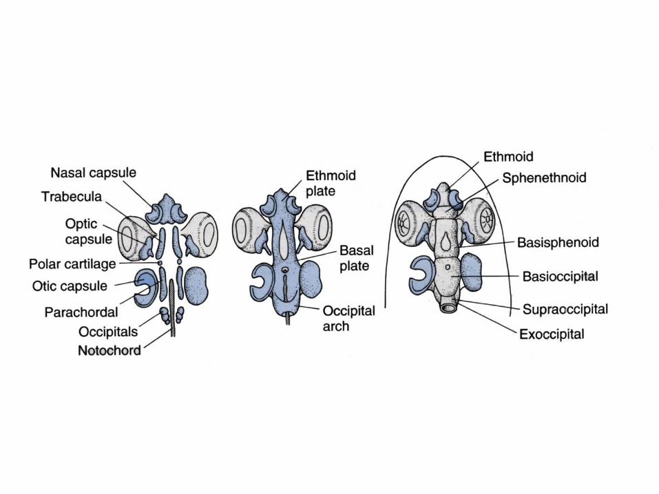
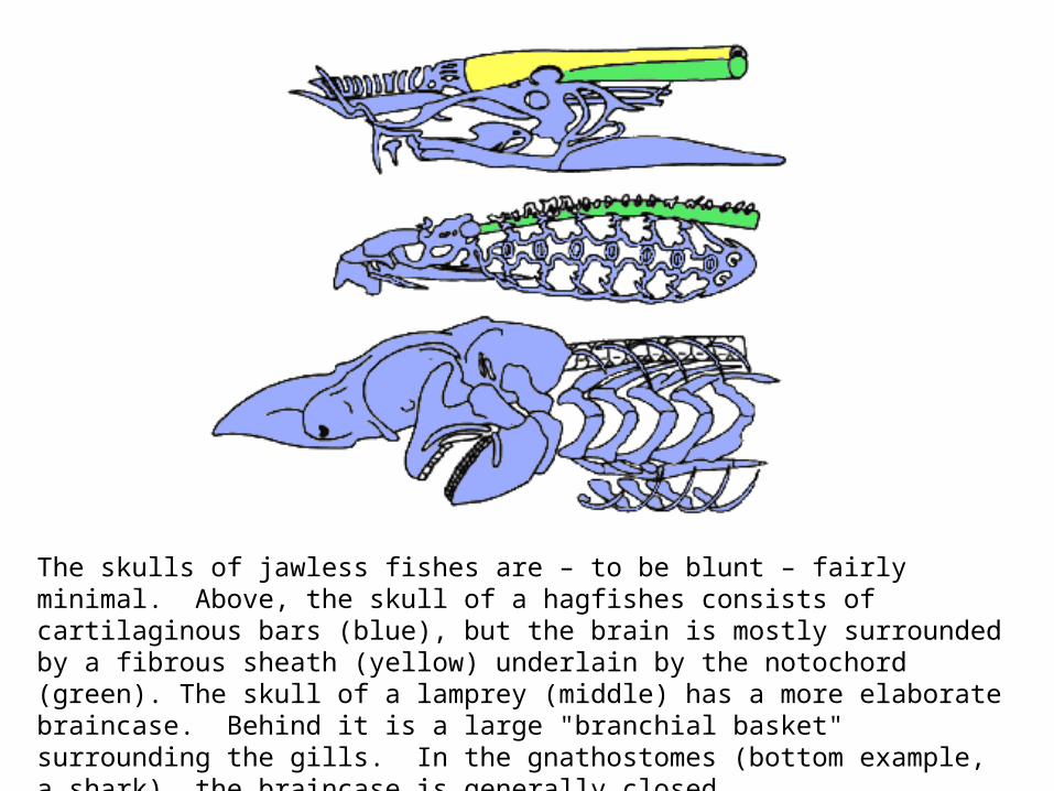
The skulls of jawless fishes are – to be blunt – fairly minimal. Above, the skull of a hagfishes consists of cartilaginous bars (blue), but the brain is mostly surrounded by a fibrous sheath (yellow) underlain by the notochord (green). The skull of a lamprey (middle) has a more elaborate braincase. Behind it is a large "branchial basket" surrounding the gills. In the gnathostomes (bottom example, a shark), the braincase is generally closed.

A cephalaspidomorph, a fossil jawless fish of the Devonian (~ 400 million years old)

Shark Chondrocranium
The shark is often used as a classic example of a gnathostome, but their braincases are unusual. All of the embryonic components fuse into a single cartilaginous structures with not sutural distinctions. Therefore, one can only name regions and foramina.


Sphen-ethmoid region Orbito-occipital region

Ethmoid Sphenoid Occipitalregion region region
Otic capsuleregion
Prootic Opisthotic
Supraoccipital
Exoccipitals
Basioccipital
Basisphenoid

TETRAPODSFollowing is an illustration of a basal tetrapod (often called “labyrinthodont amphibians” because the infolded ridges of their teeth were called “labyrinthine). Note that the components of the skull are extraordinarily similar to those of the crossopterygian.
• The ethmoid region may or may not be ossified but if not it remains as cartilage.
• The basipterygoid process (for articulation/connection with the palate) is more robust, but remains a mobile joint).
• The basioccipital, exoccipitals, and suprocciopital remain distinct structures.
• Note particularly that the jugular foramen for exit of the jugular vein and cranial nerve X is between otic and occipital region.
• The extension rostrally of the presphenoid is called the “cultriform process”.
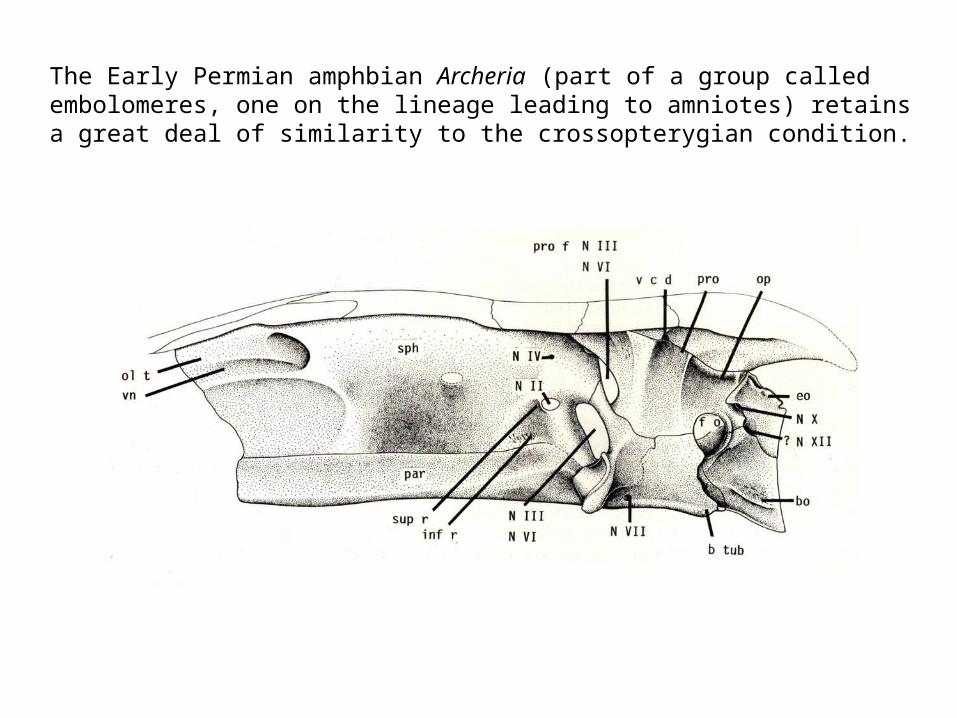
The Early Permian amphbian Archeria (part of a group called embolomeres, one on the lineage leading to amniotes) retains a great deal of similarity to the crossopterygian condition.
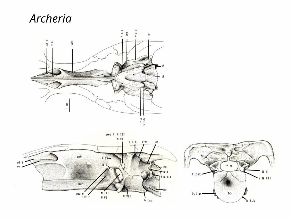
Archeria

At the upper left of an advanced amphibian, Seymouria.
• Note the basioccipital ventral to the foramen magnum. • Significantly, it is the basioccipital that will be the location of the occipital condyle. • The exoccipitals can be seen on either side of the foramen magnum.• (Seymouria is unusual in that its supraoccipital does not ossify.)• Just lateral to the exoccipitals the opisthotic – the back part of the otic capsule – is well
developed. Remember, this is the otic capsule, and thus capsule for the inner ear. Significantly, it is here that the stapes will articulate. In the case of these animals, it stapes acts as a brace between the braincase and the cheek portion dermal skull roof.
Diadectes
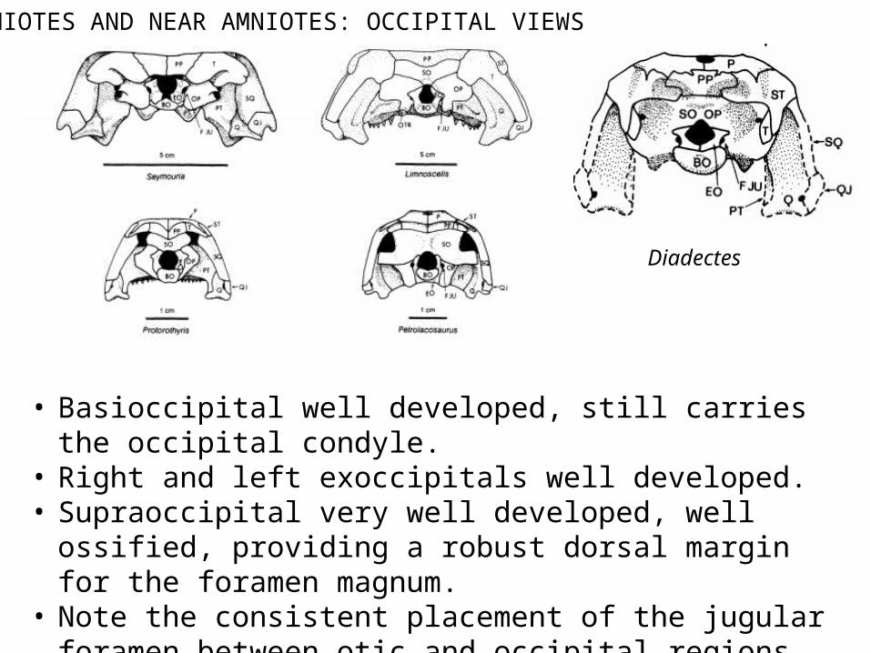
AMNIOTES AND NEAR AMNIOTES: OCCIPITAL VIEWS
• Basioccipital well developed, still carries the occipital condyle.• Right and left exoccipitals well developed.• Supraoccipital very well developed, well ossified, providing a
robust dorsal margin for the foramen magnum.• Note the consistent placement of the jugular foramen between
otic and occipital regions.

Occipital view of another diadectomorph, Diadectes. As with Limnoscelis, it is very amniote-like. In this case it has taken consolidation of the skull to an extreme by fusing the opisthotic and supraoccipital. But note the jugular foramen remains exactly where you would expect it.

This will show you the position of the braincase in the skull.In this case, in the basal reptile, Captorhinus.


Illustrations of the isolated braincase of Captorhinus
Dorsalview
Ventralview
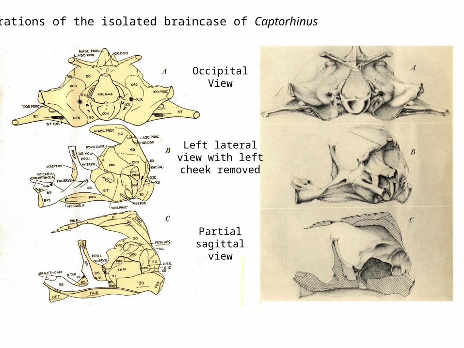
Illustrations of the isolated braincase of Captorhinus
OccipitalView
Left lateralview with left
cheek removed
Partialsagittal
view
• Generally the ethmoid region is only present in cartilage.• Sphenethmoid (derived from the trabeculae) is present, sometimes as a vertical plate. • Basipterygoid process well developed, thus a mobile basipterygoid articulation persists.• Both the basisphenoid and orbitosphenoid components ossify. • Both the opisthotic and prootic remain distinct structures.• The same set of occipital components are present: basi-, ex-, and supraoccipital.
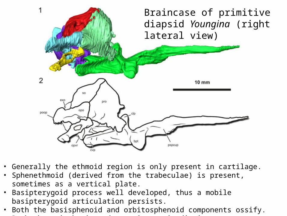
Braincase of primitive diapsid Youngina (right lateral view)

views of a Yougina braincase, in dorsal view (1-2), then ventral view (3-4). So, in 1-2, the brain would lie between the braincase and the viewer, as you are looking at the internal surface of the cradle for the brain. Why does the braincase appear to be getting smaller? In part because a component of the palatoquadrate (upper jaw) is joining to the sidewall of the braincase – an element called the epipterygoid.

Ethmoid Sphenoid Occipitalregion region region
Otic capsuleregion
Prootic Opisthotic
Supraoccipital
Exoccipitals
Basioccipital
Basisphenoid
Recall:

Here we have a comparison of proportions of braincase components between a crossopterygian fish, Eusthenopteron, the embolomorous amphibian Archeria, and the basal reptile Captorhinus.


• Many skull structures tend to fuse, obliterating the sutural boundaries between elements. However, basal birds still clearly possess the standard braincase elements:
• All occipital structures are present, but fuse into a single element around foramen magnum.• Braincase shortens, presumably to help accommodate the function of the bill.• The basisphenoid can often still be seen as a distinct element in palatal/ventral view• The parasphenoid has only a blunt rostral extension;no extended cultriform process as was
seen in reptiles.
BIRDS

As with the earlier reptile illustration, the braincase components here are also color-coded blue.
The alisphenoid, which is frequently cited as part of the lateral part of the brincase, is actually homologous to the epiperygoid, and thus derived from the palatoquadrate cartilage.
MAMMALS
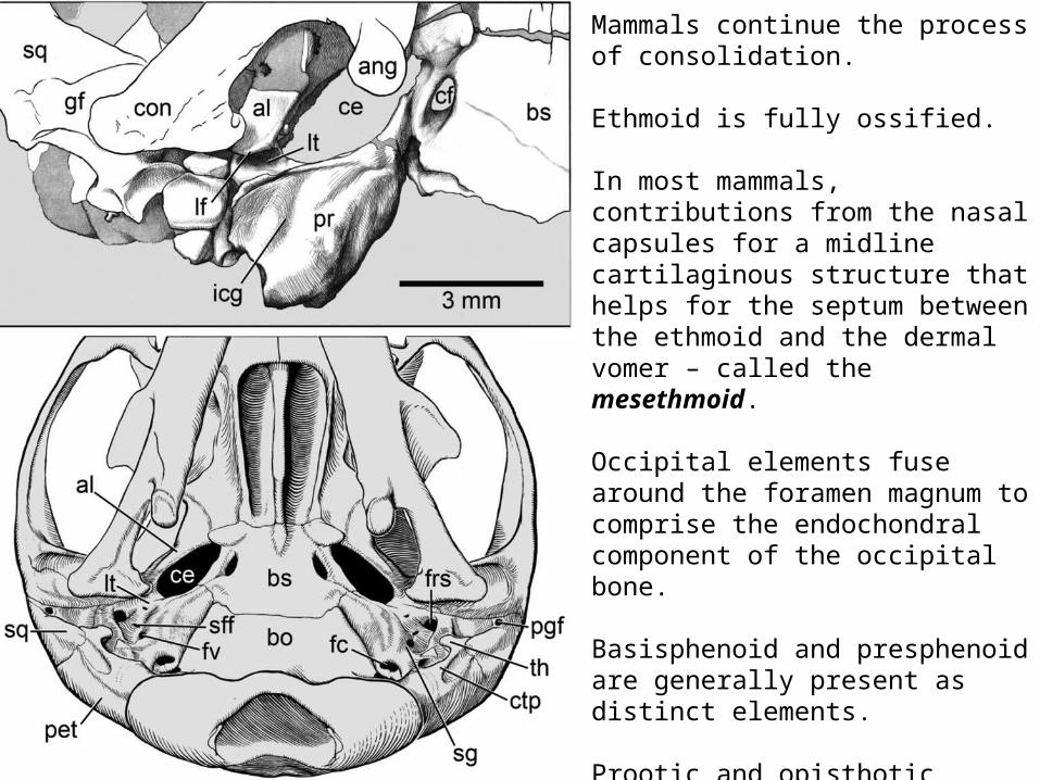
Mammals continue the process of consolidation.
Ethmoid is fully ossified.
In most mammals, contributions from the nasal capsules for a midline cartilaginous structure that helps for the septum between the ethmoid and the dermal vomer – called the mesethmoid.
Occipital elements fuse around the foramen magnum to comprise the endochondral component of the occipital bone.
Basisphenoid and presphenoid are generally present as distinct elements.
Prootic and opisthotic components of the otic casule ombine to form the petrosal portion of the temporal bone (itself a fusion of multiple components).

As synapsids become progressively more mammal-like, it appears that their braincase gets smaller. Rather, it’s that temporal fenestra and passing temporalis muscle are expanding.