Bioelectric patterning during oogenesis: stage-specific ... · PDF fileBackground Oogenesis of...
13
RESEARCH ARTICLE Open Access Bioelectric patterning during oogenesis: stage-specific distribution of membrane potentials, intracellular pH and ion-transport mechanisms in Drosophila ovarian follicles Julia Krüger and Johannes Bohrmann * Abstract Background: Bioelectric phenomena have been found to exert influence on various developmental and regenerative processes. Little is known about their possible functions and the cellular mechanisms by which they might act during Drosophila oogenesis. In developing follicles, characteristic extracellular current patterns and membrane-potential changes in oocyte and nurse cells have been observed that partly depend on the exchange of protons, potassium ions and sodium ions. These bioelectric properties have been supposed to be related to various processes during oogenesis, e. g. pH-regulation, osmoregulation, cell communication, cell migration, cell proliferation, cell death, vitellogenesis and follicle growth. Analysing in detail the spatial distribution and activity of the relevant ion-transport mechanisms is expected to elucidate the roles that bioelectric phenomena play during oogenesis. Results: To obtain an overview of bioelectric patterning along the longitudinal and transversal axes of the developing follicle, the spatial distributions of membrane potentials (V mem ), intracellular pH (pH i ) and various membrane-channel proteins were studied systematically using fluorescent indicators, fluorescent inhibitors and antisera. During mid-vitellogenic stages 9 to 10B, characteristic, stage-specific V mem -patterns in the follicle-cell epithelium as well as anteroposterior pH i -gradients in follicle cells and nurse cells were observed. Corresponding distribution patterns of proton pumps (V-ATPases), voltage-dependent L-type Ca 2+ -channels, amiloride-sensitive Na + -channels and Na + ,H + -exchangers (NHE) and gap-junction proteins (innexin 3) were detected. In particular, six morphologically distinguishable follicle-cell types are characterized on the bioelectric level by differences concerning V mem and pH i as well as specific compositions of ion channels and carriers. Striking similarities between V mem -patterns and activity patterns of voltage-dependent Ca 2+ -channels were found, suggesting a mechanism for transducing bioelectric signals into cellular responses. Moreover, gradients of electrical potential and pH were observed within single cells. Conclusions: Our data suggest that spatial patterning of V mem , pH i and specific membrane-channel proteins results in bioelectric signals that are supposed to play important roles during oogenesis, e. g. by influencing spatial coordinates, regulating migration processes or modifying the cytoskeletal organization. Characteristic stage-specific changes of bioelectric activity in specialized cell types are correlated with various developmental processes. Keywords: Drosophila melanogaster, Oogenesis, Bioelectricity, Cell communication, Pattern formation, Membrane potential, Ion channel, Ion pump, Gap junction, Live-cell imaging * Correspondence: [email protected] RWTH Aachen University, Institut für Biologie II, Abt. Zoologie und Humanbiologie, Worringerweg 3, 52056 Aachen, Germany © 2015 Krüger and Bohrmann; licensee BioMed Central. This is an Open Access article distributed under the terms of the Creative Commons Attribution License (http://creativecommons.org/licenses/by/4.0), which permits unrestricted use, distribution, and reproduction in any medium, provided the original work is properly credited. The Creative Commons Public Domain Dedication waiver (http://creativecommons.org/publicdomain/zero/1.0/) applies to the data made available in this article, unless otherwise stated. Krüger and Bohrmann BMC Developmental Biology (2015) 15:1 DOI 10.1186/s12861-015-0051-3
Transcript of Bioelectric patterning during oogenesis: stage-specific ... · PDF fileBackground Oogenesis of...
Krüger and Bohrmann BMC Developmental Biology (2015) 15:1 DOI 10.1186/s12861-015-0051-3
RESEARCH ARTICLE Open Access
Bioelectric patterning during oogenesis:stage-specific distribution of membranepotentials, intracellular pH and ion-transportmechanisms in Drosophila ovarian folliclesJulia Krüger and Johannes Bohrmann*
Abstract
Background: Bioelectric phenomena have been found to exert influence on various developmental andregenerative processes. Little is known about their possible functions and the cellular mechanisms by which theymight act during Drosophila oogenesis. In developing follicles, characteristic extracellular current patterns andmembrane-potential changes in oocyte and nurse cells have been observed that partly depend on the exchange ofprotons, potassium ions and sodium ions. These bioelectric properties have been supposed to be related to variousprocesses during oogenesis, e. g. pH-regulation, osmoregulation, cell communication, cell migration, cell proliferation,cell death, vitellogenesis and follicle growth. Analysing in detail the spatial distribution and activity of the relevantion-transport mechanisms is expected to elucidate the roles that bioelectric phenomena play during oogenesis.
Results: To obtain an overview of bioelectric patterning along the longitudinal and transversal axes of the developingfollicle, the spatial distributions of membrane potentials (Vmem), intracellular pH (pHi) and various membrane-channelproteins were studied systematically using fluorescent indicators, fluorescent inhibitors and antisera. Duringmid-vitellogenic stages 9 to 10B, characteristic, stage-specific Vmem-patterns in the follicle-cell epithelium aswell as anteroposterior pHi-gradients in follicle cells and nurse cells were observed. Corresponding distributionpatterns of proton pumps (V-ATPases), voltage-dependent L-type Ca2+-channels, amiloride-sensitive Na+-channelsand Na+,H+-exchangers (NHE) and gap-junction proteins (innexin 3) were detected. In particular, six morphologicallydistinguishable follicle-cell types are characterized on the bioelectric level by differences concerning Vmem and pHi aswell as specific compositions of ion channels and carriers. Striking similarities between Vmem-patterns and activitypatterns of voltage-dependent Ca2+-channels were found, suggesting a mechanism for transducing bioelectricsignals into cellular responses. Moreover, gradients of electrical potential and pH were observed within single cells.
Conclusions: Our data suggest that spatial patterning of Vmem, pHi and specific membrane-channel proteins results inbioelectric signals that are supposed to play important roles during oogenesis, e. g. by influencing spatial coordinates,regulating migration processes or modifying the cytoskeletal organization. Characteristic stage-specific changes ofbioelectric activity in specialized cell types are correlated with various developmental processes.
Keywords: Drosophila melanogaster, Oogenesis, Bioelectricity, Cell communication, Pattern formation, Membranepotential, Ion channel, Ion pump, Gap junction, Live-cell imaging
* Correspondence: [email protected] Aachen University, Institut für Biologie II, Abt. Zoologie undHumanbiologie, Worringerweg 3, 52056 Aachen, Germany
© 2015 Krüger and Bohrmann; licensee BioMed Central. This is an Open Access article distributed under the terms of theCreative Commons Attribution License (http://creativecommons.org/licenses/by/4.0), which permits unrestricted use,distribution, and reproduction in any medium, provided the original work is properly credited. The Creative Commons PublicDomain Dedication waiver (http://creativecommons.org/publicdomain/zero/1.0/) applies to the data made available in thisarticle, unless otherwise stated.
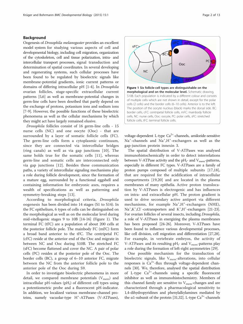
Figure 1 Six follicle-cell types are distinguishable on themorphological and on the molecular level. Schematic drawing,S10B. Each population is indicated by a different colour and consistsof multiple cells which are not shown in detail, except for the polarcells (2 cells) and the border cells (6–10 cells). Anterior is to the left.The position of the oocyte nucleus (black) marks the dorsal side. BC:border cells, cFC: centripetal follicle cells, mFC: mainbody folliclecells, NC: nurse cells, Ooc: oocyte, PC: polar cells, sFC: stretchedfollicle cells, tFC: terminal follicle cells.
Krüger and Bohrmann BMC Developmental Biology (2015) 15:1 Page 2 of 13
BackgroundOogenesis of Drosophila melanogaster provides an excellentmodel system for studying various aspects of cell anddevelopmental biology, including cell migration, organizationof the cytoskeleton, cell and tissue polarization, intra- andintercellular transport processes, signal transduction anddetermination of spatial coordinates. In several developingand regenerating systems, such cellular processes havebeen found to be regulated by bioelectric signals likemembrane-potential gradients, ionic current patterns ordomains of differing intracellular pH [1-4]. In Drosophilaovarian follicles, stage-specific extracellular currentpatterns [5,6] as well as membrane-potential changes ingerm-line cells have been desribed that partly depend onthe exchange of protons, potassium ions and sodium ions[7-9]. However, the possible functions of these bioelectricphenomena as well as the cellular mechanisms by whichthey might act have largely remained elusive.Drosophila follicles consist of 16 germ-line cells - 15
nurse cells (NC) and one oocyte (Ooc) - that aresurrounded by a layer of somatic follicle cells (FC).The germ-line cells form a cytoplasmic continuum,since they are connected via intercellular bridges(ring canals) as well as via gap junctions [10]. Thesame holds true for the somatic cells [11], whereasgerm-line and somatic cells are interconnected onlyvia gap junctions [12]. Besides these communicationpaths, a variety of intercellular signaling mechanisms playa role during follicle development, since the formation ofa mature egg, surrounded by a functional eggshell andcontaining information for embryonic axes, requires awealth of specifications as well as patterning andsymmetry-breaking steps [13].According to morphological criteria, Drosophila
oogenesis has been divided into 14 stages (S1 to S14). Inthe FC epithelium, 6 types of cells can be distinguished onthe morphological as well as on the molecular level duringmid-vitellogenic stages 9 to 10B [14-16] (Figure 1). Theterminal FC (tFC) are a population of about 200 cells atthe posterior follicle pole. The mainbody FC (mFC) forma broad band anterior to the tFC. The centripetal FC(cFC) reside at the anterior end of the Ooc and migrate inbetween NC and Ooc during S10B. The stretched FC(sFC) become flattened and cover the NC. A pair of polarcells (PC) resides at the posterior pole of the Ooc. Theborder cells (BC), a group of 6–10 anterior FC, migratebetween the NC from the anterior follicle pole to theanterior pole of the Ooc during S9.In order to investigate bioelectric phenomena in more
detail, we compared membrane potentials (Vmem) andintracellular pH-values (pHi) of different cell types usinga potentiometric probe and a fluorescent pH-indicator.In addition, we localized various membrane-channel pro-teins, namely vacuolar-type H+-ATPases (V-ATPases),
voltage-dependent L-type Ca2+-channels, amiloride-sensitiveNa+-channels and Na+,H+-exchangers as well as thegap-junction protein innexin 3.The spatial distribution of V-ATPases was analysed
immunohistochemically in order to detect interrelationsbetween V-ATPase activity and the pHi- and Vmem-patterns,especially in different FC types. V-ATPases are a family ofproton pumps composed of multiple subunits [17,18],that are required for the acidification of intracellularcompartments [19,20] and are located in the plasmamembranes of many epithelia. Active proton transloca-tion by V-ATPases is electrogenic and has influenceson intra- and extracellular pH. The proton gradient isused to drive secondary active antiport via differentmechanisms, for example Na+,H+-exchangers (NHE),Na+,K+,Cl−-cotransporters and K+,H+-exchangers [21-23].For ovarian follicles of several insects, including Drosophila,a role of V-ATPases in energizing the plasma membraneshas been proposed [24-26]. Moreover, V-ATPases havebeen found to influence various developmental processes,like cell division, cell migration and differentiation [27,28].For example, in vertebrate embryos, the activity ofV-ATPases and its resulting pHi- and Vmem-patterns playa role during the formation of left-right asymmetries [29].One possible mechanism for the transduction of
bioelectric signals, like Vmem-alterations, into cellularresponses is Ca2+-flux through voltage-dependent chan-nels [30]. We, therefore, analysed the spatial distributionof L-type Ca2+-channels using a specific fluorescentinhibitor as well as immunohistochemistry. Members ofthis channel family are sensitive to Vmem-changes and arecharacterized through a pharmacological sensitivity to1,4-dihydropyridines and phenylalkylamines mediated bythe α1-subunit of the protein [31,32]. L-type Ca2+-channels
Krüger and Bohrmann BMC Developmental Biology (2015) 15:1 Page 3 of 13
appear in three functionally distinct states: resting, orclosed, channels, activated, or open, channels, andinactivated channels. In nerve cells, depolarizationresults in a conformational change from the restingto the activated state and then to rapid inactivation,while repolarization is necessary to return to the restingstate [32]. The binding affinity of phenylalkylamines, likeverapamil, depends on the channel’s state in the fol-lowing order: inactivated > > activated > resting [33,34].Voltage-dependent L-type Ca2+-channels were originallyconsidered to be unique to excitable cells [35], but nowthey are known to be present in non-excitable cells aswell. For example, they play a role in Ca2+-reabsorption inmammalian renal epithelia [36] and in epithelial fluidtransport in Drosophila Malpighian tubules [37].The spatial distribution of amiloride-sensitive NHE
and Na+-channels was investigated using fluorescentamiloride. NHE are important for pHi-homeostasis,cell-volume regulation and transepithelial Na+-transport[38]. In insect epithelia, the electroneutral exchange ofNa+ against H+ through NHE is driven by the H+-gradientgenerated by V-ATPases [21,39]. Amiloride-sensitiveNa+-channels have been found to play a role duringreproductive as well as developmental processes. Forexample, they help to block polyspermy in Xenopusoocytes [40] and, in Drosophila, the amiloride-sensitiveNa+-channel dGNaC1 is expressed in gonads and earlyembryos. This channel has been proposed to be involvedin cytoplasmic transport processes and in water uptakeduring final maturation of the Ooc [41].Members of the innexin family are the main gap-junction
proteins in invertebrates [42,43], although other proteins,like pannexins [44] and ductins [45-48], have also beenobserved in gap junctions. In the Drosophila ovary, themRNAs of innexins 1, 2, 3, 4 and 7 were detected [49] andinnexins 1 to 4 have been shown immunohistochemicallyto be involved in the formation of different types of gapjunctions [50]. Since innexins 1, 2 and 4 did not showconspicuous spatial distribution patterns, we only presentthe distribution of innexin 3 in more detail.Using fluorescent indicators, inhibitors and antisera,
we found corresponding asymmetries in the distributionpatterns along the longitudinal and transversal axes of thefollicle during S9 to S10B. Characteristic stage-specificchanges observed in different cell types are correlated withvarious cellular and developmental processes.
MethodsPreparation of folliclesDrosophila melanogaster wild-type Oregon R flies werereared at about 20°C on standard food with additionalfresh yeast. Individual 2–3 days old females were killedby crushing the thorax with tweezers without previousetherization or chilling. The ovaries were dissected with
tweezers, and single follicles of S9 to S10B were isolatedby pulling at the anterior tip of an ovariole. For immuno-histochemistry, dissection was carried out in DrosophilaPBS [51], while for in-vitro experiments with fluorescentinhibitors and indicators R-14 medium [51] was used,which ensures optimal culture conditions during live-cellimaging of follicles [52].
AntiseraTo localize V-ATPases, we used two antisera: (1) arabbit antiserum (Anti-ductin; AB5496, ChemiconInternational, USA) raised against a highly conservedregion of the 16 kDa-protein ductin, which formssubunit c of V-ATPases and is also part of gap junctions[48], and (2), as a control, a guinea-pig antiserum(Anti-V-ATPase) raised against an N-terminal regionof subunit a of the Manduca sexta V-ATPase, kindlyprovided by B. Walz and O. Baumann (Potsdam,Germany). For the localization of innexin 3, we useda guinea-pig antiserum (Anti-Inx3 [50]) raised againstthe C-terminus of innexin 3 from Drosophila, kindlyprovided by R. Bauer and M. Hoch (Bonn, Germany). Incontrol experiments, L-type Ca2+-channels were localizedusing a rabbit antiserum (Anti-Cavα1; Anti-Cavpanα1 sub-unit, Almone Labs, Israel) raised against an intracellularepitope of subunit α1.
Indirect immunofluorescence preparationsFollicles were fixed for 30 minutes at 4°C in 4% formal-dehyde dissolved in PBS, washed in PBS and blocked for1 hour at 20°C with 2% BSA/0,1% Triton X-100 in PBS.Thereafter, the follicles were incubated overnight at 4°Cin PBS containing 0,5% BSA/0,1% Triton X-100 andthe respective antiserum (Anti-ductin diluted 1:100,Anti-V-ATPase diluted 1:1000, Anti-Inx3 diluted 1:20,Anti-Cavα1 diluted 1:100, controls without antiserum).After washing 6 times for 10 minutes, the follicles were
either treated with goat-anti-rabbit-Cy3 (Jackson, USA;diluted 1:2000) or with donkey-anti-guinea-pig-FP488(FluoProbes/Interchim, France; diluted 1:100) for 1 hour inPBS containing 0,5% BSA/0,1% Triton X-100. Washing wasrepeated 6 times and the nuclei were stained with 0,2 μg/mlDAPI (Sigma, Germany) in PBS for 3 minutes. Thereafter,the follicles were mounted in Fluoromount G (Interchim)and viewed, by using ×20 or ×40 objectives and theappropriate filter sets, either on a Zeiss Axiovert 200 wide-field fluorescence microscope (WFM), equipped with aHamamatsu Orca ER camera, or on a Zeiss AxioImager.M2structured-illumination microscope (SIM), equipped with aZeiss ApoTome and a Zeiss AxioCamMRm camera.
Fluorescent inhibitorsStaining of living follicles with fluorescent inhibitorswas carried out in R-14 medium. Stock solutions were
Krüger and Bohrmann BMC Developmental Biology (2015) 15:1 Page 4 of 13
prepared in DMSO, and the follicles were incubatedeither in 2 μM amiloride-FL (BODIPY-FL amiloride;Molecular Probes/Thermo Fisher Scientific, USA) or in2 μM verapamil-FL (BODIPY-FL verapamil, hydrochloride;Molecular Probes) for 15 minutes. Control follicles werepreincubated before labeling with the unlabeled inhibitors(Sigma, Germany; stock solutions in ethanol) for 15 minutesusing 10 μM amiloride or 100 μM verapamil-HCl (or theywere labeled with Anti-Cavα1). Thereafter, the follicles weremounted in R-14 medium and viewed immediately asdescribed above.
Fluorescent membrane-potential indicatorIn order to analyse Vmem-patterns, we used the fluorescentpotentiometric probe DiBAC (bis-(1,3-dibutylbarbituricacid) trimethine oxonol, DiBAC4(3); Molecular Probes).The anionic dye enters cells and binds to intracellularmembranes and proteins in a Vmem-dependent manner:depolarization leads to an accumulation and to an increasein fluorescence intensity. Relative fluorescence differencesbetween cells of comparable size were stated, i. e. strongerfluorescence: more depolarized vs. weaker fluorescence:more hyperpolarized. Living follicles were incubatedfor 15 minutes in R-14 medium containing 1–3 μMDiBAC (dissolved in 70% ethanol). Thereafter, they weremounted in R-14 medium and viewed immediately asdescribed above.
Fluorescent intracellular pH-indicatorFor the analysis of pHi-patterns, we used the fluorescentpH-indicator CFDA (5-carboxyfluorescein diacetate,acetoxymethyl ester, 5-CFDA, AM; Molecular Probes). Theanionic dye enters cells and reports pHi by fluorescence-intensity differences, since protonation leads to fluores-cence loss. Relative fluorescence differences between cellsof comparable size were stated, i.e. stronger fluorescence:more alkaline vs. weaker fluorescence: more acidic. Livingfollicles were incubated for 15 minutes in R-14 mediumcontaining 4 μM CFDA (dissolved in DMSO), and thenmounted and viewed as described above.
Analysis of staining patternsTo facilitate interpretation and comparability of thepatterns obtained with different staining methods, weanalysed median optical sections through the follicles.Representative grey-scale images were transferred intopseudocolour images using ImageJ Fire-LUT (NIH, USA).For immunostaining as well as fluorescent inhibitors,brighter colours represent higher concentrations of therespective membrane-channel protein. For DiBAC,brighter colours refer to more depolarized Vmem, and forCFDA, brighter colours refer to more alkaline pHi. Eachexperiment was repeated at least three times.
ResultsGradients of membrane potentials in follicle cells andoocyteUsing the potentiometric probe DiBAC, we revealed stagespecific Vmem-patterns during S9 to S10B by comparingfluorescence intensities in different follicle regions.Stronger intensities refer to more depolarized Vmem,weaker intensities to more hyperpolarized Vmem. In S9(Figure 2A) we found a complex Vmem-pattern in the FCepithelium: mainbody FC (mFC) are characterized by ahyperpolarized Vmem in relation to the neighboringterminal FC (tFC, posterior), centripetal FC (cFC, anterior)and stretched FC (sFC, further anterior). In each mFC, anapicobasal gradient was observed, the apical region beingmore hyperpolarized than the basal region. The strongestdepolarization was found in migrating border cells (BC)and in posterior polar cells (PC). In the oocyte (Ooc), ananteroposterior gradient was observed, the anterior regionbeing more hyperpolarized than the posterior region.During S10B (and S11, data not shown) we found atransversal Vmem-pattern, where one side of the FCepithelium (including tFC, mFC and cFC) was depolarizedin relation to the opposite side. In most follicles, thedepolarized side could be identified as the ventral side(25 vs. 3 follicles) according to the location of theOoc nucleus (DIC-microscopy). S10A showed characteris-tics of both S9 and S10B: mFC are hyperpolarized in rela-tion to cFC and tFC, but one side of the FC epithelium isalready more depolarized than the other (Figure 2B and C).
Gradients of intracellular pH in nurse cells and folliclecellsUsing the pH-indicator CFDA, we analysed pHi-patternsin the developing follicle. A more acidic pHi is indicatedby weaker, a more alkaline pHi by stronger fluorescenceintensity. During S9 to S10B, we found stage-specificpHi-patterns in the NC and in the FC (Figure 3). In theNC cluster, remarkable anteroposterior and dorsoventralgradients emerged, where each NC is characterizedthrough its own pHi: the NC adjacent to the anterodor-sal region of the Ooc (location of the nucleus) has themost acidic pHi, and the anterior-most NC has the mostalkaline pHi. Both gradients develop in S9 and becomefully established during S10. In the FC epithelium, wefound greater variability: most examined folliclesshowed an anteroposterior pHi-gradient, the sFC, cFCand mFC being most acidic and the tFC being mostalkaline. Some follicles, especially in S9, also showeda transversal pHi-gradient in the FC epithelium, withone side being more acidic than the other (data notshown). In addition, in S9 the migrating BC are char-acterized through acidic pHi, and the posterior regionof the Ooc is more alkaline than the anterior region(Figure 3A).

S9
S10B
A
B
C C’ D
A’
B’
Figure 2 Characteristic patterns of membrane potentials (Vmem). DiBAC staining, SIM, S9 to S10B, A, B, C: representative grey-scale images,A´, B´, C´: corresponding pseudocolour images. Depolarization is indicated by strong, hyperpolarization by weak fluorescence intensity (see scalebar). D: schematic drawings of characteristic Vmem-patterns; depolarized regions are shown in lilac (staining in NC is not considered). In S9 (A, A´)the FC epithelium is patterned along the anteroposterior axis. Strongest depolarization is found in cFC, sFC, tFC, BC and PC. The mFC are characterizedby a hyperpolarized Vmem and an intracellular apicobasal gradient (apical region near Ooc is hyperpolarized compared to basal region), while theanterior region of the Ooc is hyperpolarized relative to the posterior region. In S10B (C, C´) the ventral side of the follicle is depolarized (arrow) inrelation to the other side. S10A (B, B´) shows characteristics of both S9 and S10B. For abbreviations, see Figure 1.
Krüger and Bohrmann BMC Developmental Biology (2015) 15:1 Page 5 of 13
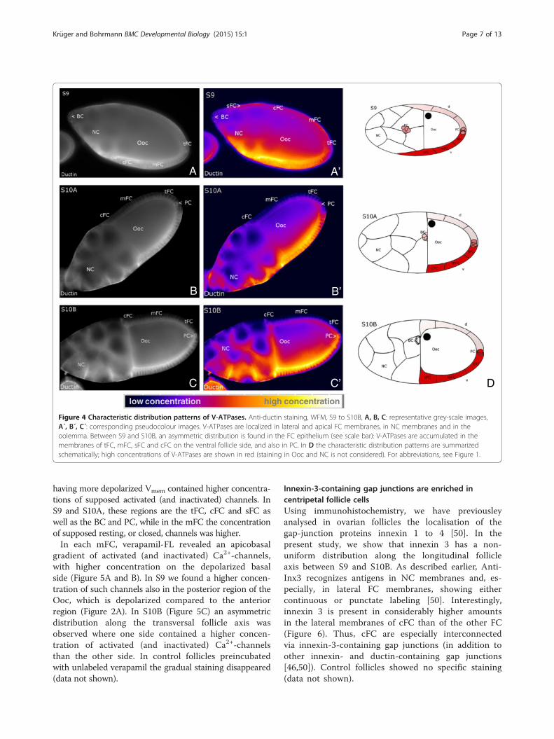
Asymmetric V-ATPase distribution in the follicle-cellepitheliumBy indirect immunofluorescence microscopy using twodifferent antisera (Anti-ductin and Anti-V-ATPase) we in-vestigated the localization of V-ATPases during mid-vitellogenic stages. Since the 16 kDa-protein ductin isknown to be part of both V-ATPases and gap junctions[48], we used Anti-V-ATPase as a control to decide,whether the observed distribution of ductin results fromthe distribution of V-ATPases or gap junctions. Bothantisera recognized cytoplasmic as well as membran-ous antigens. Membrane labeling with Anti-ductinwas either punctate or continuous (Figure 4), whilewith Anti-V-ATPase it was always continuous (datanot shown). Punctate membrane labeling is presumed to ori-ginate from ductin as part of gap junctions, whereas continu-ous membrane labeling is presumed to originate from ductinas part of V-ATPases [26]. We found V-ATPases in lateral
and in apical FC membranes, in NC membranes and in theoolemma, while in the FC epithelium, characteristic asym-metries in the spatial distribution were observed with bothantisera (for Anti-V-ATPase, data not shown). Accordingly,V-ATPases (not ductin-containing gap junctions, which showuniform punctate distribution) are enriched in FC on oneside of the follicle, including tFC, mFC, cFC and sFC,whereas FC on the opposite side contain considerably less.In most follicles, the enriched side could be identified as theventral side (13 vs. 1 follicles) according to the location ofthe Ooc nucleus (DIC-microscopy). Also the membranes ofPC showed accumulation of V-ATPases (Figure 4). Controlfollicles showed no specific staining (data not shown).
L-type Ca2+-channel patterns correspond tomembrane-potential patternsUsing the fluorescent inihibitor verapamil-FL, we analysedthe distribution of L-type Ca2+-channels. Since binding of
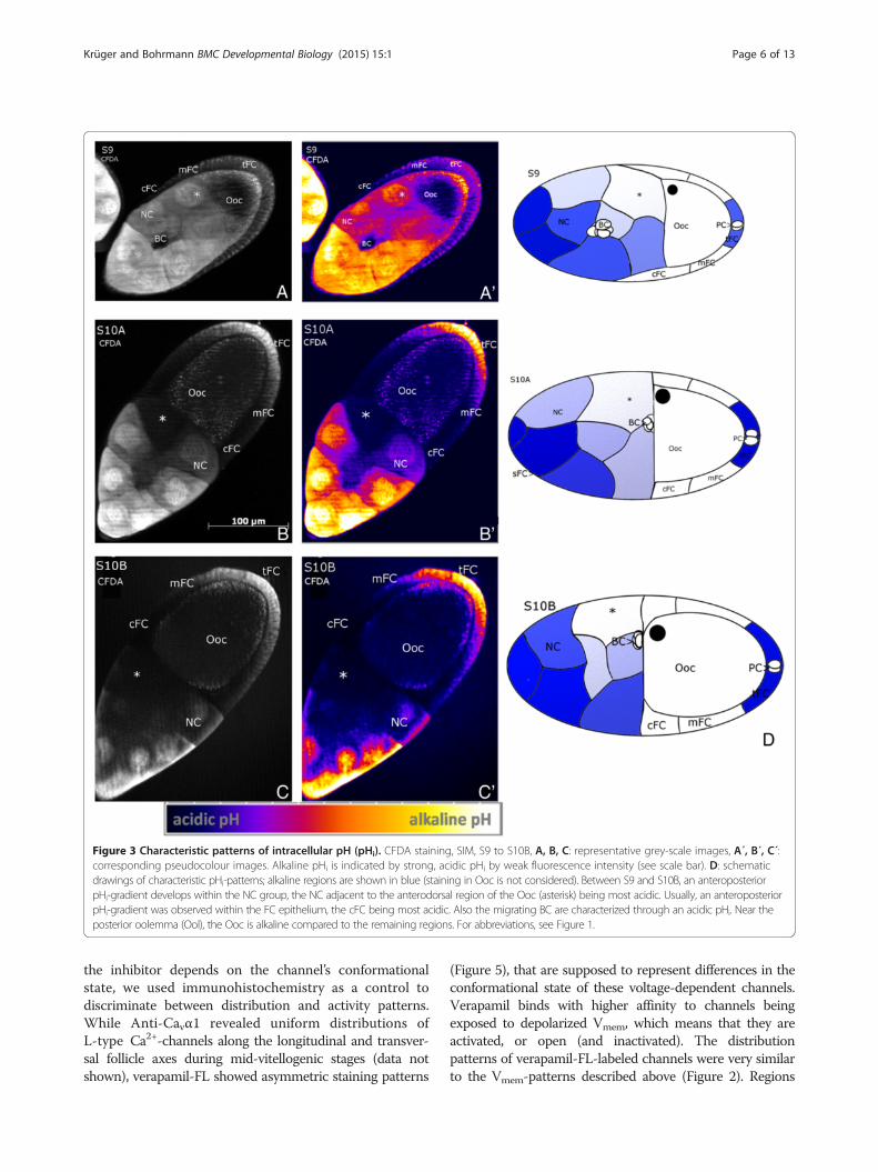
Figure 3 Characteristic patterns of intracellular pH (pHi). CFDA staining, SIM, S9 to S10B, A, B, C: representative grey-scale images, A´, B´, C´:corresponding pseudocolour images. Alkaline pHi is indicated by strong, acidic pHi by weak fluorescence intensity (see scale bar). D: schematicdrawings of characteristic pHi-patterns; alkaline regions are shown in blue (staining in Ooc is not considered). Between S9 and S10B, an anteroposteriorpHi-gradient develops within the NC group, the NC adjacent to the anterodorsal region of the Ooc (asterisk) being most acidic. Usually, an anteroposteriorpHi-gradient was observed within the FC epithelium, the cFC being most acidic. Also the migrating BC are characterized through an acidic pHi. Near theposterior oolemma (Ool), the Ooc is alkaline compared to the remaining regions. For abbreviations, see Figure 1.
Krüger and Bohrmann BMC Developmental Biology (2015) 15:1 Page 6 of 13
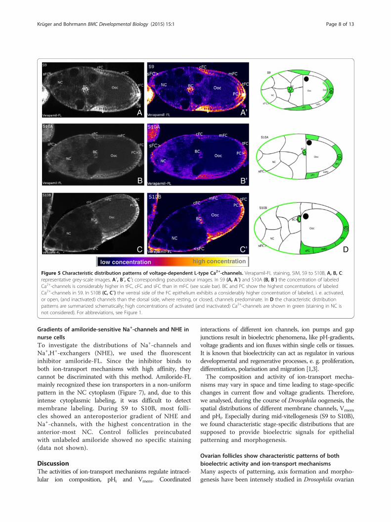
the inhibitor depends on the channel’s conformationalstate, we used immunohistochemistry as a control todiscriminate between distribution and activity patterns.While Anti-Cavα1 revealed uniform distributions ofL-type Ca2+-channels along the longitudinal and transver-sal follicle axes during mid-vitellogenic stages (data notshown), verapamil-FL showed asymmetric staining patterns
(Figure 5), that are supposed to represent differences in theconformational state of these voltage-dependent channels.Verapamil binds with higher affinity to channels beingexposed to depolarized Vmem, which means that they areactivated, or open (and inactivated). The distributionpatterns of verapamil-FL-labeled channels were very similarto the Vmem-patterns described above (Figure 2). Regions
low concentration high concentration
A
B B’
C’ DC
A’
Figure 4 Characteristic distribution patterns of V-ATPases. Anti-ductin staining, WFM, S9 to S10B, A, B, C: representative grey-scale images,A´, B´, C´: corresponding pseudocolour images. V-ATPases are localized in lateral and apical FC membranes, in NC membranes and in theoolemma. Between S9 and S10B, an asymmetric distribution is found in the FC epithelium (see scale bar): V-ATPases are accumulated in themembranes of tFC, mFC, sFC and cFC on the ventral follicle side, and also in PC. In D the characteristic distribution patterns are summarizedschematically; high concentrations of V-ATPases are shown in red (staining in Ooc and NC is not considered). For abbreviations, see Figure 1.
Krüger and Bohrmann BMC Developmental Biology (2015) 15:1 Page 7 of 13
having more depolarized Vmem contained higher concentra-tions of supposed activated (and inactivated) channels. InS9 and S10A, these regions are the tFC, cFC and sFC aswell as the BC and PC, while in the mFC the concentrationof supposed resting, or closed, channels was higher.In each mFC, verapamil-FL revealed an apicobasal
gradient of activated (and inactivated) Ca2+-channels,with higher concentration on the depolarized basalside (Figure 5A and B). In S9 we found a higher concen-tration of such channels also in the posterior region of theOoc, which is depolarized compared to the anteriorregion (Figure 2A). In S10B (Figure 5C) an asymmetricdistribution along the transversal follicle axis wasobserved where one side contained a higher concen-tration of activated (and inactivated) Ca2+-channelsthan the other side. In control follicles preincubatedwith unlabeled verapamil the gradual staining disappeared(data not shown).
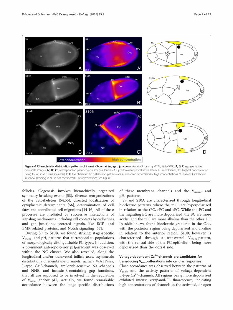
Innexin-3-containing gap junctions are enriched incentripetal follicle cellsUsing immunohistochemistry, we have previousleyanalysed in ovarian follicles the localisation of thegap-junction proteins innexin 1 to 4 [50]. In thepresent study, we show that innexin 3 has a non-uniform distribution along the longitudinal follicleaxis between S9 and S10B. As described earlier, Anti-Inx3 recognizes antigens in NC membranes and, es-pecially, in lateral FC membranes, showing eithercontinuous or punctate labeling [50]. Interestingly,innexin 3 is present in considerably higher amountsin the lateral membranes of cFC than of the other FC(Figure 6). Thus, cFC are especially interconnectedvia innexin-3-containing gap junctions (in addition toother innexin- and ductin-containing gap junctions[46,50]). Control follicles showed no specific staining(data not shown).
low concentration high concentration
A A’
B
C C’ D
B’
Figure 5 Characteristic distribution patterns of voltage-dependent L-type Ca2+-channels. Verapamil-FL staining, SIM, S9 to S10B, A, B, C:representative grey-scale images, A´, B´, C´: corresponding pseudocolour images. In S9 (A, A´) and S10A (B, B´) the concentration of labeledCa2+-channels is considerably higher in tFC, cFC and sFC than in mFC (see scale bar). BC and PC show the highest concentrations of labeledCa2+-channels in S9. In S10B (C, C´) the ventral side of the FC epithelium exhibits a considerably higher concentration of labeled, i. e. activated,or open, (and inactivated) channels than the dorsal side, where resting, or closed, channels predominate. In D the characteristic distributionpatterns are summarized schematically; high concentrations of activated (and inactivated) Ca2+-channels are shown in green (staining in NC isnot considered). For abbreviations, see Figure 1.
Krüger and Bohrmann BMC Developmental Biology (2015) 15:1 Page 8 of 13
Gradients of amiloride-sensitive Na+-channels and NHE innurse cellsTo investigate the distributions of Na+-channels andNa+,H+-exchangers (NHE), we used the fluorescentinhibitor amiloride-FL. Since the inhibitor binds toboth ion-transport mechanisms with high affinity, theycannot be discriminated with this method. Amiloride-FLmainly recognized these ion transporters in a non-uniformpattern in the NC cytoplasm (Figure 7), and, due to thisintense cytoplasmic labeling, it was difficult to detectmembrane labeling. During S9 to S10B, most folli-cles showed an anteroposterior gradient of NHE andNa+-channels, with the highest concentration in theanterior-most NC. Control follicles preincubatedwith unlabeled amiloride showed no specific staining(data not shown).
DiscussionThe activities of ion-transport mechanisms regulate intracel-lular ion composition, pHi and Vmem. Coordinated
interactions of different ion channels, ion pumps and gapjunctions result in bioelectric phenomena, like pH-gradients,voltage gradients and ion fluxes within single cells or tissues.It is known that bioelectricity can act as regulator in variousdevelopmental and regenerative processes, e. g. proliferation,differentiation, polarisation and migration [1,3].The composition and activity of ion-transport mecha-
nisms may vary in space and time leading to stage-specificchanges in current flow and voltage gradients. Therefore,we analysed, during the course of Drosophila oogenesis, thespatial distributions of different membrane channels, Vmem
and pHi. Especially during mid-vitellogenesis (S9 to S10B),we found characteristic stage-specific distributions that aresupposed to provide bioelectric signals for epithelialpatterning and morphogenesis.
Ovarian follicles show characteristic patterns of bothbioelectric activity and ion-transport mechanismsMany aspects of patterning, axis formation and morpho-genesis have been intensely studied in Drosophila ovarian
low concentration high concentration
A A’
B’B
C C’ D
Figure 6 Characteristic distribution patterns of innexin-3-containing gap junctions. Anti-Inx3 staining, WFM, S9 to S10B, A, B, C: representativegrey-scale images, A´, B´, C´: corresponding pseudocolour images. Innexin 3 is predominantly localized in lateral FC membranes, the highest concentrationbeing found in cFC (see scale bar). In D the characteristic distribution patterns are summarized schematically; high concentrations of innexin 3 are shownin yellow (staining in NC is not considered). For abbreviations, see Figure 1.
Krüger and Bohrmann BMC Developmental Biology (2015) 15:1 Page 9 of 13
follicles. Oogenesis involves hierarchically organizedsymmetry-breaking events [53], diverse reorganizationsof the cytoskeleton [54,55], directed localization ofcytoplasmic determinants [56], determination of cellfates and coordinated cell migrations [14-16]. All of theseprocesses are mediated by successive interactions ofsignaling mechanisms, including cell contacts by cadherinesand gap junctions, secreted signals, like EGF- andBMP-related proteins, and Notch signaling [57].During S9 to S10B, we found striking stage-specific
Vmem- and pHi-patterns that correspond to populationsof morphologically distinguishable FC types. In addition,a prominent anteroposterior pHi-gradient was observedwithin the NC cluster. We also revealed, along thelongitudinal and/or transversal follicle axes, asymmetricdistributions of membrane channels, namely V-ATPases,L-type Ca2+-channels, amiloride-sensitive Na+-channelsand NHE, and innexin-3-containing gap junctions,that all are supposed to be involved in the regulationof Vmem and/or pHi. Actually, we found remarkableaccordance between the stage-specific distributions
of these membrane channels and the Vmem- andpHi-patterns.S9 and S10A are characterized through longitudinal
bioelectric patterns, where the mFC are hyperpolarizedin relation to the tFC, cFC and sFC. While the PC andthe migrating BC are more depolarized, the BC are moreacidic, and the tFC are more alkaline than the other FC.In addition, we found bioelectric gradients in the Ooc,with the posterior region being depolarized and alkalinein relation to the anterior region. S10B, however, ischaracterized through a transversal Vmem-pattern,with the ventral side of the FC epithelium being moredepolarized than the dorsal side.
Voltage-dependent Ca2+-channels are candidates fortransducing Vmem-alterations into cellular responsesClose accordance was observed between the patterns ofVmem and the activity patterns of voltage-dependentL-type Ca2+-channels. All regions being more depolarizedexhibited intense verapamil-FL fluorescence, indicatinghigh concentrations of channels in the activated, or open
low concentration high concentration
A A’
B’B
C C’ D
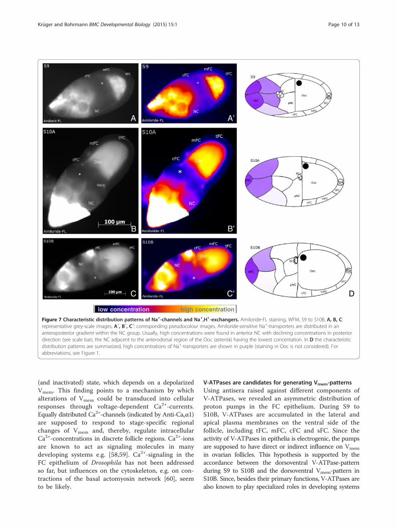
Figure 7 Characteristic distribution patterns of Na+-channels and Na+,H+-exchangers. Amiloride-FL staining, WFM, S9 to S10B, A, B, C:representative grey-scale images, A´, B´, C´: corresponding pseudocolour images. Amiloride-sensitive Na+-transporters are distributed in ananteroposterior gradient within the NC group. Usually, high concentrations were found in anterior NC with declining concentrations in posteriordirection (see scale bar), the NC adjacent to the anterodorsal region of the Ooc (asterisk) having the lowest concentation. In D the characteristicdistribution patterns are summarized, high concentrations of Na+-transporters are shown in purple (staining in Ooc is not considered). Forabbreviations, see Figure 1.
Krüger and Bohrmann BMC Developmental Biology (2015) 15:1 Page 10 of 13
(and inactivated) state, which depends on a depolarizedVmem. This finding points to a mechanism by whichalterations of Vmem could be transduced into cellularresponses through voltage-dependent Ca2+-currents.Equally distributed Ca2+-channels (indicated by Anti-Cavα1)are supposed to respond to stage-specific regionalchanges of Vmem and, thereby, regulate intracellularCa2+-concentrations in discrete follicle regions. Ca2+-ionsare known to act as signaling molecules in manydeveloping systems e.g. [58,59]. Ca2+-signaling in theFC epithelium of Drosophila has not been addressedso far, but influences on the cytoskeleton, e.g. on con-tractions of the basal actomyosin network [60], seemto be likely.
V-ATPases are candidates for generating Vmem-patternsUsing antisera raised against different components ofV-ATPases, we revealed an asymmetric distribution ofproton pumps in the FC epithelium. During S9 toS10B, V-ATPases are accumulated in the lateral andapical plasma membranes on the ventral side of thefollicle, including tFC, mFC, cFC and sFC. Since theactivity of V-ATPases in epithelia is electrogenic, the pumpsare supposed to have direct or indirect influence on Vmem
in ovarian follicles. This hypothesis is supported by theaccordance between the dorsoventral V-ATPase-patternduring S9 to S10B and the dorsoventral Vmem-pattern inS10B. Since, besides their primary functions, V-ATPases arealso known to play specialized roles in developing systems
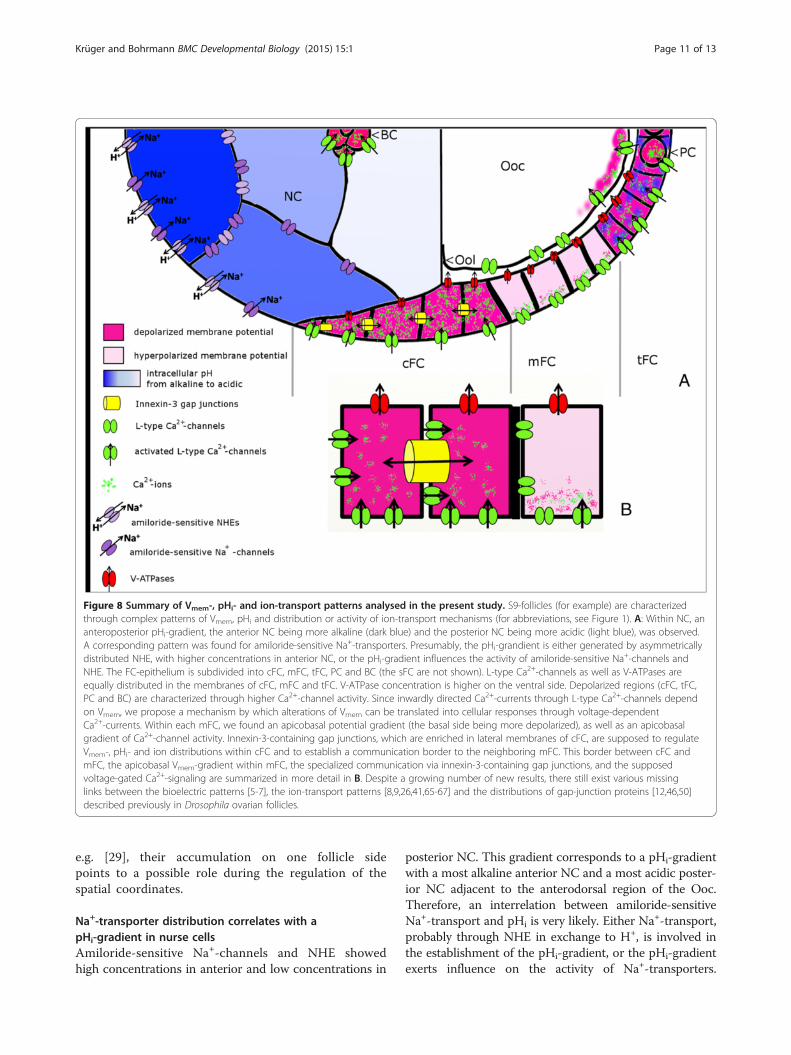
Figure 8 Summary of Vmem-, pHi- and ion-transport patterns analysed in the present study. S9-follicles (for example) are characterizedthrough complex patterns of Vmem, pHi and distribution or activity of ion-transport mechanisms (for abbreviations, see Figure 1). A: Within NC, ananteroposterior pHi-gradient, the anterior NC being more alkaline (dark blue) and the posterior NC being more acidic (light blue), was observed.A corresponding pattern was found for amiloride-sensitive Na+-transporters. Presumably, the pHi-grandient is either generated by asymmetricallydistributed NHE, with higher concentrations in anterior NC, or the pHi-gradient influences the activity of amiloride-sensitive Na+-channels andNHE. The FC-epithelium is subdivided into cFC, mFC, tFC, PC and BC (the sFC are not shown). L-type Ca2+-channels as well as V-ATPases areequally distributed in the membranes of cFC, mFC and tFC. V-ATPase concentration is higher on the ventral side. Depolarized regions (cFC, tFC,PC and BC) are characterized through higher Ca2+-channel activity. Since inwardly directed Ca2+-currents through L-type Ca2+-channels dependon Vmem, we propose a mechanism by which alterations of Vmem can be translated into cellular responses through voltage-dependentCa2+-currents. Within each mFC, we found an apicobasal potential gradient (the basal side being more depolarized), as well as an apicobasalgradient of Ca2+-channel activity. Innexin-3-containing gap junctions, which are enriched in lateral membranes of cFC, are supposed to regulateVmem-, pHi- and ion distributions within cFC and to establish a communication border to the neighboring mFC. This border between cFC andmFC, the apicobasal Vmem-gradient within mFC, the specialized communication via innexin-3-containing gap junctions, and the supposedvoltage-gated Ca2+-signaling are summarized in more detail in B. Despite a growing number of new results, there still exist various missinglinks between the bioelectric patterns [5-7], the ion-transport patterns [8,9,26,41,65-67] and the distributions of gap-junction proteins [12,46,50]described previously in Drosophila ovarian follicles.
Krüger and Bohrmann BMC Developmental Biology (2015) 15:1 Page 11 of 13
e.g. [29], their accumulation on one follicle sidepoints to a possible role during the regulation of thespatial coordinates.
Na+-transporter distribution correlates with apHi-gradient in nurse cellsAmiloride-sensitive Na+-channels and NHE showedhigh concentrations in anterior and low concentrations in
posterior NC. This gradient corresponds to a pHi-gradientwith a most alkaline anterior NC and a most acidic poster-ior NC adjacent to the anterodorsal region of the Ooc.Therefore, an interrelation between amiloride-sensitiveNa+-transport and pHi is very likely. Either Na+-transport,probably through NHE in exchange to H+, is involved inthe establishment of the pHi-gradient, or the pHi-gradientexerts influence on the activity of Na+-transporters.
Krüger and Bohrmann BMC Developmental Biology (2015) 15:1 Page 12 of 13
Despite cytoplasmic coupling via ring canals and gapjunctions, each NC regulates its pHi independently.The resulting anteroposterior pHi-gradient is supposed toplay a role in various processes during oogenesis. In gen-eral, pHi is known to exert influence on cell metabolism,enzyme activity, contractibility of cytoskeletal structures,cell polarity and proliferation rates of cells [61-63]. DuringS9, the pHi-gradient might also provide guidance cues forBC migration.
Communication via specialized gap junctions is likely togenerate and maintain boundaries between different cellpopulationsOf the ovarian gap-junction proteins innexin 1 to 4, onlyinnexin 3 showed a striking non-uniform distributionwithin the FC epithelium: it is enriched in the lateralmembranes of cFCs. This finding indicates distinctcoupling conditions between these cells, concerning e.g.regulatory signals, Vmem and pHi. Moreover, innexin 3seems to be involved in maintaining tissue integrity inresponse to tension [64], a function that is particularlyimportant for cFC.The cFC are specialized in several further respects:
they are longer dye-coupled to the Ooc by gap junctions[12], they contain higher amounts of Na+,K+-ATPase[26,65] (which becomes activated under alkaline con-ditions [66]), and they possess a higher [Ca2+]i thanthe remaining FC [67]. The permeability of innexinchannels in general has been reported to be Vmem-, pHi-,K+- and Ca2+-sensitive [68,69]. Restriction to distinctcell populations and independent regulation of permeabilityof specialized gap-junction channels (formed by differentproteins in either homomeric, heteromeric or hetero-typic combinations [50]) could provide mechanismsto generate or maintain boundaries between differentFC populations.
ConclusionsSpatial patterns of Vmem and pHi related to non-uniformdistribution and activity patterns of membrane channels(for summary, see Figure 8) are supposed to generatebioelectric signals during several important steps ofoogenesis, e.g. for the regulation of spatial coordinates,migration processes, or reorganization of the cytoskeleton.In recent years, the complex interrelations as well as thephysiological and cellular functions of bioelectricphenomena have become increasingly challenging [70].Analysing the distribution and activity patterns offurther ion-transport mechanisms - also in relevantDrosophila mutants e.g. [53,71,72] - as well as specificmanipulations of Vmem and pHi by using appropriateinhibitors will be promising ways to help cracking thebioelectric code [73].
AbbreviationsBC: Border cells; BSA: Bovine serum albumine; cFC: Centripetal follicle cells;CFDA: 5-carboxyfluorescein diacetate, acetoxymethyl ester; DAPI: 4′,6-diamidino-2-phenylindole; DiBAC: Bis-(1,3-dibutylbarbituric acid) trimethineoxonol; DIC: Differential interference contrast; DMSO: Dimethyl sulfoxide;FC: Follicle cells; Inx3: Innexin 3; mFC: Mainbody follicle cells; NC: Nurse cells;NHE: Na+,H+-exchangers; Ooc: Oocyte; Ool: Oolemma; PBS: Phosphatebuffered saline; PC: Posterior polar cells; pHi: Intracellular pH; S: Stage;sFC: stretched follicle cells; SIM: structured-illumination microscopy;tFC: terminal follicle cells; Vmem: Membrane potential; WFM: Wide-fieldmicroscopy.
Competing interestsThe authors declare that they have no competing interests.
Authors’ contributionsJK carried out the experiments and analysed the data under the supervisionof JB. JB conceived the study and reviewed the data. Both authors wrote themanuscript and read and approved the final version.
AcknowledgementsWe are indebted to Reinhard Bauer and Michael Hoch (Bonn, Germany) forproviding the innexin antisera, to Bernd Walz and Otto Baumann (Potsdam,Germany) for providing the V-ATPase antiserum, and to Maria Bugaro,Andrzej Steckiewicz and Isabel Weiß for technical assistance. Financialsupport by RWTH Aachen University is acknowledged.
Received: 28 September 2014 Accepted: 5 January 2015
References1. McCaig CD, Rajnicek AM, Song B, Zhao M. Controlling cell behaviour
electrically: current views and future potential. Physiol Rev. 2005;85:943–78.2. Zhao M, Song B, Pu J, Wada T, Reid B, Tai G, et al. Electrical signals control
wound healing through phosphatidylinositol-3-OH kinase-gamma andPTEN. Nature. 2006;442:457–60.
3. Levin M, Stevenson CG. Regulation of cell behavior and tissue patterning bybioelectric signals: challenges and opportunities for biomedical engineering.Annu Rev Biomed Eng. 2012;14:295–323.
4. Campetelli A, Bonazzi D, Minc N. Electrochemical regulation of cell polarityand the cytoskeleton. Cytoskeleton. 2012;69:601–12.
5. Overall R, Jaffe LF. Patterns of ionic currents through Drosophila follicles andeggs. Dev Biol. 1985;108:102–19.
6. Bohrmann J, Dorn A, Sander K, Gutzeit H. The extracellular electrical currentpattern and its variability in vitellogenic Drosophila follicles. J Cell Sci.1986;81:189–206.
7. Bohrmann J, Huebner E, Sander K, Gutzeit H. Intracellular electrical potentialmeasurements in Drosophila follicles. J Cell Sci. 1986;81:207–21.
8. Munley SM, Kinzeler S, Lizzano R, Woodruff RI. Fractional contribution ofmajor ions to the membrane potential of Drosophila melanogaster oocytes.Arch Insect Biochem Physiol. 2009;70:230–43.
9. Sun YA, Wyman RJ. Reevaluation of electrophoresis in the Drosophila eggchamber. Dev Biol. 1993;155:206–15.
10. Spradling A. Developmental genetics of oogenesis. In: Bate M, MartinezArias A, editors. The Development of Drosophila melanogaster. Cold SpringHarbor, New York: Cold Spring Harbor Laboratory Press; 1993. p. 1–70.
11. Airoldi SJ, McLean PF, Shimada Y, Cooley L. Intercellular protein movementin syncytial Drosophila follicle cells. J Cell Sci. 2011;124:4077–86.
12. Bohrmann J, Haas-Assenbaum A. Gap junctions in ovarian follicles ofDrosophila melanogaster: inhibition and promotion of dye-coupling betweenoocyte and follicle cells. Cell Tissue Res. 1993;273:163–73.
13. Ray RP, Schüpbach T. Intercellular signaling and the polarization of bodyaxes during Drosophila oogenesis. Genes Dev. 1996;10:1711–23.
14. González-Reyes A, St Johnston D. Patterning of the follicle cell epitheliumalong the anterior-posterior axis during Drosophila oogenesis. Development.1998;125:2837–46.
15. Grammont M, Irvine KD. Organizer activity of the polar cells duringDrosophila oogenesis. Development. 2002;129:5131–40.
16. Cavaliere V, Bernardi F, Romani P, Duchi S, Gargiulo G. Building up theDrosophila eggshell: first of all the eggshell genes must be transcribed.Dev Dyn. 2008;237:2061–72.
Krüger and Bohrmann BMC Developmental Biology (2015) 15:1 Page 13 of 13
17. Nishi T, Forgac M. The vacuolar (H+)-ATPases – nature’s most versatileproton pumps. Nat Rev Mol Cell Biol. 2002;3:94–103.
18. Kawasaki-Nishi S, Nishi T, Forgac M. Proton translocation driven by ATPhydrolysis in V-ATPases. FEBS Lett. 2003;545:76–85.
19. Nelson N. Organellar proton-ATPases. Curr Opin Cell Biol. 1992;4:654–60.20. Finbow ME, Harrison MA. The vacuolar H+-ATPase: a universal proton pump
of eukaryotes. Biochem J. 1997;324:697–712.21. Wieczorek H. The insect V-ATPase, a plasma membrane proton pump energizing
secondary active transport: molecular analysis of electrogenic potassiumtransport in the tobacco hornworm midgut. J Exp Biol. 1992;172:335–43.
22. Harvey WR, Wieczorek H. Animal plasma membrane energization bychemiosmotic H+ V-ATPases. J Exp Biol. 1997;200:203–16.
23. Pullikuth AK, Filippov V, Gill SS. Phylogeny and cloning of ion transporters inmosquitoes. J Exp Biol. 2003;206:3857–68.
24. O´Donnell MJ, Sharda RK. Membrane potential and pH regulation invitellogenic oocytes of an insect, Rhodnius prolixus. Physiol Zool. 1994;67:7–28.
25. Wang Y, Telfer WH. Cyclic-AMP-induced water uptake in a moth ovary:inhibition by bafilomycin and anthracene-9-carboxylic acid. J Exp Biol.1998;201:1627–35.
26. Bohrmann J, Braun B. Na, K-ATPase and V-ATPase in ovarian follicles ofDrosophila melanogaster. Biol Cell. 1999;91:85–98.
27. Nuccitelli R. Endogenous electric fields in embryos during development,regeneration and wound healing. Radiat Prot Dosimetry. 2003;106:375–83.
28. Levin M. Endogenous bioelectric networks store non-genetic patterninginformation during development and regeneration. J Physiol. 2014;592:2295–305.
29. Adams DS, Robinson KR, Fukumoto T, Yuan S, Albertson RC, Yelick P, et al.Early, H+-V-ATPase-dependent proton flux is necessary for consistentleft-right patterning of non-mammalian vertebrates. Development.2006;133:1657–71.
30. Cho MR, Thatte HS, Silvia MT, Golan DE. Transmembrane calcium influxinduced by ac electric fields. FASEB J. 1999;13:677–83.
31. Schuster A, Lacinová L, Klugbauer N, Ito H, Birnbaumer I, Hofmann F. TheIV6 segment of the L-type calcium channel is critical for the action ofdihydropyridines and phenylalkylamines. EMBO J. 1996;15:2365–70.
32. Hockerman GH, Peterson BZ, Johnson BD, Catterall WA. Moleculardeterminants of drug binding and action on L-type calcium channels.Annu Rev Pharmacol Toxicol. 1997;37:361–96.
33. Lee KS, Tsien RW. Mechanism of calcium channel blockade by verapamil, D600,diltiazem and nitrendipine in single dialysed heart cells. Nature. 1983;302:790–4.
34. Rakotoarisoa L, Sayet I, Mironneau C, Mironneau J. Selective modulation bymembrane potential of desmethoxyverapamil binding to calcium channelsin rat portal vein. J Pharmacol Exp Ther. 1990;255:942–7.
35. Armstrong CM, Hille B. Voltage-gated ion channels and electrical excitability.Neuron. 1998;20:371–80.
36. Zhang MI, O´Neil RG. An L-type calcium channel in renal epithelial cells. JMembr Biol. 1996;154:259–66.
37. MacPherson MR, Pollock VP, Broderick KE, Kean L, O´Connell FC, Dow JAT,et al. Model organisms: new insights into ion channel and transportfunction. L-type calcium channels regulate epithelial fluid transport inDrosophila melanogaster. Am J Physiol Cell Physiol. 2001;280:394–407.
38. Wakabayashi S, Shigekawa M, Pouyssegur J. Molecular physiology ofvertebrate Na+/H+ exchangers. Physiol Rev. 1997;77:51–74.
39. Giannakou ME, Dow JAT. Characterization of the Drosophila melanogasteralkali-metal/proton exchanger (NHE) gene family. J Exp Biol. 2001;204:3703–16.
40. Kupitz Y, Atlas D. A putative ATP-activated Na+ channel involved insperm-induced fertilization. Science. 1993;261:484–6.
41. Darboux I, Lingueglia E, Champigny G, Coscoy S, Barbry P, Lazdunsky M.dGNaC1, a gonad-specific amiloride-sensitive Na+ channel. J Biol Chem.1998;273:9424–9.
42. Phelan P. Innexins: members of an evolutionarily conserved family ofgap-junction proteins. Biochim Biophys Acta. 2005;1711:225–45.
43. Bauer R, Löer B, Ostrowski K, Martini J, Weimbs A, Lechner H, et al.Intercellular communication: the Drosophila innexin multiprotein family ofgap junction proteins. Chem Biol. 2005;12:515–26.
44. Barbe MT, Monyer H, Bruzzone R. Cell-cell communication beyondconnexins: the pannexin channels. Physiology. 2006;21:103–14.
45. Finbow ME, Pitts JD. Structure of the ductin channel. Biosci Rep.1998;18:287–97.
46. Bohrmann J. Antisera against a channel-forming 16 kDa protein inhibitdye-coupling and bind to cell membranes in Drosophila ovarian follicles.J Cell Sci. 1993;105:513–8.
47. Bohrmann J, Lämmel H. Microinjected antisera against ductin affectgastrulation in Drosophila melanogaster. Int J Dev Biol. 1998;42:709–21.
48. Bohrmann J, Bonafede A. Tissue-specific distribution and variation of thechannel-forming protein ductin during development of Drosophilamelanogaster. Int J Dev Biol. 2000;44:883–90.
49. Stebbings LA, Todman MG, Phillips R, Greer CE, Tam J, Phelan P, et al. Gapjunctions in Drosophila: developmental expression of the entire innexingene family. Mech Dev. 2002;113:197–205.
50. Bohrmann J, Zimmermann J. Gap junctions in the ovary of Drosophilamelanogaster: localization of innexins 1, 2, 3 and 4 and evidence for intercellularcommunication via innexin-2 containing channels. BMC Dev Biol. 2008;8:111.
51. Robb JA. Maintenance of imaginal discs of Drosophila melanogaster inchemically defined media. J Cell Biol. 1969;41:876–85.
52. Bohrmann J. In vitro culture of Drosophila ovarian follicles: the influence ofdifferent media on development, RNA synthesis, protein synthesis andpotassium uptake. Rouxs Arch Dev Biol. 1991;199:315–26.
53. Roth S, Lynch JA. Symmetry breaking during Drosophila oogenesis.Cold Spring Harb Perspect Biol. 2009;1:a001891.
54. Leibfried A, Müller S, Ephrussi A. A Cdc42-regulated actin cytoskeletonmediates Drosophila oocyte polarization. Development. 2013;140:362–71.
55. Viktorinová I, Dahmann C. Microtubule polarity predicts direction of eggchamber rotation in Drosophila. Curr Biol. 2013;23:1472–7.
56. Grünert S, St Johnston D. RNA localization and the development ofasymmetry during Drosophila oogenesis. Curr Opin Genet Dev. 1996;6:395–402.
57. Dobens LL, Raftery LA. Integration of epithelial patterning andmorphogenesis in Drosophila ovarian follicle cells. Dev Dyn. 2000;218:80–93.
58. Jaffe LF. Organization of early development by calcium patterns. Bioessays.1999;21:657–67.
59. Créton R, Kreiling JA, Jaffe LF. Presence and roles of calcium gradients alongthe dorso-ventral axis in Drosophila embryos. Dev Biol. 2000;217:375–85.
60. He L, Wang X, Tang HL, Montell DJ. Tissue elongation requires oscillatingcontractions of a basal actomyosin network. Nat Cell Biol. 2010;12:1133–42.
61. Busa WB, Nuccitelli R. Metabolic regulation via intracellular pH. Am J Physiol.1984;246:409–38.
62. Madshus IH. Regulation of intracellular pH in eukaryotic cells. Biochem J.1988;250:1–8.
63. Simons M, Gault WJ, Gotthardt D, Rohatgi R, Klein TJ, Shao Y, et al.Electrochemical cues regulate assembly of the Frizzled/Dishevelled complexat the plasma membrane during planar epithelial polarization. Nat Cell Biol.2009;11:286–94.
64. Giuliani F, Giuliani G, Bauer R, Rabouille C. Innexin 3, a new gene requiredfor dorsal closure in Drosophila embryo. PLoS One. 2013;8:e69212.
65. Bohrmann J, Heinrich U-R. Localisation of potassium pumps in Drosophilaovarian follicles. Zygote. 1994;2:189–99.
66. Bohrmann J. Potassium uptake into Drosophila ovarian follicles: relevance tophysiological and developmental processes. J Insect Physiol. 1991;37:937–46.
67. Heinrich U-R, Gutzeit HO. Characterization of cation-rich follicle cells in vitellogenicfollicles of Drosophila melanogaster. Differentiation. 1985;28:237–43.
68. Stebbings LA, Todman MG, Phelan P, Bacon JP, Davies JA. Two Drosophilainnexins are expressed in overlapping domains and cooperate to formgap-junction channels. Mol Biol Cell. 2000;11:2459–70.
69. Dahl G, Muller KJ. Innexin and pannexin channels and their signaling.FEBS Lett. 2014;588:1396–402.
70. Levin M. Molecular bioelectricity in developmental biology: new tools andrecent discoveries. Bioessays. 2012;34:205–17.
71. Yakoby N, Lembong J, Schüpbach T, Shvartsman SY. Drosophila eggshell ispatterned by sequential action of feedforward and feedback loops.Development. 2008;135:343–51.
72. Dahal GR, Rawson J, Gassaway B, Kwok B, Tong Y, Ptáček LJ, et al. Aninwardly rectifying K+ channel is required for patterning. Development.2012;139:3653–64.
73. Adams DS, Levin M. Endogenous voltage gradients as mediators of cell-cellcommunication: strategies for investigating bioelectrical signals duringpattern formation. Cell Tissue Res. 2013;352:95–122.