Oogenesis -...
8
Oogenesis Dear fellow colleagues, the embryology course you’ve been submitted to requires the slides that the doctor uploads as a material for the exam, these sheets are made for understanding the lectures and are not required in the exam, thank you. A very important note: This chapter specifically depends on the differentiation between the different structures throughout the progress of producing the oocytes. So I have used this tip where I highlighted the oocytes stages with pink while the developments of the follicular cells with grey. What is oogenesis? Oogenesis is the production of female gametes (oocytes) in the ovaries. It begins in the intra-uterine (inside the uterus) of a pregnant mother while the female is still an embryo in the womb (before birth) and continues after birth. Let’s move on to the progress of oogenesis: An embryo has a few cells at first created, for example, at one week old, the embryo contains less than 50 cells as whole and they multiple as the embryo grows older. Some of these cells migrate from the epiblast (the origin of all cells including the germ/sex cells) and move to the wall of yolk sac (which is similar to the yolk sac in the chicken eggs that is used for feeding the chicks but here it doesn’t have the same function and it regresses). During the fourth week till the end of fifth week, those cells which are called the Primordial Germ Cells begin to migrate from the yolk sac toward the developing gonads ovaries. Once they reach the ovaries, they start mitotic divisions to increase in number and get called Oogonia. Oogonia = Primordial Germ Cells gone through mitotic divisions The journey of the Primordial Germ Cells can be summarized as the following: Epiblast (the origin of all cells) The Yolk Sac (there is not much about them staying here) Ovaries (the gonads of a female)
Transcript of Oogenesis -...
Oogenesis Dear fellow colleagues, the embryology course you’ve been submitted to requires the slides that the
doctor uploads as a material for the exam, these sheets are made for understanding the lectures and are not required in the exam,
thank you.
A very important note: This chapter specifically depends on the differentiation between the different structures throughout the
progress of producing the oocytes. So I have used this tip where I highlighted the oocytes stages with pink while the developments of the follicular cells with grey.
What is oogenesis?
Oogenesis is the production of female gametes (oocytes) in the ovaries. It begins in the
intra-uterine (inside the uterus) of a pregnant mother while the female is still an embryo
in the womb (before birth) and continues after birth.
Let’s move on to the progress of oogenesis:
An embryo has a few cells at first created, for example, at one week old, the embryo
contains less than 50 cells as whole and they multiple as the embryo grows older. Some
of these cells migrate from the epiblast (the origin of all cells including the germ/sex
cells) and move to the wall of yolk sac (which is similar to the yolk sac in the chicken
eggs that is used for feeding the chicks but here it doesn’t have the same function and it
regresses).
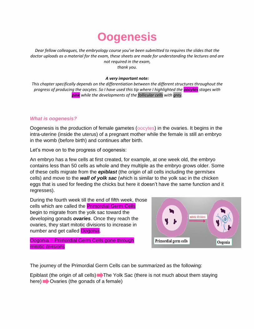
During the fourth week till the end of fifth week, those
cells which are called the Primordial Germ Cells
begin to migrate from the yolk sac toward the
developing gonads ovaries. Once they reach the
ovaries, they start mitotic divisions to increase in
number and get called Oogonia.
Oogonia = Primordial Germ Cells gone through
mitotic divisions
The journey of the Primordial Germ Cells can be summarized as the following:
Epiblast (the origin of all cells) The Yolk Sac (there is not much about them staying
here) Ovaries (the gonads of a female)
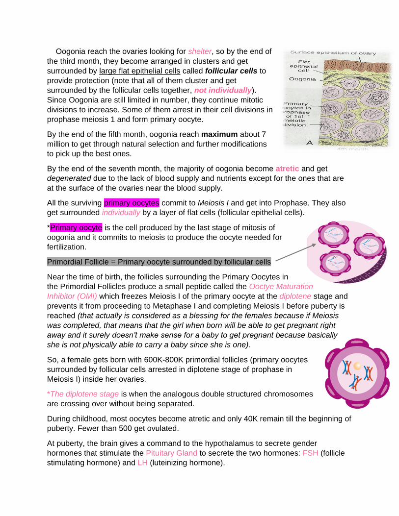
Oogonia reach the ovaries looking for shelter, so by the end of
the third month, they become arranged in clusters and get
surrounded by large flat epithelial cells called follicular cells to
provide protection (note that all of them cluster and get
surrounded by the follicular cells together, not individually).
Since Oogonia are still limited in number, they continue mitotic
divisions to increase. Some of them arrest in their cell divisions in
prophase meiosis 1 and form primary oocyte.
By the end of the fifth month, oogonia reach maximum about 7
million to get through natural selection and further modifications
to pick up the best ones.
By the end of the seventh month, the majority of oogonia become atretic and get
degenerated due to the lack of blood supply and nutrients except for the ones that are
at the surface of the ovaries near the blood supply.
All the surviving primary oocytes commit to Meiosis I and get into Prophase. They also
get surrounded individually by a layer of flat cells (follicular epithelial cells).
*Primary oocyte is the cell produced by the last stage of mitosis of
oogonia and it commits to meiosis to produce the oocyte needed for
fertilization.
Primordial Follicle = Primary oocyte surrounded by follicular cells
Near the time of birth, the follicles surrounding the Primary Oocytes in
the Primordial Follicles produce a small peptide called the Ooctye Maturation
Inhibitor (OMI) which freezes Meiosis I of the primary oocyte at the diplotene stage and
prevents it from proceeding to Metaphase I and completing Meiosis I before puberty is
reached (that actually is considered as a blessing for the females because if Meiosis
was completed, that means that the girl when born will be able to get pregnant right
away and it surely doesn’t make sense for a baby to get pregnant because basically
she is not physically able to carry a baby since she is one).
So, a female gets born with 600K-800K primordial follicles (primary oocytes
surrounded by follicular cells arrested in diplotene stage of prophase in
Meiosis I) inside her ovaries.
*The diplotene stage is when the analogous double structured chromosomes
are crossing over without being separated.
During childhood, most oocytes become atretic and only 40K remain till the beginning of
puberty. Fewer than 500 get ovulated.
At puberty, the brain gives a command to the hypothalamus to secrete gender
hormones that stimulate the Pituitary Gland to secrete the two hormones: FSH (follicle
stimulating hormone) and LH (luteinizing hormone).
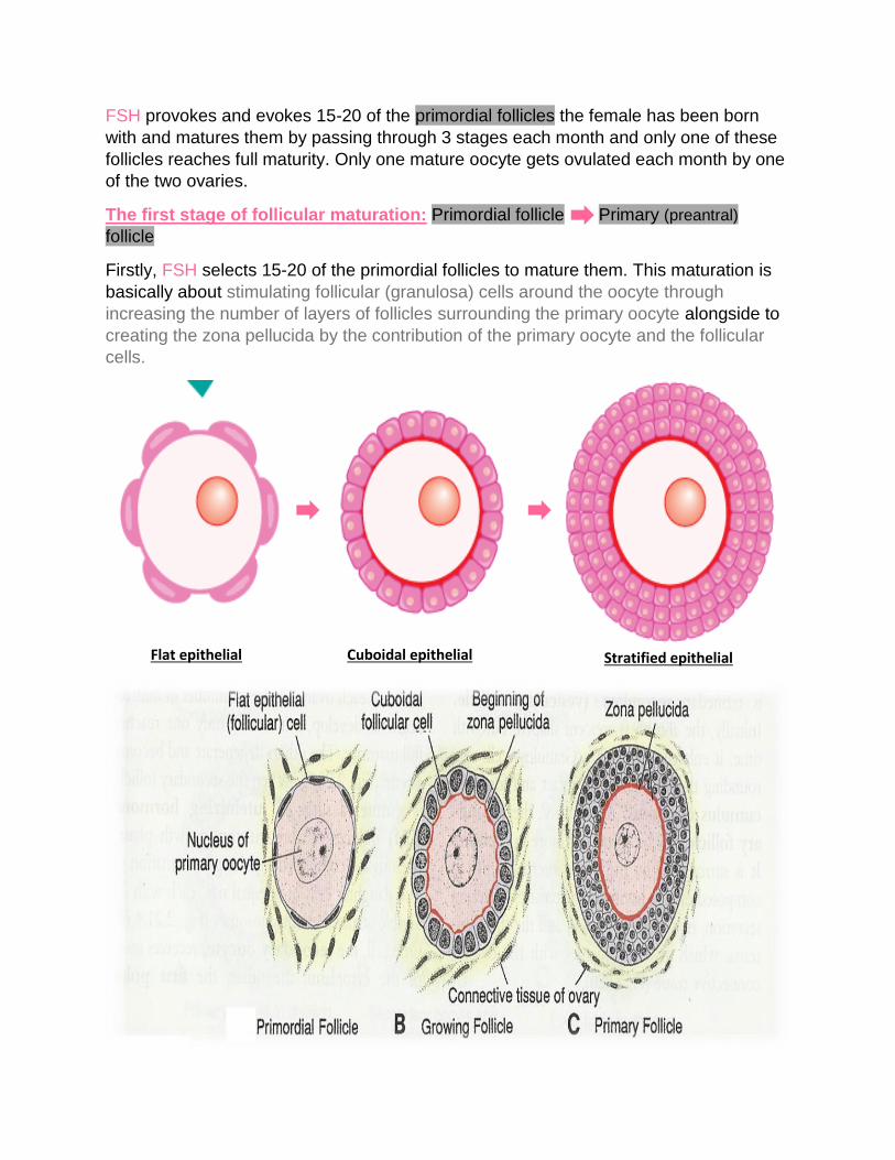
FSH provokes and evokes 15-20 of the primordial follicles the female has been born
with and matures them by passing through 3 stages each month and only one of these
follicles reaches full maturity. Only one mature oocyte gets ovulated each month by one
of the two ovaries.
The first stage of follicular maturation: Primordial follicle Primary (preantral)
follicle
Firstly, FSH selects 15-20 of the primordial follicles to mature them. This maturation is
basically about stimulating follicular (granulosa) cells around the oocyte through
increasing the number of layers of follicles surrounding the primary oocyte alongside to
creating the zona pellucida by the contribution of the primary oocyte and the follicular
cells.
Flat epithelial
Cuboidal epithelial
Stratified epithelial
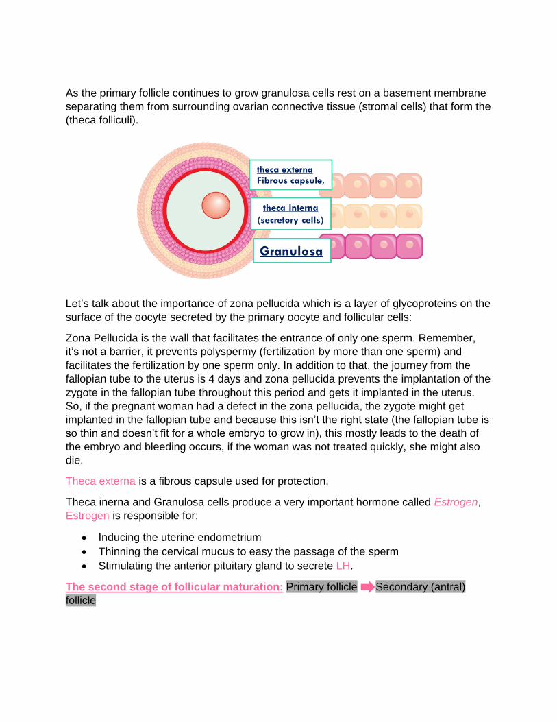
As the primary follicle continues to grow granulosa cells rest on a basement membrane
separating them from surrounding ovarian connective tissue (stromal cells) that form the
(theca folliculi).
Let’s talk about the importance of zona pellucida which is a layer of glycoproteins on the
surface of the oocyte secreted by the primary oocyte and follicular cells:
Zona Pellucida is the wall that facilitates the entrance of only one sperm. Remember,
it’s not a barrier, it prevents polyspermy (fertilization by more than one sperm) and
facilitates the fertilization by one sperm only. In addition to that, the journey from the
fallopian tube to the uterus is 4 days and zona pellucida prevents the implantation of the
zygote in the fallopian tube throughout this period and gets it implanted in the uterus.
So, if the pregnant woman had a defect in the zona pellucida, the zygote might get
implanted in the fallopian tube and because this isn’t the right state (the fallopian tube is
so thin and doesn’t fit for a whole embryo to grow in), this mostly leads to the death of
the embryo and bleeding occurs, if the woman was not treated quickly, she might also
die.
Theca externa is a fibrous capsule used for protection.
Theca inerna and Granulosa cells produce a very important hormone called Estrogen,
Estrogen is responsible for:
Inducing the uterine endometrium
Thinning the cervical mucus to easy the passage of the sperm
Stimulating the anterior pituitary gland to secrete LH.
The second stage of follicular maturation: Primary follicle Secondary (antral)
follicle
theca externa Fibrous capsule,
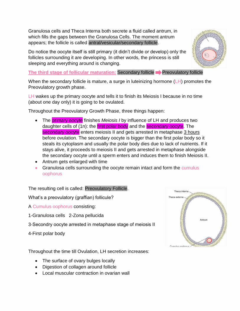
Granulosa cells and Theca Interna both secrete a fluid called antrum, in
which fills the gaps between the Granulosa Cells. The moment antrum
appears; the follicle is called antral/vesicular/secondary follicle.
Do notice the oocyte itself is still primary (it didn’t divide or develop) only the
follicles surrounding it are developing. In other words, the princess is still
sleeping and everything around is changing.
The third stage of follicular maturation: Secondary follicle Preovulatory follicle
When the secondary follicle is mature, a surge in luteinizing hormone (LH) promotes the
Preovulatory growth phase.
LH wakes up the primary oocyte and tells it to finish its Meiosis I because in no time
(about one day only) it is going to be ovulated.
Throughout the Preovulatory Growth Phase, three things happen:
The primary oocyte finishes Meiosis I by influence of LH and produces two
daughter cells of (1n): the first polar body and the secondary oocyte. The
secondary oocyte enters meiosis II and gets arrested in metaphase 3 hours
before ovulation. The secondary oocyte is bigger than the first polar body so it
steals its cytoplasm and usually the polar body dies due to lack of nutrients. If it
stays alive, it proceeds to meiosis II and gets arrested in metaphase alongside
the secondary oocyte until a sperm enters and induces them to finish Meiosis II.
Antrum gets enlarged with time
Granulosa cells surrounding the oocyte remain intact and form the cumulus
oophorus
The resulting cell is called: Preovulatory Follicle.
What’s a preovulatory (graffian) follicule?
A Cumulus oophorus consisting:
1-Granulosa cells 2-Zona pellucida
3-Secondry oocyte arrested in metaphase stage of meiosis ΙΙ
4-First polar body
Throughout the time till Ovulation, LH secretion increases:
The surface of ovary bulges locally
Digestion of collagen around follicle
Local muscular contraction in ovarian wall
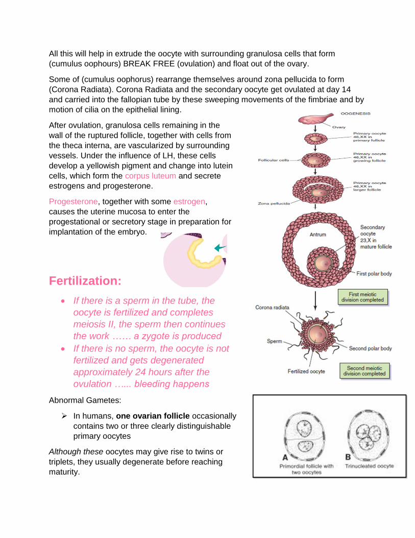
All this will help in extrude the oocyte with surrounding granulosa cells that form
(cumulus oophours) BREAK FREE (ovulation) and float out of the ovary.
Some of (cumulus oophorus) rearrange themselves around zona pellucida to form
(Corona Radiata). Corona Radiata and the secondary oocyte get ovulated at day 14
and carried into the fallopian tube by these sweeping movements of the fimbriae and by
motion of cilia on the epithelial lining.
After ovulation, granulosa cells remaining in the
wall of the ruptured follicle, together with cells from
the theca interna, are vascularized by surrounding
vessels. Under the influence of LH, these cells
develop a yellowish pigment and change into lutein
cells, which form the corpus luteum and secrete
estrogens and progesterone.
Progesterone, together with some estrogen,
causes the uterine mucosa to enter the
progestational or secretory stage in preparation for
implantation of the embryo.
Fertilization:
If there is a sperm in the tube, the
oocyte is fertilized and completes
meiosis II, the sperm then continues
the work …… a zygote is produced
If there is no sperm, the oocyte is not
fertilized and gets degenerated
approximately 24 hours after the
ovulation …... bleeding happens
Abnormal Gametes:
In humans, one ovarian follicle occasionally
contains two or three clearly distinguishable
primary oocytes
Although these oocytes may give rise to twins or
triplets, they usually degenerate before reaching
maturity.
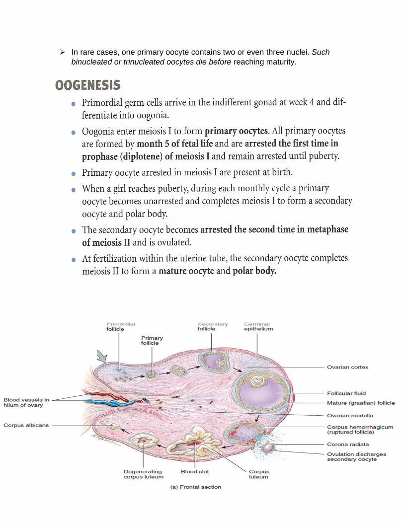
In rare cases, one primary oocyte contains two or even three nuclei. Such
binucleated or trinucleated oocytes die before reaching maturity.
Some conceptual notes:
You may ask yourself why does the secondary oocyte steal the cytoplasm of the
first polar body? Well, that’s because the secondary oocyte is bigger and is the
one that’s going to get fertilized so it prepares itself for the fertilization because
the sperm basically will have no food or mitochondria when it arrives to the
oocyte.
If one oocyte gets ovulated only, why does FSH maturate 15-20 follicles? These
follicles have a really important role in secreting Estrogen and Progesterone (the
functions written upwards). That’s why these follicles grow and through natural
selection, the best oocyte gets ovulated.
The main difference between the female and the male is mainly the fact that the
woman takes pregnancy in addition to producing gametes while the male
produces gametes only.
The male can produce gametes starting from puberty till the moment he dies
(although the fertility decreases with the increase of age) but the woman has got
a finite productive life approx. 35 years (from age 15 till age 45~50), that’s also
because of the fact that she is the one who gets pregnant and only throughout
this time, she is physically able to do that.
A woman produces one oocyte from one of the two ovaries each month, she
produces 10~12 oocytes in the year, about 250 oocytes are produced by each
ovary throughout her life, yet she has 7 million oocytes at the beginning to get
through natural selection and select the best out of the best.
The only time a female has a haploid cell (1n) is the secondary oocyte.
For females who aren’t in a sexual interaction, they never complete Meiosis II.
If the first polar body didn’t die, it completes meiosis and produces 2 secondary
bodies.
If fertilization and the previous point happened, the final produced cells are: 3
secondary polar bodies and one fertilized egg.
Because it may take too long for a female to get her puberty (secretion of LH)
and the chromosomes stay undivided in the diplotene stage, some abnormalities
when dividing may occur such as Monosomy and Trisomy.
The gender of a baby can’t be known until the outer genital parts develop.
The follicles under FSH secrete Estrogen and under LH they secrete
Progesterone.
EXTREMELY IMPORTANT: DO DIFFERENTIATE BETWEEN THE
DIFFERENT STAGES OF OOCYTES AND FOLLICLAR CELLS AND KNOW
THE DIFFERENCES IN STRUCTURE BETWEEN EACH OF THEM BECAUSE
THE DOCTOR SAID IT’S A SURE QUESTION IN THE EXAM. GOOD LUCK!
Done by: Zina Smadi
Corrected by: Sabina Khdairi & Abdulrahman Jabr