Biodegradable Magnesium Alloys Developed as Bone Repair ...
16
Review Article Biodegradable Magnesium Alloys Developed as Bone Repair Materials: A Review Chen Liu, 1,2 Zheng Ren, 2 Yongdong Xu, 2 Song Pang, 2 Xinbing Zhao, 1 and Ying Zhao 3 1 Department of Materials Science and Engineering, Zhejiang University, Hangzhou, China 2 Ningbo Branch of China Academy of Ordnance Science, Ningbo, China 3 Shenzhen Institutes of Advanced Technology, Chinese Academy of Sciences, Shenzhen, China Correspondence should be addressed to Ying Zhao; [email protected] Received 28 July 2017; Revised 3 November 2017; Accepted 5 February 2018; Published 13 March 2018 Academic Editor: Daniele Passeri Copyright © 2018 Chen Liu et al. is is an open access article distributed under the Creative Commons Attribution License, which permits unrestricted use, distribution, and reproduction in any medium, provided the original work is properly cited. Bone repair materials are rapidly becoming a hot topic in the field of biomedical materials due to being an important means of repairing human bony deficiencies and replacing hard tissue. Magnesium (Mg) alloys are potentially biocompatible, osteoconductive, and biodegradable metallic materials that can be used in bone repair due to their in situ degradation in the body, mechanical properties similar to those of bones, and ability to positively stimulate the formation of new bones. However, rapid degradation of these materials in physiological environments may lead to gas cavities, hemolysis, and osteolysis and thus, hinder their clinical orthopedic applications. is paper reviews recent work on the use of Mg alloy implants in bone repair. Research to date on alloy design, surface modification, and biological performance of Mg alloys is comprehensively summarized. Future challenges for and developments in biomedical Mg alloys for use in bone repair are also discussed. 1. Introduction As the largest dynamic biological tissue in the body, bones are composed of inorganic minerals and metabolically active cells surrounded by a large volume of extracellular matrix, and they form a rigid framework that has an irreplaceable role in maintaining life activities, including supporting the body and protecting visceral organs [1, 2]. Surgical treatment of bone injuries has become common, where there are millions of bone injury patients in emergency departments worldwide each year due to involvement in vigorous athletic activities, social instability, traffic accidents, and prolonged human lifespan [3–5]. Bone defects, mainly induced by traumatic avulsions, sequelae of infection-induced bony sequestration, congenital malformations, or neoplastic resections, confront us with an extreme challenge for reconstructive surgery. e need to induce bone regeneration to repair structural bone deficiencies has inspired research on and development of a vast number of bone repair materials [2, 6]. Bone repair is a physiological process influenced by a variety of biomechanical, biochemical, cellular, hormonal, and pathological factors. Continuous bone deposition, resorption, and remodeling and sufficient blood supply promote bone repair [7]. Based on the basic principles of bone tissue healing, different bone repair materials have been developed. For a long time, autograſt bones have been considered the gold standard of bone repair materials when replacing damaged or lost bones because they have all the characteristics necessary to stimulate new bone growth of osteoconductivity, osteogenicity, and osteoinductivity. However, resources for these autograſts are scarce and sec- ondary surgeries increase the pain experienced by patients. Furthermore, donor-site complications can occur, clinical benefits are not guaranteed, and there is a high rate of asso- ciated complications [4, 8, 9]. A large number of alternative bone repair materials have been increasingly used to replace autograſt bones and are commercially available as bone substitutes. e most commonly used products are composed of calcium (Ca) phosphate ceramics, Ca sulfate, bioactive glass, natural materials, and biological/synthetic composites [10–15]. However, the clinical performance of these materials is unsatisfactory. For example, some have poor mechanical Hindawi Scanning Volume 2018, Article ID 9216314, 15 pages https://doi.org/10.1155/2018/9216314
Transcript of Biodegradable Magnesium Alloys Developed as Bone Repair ...

Review ArticleBiodegradable Magnesium Alloys Developed asBone Repair Materials: A Review
Chen Liu,1,2 Zheng Ren,2 Yongdong Xu,2 Song Pang,2 Xinbing Zhao,1 and Ying Zhao 3
1Department of Materials Science and Engineering, Zhejiang University, Hangzhou, China2Ningbo Branch of China Academy of Ordnance Science, Ningbo, China3Shenzhen Institutes of Advanced Technology, Chinese Academy of Sciences, Shenzhen, China
Correspondence should be addressed to Ying Zhao; [email protected]
Received 28 July 2017; Revised 3 November 2017; Accepted 5 February 2018; Published 13 March 2018
Academic Editor: Daniele Passeri
Copyright © 2018 Chen Liu et al.This is an open access article distributed under the Creative Commons Attribution License, whichpermits unrestricted use, distribution, and reproduction in any medium, provided the original work is properly cited.
Bone repair materials are rapidly becoming a hot topic in the field of biomedical materials due to being an importantmeans of repairing human bony deficiencies and replacing hard tissue. Magnesium (Mg) alloys are potentially biocompatible,osteoconductive, and biodegradable metallic materials that can be used in bone repair due to their in situ degradation in the body,mechanical properties similar to those of bones, and ability to positively stimulate the formation of new bones. However, rapiddegradation of these materials in physiological environments may lead to gas cavities, hemolysis, and osteolysis and thus, hindertheir clinical orthopedic applications. This paper reviews recent work on the use of Mg alloy implants in bone repair. Researchto date on alloy design, surface modification, and biological performance of Mg alloys is comprehensively summarized. Futurechallenges for and developments in biomedical Mg alloys for use in bone repair are also discussed.
1. Introduction
As the largest dynamic biological tissue in the body, bonesare composed of inorganic minerals and metabolically activecells surrounded by a large volume of extracellular matrix,and they form a rigid framework that has an irreplaceable rolein maintaining life activities, including supporting the bodyand protecting visceral organs [1, 2]. Surgical treatment ofbone injuries has become common, where there are millionsof bone injury patients in emergency departments worldwideeach year due to involvement in vigorous athletic activities,social instability, traffic accidents, and prolonged humanlifespan [3–5]. Bone defects, mainly induced by traumaticavulsions, sequelae of infection-induced bony sequestration,congenital malformations, or neoplastic resections, confrontus with an extreme challenge for reconstructive surgery. Theneed to induce bone regeneration to repair structural bonedeficiencies has inspired research on and development of avast number of bone repair materials [2, 6].
Bone repair is a physiological process influenced by avariety of biomechanical, biochemical, cellular, hormonal,
and pathological factors. Continuous bone deposition,resorption, and remodeling and sufficient blood supplypromote bone repair [7]. Based on the basic principles ofbone tissue healing, different bone repair materials havebeen developed. For a long time, autograft bones have beenconsidered the gold standard of bone repair materials whenreplacing damaged or lost bones because they have all thecharacteristics necessary to stimulate new bone growthof osteoconductivity, osteogenicity, and osteoinductivity.However, resources for these autografts are scarce and sec-ondary surgeries increase the pain experienced by patients.Furthermore, donor-site complications can occur, clinicalbenefits are not guaranteed, and there is a high rate of asso-ciated complications [4, 8, 9]. A large number of alternativebone repair materials have been increasingly used to replaceautograft bones and are commercially available as bonesubstitutes.Themost commonly used products are composedof calcium (Ca) phosphate ceramics, Ca sulfate, bioactiveglass, natural materials, and biological/synthetic composites[10–15]. However, the clinical performance of these materialsis unsatisfactory. For example, some have poor mechanical
HindawiScanningVolume 2018, Article ID 9216314, 15 pageshttps://doi.org/10.1155/2018/9216314

2 Scanning
properties and display limited osteoinduction in the clinic[16, 17]. Metallic materials are another alternative for use inthe repair or replacement of diseased or damaged bone tissue.Metallic materials currently widely used in orthopedicsinclude stainless steel and titanium alloys because theyare mechanically strong and resistant to fracture [18–21].However, there is a potential for the release of metallic ionsand/or particles through corrosion and/or wear that triggerinflammatory responses that can reduce biocompatibilityand lead to tissue loss. Furthermore, the elastic moduli andtensile strength of metals and bone are significantly different,which can cause stress shielding and result in weakening ofsurrounding bone. These inert implants also often need tobe removed via invasive secondary surgeries once the bonefracture has completely healed. To minimize trauma to thepatients and decrease medical costs, biodegradable implantscould be used to replace traditional metal implants andremove the need for secondary surgeries [22–26].
Magnesium (Mg) alloys have a reputation for being revo-lutionary biodegradable metal materials in orthopedic appli-cations due to their good biocompatibility, biodegradability,and acceptable mechanical properties [27–30]. The fourthmost plentiful cation in the human body, Mg is an elementessential in manymetabolic processes and is primarily storedin bone tissue. Mg is taken into the body daily in substantialamounts, stimulates the growth of bone cells, and acceleratesthe healing of bone tissue. Mg alloys are degraded in vivodue to the presence of Cl− in the physiological environment,thereby eliminating the need for secondary surgeries toremove the implant. Mg2+, a corrosion product of Mg alloyimplants, does not cause unexpected complications becauseexcessive Mg cations are easily eliminated in the urine [31–34]. Moreover, Mg alloys have mechanical properties similarto those of bone. Mg alloys are lightweight with densities(1.7–1.9 g/cm3) very similar to those of human cortical bone(1.75 g/cm3), unlike titanium alloys (Ti-6Al-4V 4.47 g/cm3)and stainless steel (about 7.8 g/cm3). The elastic modulusof Mg alloys, about 45GPa, is relatively close to that ofnatural bone, 3–20GPa, compared to the elastic moduli oftitanium alloys and stainless steel (110 and 200GPa, resp.).Therefore, the stress shielding from the notable mechanicalmismatch between natural bone and metal implants shouldbe mitigated [35–37]. Therefore, Mg alloys are expected tobecome biocompatible, biodegradable, lightweight, and load-bearing orthopedic implants [22, 38–40].
While research on Mg alloys as bone implants has led tosignificant progress over the past 20 years, rapid degradationof these materials inside the human body is still a majorobstacle hampering their use in the clinic. As biodegradablematerials, it is important that the rate of implant degradationmatches the rate of healing of the bone tissue, which generallyconsists of an early inflammatory stage lasting from 3 to7 days, a reparative stage that leads to a strong healingunion lasting about 3-4 months, and then a remodelingphase that can last months to years [41–43]. Therefore, itis necessary for the implant to remain stable for at least12 weeks [22]. However, the currently available Mg alloysdegrade too quickly to hold well during implantation. Thisfast degradation results in the formation of hydrogen gas
cavities, rapid loss of mechanical integrity of the implants,and adverse host tissue reactions, such as local swelling andsignificant pain within the first week after surgery [44–46].
There have been a number of recent opportunities andchallenges in the development of Mg alloys for use in bonerepair. Therefore, it is necessary to summarize the findings ofthe researchers in this field. Compared to recently publishedreviews [27, 47–53], this paper is more targeted and specif-ically discusses biodegradable Mg alloys to be used in bonerepair. We review the alloying design, surface modifications,and the in vitro and in vivo biological performance of Mg inbone repair. Novel insights that have been used to improvethe compatibility and reliability of biomedical Mg alloys inthe bone reconstruction field are also discussed.
2. Alloying Design of Magnesium Alloys
Adequate strength, ductility, fatigue resistance, and biocorro-sion resistance are important characteristics for biodegrad-able implants to be used in orthopedic applications. Becauseadding alloying elements can improve mechanical propertiesand decrease the corrosion rate of Mg by modifying thestructure and phase distribution, several Mg alloys have beendesigned to meet the requirements of bone repair implantmaterials [30, 32, 60].
2.1. Alloying Elements. Careful selection of alloying elementsis the first step in designing Mg alloys. To strengthen Mg-based materials, adding elements such as Al, Zn, Ca, Ag, Ce,and Th can generate different microstructures and improvethe mechanical properties of the resulting Mg alloy [71–74].In terms of corrosion, alloying elements that have electro-chemical potentials similar to that of Mg (−2.37V), such as Y(−2.37V),Nd (−2.43V), andCe (−2.48V), and have relativelyhigh solid solubility in Mg, such as Sc (25.9 wt.% limit), Gd(23.5 wt.% limit), and Dy (25.3 wt.% limit), can enhance thecorrosion resistance by reducing internal galvanic corrosionin physiological environments [37, 75, 76]. Biocompatibilityalso needs to be considered. Previous reports have shownthat biological nutrients (e.g., Ca, Sr, Zn, Si, and Mn) andtrace nontoxic elements (e.g., Zr, Nd, and Y) added eitherindependently or together to the Mg matrix do not causedetrimental local tissue responses and can be easily absorbedby surrounding tissues [29, 30, 35, 77–80]. With the devel-opment of biodegradable Mg alloys, researchers have startedtrying to endow Mg alloys with new biomedical functionsthrough alloying. Ca, Sr, Ag, and Cu as biofunctional tracemetallic elements have been confirmed to promote bone cellactivation and stimulate new bone formation. In addition topromoting osteogenesis, these elements also inhibit bacterialinfection after implantation, thereby effectively decreasingmorbidity andmortality, bymaking the environment alkalineand releasing antimicrobial metallic ions [81–86].
2.2. Alloy Systems. Due to having a combination of goodmechanical properties and corrosion resistance, some com-mercialMg alloy systems have been selected as biodegradableMg alloys at an early stage. Commercial Mg alloys used inbiological research include theAZ (Mg-Al-Zn),WE (Mg-RE-Zr), and ZK (Mg-Zn-Zr) series alloys.
Scanning 3
AZ series alloys, particularly AZ31 (Mg-3Al-1Zn) andAZ91 (Mg-9Al-1Zn) alloys, have been extensively studiedboth in vitro and in vivo in recent years [46, 87–89]. It hasbeen reported that AZ31 and AZ91 alloys release hydrogenupon degradation in physiological environments, leading toa significant increase in both pH and Mg ion concentration[90]. In Hank’s solution, the AZ31 alloy degrades more slowlythan the AZ91 alloy, but there is no significant difference invivo [91, 92]. Short-term in vivo studies of AZ31 and AZ91alloys have also revealed that a biocompatible Ca phosphateprotective film layer covers their surfaces and increases theformation of new bone mass around the implants [92, 93].
WE series alloys have good biocorrosion resistancebecause they form a rare-earth (RE) oxide film in aqueousenvironments. It has been reported that WE54 (1.58 Nd,4.85 Y, 0.28 Zr, 0.08 Ce, 0.13 Gd, 0.16 Er, 0.13 Yb, andbalanced Mg in wt.%) has marginally higher resistanceto degradation in vitro than pure Mg and heat treatmentimpacts its degradation [94]. Witte et al. analyzed the in vivodegradation of four different Mg alloys and confirmed thatWE43 (4.16 Y, 3.80 RE, 0.36 Zr, 0.20 Zn, and 0.13 Mn, all inwt.%) has good biocompatibility [93]. However, an increasein Al ion concentration in the brain is associated with theoccurrence of Alzheimer’s disease and severe hepatotoxicityhas occurred after the administration of RE elements, such asY, Ce, and Pr [6].
Recently, ZK series alloys, especially ZK40 (Mg-4Zn-0.5Zr) and ZK60 (Mg-6Zn-0.5Zr), have attracted the atten-tion of researchers because of the good biocompatibility ofthe component elements [95–97]. A daily intake of 11mg Znand 50 𝜇g Zr is permissible, so Mg-Zn-Zr alloys are moreattractive than Mg-Al-Zn and Mg-RE-Zr alloys in terms ofelement biocompatibility and biosafety and are candidatebiodegradable metals for use in bone repair devices [25].However, the extremely high rates of degradation of Mg-Zn-Zr alloys are alarming and restrict their future development.
In addition to the above commercial Mg alloy systems,newMg alloys have also been developed for use in orthopedicapplications, including Mg-Ca, Mg-Sr, Mg-Zn, and Mg-REalloy systems.
Ca, acting as a grain-refining agent in Mg alloys, canstabilize grain size at levels up to 0.5% of the Ca contentand cause slight decreases with further addition [98]. As amajor component of human bone, Ca is essential for bonecell signaling and beneficial to bone healing. It has beenreported that Mg-1Ca alloy does not induce cytotoxicityand osteoblasts and osteocytes are highly active aroundMg-1Ca alloy pins implanted in rabbit femoral shafts, thusdemonstrating good biocompatibility and bioactivity [84].
Strontium (Sr) and Ca belong to the same family andhave similar physical and chemical properties and biologicalfunctions. Brar et al. studied Mg-𝑥wt.% Sr (𝑥 = 0.5, 1.0, and1.5 wt.%) alloys and found that the Mg-0.5Sr alloy degradedthe slowest [35]. Zhao et al. and Gu et al., respectively,reported that the as-extrudedMg-0.5Sr and as-rolledMg-2Sralloys had the best combination of corrosion resistance, highstrength, and in vivo biocompatibility [86, 99].
Zinc (Zn) is one of the most abundant essential nutrientsin the human body and is safe for use in biomedical
Table 1: Common second phases of select biodegradable Mg alloys.
Biodegradable magnesiumalloys
The second phases in magnesiummatrix
AZ31B [61], AZ61D [62] Mg17Al12
AZ91D [63, 64] Mg17Al12, Al8Mn5
Mg-Ca [65] Mg2Ca
Mg-Sr [4, 65] Mg17Sr2, Mg2Sr
Mg-Zn [66] MgZn2
Mg-Zn-Ca [67] Mg2Zn3
Mg-Si [66] Mg2Si
Mg-Al-Si [68] Mg2Si
WE43 [69] Mg24Y5, Mg41Nd5, Mg12Nd
ZK60 [70] MgZn, MgZn2
applications [100]. The rate of Mg corrosion can be reducedby increasing the mass fraction of Zn mixed with Mg,thus strengthening the mechanical properties of Mg throughsolid solution hardening [101]. Cai et al. reported that a Zncontent of up to 5wt.% in Mg-Zn binary alloys exhibits grainboundary, solid solution, and secondary phase strengthening,resulting in improved resistance to corrosion andmechanicalproperties [102]. Mg-6Zn alloy has good biocompatibility invitro based on hemolysis and MC3T3-E1 cell adhesion assays[103].
Because Mg-RE alloys have good mechanical propertiesand corrosion resistance, new Mg-RE alloys, such as Mg-Y, Mg-Nd, Mg-Gd, Mg-Ce, and Mg-Ld, have been studied.Among these, Mg-Nd alloy has a much slower corrosionrate than the other alloys [74]. Mg-Y alloy was preparedusing a zone solidification method and improved corrosionresistance and mechanical properties [104]. Mg-Y-Zn alloycontains an interesting combination of preferred microstruc-tural, mechanical, electrochemical, and biological properties,making it very promising for use as a biodegradable implantmaterial [105].
2.3. Alloy Microstructures. Alloying elements in Mg alloysmay exist in the form of second-phase particles and precip-itate in grains or grain boundaries, substantially enhancingmechanical properties through second-phase strengthening.
Figure 1 presents the typical morphologies of secondphases for Mg alloys and Table 1 presents the second phasesof biodegradable Mg alloys. Compared to Mg matrix, secondphases have higher potentials and may facilitate corrosion,leaching into the physiological environment accompaniedwith the degradation of the matrix. Kannan investigatedthe degradability of Mg
17Al12
phase in simulated body fluid(SBF) using electrochemical measurements and found thatthe degradation rate of Mg
17Al12was lower than that of bare
Mg. Our previous study demonstrated that pitting corrosionoccurs with crackings for Mg
17Al12phase in Hank’s solution
and degrades much slower than AZ31 alloy and pure Mg[106].
When assessing Mg alloy implants for use in bone repair,the stability of second phases and Mg matrix under different

4 Scanning
20 G
(a)
(a)
10 G
(b)
(b)
10 G
(c)
(c)
50 G
(d)
(d)
Figure 1: Typical morphologies of second phases in (a) as-cast ZE41, (b) as-cast WE43, (c) as-forged WE43 [54], and (d) AZ91D alloys [55].
conditions may have significantly influenced degradationand biological responses to the implant in the body. Yanget al. theoretically investigated the thermodynamic stabilityof four conventional second phases for Mg-Zn-Zr, Mg-Ca, Mg-Sr, and Mg-Al-Zn alloys, as well as Mg matrix inbioabsorbable Mg alloys, via the Dmol3 calculation method.The second phases had higher phase stability thanMgmatrix,but the phase stability was quite different for different typesof second phases and second-phase-4H
2O systems [71]. In
order to evaluate the effect of second phases on the bio-logical safety of biodegradable Mg alloy implants, Mg
17Al12
second phase from Mg-Al-Zn alloys was investigated forin vitro biocompatibility and phagocytosis by macrophages.Mg17Al12
second phase did not induce hemolysis and hadexcellent cytocompatibility. Mg
17Al12particles are processed
in endolysosomal compartments and lysosomes play a majorrole in digesting Mg
17Al12particles [107].
However, not all the alloying elements in Mg alloysform second-phase particles. Asmentioned above, some alloyelements have relatively high solid solubility in Mg, such asY (12 wt.% limit), Sc (25.9 wt.% limit), Gd (23.5 wt.% limit),and Dy (25.3 wt.% limit), and can exist in the form of solidsolutions, thus achieving solid solution strengthening. In thesolution, the original crystal structure of magnesium remainsunchanged, but a lattice distortion is produced and thus themotion of dislocations becomes impeded, which leads tothe enhancement of strength of Mg. Gao et al. explored the
effects of solid solutions on themechanical behavior of binaryMg-Y single-phase alloys. They found enhanced hardnessas the Y content increased at room temperature becauseof large differences in the atomic radii of Y and Mg anda relatively wide range of solubilities [108]. Moreover, solidsolution alloying also potentially affects degradation of Mgalloys by improving corrosion resistance by reducing internalgalvanic corrosion between the second phase andMgmatrix.Zhang et al. studied the effect of solid solution treatment onthe corrosion and electrochemical behaviors of Mg-15Y alloyand found that solution treatment decreased the extent ofgalvanic corrosion due to the dissolution of Mg
24Y5second
phase into the matrix [109]. Therefore, solid solution mightbe a feasible alternative for generating a single-phaseMg alloyand can help improve the corrosion resistance ofMg alloys inorthopedic applications.
2.4. Impurities in Magnesium Alloys. During casting andrefining, magnesium always introduces superfluous amountsof impurity elements. Impurity elements in Mg alloys usuallyinclude iron (Fe), nickel (Ni), and copper (Cu) [66, 110].Theseelements can significantly accelerate Mg corrosion whentheir concentrations exceed the limits of tolerance [111–113].Standards for Mg impurity elements are 35–50 ppm for Fe,20–50 ppm forNi, and 100–300 ppm for Cu (wt.%). Below thetolerance limits, no impurity particles are formed and, thus,no electrochemically active cathodic sites exist to accelerate

Scanning 5
5 G
(a)
MgO
(a)
5 G
(b)
MgO
Pore
(b)
10 G
(c)
MgO
(c)
20 G
(d)
MgO
(d)
20 G
(e)
MgO
(e)
20 G
(f)
MgO
(f)
Figure 2: Scanning electronmicroscope (SEM) images ofMgO inclusions inMg-Gd-Y-Zr: (a) Z-shaped, (b) spherical, (c) block, (d) rod-like,(e) needle-like, and (f) lamellar MgO [56].
corrosive attack, which keeps the corrosion rate very slow.When levels are above the tolerance limits, Fe, Ni, and Cuin Mg alloys significantly increase the corrosion rate dueto the low solubility of these elements and their distinctlymore noble position in the electrochemical series [66]. Atrenset al. found that impurity elements notably accelerate salt-water corrosion ofMg binary alloys [114]. Recent studies haveshown that adding silicon (Si) to the reactive impurity ele-ments Fe, Ni, and Cu is detrimental to corrosion, as it plays acritical role in promoting the formation and growth of Fe-richparticles. Lee et al. suggested that corrosion of Mg is depen-dent on the content ratio of impurities, such as the Fe/Mnratio, rather than their absolute content. As the Fe/Mn ratioincreases, the high rate of corrosion stage extends [5]. In addi-tion to accelerating corrosion, excessive impurity elementsare also harmful to biocompatibility. For example,Ni leachinginto the body has toxic biological effects and high levels ofCu exert a toxic effect at cell surfaces [115]. In order to reduceimpurity during casting and refining, the crucible, stirrer, andmold containing no such elements are prudently utilized [31].
As the chemical properties of Mg alloys are very active,a large amount of nonmetallic inclusions is also producedduring casting and refining which act as additional majorimpurities in Mg alloys [116]. The main nonmetallic inclu-sions include MgO, Mg
3N2, MgF
2, MgS
2, and AlF
3. These
nonmetallic impurities primarily come from the oxidationof Mg alloys in ambient atmospheres. For example, MgO, acommon Mg alloy inclusion, is produced when Mg and O
2
react in the air. Figure 2 illustrates the different morphologies
of MgO impurities in Mg-Gd-Y-Zr alloy [56]. Mg3N2is
attributed to Mg and N2combining in the air. When Mg
alloys smelt under the protection of SF6gas, MgF
2and MgS
inclusions may form from reactions between SF6and liquid
Mg. As the nonmetallic impurities significantly reduce thecastability, mechanical properties, and corrosion resistance ofMg alloys, purification technology is undergoing continuousdevelopment [117]. The common methods of purifying Mgalloys include gas purge, flux purification, filtering purifi-cation, RE purification, and electromagnetic purificationmethods [116].
3. Surface Modifications of Magnesium Alloys
In order to efficiently improve the corrosion resistance ofMg alloys in physiological environments, as well as main-tain their mechanical integrity and ameliorate interfacialbiocompatibility, various surface modifications have beendeveloped. Distinct from alloying techniques, surface mod-ifications directly insulate Mg alloys from the surroundingbiological environment and prevent the penetration of bodyfluid into substrates [100, 118, 119]. Based on whether a newphase is generated on the surface of the Mg alloys, themethods of surface modification can be classified into threecategories: chemical modifications, physical modifications,and a combination of these two methods [120].
3.1. Chemical Modifications. Chemical modifications aredefined as new phases covering the surface of Mg alloys
6 Scanning
(a)
(a)
(b)
(b)
(c)
(c)
(d)
(d)
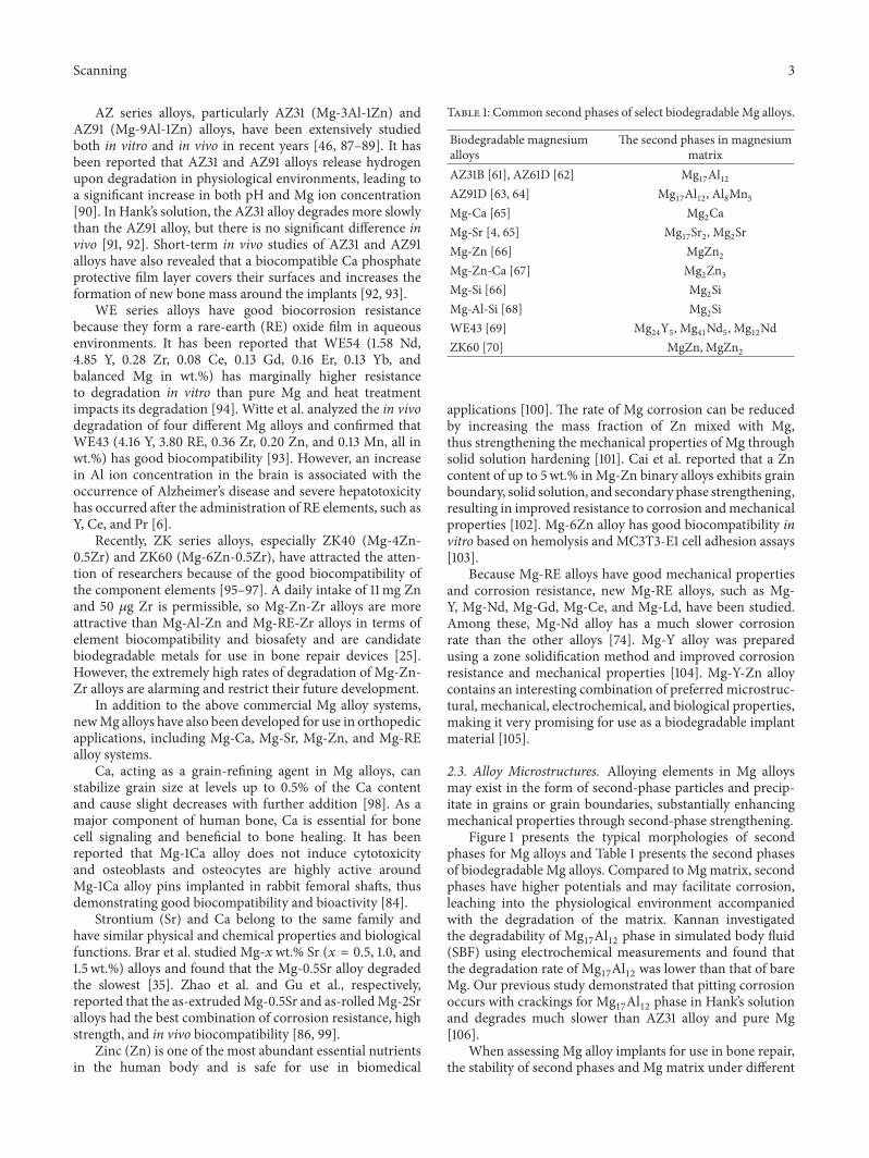
Figure 3: Surface morphologies of ZK60 alloy coated by microarc oxidation at voltages of (a) 230V, (b) 300V, (c) 370V, and (d) 450V [29].Some microcracks can be found on the 230V coating, as marked by the black arrow in (a).
that are synthesized through chemical or electrochemicalreactions.Thismethod removes the native oxide layer that hasfewer passive properties, such as an inability to efficiently pro-tect against corrosion, but forms easily due to the high reac-tivity ofMgmatrix. Chemicalmodifications generally includeacid etching, alkaline heat treatment, fluoride treatment,anodic oxidation, and microarc oxidation (MAO) [120].
Acid etching is a pretreatment method commonly usedto remove the coarse scale produced during manufacturingand replace the native oxide layer with a more compactpassivated layer [121]. Turhan et al. reported that acid etchingwith a 2.5% H
2SO4solution greatly enhances the resistance
of AZ91D alloys to degradation [122]. In addition, alkalineheat treatment, a simple and economical method, creates aMg(OH)
2barrier layer on substrate surface that slows down
the corrosion rate of Mg alloy [123]. It has been reportedthat the corrosion rate of Mg is decreased through NaOHtreatment, where an NaOH concentration of 1M leads to theslowest corrosion rate, through the formation of a protectivelayer [123, 124]. Fluoride treatment of Mg alloys replaces theoriginal oxide film with a thin and more homogeneousMgF
2
layer with higher polarization resistance. The advantages ofthe MgF
2layer include a high density, low water solubility,
and nontoxicity when fluorine ions are released into thehost organism. Witte found out that MgF
2coating slows in
vivo corrosion of LAE442 alloy without observably elevatingfluoride concentrations in the adjacent bone [125]. Moreover,fluoride can stimulate osteoblast proliferation, increase new
mineral deposition in cancellous bones, and decrease the sol-ubility of bone tissue upon incorporation into the bone [88].An experimental study in dogs found that fluoride-modifiedimplant surfaces promote osteointegration during the earlyphase of healing following installation of the implant [126].
Anodic oxidation is an electrochemical process that pro-duces a thick and stable oxide film onmetals. Lei et al. createdan Mg oxide film on AZ31B Mg alloy by anodic oxidationat a constant current. This film efficiently delays degradationof AZ31B Mg alloy without having any adverse effects onosteoblast proliferation or new bone formation [127]. MAO isa high-voltage plasma-assisted anodic oxidation process thatis widely employed to modify the surface of biodegradableMg alloys. MAO coatings are very hard and have goodwear resistance, moderate corrosion resistance, and betterthermal stability and dielectric properties [128]. Lin et al.prepared forsterite-containing MAO coatings on ZK60 Mgalloy to slow down degradation and improve the biologicalproperties of the alloy. It was found that the resistance tocorrosion from theMAOcoating increased as the preparationvoltage increased. Compared to bare ZK60 Mg alloy, MAO-coated ZK60 has a dramatically lower hemolytic ratio andno cytotoxicity to L929 cells. Figure 3 presents the surfacemorphologies of ZK60 alloy with MAO coatings generatedat different voltages [29].
3.2. Physical Modifications. Different from the chemicalmethods, no chemical bonds were formed between the

Scanning 7
surface and the substrates for physical modifications. Themodifications aim to offer a physical barrier to improve thecorrosion resistance of magnesium substrates. The physicalmodifications can be performed by introducing apatite coat-ings, polymer coatings, laser surface processing, or cold spraycoatings [120, 129].
Apatite is a main inorganic component of natural bone. Itcan remarkably promote the recovery of bone fracture due toits excellent bioactivity. Besides, apatite also could improvethe degradation resistance of implants as a protective layerdue to its relatively low solubility and high thermal stability[130].
As one important member of the apatite family, hydrox-yapatite (HA) shows the closest chemical composition withbone mineral and is widely used to coat magnesium alloysfor bone repair [120]. Wang et al. developed an HA coatingon ZK60 Mg alloy with HA and found that it prevented thedegradation of the alloy and increased cytocompatibility forL929 cells, rendering ZK60 alloymore suitable for orthopedicapplications. In addition, no significant deterioration incompression strengthwas noted in the coated alloy comparedto the uncoated one [131].
Polymer coatings are also promising Mg alloy modi-fications for use in orthopedic applications. Gray-Munroet al. explored the influence of polymer coating on thecorrosion rate of AZ31 Mg alloy in SBF using PLA, whichis a semicrystalline biodegradable polymer, and found thatthe coating prevented corrosion, especially during the earlystages of implantation [90].
Laser surface processing, which uses a high-energy laserbeam, has also been employed to regulate biodegradationof Mg alloys and has been found to cause secondary phasedissolution and create a fine grained structure. Coy et al.found significant dissolution of the second phase ofMg
17Al12
in AZ91D when using laser surface processing [132]. Similarresults were reported by Guo et al. and Khalfaui et al. forWE43 and ZE41 alloys using laser processing [133, 134].Appreciable improvements in resistance to corrosion havealso been observed for the aforementioned modified alloys[135].
Cold spray technology is a viablemethod for surface engi-neering of Mg alloys. The deposition of cold spray coatingsinvolves ballistic impingement of particles, usually rangingin size from 1 to 100 𝜇m, accelerated by a high-velocity gasstream and sprayed towards the substrate surface. A lowtemperature process, cold spray is particularly suitable forthe deposition of bioactive coatings on Mg alloys, making itpossible to depress oxidation and phase transformation of thesubstrate. Noorakma et al. recently studied the deposition ofHA on an AZ51 alloy using a modified cold spray process andfound that this modification helped retain the characteristicsof HA. Immersion in SBF for up to 14 days revealed thatHA-coated AZ51 alloy was bioactive and facilitated apatiteformation [136].
3.3. Chemical and Physical Modifications. Considering thelimitations of single chemical and physical treatments, com-posite modifications that involve both chemical and physicaltreatments have been gaining increasing attention. It has been
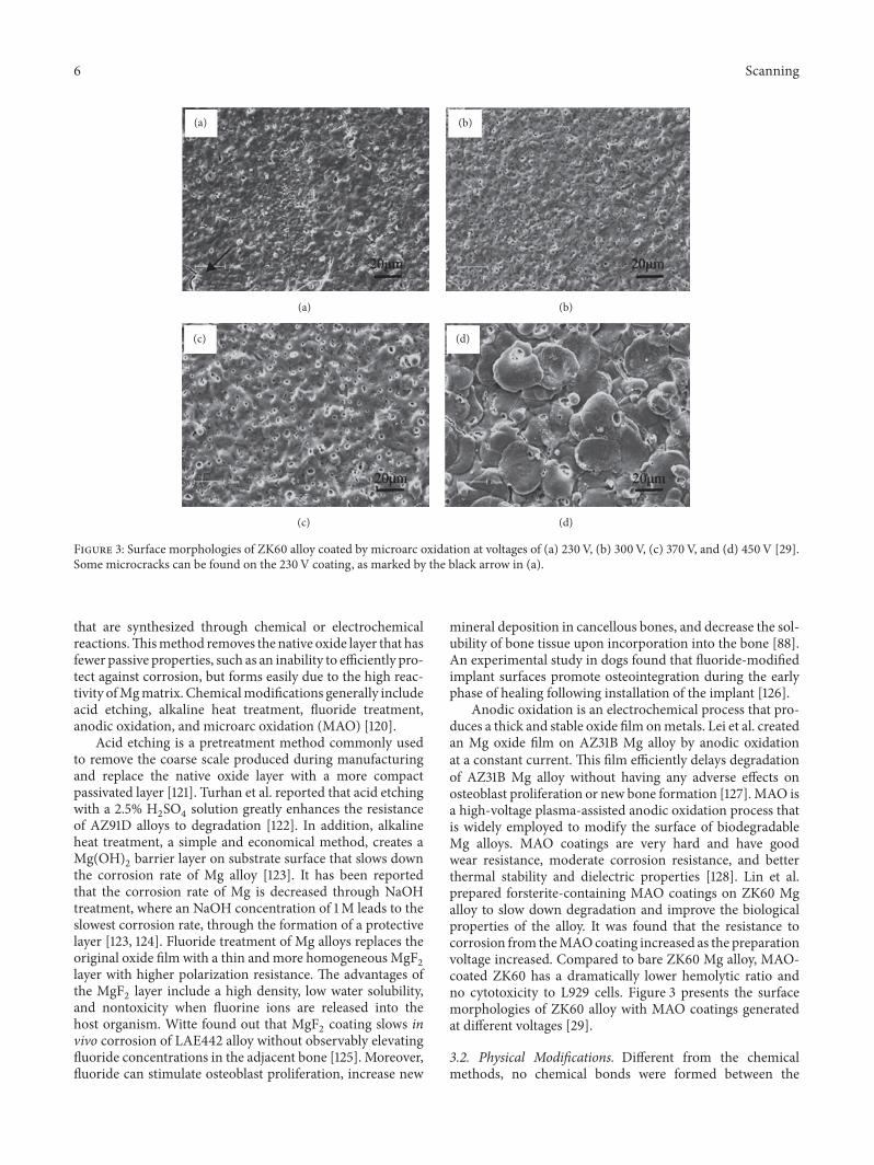
reported that double-modified layers effectively improvebiodegradation resistance of substrates and control degra-dation rates over a larger range [120]. Guo et al. fabricatedan MAO/poly-L-lactic acid (PLLA) composite coating onWE42 alloy surfaces by sealing PLLA to the MAO coatingthrough physical interlocking. This MAO/PLLA-modifiedWE42 alloy was found to have good corrosion resistance andcytocompatibility. Figure 4 presents the surface morpholo-gies of WE42, WE42-MAO, and WE42-MAO/PLLA beforeand after being submerged in Hank’s solution for four days[57]. As shown in Figures 4(a) and 4(d), WE42 Mg alloywas severely corroded by Hank’s solution. The surface of theWE42 experienced strong corrosion as shown in Figure 4(d)based on deeper and wider cracks and holes, as well as thedeposition of white flocculent accumulations. Microporesandmicrocracks were randomly distributed on the surface ofthe MAO coating (Figure 4(b)). After submersion, the MAOcoating was corroded with little white flocculent deposits onthe surface (Figure 4(e)). The biocompatible PLLA sealinglayer was smooth and uniform, overlaying cracks and poreson the surface of the MAO coating (Figure 4(c)). As shownin Figure 4(f), there were no notable changes to the surface oftheMAO/PLLA, where the surface of theWE42-MAO/PLLAsample remained covered with an intact layer that displayedno signs of corrosion.
4. Biological Performance of BiodegradableMagnesium Alloys as Bone Implants
It is critical for biodegradable Mg alloys to have goodbiocompatibility in the body in order to be used in theclinic [130]. Therefore, the in vitro and in vivo biologicalperformance of biodegradable Mg alloys has been examinedfor many years [137].
4.1. In Vitro Biological Performance. In vitro experimentscan be used to simulate and predict corrosion and bio-compatibility of Mg alloys in vivo [138]. Compared to invivo experiments, in vitro experiments are more convenientand can provide quick and reasonable feedback concerningefficacy [139]. Gu et al. studied the in vitro corrosion andbiocompatibility of nine binary Mg-1X (wt.%, X = Al, Ag, In,Mn, Si, Sn, Y, Zn, and Zr) alloys using SEM, X-ray diffraction,tensile tests, immersion tests, electrochemical corrosion tests,cell culture, and platelet adhesion. The addition of alloyingelements influenced the strength and corrosion resistance ofMg. Al, Si, Sn, Zn, and Zr improved the strength ofMg, whileAl, In, Mn, Zn, and Zr slowed down corrosion of as-cast Mg-X alloys in both SBF and Hank’s solutions. Conversely, Siand Y negatively impacted Mg corrosion. Cytotoxicity assaysindicate that Mg-1Al, Mg-1Sn, and Mg-1Zn alloy extractsdo not significant reduce the viability of fibroblasts (L-929and NIH3T3), Mg-1Al, Mg-1Si, Mg-1Sn, Mg-1Y, Mg-1Zn, andMg-1Zr alloy extracts do not have significant toxicity againstosteoblasts (MC3T3-E1), and Mg-1Al and Mg-1Zn have nonegative effects on the viability of blood vessel-related cells(ECV304 andVSMC). In hemolysis assays,Mg-1In,Mg-1Mn,Mg-1Si, and Mg-1Y alloys had low ratios of hemolysis ofless than 5%. Adhered platelets are approximately round in

8 Scanning
(a)
(a)
(b)
(b)
(c)
(c)
(d)
(d)
(e)
(e)
(f)
(f)
Figure 4: SEM images of sample surface morphology before (a)WE42, (b)WE42-MAO, and (c)WE42-MAO/PLLA and after (d)WE42, (e)WE42-MAO, and (f) WE42-MAO/PLLA were submerged in Hank’s solution at 37∘C (pH = 7.4) [57].
shape and have slight spreading of pseudopodia, but fewerwere adhered for alloys compared to the pure Mg control[140]. Wang et al. investigated in vitro cellular responsesand degradation of the Mg alloy M1A (Mg-1.42wt.% Mn)in SBF and albumin-containing SBF (A-SBF, 40 g/L). Theyfound that the corrosion of M1A was strongly affected by thepresence of albumin due to the synergistic effects of albuminadsorption and chelation. M1A samples had well-spread cellsand good cell viability, implying that M1A Mg alloy hasthe potential to serve in biodegradable implants. Figure 5presents the surface morphology of M1A after soaking in A-SBF for 30min [58]. Figure 5(a) suggests that the presenceof albumin does not significantly influence the formationof the passivation layer within the first 0.5 h of immersion.However, assessments of the surface after cleaning (Figures5(b) and 5(c)) reveal that the grain boundaries are still thepreferred sites for initiation of corrosion and the corrosionwas relatively uniform across the test surface. However, invitro assays cannot completely recapitulate in vivo experi-ments because in vivo environments are more complex [141].Witte et al. investigated the effects of in vitro and in vivo cor-rosive environments on the corrosion rates of gravity-castedAZ91D and LAE442 Mg alloys and found that corrosion wasabout four orders of magnitude slower in vivo than in vitro[92].
4.2. In Vivo Biological Performance. In vivo animal experi-ments must be performed to optimally mimic physiologicalenvironments of human body prior to clinical experiments.In vivo animal experiments help characterize local tissuereactions to Mg-based implants through follow-up testing,including serum analysis, radiographic examination, micro-CT investigations, histology analysis, and implant examina-tion [142]. Local bone responses to biodegradable Mg alloysdepend on the rate of degradation, corrosion products, andstability of the Mg alloys.
Zhang et al. implanted Mg-Zn-Mn alloy into rats toinvestigate the in vivo degradation of Mg alloy, response ofthe bone to the biodegradable Mg implant, and effect of thedegradation of Mg alloy on blood composition and organs.Mg-Zn-Mn alloy was found to degrade at different ratesin the marrow cavity and cortical bone. New bone tissue,but not fibrous capsule, formed around the Mg implants6 weeks after implantation. More new bone tissue, as wellas membrane, was found around the implant 10 and 26weeks after implantation.The degradation of the Mg-Zn-Mnimplant caused little change to the blood composition, liver,and kidneys [143]. Dziuba et al. developed a new degradableMg alloy, ZEK100, and explored its long-term degrada-tion and biocompatibility in adult female New Zealandwhite rabbits. Importantly, ZEK100 degrades slowly in vivo.

Scanning 9
(a)
(a)
(b)
(b)
(c)
(c)
Figure 5: Surface morphology of M1A after soaking in A-SBF for 30min: (a) original surface, (b) surface after cleaning, and (c) high-magnification view of surface after cleaning [58].
However, favorable in vivo degradation is not necessarilyassociated with good biocompatibility and the absence ofgeneral pathological disorders does not definitively indicatethat Mg implants have acceptable biocompatibility. In thisstudy, ZEK100 caused various local pathological effects in theform of severe bone alterations [142]. Chai et al. implanted𝛽-tricalcium phosphate- (TCP-) coated AZ31 Mg alloys intothe femurs of rats after predrilling with 1mm hand-operateddrills to evaluate implant osteogenesis and biodegradability.Figure 6 shows the SEM of the rod samples of 𝛽-TCP-coatedAZ31, naked AZ31, and Ti-6Al-4V alloys after implantationfor 1, 4, and 12 weeks [59]. For the 𝛽-TCP-coated Mg alloy,cells and cell secretion proteins were found on the surfaceafter the 1st week. After 4 weeks, the rod implant was coveredwith a large amount of organic proteins. After 12 weeks,degradation products and cracks were thicker on the surfacethan at the previous timepoint. On the naked Mg alloy,many cracks were clearly seen after 1 week. After 4 weeks,cell secretion proteins were found on the surface. After 12weeks, a thin excreted matrix layer that almost coveredthe naked Mg alloy sample was observed. By comparison,the Ti alloy surface morphology was the same at differenttimepoints. This demonstrates that the 𝛽-TCP coating slowsdown degradation of naked Mg alloy at the early stages ofimplantation and confirms that the 𝛽-TCP coating greatlyimproved osteoconductivity and osteogenesis in the early 12-week postoperation period.
5. Conclusion and Suggestions
This review presented and discussed recent research anddevelopments on Mg alloy for use in bone repair. Signif-icant efforts have been made to improve the mechanicalproperties, corrosion resistance, and biocompatibility of Mgalloys through alloying design and surface modification. Insummary, there is great potential for the future use of Mgalloys in bone repair as surgical implant materials. Althougha vast number of studies have focused on biodegradable Mgalloy implants, which are expected to reduce the need forfollow-up surgeries and lead to safer, more effective bonerepair, improvements are needed and suggestions for futureresearch are presented in this article.
To better mimic the performance of Mg alloys in phys-iological environments, targeted animal models need to becreated. For example, an ovariectomized rat model was builtto explore the effects of 10% SrHA coatings on implantfixation and prophylaxis of postmenopausal osteoporosis[144]. Waselau et al. created triangular fragments with 1 cmlong arms using a Y-shaped osteotomy of the second andfourthmetatarsal bones in horses and compared the effects ofbiodegradable Mg phosphate cement, Ca phosphate cement,and no cement on bone repair, biocompatibility, and boneadhesion [145]. The above-described animal models, as wellas traditional bone damage models, should be adapted forfuture studies on the use of Mg alloys for bone repair.
With regard to the feasibility of using biodegradableMg alloys in bone repair surgery, the interlocking of boneimplants, such as nails, screws, needles, and plates, intothe surrounding bone must be biomechanically tested. It isimportant to assess the strength of bone-implant fixation invivo by comparing the implants of interest with commonlyused implants. Erdmann et al. compared the biomechanicalproperties of degradableMg-0.8Ca alloy and commonly usedstainless steel (S316L) screws using uniaxial pull-out tests inan MTS 858 Mini Bionix at a rate of 0.1mm/s. Mg-0.8Ca hadgood tolerability and biomechanical properties comparableto S316L during the first 2-3 weeks after implantation.Therefore, its use as a biodegradable implant is conceivable[23]. Castellani et al. investigated the bone-implant interfacestrength and osseointegration of a novel biodegradable Mgalloy (Mg-Y-Nd-HRE, based on WE43) and compared itto a titanium control (Ti-6Al-7Nb). By comparison, Mg-Y-Nd-HRE alloy not only enhanced the response of the bonebut also had excellent interfacial strength, thus fulfilling twocritical requirements for use in bone implants [146]. Creatinga mechanically stable bone-implant interface is particularlycritical to the successful clinical use of bone repair implants.Therefore, additional biomechanical research is required inthe future.
Because of the complexity of the physiological environ-ment of the human body, long-term studies are requiredto investigate in vivo degradation and biocompatibility ofbiodegradable Mg alloys. In addition to the above sugges-tions, future work should focus on the topics described

10 Scanning
17
47
127
-TCP-coated Mg alloy Naked Mg alloy Ti alloy
Figure 6: SEM images of 𝛽-tricalcium phosphate-coated AZ31, naked AZ31, and Ti-6Al-4V alloy rod samples after implantation for 1, 4, and12 weeks. Scale bar = 5 𝜇m [59].
below. The development of controllable degradation ofbiodegradable Mg alloys via either novel or traditionalstrategies, such as processing control and bionic coating,is required. An example is the development of biofunc-tional alloy systems using human essential nutrients inalloying [81]. In addition, because bone vasculature playsa vital role in bone development, remodeling, and home-ostasis, angiogenesis of Mg-based implants should also bea focus of research [147]. In order to obtain more reli-able biosafety information and prepare for clinical trials,it is necessary to investigate the longer-term effects of Mgalloy implants on tissues and organs. The in vivo per-formance of biodegradable Mg alloys will likely improvein the near future and, therefore, Mg alloy implants willplay more important roles in the treatment of orthopedicdiseases.
Conflicts of Interest
The authors declare that they have no conflicts of interest.
Acknowledgments
The present work was supported by Zhejiang Projectsfor Postdoctoral Research Preferred Funds (2017), theNational Natural Science Foundation of China (81572113,51501218), Guangdong Provincial Science and TechnologyProjects (2016A020222007), Shenzhen Science andTechnology Research Funding (JCYJ20160229195249481,JCYJ20160429185449249, and JCYJ20160608153641020),Shenzhen-Hong Kong Technology Cooperation FundingScheme (SGLH20150213143207910), and Shenzhen PeacockPrograms 110811003586331.
References
[1] I. H. Kalfas, “Principles of bone healing,” Neurosurgical Focus,vol. 10, no. 4, p. E1, 2001.
[2] K. M. Nuss and B. v. Rechenberg, “Biocompatibility Issues withModern Implants in Bone - AReview for Clinical Orthopedics,”The Open Orthopaedics Journal , vol. 2, no. 1, pp. 66–78, 2008.

Scanning 11
[3] R. W. Westerman and B. E. Scammell, “Principles of bone andjoint injuries and their healing,” Surgery, vol. 30, no. 2, pp. 54–60, 2012.
[4] C. Liu, P. Wan, L. L. Tan, K. Wang, and K. Yang, “Preclinicalinvestigation of an innovative magnesium-based bone graftsubstitute for potential orthopaedic applications,” Journal ofOrthopaedic Translation, vol. 2, no. 3, pp. 139–148, 2014.
[5] J.-Y. Lee, G. Han, Y.-C. Kim et al., “Effects of impurities onthe biodegradation behavior of pure magnesium,” Metals andMaterials International, vol. 15, no. 6, pp. 955–961, 2009.
[6] L. Tan, X. Yu, P. Wan, and K. Yang, “Biodegradable Materialsfor Bone Repairs: A Review,” Journal of Materials Science andTechnology, vol. 29, no. 6, pp. 503–513, 2013.
[7] J. G. Stark, “Use of selective estrogen receptor modulator forjoint fusion and other healing,” WO 136956, 2001.
[8] G. M. Calori, E. Mazza, M. Colombo, and C. Ripamonti, “Theuse of bone-graft substitutes in large bone defects: any specificneeds?” Injury, vol. 42, supplement 2, pp. S56–S63, 2011.
[9] J. Van der Stok, E. M. M. Van Lieshout, Y. El-Massoudi,G. H. Van Kralingen, and P. Patka, “Bone substitutes in theNetherlands—a systematic literature review,” Acta Biomateri-alia, vol. 7, no. 2, pp. 739–750, 2011.
[10] J. A. Auer, B. V. Rechenberg, M. Bohner, and M. Hofmann-Amtenbrink, “Bone Grafts and Bone Replacements,” EquineSurgery, pp. 1081–1096, 2012.
[11] H.-M. Kim, “Ceramic bioactivity and related biomimetic strat-egy,” Current Opinion in Solid State & Materials Science, vol. 7,no. 4-5, pp. 289–299, 2003.
[12] A. G. Dias, M. A. Lopes, I. R. Gibson, and J. D. Santos, “In vitrodegradation studies of calcium phosphate glass ceramics pre-pared by controlled crystallization,” Journal of Non-CrystallineSolids, vol. 330, no. 1-3, pp. 81–89, 2003.
[13] S. Aoki, S. Yamaguchi, A. Nakahira, and K. Suganuma, “A newapproach to an artificial joint based on bio-cartilage/porous ß-tricalcium phosphate system,” Journal of the European CeramicSociety, vol. 23, no. 15, pp. 2939–2946, 2003.
[14] K. R. Mohamed, H. H. Beherei, and Z. M. El-Rashidy, “In vitrostudy of nano-hydroxyapatite/chitosan-gelatin composites forbio-applications,” Journal of Advanced Research, vol. 5, no. 2, pp.201–208, 2014.
[15] C. P. Yoganand, V. Selvarajan, V. Cannillo et al., “Characteri-zation and in vitro-bioactivity of natural hydroxyapatite basedbio-glass-ceramics synthesized by thermal plasma processing,”Ceramics International, vol. 36, no. 6, pp. 1757–1766, 2010.
[16] P. V. Giannoudis, H. Dinopoulos, and E. Tsiridis, “Bone sub-stitutes: an update,” Injury, vol. 36, supplement 3, pp. S20–S27,2005.
[17] O. Bostman and H. Pihlajamaki, “Clinical biocompatibilityof biodegradable orthopaedic implants for internal fixation: areview,” Biomaterials, vol. 21, no. 24, pp. 2615–2621, 2000.
[18] H. Chai, L. Guo, X. Wang et al., “Antibacterial effect of 317Lstainless steel contained copper in prevention of implant-relatedinfection in vitro and in vivo,” Journal of Materials Science:Materials in Medicine, vol. 22, no. 11, pp. 2525–2535, 2011.
[19] L. Nan and K. Yang, “Cu Ions Dissolution from Cu-bearingAntibacterial Stainless Steel,” Journal of Materials Science andTechnology, vol. 26, no. 10, pp. 941–944, 2010.
[20] T. Shirai, H. Tsuchiya, T. Shimizu, K. Ohtani, Y. Zen, and K.Tomita, “Prevention of pin tract infection with titanium-copperalloys,” Journal of Biomedical Materials Research Part B: AppliedBiomaterials, vol. 91, no. 1, pp. 373–380, 2009.
[21] M. Nakai, M. Niinomi, and D. Ishii, “Mechanical andbiodegradable properties of porous titanium filled with poly-L-lactic acid by modified in situ polymerization technique,”Journal of the Mechanical Behavior of Biomedical Materials, vol.4, no. 7, pp. 1206–1218, 2011.
[22] M. P. Staiger, A. M. Pietak, J. Huadmai, and G. Dias, “Mag-nesium and its alloys as orthopedic biomaterials: a review,”Biomaterials, vol. 27, no. 9, pp. 1728–1734, 2006.
[23] N. Erdmann, N. Angrisani, J. Reifenrath et al., “Biomechanicaltesting and degradation analysis of MgCa0.8 alloy screws: acomparative in vivo study in rabbits,” Acta Biomaterialia, vol.7, no. 3, pp. 1421–1428, 2011.
[24] H. Tschernitschek, L. Borchers, and W. Geurtsen, “Nonalloyedtitanium as a bioinert metal-a review,” Quintessence Interna-tional, vol. 36, no. 7, pp. 523–530, 2005.
[25] D. Hong, P. Saha, D.-T. Chou et al., “In vitro degradation andcytotoxicity response of Mg-4% Zn-0.5% Zr (ZK40) alloy as apotential biodegradablematerial,”Acta Biomaterialia, vol. 9, no.10, pp. 8534–8547, 2013.
[26] K. W. K. Yeung and K. H. M. Wong, Biodegradable MetallicMaterials for Orthopaedic Implantations: A Review, vol. 20, IOSPress, 2012.
[27] S. Shadanbaz and G. J. Dias, “Calcium phosphate coatings onmagnesium alloys for biomedical applications: a review,” ActaBiomaterialia, vol. 8, no. 1, pp. 20–30, 2012.
[28] H. Yang, X. Yan, M. Ling, Z. Xiong, C. Ou, andW. Lu, “In vitrocorrosion and cytocompatibility properties of nano-Whiskerhydroxyapatite coating on magnesium alloy for bone tissueengineering applications,” International Journal of MolecularSciences, vol. 16, no. 3, pp. 6113–6123, 2015.
[29] X. Lin, L. Tan, Q. Zhang et al., “The in vitro degradationprocess and biocompatibility of a ZK60 magnesium alloywith a forsterite-containing micro-arc oxidation coating,” ActaBiomaterialia, vol. 9, no. 10, pp. 8631–8642, 2013.
[30] D. Persaud-Sharma and A. McGoron, “Biodegradable magne-sium alloys: a review ofmaterial development and applications,”Journal of Biomimetics, Biomaterials and Tissue Engineering, vol.12, no. 1, pp. 25–39, 2011.
[31] H. Waizy, J.-M. Seitz, J. Reifenrath et al., “Biodegradablemagnesium implants for orthopedic applications,” Journal ofMaterials Science, vol. 48, no. 1, pp. 39–50, 2013.
[32] M. Bornapour, M. Celikin, M. Cerruti, and M. Pekguleryuz,“Magnesium implant alloy with low levels of strontium and cal-cium:The third element effect and phase selection improve bio-corrosion resistance and mechanical performance,” MaterialsScience and Engineering C: Materials for Biological Applications,vol. 35, no. 1, pp. 267–282, 2014.
[33] D. Xue, Y. Yun, M. J. Schulz, and V. Shanov, “Corrosion protec-tion of biodegradable magnesium implants using anodization,”Materials Science and Engineering C: Materials for BiologicalApplications, vol. 31, no. 2, pp. 215–223, 2011.
[34] C. K. Seal, K. Vince, and M. A. Hodgson, “Biodegradablesurgical implants based on magnesium alloys - A review ofcurrent research,” in Proceedings of the IOP Conference Series:Materials Science and Engineering, IOP Publishing, 2009.
[35] H. S. Brar, J. Wong, and M. V. Manuel, “Investigation of themechanical and degradation properties of Mg-Sr and Mg-Zn-Sr alloys for use as potential biodegradable implant materials,”Journal of the Mechanical Behavior of Biomedical Materials, vol.7, pp. 87–95, 2012.

12 Scanning
[36] N. Li and Y. Zheng, “Novel Magnesium Alloys Developed forBiomedical Application: A Review,” Journal of Materials Scienceand Technology, vol. 29, no. 6, pp. 489–502, 2013.
[37] Y. Chen, Z. Xu, C. Smith, and J. Sankar, “Recent advances on thedevelopment of magnesium alloys for biodegradable implants,”Acta Biomaterialia, vol. 10, no. 11, pp. 4561–4573, 2014.
[38] Y. Yun, Z. Dong, D. Yang et al., “Biodegradable Mg corrosionand osteoblast cell culture studies,” Materials Science andEngineering C: Materials for Biological Applications, vol. 29, no.6, pp. 1814–1821, 2009.
[39] S. Agarwal, J. Curtin, B. Duffy, and S. Jaiswal, “Biodegradablemagnesium alloys for orthopaedic applications: A review oncorrosion, biocompatibility and surface modifications,” Mate-rials Science and Engineering C: Materials for Biological Applica-tions, vol. 68, pp. 948–963, 2016.
[40] G. Song and S. Song, “A possible biodegradable magnesiumimplant material,” Advanced Engineering Materials, vol. 9, no.4, pp. 298–302, 2007.
[41] X. N. Gu, W. Zheng, Y. Cheng, and Y. F. Zheng, “A studyon alkaline heat treated Mg-Ca alloy for the control of thebiocorrosion rate,” Acta Biomaterialia, vol. 5, no. 7, pp. 2790–2799, 2009.
[42] A. W. Lloyd, “Interfacial bioengineering to enhance surfacebiocompatibility,” Medical Device Technology, vol. 13, no. 1, pp.18–21, 2002.
[43] R. M. Smith, “AO Principles of Fracture Management,” TheJournal of Bone & Joint Surgery, vol. 84, no. 7, p. 1293, 2002.
[44] T. Hassel, F.-W. Bach, C. Krause, and P. Wilk, “Corrosion pro-tection and repassivation after the deformation of magnesiumalloys coated with a protective magnesium fluoride layer,” inProceedings of the 2005 TMS Annual Meeting, pp. 485–490, usa,February 2005.
[45] J. Kuhlmann, I. Bartsch, E.Willbold et al., “Fast escape of hydro-gen from gas cavities around corroding magnesium implants,”Acta Biomaterialia, vol. 9, no. 10, pp. 8714–8721, 2013.
[46] H. Wang and Z. Shi, “In vitro biodegradation behavior of mag-nesium and magnesium alloy,” Journal of Biomedical MaterialsResearch Part B: Applied Biomaterials, vol. 98, no. 2, pp. 203–209, 2011.
[47] J. J. Huang and K. Yang, “Research on magnesium alloys forbio-medical applications,”Materials Review, vol. 20, pp. 67–69,2006.
[48] F. Witte, N. Hort, C. Vogt et al., “Degradable biomaterials basedon magnesium corrosion,” Current Opinion in Solid State &Materials Science, vol. 12, no. 5-6, pp. 63–72, 2008.
[49] P. Gill, N. Munroe, R. Dua, and S. Ramaswamy, “Corrosion andBiocompatibility Assessment of Magnesium Alloys,” Journal ofBiomaterials and Nanobiotechnology, vol. 03, no. 01, pp. 10–13,2012.
[50] H. Hornberger, S. Virtanen, and A. R. Boccaccini, “Biomedicalcoatings on magnesium alloys - A review,” Acta Biomaterialia,vol. 8, no. 7, pp. 2442–2455, 2012.
[51] J. Chen, L. Tan, and K. Yang, “Recent advances on the develop-ment of biodegradable magnesium alloys: a review,” MaterialsTechnology, vol. 31, no. 12, pp. 681–688, 2016.
[52] M. Paramsothy and S. Ramakrishna, “Biodegradable Materialsfor Clinical Applications: A Review,” Reviews in AdvancedSciences and Engineering, vol. 4, no. 3, pp. 221–238, 2015.
[53] M. Pogorielov, E. Husak, A. Solodivnik, and S. Zhdanov,“Magnesium-based biodegradable alloys: Degradation, appli-cation, and alloying elements,” Interventional Medicine andApplied Science, vol. 9, no. 1, pp. 27–38, 2017.
[54] A. E. Coy, F. Viejo, P. Skeldon, and G. E. Thompson, “Sus-ceptibility of rare-earth-magnesium alloys to micro-galvaniccorrosion,” Corrosion Science, vol. 52, no. 12, pp. 3896–3906,2010.
[55] L.Wang, B.-P. Zhang, and T. Shinohara, “Corrosion behavior ofAZ91 magnesium alloy in dilute NaCl solutions,”Materials andCorrosion, vol. 31, no. 2, pp. 857–863, 2010.
[56] M.-J. Liang, G.-H. Wu, W.-J. Ding, and W. Wang, “Effect ofinclusion on service properties of GW103K magnesium alloy,”Transactions of Nonferrous Metals Society of China, vol. 21, no.4, pp. 717–724, 2011.
[57] M. Guo, L. Cao, P. Lu, Y. Liu, and X. Xu, “Anticorrosion andcytocompatibility behavior of MAO/PLLA modified magne-sium alloy WE42,” Journal of Materials Science: Materials inMedicine, vol. 22, no. 7, pp. 1735–1740, 2011.
[58] Y. Wang, C. S. Lim, C. V. Lim, M. S. Yong, E. K. Teo, and L. N.Moh, “In vitro degradation behavior of M1A magnesium alloyin protein-containing simulated body fluid,” Materials Scienceand Engineering C: Materials for Biological Applications, vol. 31,no. 3, pp. 579–587, 2011.
[59] H. Chai, L. Guo, X.Wang et al., “In vitro and in vivo evaluationson osteogenesis and biodegradability of a 𝛽-tricalcium phos-phate coated magnesium alloy,” Journal of Biomedical MaterialsResearch Part A, vol. 100, no. 2, pp. 293–304, 2012.
[60] Q. Li, Q. Wang, Y. Wang, X. Zeng, and W. Ding, “Effect of Ndand Y addition onmicrostructure andmechanical properties ofas-cast Mg-Zn-Zr alloy,” Journal of Alloys and Compounds, vol.427, no. 1-2, pp. 115–123, 2007.
[61] Z. Huang, Q. Huang, L. Ma, J. Lin, and Z. Pang, “Effects of 𝛽-Mg17A112 on edge crack of roll-castingAZ31Bmagnesiumalloyplate,” Rare Metal Materials and Engineering, vol. 43, no. 5, pp.1199–1203, 2014.
[62] H. S. Kim and W. J. Kim, “Enhanced corrosion resistance ofultrafine-grained AZ61 alloy containing very fine particles ofMg17Al12phase,” Corrosion Science, vol. 75, pp. 228–238, 2013.
[63] Y. Wang, M. Xia, Z. Fan, X. Zhou, and G. E. Thompson, “Theeffect of Al8Mn5 intermetallic particles on grain size of as-castMg-Al-Zn AZ91D alloy,” Intermetallics, vol. 18, no. 8, pp. 1683–1689, 2010.
[64] M. Jonsson, D. Thierry, and N. LeBozec, “The influence ofmicrostructure on the corrosion behaviour ofAZ91D studied byscanning Kelvin probe force microscopy and scanning Kelvinprobe,” Corrosion Science, vol. 48, no. 5, pp. 1193–1208, 2006.
[65] M. Aljarrah andM.Medraj, “Thermodynamic modelling of theMg-Ca,Mg-Sr, Ca-Sr andMg-Ca-Sr systems using themodifiedquasichemical model,” Calphad, vol. 32, no. 2, pp. 240–251,2008.
[66] A. D. Sudholz, N. T. Kirkland, R. G. Buchheit, and N. Birbilis,“Electrochemical properties of intermetallic phases and com-mon impurity elements in magnesium alloys,” Electrochemicaland Solid-State Letters, vol. 14, no. 2, pp. C5–C7, 2011.
[67] J. H.Gao, S. K. Guan, Z.W. Ren, Y. F. Sun, S. J. Zhu, andB.Wang,“Homogeneous corrosion of high pressure torsion treated Mg-Zn-Ca alloy in simulated body fluid,”Materials Letters, vol. 65,no. 4, pp. 691–693, 2011.
[68] M. B. Yang, F. S. Pan, L. Bai et al., “Development of effectsof alloy elements on morphology of Mg2Si phase in Mg-Al-Sibased magnesium alloys,” Hot Working Technology, vol. 1, pp.21–23, 2007.
[69] J. Liu, C. Xia, A. Wu et al., “Microstructure and mechanicalproperties of Mg-5.0Y-3.0Nd-0.5Zr alloy,” Special Casting &Nonferrous Alloys, vol. 49, pp. 1021–1024, 2007.

Scanning 13
[70] S. M. He, L. M. Peng, X. Q. Zeng, W. J. Ding, and Y. P. Zhu,“Comparison of the microstructure and mechanical propertiesof a ZK60 alloy with andwithout 1.3 wt.% gadolinium addition,”Materials Science and Engineering: A Structural Materials:Properties, Microstructure and Processing, vol. 433, no. 1-2, pp.175–181, 2006.
[71] H. Yang, C. Liu, P. Wan, L. Tan, and K. Yang, “Study of secondphase in bioabsorbablemagnesium alloys: Phase stability evalu-ation via Dmol3 calculation,”APLMaterials, vol. 1, no. 5, ArticleID 052104, 2013.
[72] D. Tie, F. Feyerabend, N. Hort et al., “In vitro mechanical andcorrosion properties of biodegradable Mg-Ag alloys,”Materialsand Corrosion, vol. 65, no. 6, pp. 569–576, 2014.
[73] L. J. M. Hirvinen, A. S. Litsky, V. F. Samii, S. E. Weisbrode,and A. L. Bertone, “Influence of bone cements on bone-screwinterfaces in the third metacarpal and third metatarsal bones ofhorses,” American Journal of Veterinary Research, vol. 70, no. 8,pp. 964–972, 2009.
[74] Y. Zheng and X. Gu, “Research activities of biomedical magne-sium alloys in China,” JOM:The Journal of TheMinerals, Metals& Materials Society (TMS), vol. 63, no. 4, pp. 105–108, 2011.
[75] L. Yang, Y. Huang, F. Feyerabend et al., “Microstructure,mechanical and corrosion properties of Mg-Dy-Gd-Zr alloysfor medical applications,” Acta Biomaterialia, vol. 9, no. 10, pp.8499–8508, 2013.
[76] N. Hort, Y. Huang, D. Fechner et al., “Magnesium alloys asimplant materials–principles of property design for Mg-REalloys,” Acta Biomaterialia, vol. 6, no. 5, pp. 1714–1725, 2010.
[77] H. R. B. Rad, M. H. Idris, M. R. A. Kadir, and S. Farahany,“Microstructure analysis and corrosion behavior of biodegrad-ableMg-Ca implant alloys,”Materials and Corrosion, vol. 33, no.1, pp. 88–97, 2012.
[78] E. Zhang, L. Yang, J. Xu, and H. Chen, “Microstructure,mechanical properties and bio-corrosion properties of Mg-Si(-Ca, Zn) alloy for biomedical application,” Acta Biomaterialia,vol. 6, no. 5, pp. 1756–1762, 2010.
[79] L. Xu, G. Yu, E. Zhang, F. Pan, and K. Yang, “In vivo corrosionbehavior of Mg-Mn-Zn alloy for bone implant application,”Journal of Biomedical Materials Research Part A, vol. 83, no. 3,pp. 703–711, 2007.
[80] E. Aghion and G. Levy, “The effect of Ca on the in vitrocorrosion performance of biodegradable Mg-Nd-Y-Zr alloy,”Journal of Materials Science, vol. 45, no. 11, pp. 3096–3101, 2010.
[81] C. Liu, X. Fu, H. Pan et al., “Biodegradable Mg-Cu alloyswith enhanced osteogenesis, angiogenesis, and long-lastingantibacterial effects,” Scientific Reports, vol. 6, Article ID 27374,2016.
[82] M. Bornapour, N. Muja, D. Shum-Tim, M. Cerruti, and M.Pekguleryuz, “Biocompatibility and biodegradability of Mg-Sralloys: The formation of Sr-substituted hydroxyapatite,” ActaBiomaterialia, vol. 9, no. 2, pp. 5319–5330, 2013.
[83] Y. Zhao, M. I. Jamesh, W. K. Li et al., “Enhanced antimi-crobial properties, cytocompatibility, and corrosion resistanceof plasma-modified biodegradable magnesium alloys,” ActaBiomaterialia, vol. 10, no. 1, pp. 544–556, 2014.
[84] Z. Li, X. Gu, S. Lou, and Y. Zheng, “The development of binaryMg-Ca alloys for use as biodegradable materials within bone,”Biomaterials, vol. 29, no. 10, pp. 1329–1344, 2008.
[85] D. Tie, F. Feyerabend, W.-D. Muller et al., “AntibacterialbiodegradableMg-Ag alloys,” European Cells andMaterials, vol.25, pp. 284–298, 2012.
[86] C. Zhao, F. Pan, L. Zhang, H. Pan, K. Song, and A. Tang,“Microstructure, mechanical properties, bio-corrosion proper-ties and cytotoxicity of as-extruded Mg-Sr alloys,” MaterialsScience and Engineering C: Materials for Biological Applications,vol. 70, pp. 1081–1088, 2017.
[87] Y. Song,D. Shan, R.Chen, F. Zhang, andE.-H.Han, “Biodegrad-able behaviors of AZ31 magnesium alloy in simulated bodyfluid,” Materials Science and Engineering C: Materials for Bio-logical Applications, vol. 29, no. 3, pp. 1039–1045, 2009.
[88] T. Yan, L. Tan, D. Xiong, X. Liu, B. Zhang, and K. Yang,“Fluoride treatment and in vitro corrosion behavior of anAZ31B magnesium alloy,” Materials Science and Engineering C:Materials for Biological Applications, vol. 30, no. 5, pp. 740–748,2010.
[89] Y. Ding, C. Wen, P. Hodgson, and Y. Li, “Effects of alloyingelements on the corrosion behavior and biocompatibility ofbiodegradablemagnesiumalloys: A review,” Journal ofMaterialsChemistry B, vol. 2, no. 14, pp. 1912–1933, 2014.
[90] J. E. Gray-Munro, C. Seguin, and M. Strong, “Influence of sur-face modification on the in vitro corrosion rate of magnesiumalloyAZ31,” Journal of BiomedicalMaterials Research Part A, vol.91, no. 1, pp. 221–230, 2009.
[91] A. A. Ghoneim, A. M. Fekry, and M. A. Ameer, “Electrochemi-cal behavior of magnesium alloys as biodegradable materials inHank’s solution,” Electrochimica Acta, vol. 55, no. 20, pp. 6028–6035, 2010.
[92] F. Witte, J. Fischer, J. Nellesen et al., “In vitro and in vivocorrosion measurements of magnesium alloys,” Biomaterials,vol. 27, no. 7, pp. 1013–1018, 2006.
[93] F. Witte, V. Kaese, H. Haferkamp et al., “In vivo corrosionof four magnesium alloys and the associated bone response,”Biomaterials, vol. 26, no. 17, pp. 3557–3563, 2005.
[94] R. Walter and M. B. Kannan, “In-vitro degradation behaviourof WE54 magnesium alloy in simulated body fluid,” MaterialsLetters, vol. 65, no. 4, pp. 748–750, 2011.
[95] M. Shahzad and L. Wagner, “The role of Zr-rich cores instrength differential effect in an extruded Mg-Zn-Zr alloy,”Journal of Alloys and Compounds, vol. 486, no. 1-2, pp. 103–108,2009.
[96] X. N. Gu, N. Li, Y. F. Zheng, and L. Ruan, “In vitro degradationperformance and biological response of a Mg-Zn-Zr alloy,”Materials Science and Engineering: BAdvanced Functional Solid-State Materials, vol. 176, no. 20, pp. 1778–1784, 2011.
[97] Z. G. Huan, M. A. Leeflang, J. Zhou, L. E. Fratila-Apachitei, andJ.Duszczyk, “In vitro degradation behavior and cytocompatibil-ity of Mg-Zn-Zr alloys,” Journal of Materials Science: Materialsin Medicine, vol. 21, no. 9, pp. 2623–2635, 2010.
[98] Y. Li, M. Li, W. Hu, P. Hodgson, and C. Wen, “BiodegradableMg-Ca andMg-Ca-Y alloys for regenerative medicine,”Materi-als Science Forum, vol. 654-656, pp. 2192–2195, 2010.
[99] X. N. Gu, X. H. Xie, N. Li, Y. F. Zheng, and L. Qin, “In vitro andin vivo studies on a Mg-Sr binary alloy system developed as anew kind of biodegradablemetal,”Acta Biomaterialia, vol. 8, no.6, pp. 2360–2374, 2012.
[100] J. Li, Y. Song, S. Zhang et al., “In vitro responses of human bonemarrow stromal cells to a fluoridated hydroxyapatite coatedbiodegradable Mg-Zn alloy,” Biomaterials, vol. 31, no. 22, pp.5782–5788, 2010.
[101] S. Zhang, X. Zhang, C. Zhao et al., “Research on anMg-Zn alloyas a degradable biomaterial,”Acta Biomaterialia, vol. 6, no. 2, pp.626–640, 2010.

14 Scanning
[102] S. Cai, T. Lei, N. Li, and F. Feng, “Effects of Zn on microstruc-ture, mechanical properties and corrosion behavior of Mg-Zn alloys,” Materials Science and Engineering C: Materials forBiological Applications, vol. 32, no. 8, pp. 2570–2577, 2012.
[103] S. Zhang, J. Li, Y. Song et al., “In vitro degradation, hemolysisand MC3T3-E1 cell adhesion of biodegradable Mg-Zn alloy,”Materials Science and Engineering C: Materials for BiologicalApplications, vol. 29, no. 6, pp. 1907–1912, 2009.
[104] Q. Peng, Y. Huang, L. Zhou, N. Hort, and K. U. Kainer,“Preparation and properties of high purity Mg-Y biomaterials,”Biomaterials, vol. 31, no. 3, pp. 398–403, 2010.
[105] A. C. Hanzi, I. Gerber, M. Schinhammer, J. F. Loffler, andP. J. Uggowitzer, “On the in vitro and in vivo degradationperformance and biological response of newbiodegradableMg-Y-Zn alloys,” Acta Biomaterialia, vol. 6, no. 5, pp. 1824–1833,2010.
[106] M. B. Kannan, “Influence of microstructure on the in-vitrodegradation behaviour of magnesium alloys,”Materials Letters,vol. 64, no. 6, pp. 739–742, 2010.
[107] C. Liu, P. He, P. Wan et al., “The in vitro biocompatibilityand macrophage phagocytosis of Mg17Al12 phase in Mg-Al-Znalloys,” Journal of BiomedicalMaterials Research Part A, vol. 103,no. 7, pp. 2405–2415, 2015.
[108] L. Gao, R. S. Chen, and E. H. Han, “Solid solution strengtheningbehaviors in binary Mg-Y single phase alloys,” Journal of Alloysand Compounds, vol. 472, no. 1-2, pp. 234–240, 2009.
[109] X. Zhang, K. Zhang, X. Li et al., “Effect of solid-solutiontreatment on corrosion and electrochemical behaviors of Mg-15Y alloy in 3.5 wt.% NaCl solution,” Journal of Rare Earths, vol.30, no. 11, pp. 1158–1167, 2012.
[110] J. Hofstetter, E. Martinelli, S. Pogatscher et al., “Influence oftrace impurities on the in vitro and in vivo degradation ofbiodegradable Mg-5Zn-0.3Ca alloys,” Acta Biomaterialia, vol.23, pp. 347–353, 2015.
[111] G. Song and A. Atrens, “Understanding magnesium corro-sion—a framework for improved alloy performance,”AdvancedEngineering Materials, vol. 5, no. 12, pp. 837–858, 2003.
[112] J. Hofstetter, E. Martinelli, A. M. Weinberg et al., “Assessingthe degradation performance of ultrahigh-puritymagnesium invitro and in vivo,” Corrosion Science, vol. 91, pp. 29–36, 2015.
[113] M. Liu, P. J. Uggowitzer, A. V. Nagasekhar et al., “Calculatedphase diagrams and the corrosion of die-cast Mg-Al alloys,”Corrosion Science, vol. 51, no. 3, pp. 602–619, 2009.
[114] A. Atrens, M. Liu, and N. I. Zainal Abidin, “Corrosionmechanism applicable to biodegradable magnesium implants,”Materials Science and Engineering: BAdvanced Functional Solid-State Materials, vol. 176, no. 20, pp. 1609–1636, 2011.
[115] C. A. Flemming and J. T. Trevors, “Copper toxicity and chem-istry in the environment: a review,”Water, Air, & Soil Pollution,vol. 44, no. 1-2, pp. 143–158, 1989.
[116] G. H. Wu, M. Sun, Wang. W. et al., “New research developmenton purification technology of magnesium alloys,” InternationalJournal of Nonferrous Metallurgy, vol. 20, pp. 1021–1031, 2010.
[117] H. L. Li, R. Hu, W. T. Qu et al., “The high purification ofmagnesium and its alloys,” Special Casting & Nonferrous Alloys,1, pp. 88-89, 2001.
[118] J. Yang, F. Cui, and I. S. Lee, “Surface modifications of magne-sium alloys for biomedical applications,” Annals of BiomedicalEngineering, vol. 39, no. 7, pp. 1857–1871, 2011.
[119] J.-C. Gao, L.-Y. Qiao, and R.-L. Xin, “Corrosion and boneresponse of magnesium implants after surface modification by
heat-self-assembled monolayer,” Frontiers of Materials Sciencein China, vol. 4, no. 2, pp. 120–125, 2010.
[120] J.Wang, J. Tang, P. Zhang et al., “Surfacemodification ofmagne-sium alloys developed for bioabsorbable orthopedic implants: ageneral review,” Journal of BiomedicalMaterials Research Part B:Applied Biomaterials, vol. 100, no. 6, pp. 1691–1701, 2012.
[121] J. E. Gray and B. Luan, “ChemInform Abstract: ProtectiveCoatings on Magnesium and Its Alloys - A Critical Review,”ChemInform, vol. 33, no. 26, pp. no–no, 2002.
[122] M. C. Turhan, R. Lynch,M. S. Killian, and S. Virtanen, “Effect ofacidic etching and fluoride treatment on corrosion performancein Mg alloy AZ91D (MgAlZn),” Electrochimica Acta, vol. 55, no.1, pp. 250–257, 2009.
[123] M. Assadian, M. H. Idris, M. M. Taheri, M. Rezazadeh Shirdar,and D. Almasi, “Effects of Alkaline Treatment on CorrosionBehavior of Biodegradable Magnesium,” Advanced MaterialsResearch, vol. 1125, pp. 441–444, 2015.
[124] L. Li, J. Gao, and Y. Wang, “Evaluation of cyto-toxicity and cor-rosion behavior of alkali-heat-treated magnesium in simulatedbody fluid,” Surface and Coatings Technology, vol. 185, no. 1, pp.92–98, 2004.
[125] F. Witte, “The history of biodegradable magnesium implants: areview,” Acta Biomaterialia, vol. 6, no. 5, pp. 1680–1692, 2010.
[126] T. Berglundh, I. Abrahamsson, J.-P. Albouy, and J. Lindhe,“Bone healing at implants with a fluoride-modified surface: anexperimental study in dogs,” Clinical Oral Implants Research,vol. 18, no. 2, pp. 147–152, 2007.
[127] G. Lei, K. Liu, S. Zhang et al., “Cytotoxicity of AZ31B mag-nesium alloy covering with magnesium oxide,” Rare MetalMaterials and Engineering, vol. 37, no. 6, pp. 1027–1031, 2008.
[128] T. S. N. SankaraNarayanan, I. S. Park, andM.H. Lee, “Strategiesto improve the corrosion resistance of microarc oxidation(MAO) coated magnesium alloys for degradable implants:Prospects and challenges,” Progress in Materials Science, vol. 60,no. 1, pp. 1–71, 2014.
[129] S. N. Tsn, I. S. Park, and H. L. Min, “Surface modification ofmagnesium and its alloys for biomedical applications: oppor-tunities and challenges,” in Surface Modification of Magnesiumand its Alloys for Biomedical Applications, pp. 29–87, 2015.
[130] R. Narayanan, S. K. Seshadri, T. Y. Kwon, and K. H. Kim,“Calcium phosphate-based coatings on titanium and its alloys,”Journal of Biomedical Materials Research Part B: Applied Bioma-terials, vol. 85, no. 1, pp. 279–299, 2007.
[131] B. Wang, P. Huang, C. Ou, K. Li, B. Yan, and W. Lu, “Invitro corrosion and cytocompatibility of ZK60magnesiumalloycoated with hydroxyapatite by a simple chemical conversionprocess for orthopedic applications,” International Journal ofMolecular Sciences, vol. 14, no. 12, pp. 23614–23628, 2013.
[132] A. E. Coy, F. Viejo, F. J. Garcia-Garcia, Z. Liu, P. Skeldon, andG. E. Thompson, “Effect of excimer laser surface melting onthe microstructure and corrosion performance of the die castAZ91D magnesium alloy,” Corrosion Science, vol. 52, no. 2, pp.387–397, 2010.
[133] L. F. Guo, T. M. Yue, and H. C. Man, “Excimer laser surfacetreatment of magnesium alloy WE43 for corrosion resistanceimprovement,” Journal of Materials Science, vol. 40, no. 13, pp.3531–3533, 2005.
[134] W. Khalfaoui, E. Valerio, J. E. Masse, and M. Autric, “Excimerlaser treatment of ZE41 magnesium alloy for corrosion resis-tance and microhardness improvement,” Optics and Lasers inEngineering, vol. 48, no. 9, pp. 926–931, 2010.

Scanning 15
[135] M. A. Melia, D. C. Florian, F. W. Steuer et al., “Investigationof critical processing parameters for laser surface processing ofAZ31B-H24,” Surface and Coatings Technology, vol. 325, pp. 157–165, 2017.
[136] A. C. W. Noorakma, H. Zuhailawati, V. Aishvarya, andB. K. Dhindaw, “Hydroxyapatite-coated magnesium-basedbiodegradable alloy: Cold spray deposition and simulated bodyfluid studies,” Journal ofMaterials Engineering and Performance,vol. 22, no. 10, pp. 2997–3004, 2013.
[137] D. F. Williams, “On the mechanisms of biocompatibility,”Biomaterials, vol. 29, no. 20, pp. 2941–2953, 2008.
[138] F. L. Nie, Y. F. Zheng, S. C. Wei, C. Hu, and G. Yang, “Invitro corrosion, cytotoxicity and hemocompatibility of bulknanocrystalline pure iron,” Biomedical Materials, vol. 5, no. 6,Article ID 065015, 2010.
[139] Y. Xin, T. Hu, and P. K. Chu, “In vitro studies of biomedicalmagnesium alloys in a simulated physiological environment: Areview,” Acta Biomaterialia, vol. 7, no. 4, pp. 1452–1459, 2011.
[140] X. Gu, Y. Zheng, Y. Cheng, S. Zhong, and T. Xi, “In vitrocorrosion and biocompatibility of binary magnesium alloys,”Biomaterials, vol. 30, no. 4, pp. 484–498, 2009.
[141] A. H. M. Sanchez, B. J. C. Luthringer, F. Feyerabend, and R.Willumeit, “Mg andMg alloys: How comparable are in vitro andin vivo corrosion rates?A review,”Acta Biomaterialia, vol. 13, pp.16–31, 2015.
[142] D. Dziuba, A. Meyer-Lindenberg, J. M. Seitz, H. Waizy, N.Angrisani, and J. Reifenrath, “Long-term in vivo degradationbehaviour and biocompatibility of themagnesiumalloy ZEK100for use as a biodegradable bone implant,” Acta Biomaterialia,vol. 9, no. 10, pp. 8548–8560, 2013.
[143] E. L. Zhang, L. P. Xu, G. N. Yu et al., “In vivo evaluation ofbiodegradable magnesium alloy bone implant in the first 6months implantation,” Journal of Biomedical Materials ResearchPart A, vol. 90A, no. 3, pp. 882–893, 2009.
[144] Y. Li, Q. Li, S. Zhu et al., “The effect of strontium-substitutedhydroxyapatite coating on implant fixation in ovariectomizedrats,” Biomaterials, vol. 31, no. 34, pp. 9006–9014, 2010.
[145] M. Waselau, V. F. Samii, S. E. Weisbrode, A. S. Litsky, andA. L. Bertone, “Effects of a magnesium adhesive cement onbone stability and healing following a metatarsal osteotomy inhorses,” American Journal of Veterinary Research, vol. 68, no. 4,pp. 370–378, 2007.
[146] C. Castellani, R. A. Lindtner, P. Hausbrandt et al., “Bone-implant interface strength and osseointegration: biodegradablemagnesium alloy versus standard titanium control,” Acta Bio-materialia, vol. 7, no. 1, pp. 432–440, 2011.
[147] U. Saran, S. G. Piperni, and S. Chatterjee, “Role of angiogenesisin bone repair,”Archives of Biochemistry and Biophysics, vol. 561,pp. 109–117, 2014.

Hindawiwww.hindawi.com Volume 2018
Active and Passive Electronic Components
Hindawiwww.hindawi.com Volume 2018
Shock and Vibration
Hindawiwww.hindawi.com Volume 2018
High Energy PhysicsAdvances in
Hindawi Publishing Corporation http://www.hindawi.com Volume 2013Hindawiwww.hindawi.com
The Scientific World Journal
Volume 2018
Acoustics and VibrationAdvances in
Hindawiwww.hindawi.com Volume 2018
Hindawiwww.hindawi.com Volume 2018
Advances in Condensed Matter Physics
OpticsInternational Journal of
Hindawiwww.hindawi.com Volume 2018
Hindawiwww.hindawi.com Volume 2018
AstronomyAdvances in
Antennas andPropagation
International Journal of
Hindawiwww.hindawi.com Volume 2018
Hindawiwww.hindawi.com Volume 2018
International Journal of
Geophysics
Advances inOpticalTechnologies
Hindawiwww.hindawi.com
Volume 2018
Applied Bionics and BiomechanicsHindawiwww.hindawi.com Volume 2018
Advances inOptoElectronics
Hindawiwww.hindawi.com
Volume 2018
Hindawiwww.hindawi.com Volume 2018
Mathematical PhysicsAdvances in
Hindawiwww.hindawi.com Volume 2018
ChemistryAdvances in
Hindawiwww.hindawi.com Volume 2018
Journal of
Chemistry
Hindawiwww.hindawi.com Volume 2018
Advances inPhysical Chemistry
International Journal of
RotatingMachinery
Hindawiwww.hindawi.com Volume 2018
Hindawiwww.hindawi.com
Journal ofEngineeringVolume 2018
Submit your manuscripts atwww.hindawi.com