Bile salt biotransformations by human intestinal bacteria · bile acid 7-dehydroxylation appears...
19
review Bile salt biotransformations by human intestinal bacteria Jason M. Ridlon, Dae-Joong Kang, and Phillip B. Hylemon 1 Department of Microbiology/Immunology, Medical College of Virginia, Virginia Commonwealth University, Richmond, VA Abstract Secondary bile acids, produced solely by intesti- nal bacteria, can accumulate to high levels in the enter- ohepatic circulation of some individuals and may contribute to the pathogenesis of colon cancer, gallstones, and other gastrointestinal (GI) diseases. Bile salt hydrolysis and hy- droxy group dehydrogenation reactions are carried out by a broad spectrum of intestinal anaerobic bacteria, whereas bile acid 7-dehydroxylation appears restricted to a limited number of intestinal anaerobes representing a small frac- tion of the total colonic flora. Microbial enzymes modifying bile salts differ between species with respect to pH optima, enzyme kinetics, substrate specificity, cellular location, and possibly physiological function. Crystallization, site-directed mutagenesis, and comparisons of protein secondary struc- ture have provided insight into the mechanisms of several bile acid-biotransforming enzymatic reactions. Molecular cloning of genes encoding bile salt-modifying enzymes has facilitated the understanding of the genetic organization of these pathways and is a means of developing probes for the detection of bile salt-modifying bacteria. The poten- tial exists for altering the bile acid pool by targeting key enzymes in the 7a/b-dehydroxylation pathway through the development of pharmaceuticals or sequestering bile acids biologically in probiotic bacteria, which may result in their effective removal from the host after excretion.— Ridlon, J. M., D-J. Kang, and P. B. Hylemon. Bile salt bio- transformations by human intestinal bacteria. J. Lipid Res. 2006. 47: 241–259. Supplementary key words bile acids . deoxycholic acid . 7a- dehydroxylation . gallstone disease . colon cancer . bile salt hydrolase . probiotics . hydroxysteroid dehydrogenase . hydrogen sulfide The human large intestine harbors a complex microbial flora (1). Bacterial density in the human colon is among the highest found in nature, approaching 10 12 bacteria/g wet weight of feces (2, 3). In contrast, the host suppresses significant bacterial colonization of the small intestine by a variety of mechanisms, including rapid transit times, antimicrobial peptides, proteolytic enzymes, and bile (4). Failure of these mechanisms leads to bacterial overgrowth of the small intestine, resulting in malabsorption as bac- teria compete with the host for nutrients. Under normal conditions, bacterial fermentation in the colon represents an important salvage mechanism. Complex carbohydrates, which are intrinsically indigestible or which escape di- gestion and absorption in the proximal gut, are fermented by colonic bacteria to yield short-chain fatty acids. It has been estimated that these short-chain fatty acids consti- tute 3–9% of our daily caloric intake (4). Colonic bacteria also contribute to the salvage of bile salts that escape active transport in the distal ileum. The major bile salt modi- fications in the human large intestine include deconjuga- tion, oxidation of hydroxy groups at C-3, C-7, and C-12, and 7a/b-dehydroxylation (Fig. 1). Deconjugation and 7a/b-dehydroxylation of bile salts increases their hydro- phobicity and their Pk a , thereby permitting their recovery via passive absorption across the colonic epithelium. How- ever, the increased hydrophobicity of the transformed bile salts also is associated with increased toxic and metabolic effects. High concentrations of secondary bile acids in feces, blood, and bile have been linked to the pathogen- esis of cholesterol gallstone disease and colon cancer (5). We present here a current review of the microbiology of bile acid metabolism in the human GI tract, focusing on understanding the biochemical mechanisms and physio- logical consequences of such metabolism on both the bacterium and the human host. THE ENTEROHEPATIC CIRCULATION OF BILE ACIDS Bile acids are saturated, hydroxylated C-24 cyclopenta- nephenanthrene sterols synthesized from cholesterol in hepatocytes. The two primary bile acids synthesized in the Manuscript received 11 October 2005 and in revised form 17 November 2005. Published, JLR Papers in Press, November 18, 2005. DOI 10.1194/jlr.R500013-JLR200 Abbreviations: bai, bile acid-inducible; BSH, bile salt hydrolase; CA, cholic acid; CBAH-1, conjugated bile acid hydrolase from C. perfringens; CDCA, chenodeoxycholic acid; DCA, deoxycholic acid; GDCA, glycodeoxycholate; HSDH, hydroxysteroid dehydrogenase; LCA, litho- cholic acid; TDCA, taurodeoxycholate; UDCA, ursodeoxycholic acid. 1 To whom correspondence should be addressed. e-mail: [email protected] Copyright I 2006 by the American Society for Biochemistry and Molecular Biology, Inc. This article is available online at http://www.jlr.org Journal of Lipid Research Volume 47, 2006 241 by guest, on June 13, 2018 www.jlr.org Downloaded from
Transcript of Bile salt biotransformations by human intestinal bacteria · bile acid 7-dehydroxylation appears...

review
Bile salt biotransformations by human intestinal bacteria
Jason M. Ridlon, Dae-Joong Kang, and Phillip B. Hylemon1
Department of Microbiology/Immunology, Medical College of Virginia, Virginia CommonwealthUniversity, Richmond, VA
Abstract Secondary bile acids, produced solely by intesti-nal bacteria, can accumulate to high levels in the enter-ohepatic circulation of some individuals and may contributeto the pathogenesis of colon cancer, gallstones, and othergastrointestinal (GI) diseases. Bile salt hydrolysis and hy-droxy group dehydrogenation reactions are carried out by abroad spectrum of intestinal anaerobic bacteria, whereasbile acid 7-dehydroxylation appears restricted to a limitednumber of intestinal anaerobes representing a small frac-tion of the total colonic flora. Microbial enzymes modifyingbile salts differ between species with respect to pH optima,enzyme kinetics, substrate specificity, cellular location, andpossibly physiological function. Crystallization, site-directedmutagenesis, and comparisons of protein secondary struc-ture have provided insight into the mechanisms of severalbile acid-biotransforming enzymatic reactions. Molecularcloning of genes encoding bile salt-modifying enzymes hasfacilitated the understanding of the genetic organizationof these pathways and is a means of developing probes forthe detection of bile salt-modifying bacteria. The poten-tial exists for altering the bile acid pool by targeting keyenzymes in the 7a/b-dehydroxylation pathway throughthe development of pharmaceuticals or sequestering bileacids biologically in probiotic bacteria, which may resultin their effective removal from the host after excretion.—Ridlon, J. M., D-J. Kang, and P. B. Hylemon. Bile salt bio-transformations by human intestinal bacteria. J. Lipid Res.2006. 47: 241–259.
Supplementary key words bile acids . deoxycholic acid . 7a-dehydroxylation . gallstone disease . colon cancer . bile salt hydrolase. probiotics . hydroxysteroid dehydrogenase . hydrogen sulfide
The human large intestine harbors a complex microbialflora (1). Bacterial density in the human colon is amongthe highest found in nature, approaching 1012 bacteria/gwet weight of feces (2, 3). In contrast, the host suppressessignificant bacterial colonization of the small intestine bya variety of mechanisms, including rapid transit times,antimicrobial peptides, proteolytic enzymes, and bile (4).Failure of these mechanisms leads to bacterial overgrowth
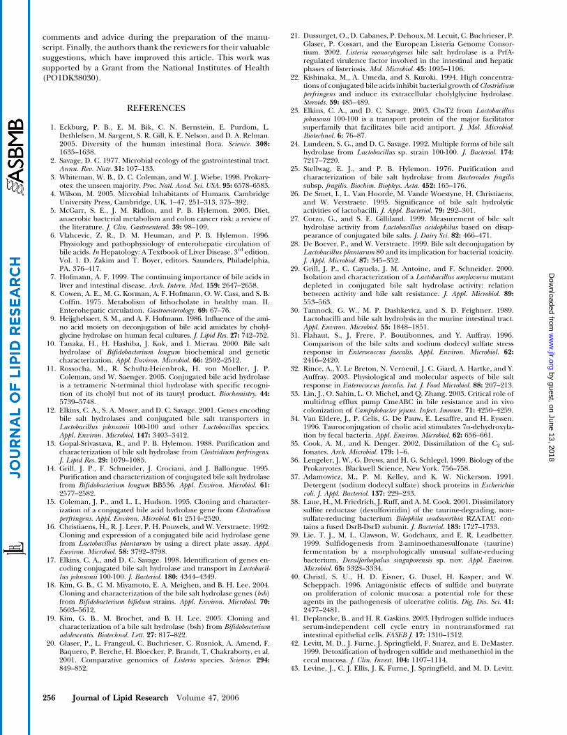
of the small intestine, resulting in malabsorption as bac-teria compete with the host for nutrients. Under normalconditions, bacterial fermentation in the colon representsan important salvage mechanism. Complex carbohydrates,which are intrinsically indigestible or which escape di-gestion and absorption in the proximal gut, are fermentedby colonic bacteria to yield short-chain fatty acids. It hasbeen estimated that these short-chain fatty acids consti-tute 3–9% of our daily caloric intake (4). Colonic bacteriaalso contribute to the salvage of bile salts that escape activetransport in the distal ileum. The major bile salt modi-fications in the human large intestine include deconjuga-tion, oxidation of hydroxy groups at C-3, C-7, and C-12,and 7a/b-dehydroxylation (Fig. 1). Deconjugation and7a/b-dehydroxylation of bile salts increases their hydro-phobicity and their Pka, thereby permitting their recoveryvia passive absorption across the colonic epithelium. How-ever, the increased hydrophobicity of the transformed bilesalts also is associated with increased toxic and metaboliceffects. High concentrations of secondary bile acids infeces, blood, and bile have been linked to the pathogen-esis of cholesterol gallstone disease and colon cancer (5).We present here a current review of the microbiology ofbile acid metabolism in the human GI tract, focusing onunderstanding the biochemical mechanisms and physio-logical consequences of such metabolism on both thebacterium and the human host.
THE ENTEROHEPATIC CIRCULATIONOF BILE ACIDS
Bile acids are saturated, hydroxylated C-24 cyclopenta-nephenanthrene sterols synthesized from cholesterol inhepatocytes. The two primary bile acids synthesized in the
Manuscript received 11 October 2005 and in revised form 17 November2005.
Published, JLR Papers in Press, November 18, 2005.DOI 10.1194/jlr.R500013-JLR200
Abbreviations: bai, bile acid-inducible; BSH, bile salt hydrolase; CA,cholic acid; CBAH-1, conjugated bile acid hydrolase from C. perfringens;CDCA, chenodeoxycholic acid; DCA, deoxycholic acid; GDCA,glycodeoxycholate; HSDH, hydroxysteroid dehydrogenase; LCA, litho-cholic acid; TDCA, taurodeoxycholate; UDCA, ursodeoxycholic acid.
1 To whom correspondence should be addressed.e-mail: [email protected]
Copyright I 2006 by the American Society for Biochemistry and Molecular Biology, Inc.
This article is available online at http://www.jlr.org Journal of Lipid Research Volume 47, 2006 241
by guest, on June 13, 2018w
ww
.jlr.orgD
ownloaded from

human liver are cholic acid (CA; 3a,7a,12a-trihydroxy-5b-cholan-24-oic acid) and chenodeoxycholic acid (CDCA;3a,7a-dihydroxy-5b-cholan-24-oic acid). Bile acids are fur-ther metabolized by the liver via conjugation (N-acyl ami-dation) to glycine or taurine, a modification that decreasesthe Pka to z5. Thus, at physiological pH, conjugated bileacids are almost fully ionized and may be termed bile salts(6). Bile salts are secreted actively across the canalicularmembrane and are carried in bile to the gallbladder,where they are concentrated during the interdigestiveperiod. After a meal, release of cholecystokinin from theduodenum stimulates the gallbladder to contract, causingbile to flow into the duodenum (7). Bile salts are highlyeffective detergents that promote solubilization, digestion,and absorption of dietary lipids and lipid-soluble vitaminsthroughout the small intestine. High concentrations ofbile salts are maintained in the duodenum, jejunum, andproximal ileum, where fat digestion and absorption takeplace. Bile salts are then absorbed through high-affinity
active transport in the distal ileum (6). Upon entering thebloodstream, bile salts are complexed to plasma proteinsand returned to the liver. Upon reaching the liver, they arecleared efficiently from the circulation by active trans-porters on the sinusoidal membrane of hepatocytes andrapidly secreted into bile. This process is known as theenterohepatic circulation. Figure 2 depicts the enterohe-patic circulation in the context of the gastrointestinalanatomy and also indicates the relative numbers andgenera of the predominant bacteria inhabiting each sec-tion of the GI tract.
During the enterohepatic circulation, bile salts encoun-ter populations of facultative and anaerobic bacteria ofrelatively low numbers and diversity in the small bowel.Bile salt metabolism by small bowel microbes consistsmainly of deconjugation and hydroxy group oxidation.Ileal bile salt transport is highly efficient (z95%), butapproximately 400–800 mg of bile salts escapes the en-terohepatic circulation daily and becomes substrate for
Fig. 1. Bacterial bile salt-biotransforming reactions in the human intestinal tract. Hydroxy group carbons of cholate are numbered and theAB rings are identified. The 3, 7, and 12 carbons of cholic acid (CA) are numbered. Nomenclature is that of Hofmann et al. (160). BSH, bilesalt hydrolase; HSDH, hydroxysteroid dehydrogenase.
242 Journal of Lipid Research Volume 47, 2006
by guest, on June 13, 2018w
ww
.jlr.orgD
ownloaded from

significant microbial biotransforming reactions in thelarge bowel (6). Comparison of bile acid composition inthe gallbladder and feces illustrates the extent of microbialbile acid metabolism in the large intestine (Fig. 3). Thesecondary bile acids deoxycholic acid (DCA; 3a,12a-dihydroxy-5b-cholan-24-oic acid) and lithocholic acid(LCA; 3a-hydroxy-5b-cholan-24-oic acid) are producedsolely by microbial biotransforming reactions in thehuman large intestine. DCA accumulates in the bile acidpool (LCA to a much lesser extent) as a result of passiveabsorption through the colonic mucosa and the inabilityof the human liver to 7a-hydroxylate DCA and LCA totheir respective primary bile acids. LCA is sulfated in thehuman liver at the 3-hydroxy position, conjugated at C-24,and excreted back into bile (6). The resultant bile acidsulfate is poorly reabsorbed from the gut. Even though 3-sulfo-LCA glycine and taurine conjugates are deconju-gated and to some extent desulfated by intestinal bacteria,
3-sulfo-LCA/LCA is lost in feces and does not normallyaccumulate in the enterohepatic circulation (8).
DECONJUGATION OF BILE SALTS
Characteristics of bile salt hydrolase(s)
Deconjugation refers to the enzymatic hydrolysis of theC-24 N-acyl amide bond linking bile acids to their aminoacid conjugates. This reaction is substrate-limiting andgoes to completion in the large bowel. Bile salt hydrolases(BSHs) are in the choloylglycine hydrolase family (EC3.5.1.24) and have been isolated and/or characterizedfrom several species of intestinal bacteria (Table 1). Theimportance of the position, charge, shape, and chiralityof various analogs of taurine/glycine conjugates on therate of hydrolysis by BSHs has also been investigated (9).BSHs differ in subunit size and composition, pH optimum,
Fig. 2. Anatomy, physiology, and microbiology of the gastrointestinal tract. Large arrows denote the enterohepatic circulation of bile acids,which begins with contraction of the gallbladder, releasing bile into the duodenum. Small arrows denote the passive absorption of bile acidsthat escape active transport. * Secondary bile acids produced by 7a-dehydroxylation are passively absorbed in the large intestine andreturned to the liver (see Fig 3). The genera of predominant bacteria isolated from each region of the lower gastrointestinal tract are listed.
Bile salt biotransformations by human intestinal bacteria 243
by guest, on June 13, 2018w
ww
.jlr.orgD
ownloaded from

kinetic properties, substrate specificity, gene organization,and regulation. BSHs do, however, share in common sev-eral conserved active site amino acids [cysteine 2 (Cys2),arginine 18 (Arg18), aspartic acid 21 (Asp21), asparagine175 (Asn175), and arginine 228 (Arg228)] and share ahigh degree of amino acid sequence similarity with thepenicillin V amidase of Bacillus sphaericus (Fig. 4). Theconservation of tyrosine 82 (Tyr82) in penicillin V amidaseand Asn82 in BSH are likely a result of differing stericrequirements for their respective substrates (10). Recently,a bsh from Clostridium perfringens was crystallized both inthe apoenzyme form and in complex with taurodeoxycho-late (TDCA; hydrolyzed product) at resolutions of 2.1 and1.7 A, respectively (11). The structure revealed that theCys2 residue is in position for nucleophilic attack of the N-acyl amide bond. Site-directed mutagenesis of the Cys2residue from the BSH of Bifidobacterium longum and Bi.bifidum (10, 18) as well as sulfhydryl inhibition of severalBSHs have shown the importance of this residue incatalysis (10, 13, 14). Alignment of amino acid sequencesfrom BSHs shows that the Cys2 residue is conserved in allBSHs characterized to date (Fig. 4). The broad substratespecificities reported (Table 1) are potentially a function
of a lack of conservation observed in residues making upthe substrate binding pocket of the conjugated bile acidhydrolase gene product of C. perfringens (CBAH-1) and thecorresponding residues predicted in amino acid multiplesequence alignment with other BSHs (Fig. 4). The sterolmoiety is bound primarily through hydrophobic inter-actions in the CBAH-1 (residues highlighted in gray inFig. 4) as well as hydrogen bonds to the carboxylate group.Although the crystal structure of CBAH-1 did not revealspecific recognition of the taurine/glycine moiety, kineticdata from several BSHs suggest that the conjugates areimportant in substrate specificity (Table 1). Therefore,additional crystallization and site-directed mutagenesis(preferably with mutagenesis of Cys2) of BSHs from dif-ferent species will be helpful in explaining the kinetic ob-servations of substrate specificity.
Distribution, genetic organization, and regulation of BSH
Genes encoding BSHs have been cloned from C. perf-ringens (15), Lactobacillus plantarum (16), La. johnsonii (12,17), Bi. longum (10), Bi. bifidum (18), Bi. adolescentis (19),and Listeria monocytogenes (20, 21). Homologs and puta-tive bsh genes have also been identified recently throughmicrobial genome analysis. The organization and regula-tion of genes encoding BSH differ between species andgenera. Monocistronic BSH genes have been reported inLa. plantarum (16),La. johnsonii (12),Li. monocytogenes (21),and Bi. bifidum (18). A gene encoding BSH (CBAH-1)cloned from C. perfringens (15) differed significantly in sizeand amino acid sequence from a BSH purified from adifferent strain ofC. perfringens (13). The inactivation of thegene encoding CBAH-1 resulted in only partial reduction inBSH activity (BSH activity was 86% of that in the wild type),suggesting multiple BSH genes in C. perfringens. Further-more, the crystal structure showed that the enzyme en-coded by the CBAH-1 gene forms an active homotetramer(11). These observations, coupled with the detection ofboth intracellular and extracellular BSHs, provide furtherevidence for multiple isoforms, although the organizationand regulation of the bsh gene(s) fromC. perfringens are notknown at present (22). Polycistronic operons encodingthree genes involved in bile salt deconjugation (cbsT1,cbsT2, and cbsHb) have been characterized in La. johnsoniiand La. acidophilus (12). Genes cbsT1 and cbsT2 appear tobe gene duplications that encode taurocholate/CA anti-port proteins of the major facilitator superfamily, whereascbsHb encodes the BSH b-isoform (23). In addition, anuncharacterized extracellular factor has been detected inLa. johnsonii 100-100, which stimulates BSH activity anduptake of conjugated bile salts during the stationary growthphase (12, 24). BSH expression is also growth phase-dependent. Stationary phase expression has been reportedin Bacteroides fragilis (25), and exponential phase expres-sion was reported for Bi. longum (10).
Benefits of BSHs to the bacterium
BSHs appear to enhance the bacterial colonization ofthe lower gastrointestinal tract of higher mammals. The
Fig. 3. Composition of bile acids in the gallbladder and feces ofhealthy individuals. ‘‘Other’’ bile acids refer to oxo- and 3b-hydroxy derivatives of secondary bile acids. Values were derivedfrom published sources (6, 93, 161, 162). CDCA, chenodeoxycholicacid; DCA, deoxycholic acid; LCA, lithocholic acid; UDCA,ursodeoxycholic acid.
244 Journal of Lipid Research Volume 47, 2006
by guest, on June 13, 2018w
ww
.jlr.orgD
ownloaded from

physiological advantages of BSHs are not fully understoodand may vary between bacterial species and genera. It hasbeen hypothesized that deconjugation may be a mecha-nism of the detoxification of bile salts. De Smet et al. (26)observed significantly higher rates of deconjugation ofglycodeoxycholate (GDCA) over TDCA in La. plantarum.Mutants lacking functional BSH (bsh�) exhibited pH- andconcentration-dependent toxicity of GDCA comparedwith wild-type cells; this effect was not demonstrated withTDCA. De Smet et al. (26) hypothesized that the differ-ence in dissociation constants between GDCA and TDCAresulted in the collapse of cellular proton motive forceby intracellular deprotonation with the glycine conjugate.The presence of a functional BSH results in the intracellu-lar accumulation of free bile acids, which become pro-tonated in a stoichiometric manner, decreasing energy-dependent H1-ATPase-driven proton efflux. BSHs fromhuman intestinal lactobacilli generally have higher affinityfor glycine conjugates (26–29). This observation may lendweight to the hypothesis of De Smet et al. (26), or thehigher affinity of BSHs for glycine conjugates may haveevolved because glycine conjugates are generally higherin proportion (3:1) than taurine conjugates in humanbile (6). Tannock, Dashkevicz, and Feighner (30) arguedagainst the hypothesis of deconjugation as a means ofdetoxification in lactobacilli, because free bile acids aremore cytotoxic than their conjugates. However, when thefree bile acids become 7-dehydroxylated by other intesti-nal bacteria in vivo, the resultant secondary bile acidstend to precipitate (with the extent depending on luminalpH) and bind to insoluble fiber, or they may be absorbedthrough the colonic membrane and may exist in low con-centrations in the bacterium’s microenvironment. There-fore, additional studies comparing various characteristicsof bsh knockouts with their isogenic parent strain willbe needed to determine the function of deconjugation inLactobacillus species.
Strategies to resist bile salt toxicity have been observed inpathogens that colonize the intestinal tract (31–33). Re-cently, a BSH from Li. monocytogenes was shown to be a novelvirulence factor (21). Comparative genome analysis revealedthe absence of a bsh gene in the closely related nonvirulentLi. innocua (20). The bsh gene is positively regulated by PrfA,which is a transcriptional activator of numerous virulencegenes in Li. monocytogenes. Deletion of the bsh gene results indecreased resistance to bile salts and significantly reducedinfectivity in vivo. These results demonstrate the importanceof BSH activity for survival in vivo and infection in the intes-tinal and hepatic phases of listeriosis. The mechanism bywhich BSH activity in Li. monocytogenes enhances survival andvirulence is currently unknown.
Deconjugation may provide a means of obtaining cel-lular carbon, nitrogen, and sulfur for some bacterial spe-cies. This has been demonstrated in bacteroides (34) andis suggested in Bi. longum (10). In fact, the bsh gene fromBi. longum is cotranscribed with the gene encoding gluta-mine synthetase adenylyltransferase (glnE), a componentof the nitrogen regulation cascade (10). In this regard,hydrolysis of the conjugated bile acid may provide aminonitrogen, providing a possible explanation for the coor-dinated regulation of these seemingly physiologically un-related genes (10). Taurine utilization is also widespreadand can serve as an energy source under both aerobic andanaerobic conditions (35). Glycine can be used as an en-ergy source by certain clostridia by the Stickland reaction(36). The Stickland reaction is a form of amino acid fer-mentation in which one amino acid donates electrons thatare accepted by another amino acid distinct from theelectron donor. Another hypothesis suggests that BSHsare detergent shock proteins enabling survival duringstress (37). De Smet et al. (26) found no evidence for thisin lactobacilli after growth with various detergents.
The widespread distribution of BSHs across Gram-negative and Gram-positive intestinal bacteria coupled
TABLE 1. Characteristics of BSHs from human intestinal bacteria
Apparent Km
OrganismNative
Molecular MassSubunit
Molecular Mass TCA TCDCA TDCA GCA GCDCA GDCA pH Optimum Reference(s)
kDa mMBacteroides fragilis 250 32.5 0.45 0.29 0.17 0.35 0.26 0.2 4.2–4.5 25Bacteroides vulgatus 140 36 1 1 1 � � � ND 163Clostridium perfringens MCV 185 250 56 37 3 3.5 3.6 14 1.2 5.8–6.4 13Clostridium perfringens 13 147 36.1 1 ND 1 1 ND ND 4.5–5.5 15Lactobacillus johnsonii 100-100 42(a), 38(b) 12, 17, 23
Isozyme A 115 42 0.76 1 1 1 ND 1 4.2–4.5Isozyme B 105 42, 38 0.95 1 1 1 ND 1 4.2–4.5Isozyme C 95 42, 38 0.45 ND ND ND ND ND 4.2–4.5Isozyme D 80 38 0.37 ND ND ND ND ND 4.2–4.5
Lactobacillus plantarum 80 ND 37.1a TR TR TR 1 1 1 4.7–5.5 28Lactobacillus acidophilus ND 35.0a 1 ND ND 1 ND ND ND 12, 27Bifidobacterium longum BB536 250 40 0.875 1.61 0.516 1.33 1.4 1.37 5.5–6.5 14Bifidobacterium longum SBT2928 125–130 35 1.12 0.33 0.79 0.16 0.13 0.28 5.0–7.0 10Bifidobacterium bifidum ATCC 11863 150 35 1 1 1 1 1 1 1 1 1 ND 18Bifidobacterium adolescentis ND 35a ND ND ND ND ND ND ND 19Listeria monocytogenes ND 36.8a ND ND ND ND ND 1 ND 20, 164
BSH, bile salt hydrolase; GCA, glycocholate; GCDCA, glycochenodeoxycholate; GDCA, glycodeoxycholate; ND, not determined; TCA,taurocholate; TCDCA, taurochenodeoxycholate; TDCA, taurodeoxycholate; TR, trace of activity; 1, activity detected; �, no activity detected.
a Value derived from the Protparam program (http://www.expasy.ch/tools/protparam.html) using the deduced amino acid sequence.
Bile salt biotransformations by human intestinal bacteria 245
by guest, on June 13, 2018w
ww
.jlr.orgD
ownloaded from

Fig. 4. Multiple sequence alignment of cholylglycine hydrolases. Protein sequences were obtained from the National Center forBiotechnology Information (http://www.ncbi.nlm.nih.gov/). Alignments were made with the ClustalW program (http://www.ebi.ac.uk/clustalw/) using the GONNET 250 matrix. Residues highlighted in yellow are predicted active site amino acids based on the crystalstructure of the BSH from C. perfringens (11) as well as on site-directed mutagenesis and biochemical data (10, 12–14). Residues highlightedin gray correspond to residues involved in substrate binding in the BSH from C. perfringens (11). The secondary structural elements, whichare based on the conjugated bile acid hydrolase from C. perfringens (CBAH-1) crystal structure, are shown above the alignment. The a and b
designations of Lactobacillus johnsonii refer to the two isoforms of the genes found in this bacterium.
246 Journal of Lipid Research Volume 47, 2006
by guest, on June 13, 2018w
ww
.jlr.orgD
ownloaded from

with a wide range of substrate specificities, genetic regu-lation, and the occurrence of multiple isoforms in certainstrains have created conflicting reports regarding thephysiological benefit to the bacterium in hydrolyzing bileacid conjugates. Determining the mechanism(s) by whichBSHs aid bacteria in the colonization of the mammalianintestine will be of great interest, especially with regard tobacterial pathogenesis.
Taurine, hydrogen sulfide production, and colon cancer
The bile acid conjugates glycine and taurine serve assubstrates in microbial metabolism. Unlike glycine, taurinecontains a sulfonic acid moiety that is reduced and dis-similated to hydrogen sulfide after deconjugation (38, 39).Hydrogen sulfide is highly toxic and has been shown toincrease colonocyte turnover (40). Activation and upregu-lation of the extracellular signal-regulated kinase 1 and 2(ERK1/2) signaling pathway has been suggested as a possi-ble mechanism for sulfide-induced colonocyte proliferation(41). Hydrogen sulfide also inhibits butyrate metabolismin colonocytes, a key nutrient and regulator of cell turnoverin the gut (40). Levitt et al. (42) demonstrated that colo-nocyteshaveevolvedahighlyefficientmechanismtodetoxifyvolatile reduced sulfides through oxidation to thiosulfate.Defects in this detoxification system are suggested to play arole in the pathogenesis of ulcerative colitis, a known riskfactor for colon cancer (42, 43). Recently, sulfide was im-plicated in preventing apoptosis in the adenocarcinomacell line HCT116 after exposure of cells to b-phenylethylisothiocyanate, a phytochemical found in cruciferous vege-tables, which has been shown to prevent colon carcinogen-esis (44, 45).
A diet high in meat has been shown to significantlyincrease both the levels of taurine conjugation to bile acids(46, 47) and the production of hydrogen sulfide in thecolon (48). A relationship exists between the generation ofhydrogen sulfide in the colon and chronic GI illness, suchas inflammatory bowel disease and colon cancer (5, 49).Populations such as native black Africans with low inci-dence of colon cancer consume low-meat diets (50). Nativeblack Africans also have low ratios of taurine to glycineconjugation (1:9) and low hydrogen sulfide productioncompared with populations consuming a ‘‘Western diet’’(46, 47). In human fecal slurries obtained from individ-uals consuming a Western diet, taurine addition generatedsome of the highest sulfide levels of any organic or in-organic sulfur source added (43). Taurine addition to acoculture of a species of bacteroides and an unidentified7a-dehydroxylating bacterium resulted in significant sul-fide production, which stimulated increased rates of DCAproduction (34).
Although the extent to which taurine metabolism con-tributes to total colonic sulfide production has yet tobe established, several key points have been made: 1)the extent of taurine conjugation in the bile acid pool islargely affected by diet; 2) the same dietary factors thatincrease taurine conjugation are hypothesized to increasecolon cancer risk; 3) taurine metabolism by intestinal bac-teria results in hydrogen sulfide generation; 4) sulfide
generation is linked to the carcinogenesis process throughenhanced cell proliferation, inhibition of butyrate me-tabolism, and activation of cell signaling pathways; and5) sulfide generation may enhance DCA formation inthe gut through stimulation of the microbial bile acid 7a-dehydroxylation pathway.
MICROBIAL BILE ACID HYDROXYSTEROIDDEHYDROGENASE(S)
Oxidation and epimerization
Oxidation and epimerization of the 3-, 7-, and 12-hydroxy groups of bile acids in the GI tract are carried outby hydroxysteroid dehydrogenase (HSDH) expressed byintestinal bacteria (Fig. 1). Epimerization of bile acidhydroxy groups is the reversible change in stereochemistryfrom a to b configuration (or vise versa) with the gen-eration of a stable oxo-bile acid intermediate. Epimeriza-tion requires the concerted effort of two position-specific,stereochemically distinct HSDHs of intraspecies or inter-species origin. For example, the presence of both 7a-and 7b-HSDH in C. absonum allows epimerization by asingle bacterium (51), whereas epimerization also can beachieved in cocultures of intestinal bacteria, one posses-sing 7a-HSDH and the other 7b-HSDH (52, 53).
The extent of the reversible oxidation and reduction ofbile acid hydroxy groups by HSDH depends in part on theredox potential of the environment. Addition of oxygen tothe culture medium increases the accumulation of oxo-bile acids (51). Generation of oxo-bile acids may be morefavorable under the higher redox potentials found on themucosal surface (4), whereas reduction of oxo-bile acidsmay be more favorable under the low redox potential(�200 to �300 mV) in the large intestinal lumen. Thus,although the redox potential of the colon is net reductive,microenvironments at the mucosa may provide oxidiz-ing conditions favorable for certain microbial reactions.HSDHs differ in their reductive and oxidative pH optima,NAD(H) or NADP(H) requirements, molecular weight,and gene regulation (Table 2).
3a- and 3b-HSDHs
3a/b-HSDHs specifically catalyze the reversible, stereo-specific oxidation/reduction between 3-oxo-bile acids and3a- or 3b-hydroxy bile acids. 3a-HSDHs have been de-tected in some of the most prevalent intestinal bacteria,including C. perfringens (54), Peptostreptococcus productus(55), and Eggerthella lenta (formerly Eubacterium lentum)(56, 57), as well as in intestinal bacteria present in lowernumbers (<105/g wet weight of feces), including C.scindens (58) and C. hiranonis (59), and in nonintestinalbacteria, including Pseudomonas testosteroni (60, 61). 3b-HSDH activity has been described in species of Clostridiumand Rumminococcus (62–64). It appears that intraspecies 3-epimerization favors the 3a-position. In fact, growingcultures of C. perfringens in the presence of 3-oxo-CDCAformed CDCA (84%) preferentially over iso-CDCA (16%)under anaerobic conditions (65).
Bile salt biotransformations by human intestinal bacteria 247
by guest, on June 13, 2018w
ww
.jlr.orgD
ownloaded from

Pyridine nucleotide cofactor requirements differ be-tween 3a/b-HSDHs. 3a-HSDHs require NAD(H), with theexception of the enzyme purified from C. perfringens,which uses NADP(H), and that purified from C. scindens,which can use either NAD(H) or NADP(H) (54, 56–58, 61). 3b-HSDHs have been shown to preferentially re-quire NADP(H), with the exception of C. innocuum, whichuses NAD(H) (62–64). Dihydroxy bile acids [DCA, CDCA,and ursodeoxycholic acid (UDCA)] are generally bettersubstrates than trihydroxy bile acids (CA) (62, 65).
3a/b-HSDHs characterized to date are constitutivelyexpressed with the exception of those from C. scindens andC. hiranonis, which are induced by the primary bile acidsCA and CDCA. In fact, three copies of 3a-HSDH genes(the bile acid-inducible genes baiA1, baiA2, and baiA3) havebeen identified from C. scindens (66, 67), and baiA1 hasbeen expressed in Escherichia coli and characterized (58).The baiA gene products are unique among 3a/b-HSDHs asa result of their high specificity toward CA-CoA and CDCA-CoA conjugates and relatively low activity toward free bileacids (58).
7a- and 7b-HSDHs
7a/b-HSDHs catalyze the reversible, stereospecificoxidation/reduction of the 7a- and 7b-hydroxyl groups
of bile acids. Although 7a/b-HSDHs are common amongintestinal bacteria, the extent of 7a/b-dehydrogenation inthe intestine, or in mixed fecal suspensions, is difficult tointerpret because of the competing, and irreversible, 7a/b-dehydroxylation of bile acids (see below) (68).
7a/b-HSDHs are widespread among the bacteroidesand clostridia as well as in E. coli and Ruminococcus species(Table 2) (54, 64, 69–74). In addition, several intestinalclostridia express both 7a- and 7b-HSDHs and have beenshown to epimerize the 7a/b-hydroxy group (75, 76–78).7a/b-HSDHs have been partially purified from intestinalbacteria, including Ba. fragilis (71, 79), Ba. thetaiotaomicron(74), C. scindens (80), C. sordellii (70), and E. coli (73), aswell as from the soil isolates Xanthomonas maltophilia (81),C. absonum (82), and C. bifermentans (83). Interestingly,several intestinal bacteria with 7a/b-HSDH activity alsopossess BSH, including Ba. fragilis (25), C. sordellii (70), C.perfringens (84), and C. innocuum (63), as well as the soilisolate C. bifermentans (83).
7a-HSDHs generally use NADP(H) as a cofactor, withthe exception of E. coli (85) and Ba. thetaiotaomicron (74).C. bifermentans, C. absonum, and Ba. fragilis 7a-HSDHs canuse either NAD(H) or NADP(H) as a cofactor (76, 79, 83).7b-HSDH enzymes characterized to date use NADP(H) asa cofactor (53, 62, 75, 76). 7a/b-HSDH enzymes have
TABLE 2. Characteristics of HSDHs from intestinal bacteria
Organism Stereospecificity Cofactor pH OptimumNative
Molecular Mass Gene Regulation Reference(s)
kDa
Eggerthella lenta 3a (CE) NAD(H) 11.3 205 R by BA 56, 5712a (CE) NAD(H) 8.0–10.5 125 R by BA
Clostridium perfringens 3a (CE) NADP(H) 11.3 ND NI 54, 6512a (CE) NAD(H) 10.5 ND NI
Peptostreptococcus productus 3a (CE) NAD(H) 8.5 132 NI 55, 165, 1663b (CE) NAD(H) 9.5 95 NI7b (PP) NADP(H) 9.8 53 I by 7-oxo-BA
Bacteroides fragilis 7a (P) NAD(H) 8.5 (oxid);6.5 (red)
110 (tetramer) I (CA) 71, 79, 167
7a (PP) NADP(H) 7.0–9.0 127 I (CA)Bacteroides thetaiotaomicron 7a (PP) NAD(H) ND 320 ND 74Escherichia coli 7a (P) NAD (H) 8.5 120 (tetramer) NI 73Clostridium sp. 25.11.C 3b (CE) NADP(H) 7.5 104 NI 62
7a (CE) NADP(H) 8.6 82 I (CA, CDCA, 7-oxo-CA)7b (CE) NADP(H) 8.6 115 I (CA, CDCA, 7-oxo-CA)
Clostridium absonum 7a (PP) NAD(H) 9.5–11.5 ND I (CDCA, DCA); R (UDCA) 76, 827b (PP) NADP(H) 9.0–10.0 200 (tetramer) I (CDCA, DCA); R (UDCA)
Clostridium sordellii 7a (P) NADP(H) ND 120 (tetramer) I (CDCA, CA, DCA) 70Clostridium innocuum 3b (PP) NAD(H) 10.0–10.2 56 NI 63Clostridium scindens
VPI 127083a (P) NAD(H)/
NADP(H)5.0–9.0 108 (tetramer) I (CA, CDCA) 58, 80
7a (P) NADP(H) ND 124 (tetramer) NIClostridium bifermentans 7a (PP) NAD(H)/
NADP(H)11.0 ND I (7-oxo-LCA, CDCA, DCA) 83
Clostridium limosum 7a (CE) NAD(H)/NADP(H)
10.5 ND I (CA, CDCA, UDCA) 75
7b (CE) NADP(H) 10.5 ND I (CA, CDCA, UDCA)Clostridium leptum 12a (PP) NADP(H) 8.5–9.0 225 NI 89Clostridium group
P strain C48-5012a (PP) NADP(H) 7.8 100 R (CDCA, CA) 90
Clostridium paraputrificum 12b (CE) NADP(H) 7.8 (oxid);10 (red)
126 NI 91
Eubacterium aerofaciens 7b (CE) NADP(H) 10.5 45 R (UDCA) 165
BA, bile acids; CA, cholic acid; CDCA, chenodeoxycholic acid; CE, cell extract; HSDH, hydroxysteroid dehydrogenase; I, induced; LCA,lithocholic acid; NI, noninduced by bile acids; ND, not determined; P, purified; PP, partially purified; R, repressed; UDCA, ursodeoxycholic acid.
248 Journal of Lipid Research Volume 47, 2006
by guest, on June 13, 2018w
ww
.jlr.orgD
ownloaded from

higher affinity for dihydroxy bile acids (CDCA and 7-oxo-LCA) than for trihydroxy bile acids (CA and 7-oxo-DCA).C. limosum cell extracts use both free and conjugated bileacids, whereas whole cells only oxidize free bile acids (75,86). This is attributable to the intracellular location ofthe 7a-HSDH and the inability of the organism to take upa conjugated bile salt. The genes encoding 7a-HSDHshave been cloned from E. coli (73), C. scindens (69), andC. sordellii (70). Sequence similarity suggests that theseenzymes belong to the short-chain polyol dehydrogenasefamily. Regulation of 7a/b-HSDH expression is generallygrowth phase-dependent and inducible by bile acid sub-strates (Table 2).C. scindens andE. coli constitutively express7a-HSDHs and are noninducible (69, 85). Unexpectedly,the nonsubstrate DCA can induce 7a-HSDH expressionin C. absonum and C. sordellii, although the reason for thisinduction remains unclear (70, 76). Macdonald, White,and Hylemon (82) observed the expression of five solu-ble and two membrane polypeptides upon exposure of C.absonum to DCA and CDCA in the culture medium, al-though the functions of these additional polypeptides havenot been determined.
Crystal structures of the E. coli 7a-HSDH binary andternary complexes have been solved and a mechanism for7a-dehydrogenation proposed (Fig. 5) (87). Binding of7a-hydroxy bile acids elicits major conformational changesat the substrate binding loop and C-terminal domain. Atwo-step mechanism is proposed in which Tyr159 actsas a catalytic base removing the C-7 hydroxy hydrogen.Hydrogen bonding by serine 146 (Ser146) is hypothesizedto stabilize the intermediate. Regeneration of the catalystoccurs through the transfer of the acquired hydride fromthe phenolic group at Tyr159 to lysine 163 (Lys163) toposition 4 of NAD+. Lys163 serves two important roles:anchoring NAD+ through bifurcated hydrogen bonding,and indirect hydride transfer from Tyr159 to position 4 ofNAD+ (87). Site-directed mutagenesis confirmed the roleof these amino acids in 7a-HSDH catalysis (88). Analysis ofthe 7-oxo-GLCA bile acid substrate/enzyme complex re-vealed tight binding of the sterol with loose associationfor the glycine conjugate. Binding of glycine and taurineconjugates of CDCA was not significantly different fromthat of the free bile acid (87).
12a- and 12b-HSDHs
12a/b-HSDHs have been detected mainly among mem-bers of the genus Clostridium. NADP-dependent 12a-HSDHs have been detected in C. leptum (89) in Clostridiumgroup P (90), whereas NAD-dependent 12a-HSDH activitywas reported in Eg. lenta (56) and C. perfringens (54). 12b-HSDHs have been detected in C. tertium, C. difficile, and C.paraputrificum (91, 92). 12a/b-HSDHs characterized todate are constitutively expressed and noninducible, withthe exception of the 12b-HSDH from C. paraputrificum,which is induced by 12-oxo-bile acid substrates (92). 12a/b-HSDHs generally have higher affinity for dihydroxy bileacids (DCA) than for trihydroxy bile acids (CA and iso-CA)and for free versus conjugated bile acids. The 12a-HSDHfrom C. leptum is an exception, demonstrating higher af-
finity for CA conjugates than for free CA (89). 12a-HSDHsappear to be repressed by the addition of bile acid sub-strates (DCA . CDCA . CA) to the growth medium at1 mM concentrations. It has been suggested that theseenzyme activities should be repressed in bacteria coloniz-ing the large intestine (56, 92), although 12-oxo-bile acidshave been detected at low levels in the feces of healthyindividuals (Fig. 3) (93, 94).
Benefits of bile acid hydroxysteroid oxidoreductases tothe bacterium
The oxidation of bile acid hydroxyl groups generatesreducing equivalents for cellular biosynthetic reactionsand possibly electron transport phosphorylation. Bile acid
Fig. 5. Proposed catalytic mechanism of bile acid 7a-dehydroge-nation based on the crystal structure and site-directed mutagenesisof active site amino acids of the 7a-dehydrogenation from E. coli.See text for a description of catalysis. Reprinted with permissionfrom Tanaka et al. (87). Copyright I 1996 American ChemicalSociety.
Bile salt biotransformations by human intestinal bacteria 249
by guest, on June 13, 2018w
ww
.jlr.orgD
ownloaded from

dehydrogenation is hypothesized to generate energy in Ba.thetaiotaomicron (74). Sherod and Hylemon (74) suggestedthat reduced pyridine nucleotides generated from 7a-hydroxy oxidation serve to generate ATP via a cytochrome-linked electron transport chain in the presence of electronacceptors (i.e., fumarate).
Bile acids are potent antimicrobial agents provided thatthe proper concentration and proportion of hydrophobicbile acids (CDCA, LCA, and DCA) are present (95). Al-teration of hydroxy group stereochemistry has a markedinfluence on the physiochemical properties of bile acids(96, 97). The epimerization of the 7a-hydoxy group ofCDCA decreases the hydrophobicity and toxicity of thebile acid (97). Macdonald, White, and Hylemon (82)observed that C. absonum grew on plates containing 1 mMUDCA, although it was unable to grow on plates contain-ing 1 mM CDCA. Furthermore, when cultured in thepresence of 7-oxo-bile acids, only low concentrations of CAand CDCA were formed, whereas the majority of 7-oxo-bileacids were reduced to 7-epicholic acid and UDCA,respectively, by log-phase C. absonum (51). UDCA hasalso been shown to act as a repressor of 7a/b-HSDHproduction in C. absonum, suggesting that UDCA is an endproduct (76). The enzyme also displays markedly higheraffinity for CDCA than for CA, the former being moretoxic. In summary, dehydrogenation may serve functionsrelated to energy generation as well as attempts tomaintain low concentrations of more hydrophobic bileacids in the bacterium’s microenvironment.
Interplay between HSDH enzymes in human liver andintestinal bacteria
The coevolution between host and gut flora is evidentwhen observing the interplay between liver and bacterialbiotransforming reactions. The liver synthesizes bile acidsin which the hydroxy groups are in the a orientation. Inthe a-hydroxy orientation, one face of the molecule ishydrophobic and the other side is hydrophilic. This trans-lates to efficient solubilization of lipid molecules throughthe formation of mixed micelles capable of efficient emul-sification while remaining soluble in aqueous environ-ments. Generation of b-hydroxy bile acids by microbialenzymes alters the efficiency of micelle formation as aresult of hydrophilic groups on both faces of the sterolmolecule. The differences observed between the compo-sition of bile acids in serum and bile are a result of thecontinual interplay between liver and bacterial enzymes.Exposure of bile salts to intestinal bacteria results in z50%of bile acids requiring reconjugation and low levels of bileacids returned to the liver in the 3b-hydroxy orientation(98). Without a means of epimerizing bile acid hydroxygroups in the human liver, b-hydroxy bile acids wouldaccumulate in the bile acid pool. Interestingly, the liverseems to ‘‘allow’’ the accumulation of UDCA (7b-hydroxy)in the biliary pool, as in the case of therapeutic admin-istration of CDCA (99). The protective effects of UDCAobserved in clinical studies (100) as well as cell culturestudies (97, 101) may provide an evolutionary explanationfor this phenomenon.
THE BIOCHEMISTRY AND MOLECULAR BIOLOGYOF BILE ACID 7a/b-DEHYDROXYLATION
Introduction
Secondary bile acids (DCA and LCA) predominate inhuman feces (Fig. 3). Therefore, 7a-dehydroxylation is themost quantitatively important bacterial bile salt biotrans-formation in the human colon. The rapid rate of conver-sion of primary to secondary bile acids is surprising givencurrent estimates that this metabolic pathway is found inz0.0001% of total colonic flora (102–104). Human in-testinal bacteria capable of bile acid 7a-dehydroxylationhave been isolated (104, 105), and 16S rDNA phylogeneticanalysis has led to their classification to the genus Clos-tridium (106–108).
Unlike bile acid oxidation and epimerization, 7a/b-dehydroxylation appears restricted to free bile acids. Re-moval of glycine/taurine bile acid conjugates via BSHenzymes is thus a prerequisite for 7a/b-dehydroxylationby intestinal bacteria (109–112). Some intestinal bacteriaare capable of both 7a/b-dehydroxylating activities (113),whereas 7b-dehydroxylation activity is absent in otherintestinal 7a-dehydroxylating bacteria (113). Epimeriza-tion of UDCA (7b-hydroxy) to CDCA (7a-hydroxy) via 7b-HSDHs produced by members of the gut flora results insubsequent 7a-dehydroxylation. In this regard, it appearsthat the presence of 7b-dehydroxylation activity is more ofa luxury than a necessity.
Elucidating the bile acid 7a/b-dehydroxylation pathway
Samuelsson (114) administered [6a-3H,6b-3H,8b-3H][24-14C]CA to bile duct-cannulated rabbits and rats. Anal-ysis of the products recovered after exposure to intestinalbacteria revealed a differential loss of the 6b-3H during 7a-dehydroxylation of CA. Previous work showed completeretention of the 7b-3H in [7b-3H][24-14C]CA during 7a-dehydroxylation in the rat intestine (115, 116). These dataled Samuelsson (114) to propose a mechanism for CA 7a-dehydroxylation involving two steps: diaxial trans-elimina-tion of the 7a-hydroxy group and 6b-hydrogen atom,followed by reduction through trans-hydrogenation of the6b and 7a positions of the cholen-6-oic acid intermediateforming DCA. Bjorkhem et al. (117) showed the formationof a 3-dehydro-4-cholenoic acid intermediate after thedifferential loss of the 5bH in vitro and in vivo using [3b-3H][2414C]CA and [5b-3H]2414C]CA. Hylemon et al. (118)subsequently observed the accumulation of multiple bileacid intermediates in cell extracts of C. scindens induced byCA (Fig. 6). These radiolabeled CA intermediates wereidentified by mass spectrometry, then chemically synthe-sized and added to cell extracts of CA-induced C. scindens.Each 24[14C]CA intermediate was converted to 2414C]DCAin cell extracts prepared from CA-induced cultures ofC. scindens. These observations suggested that the 7a-dehydroxylation mechanism was more complex than thetwo-step mechanism proposed by Samuelsson (114). Fur-thermore, these data demonstrated that bile acid 7a-dehydroxylation was a multistep pathway in C. scindens andsuggested the presence of multiple bai genes.
250 Journal of Lipid Research Volume 47, 2006
by guest, on June 13, 2018w
ww
.jlr.orgD
ownloaded from

The induction of 7a-dehydroxylation activity in C. scin-dens by unconjugated C24 primary bile acids resulted inthe appearance of several new polypeptides, as observed
by one- and two-dimensional SDS-PAGE (119, 120). Puri-fication and N-terminal sequencing of these bai polypep-tides facilitated the cloning of bai genes through the designof degenerate probes (121–123). Northern blot analysisindicated the presence of a large CA-inducible (>10 kb)mRNA transcript and a smaller transcript (<1.5 kb) in C.scindens (121, 124). These studies led to the discovery of abai regulon encoding at least 10 open reading frames(Fig. 7). Individual bai genes have been subcloned intoE. coli and the functions of many of them determined (58,67, 123, 125–130; P. B. Hylemon, unpublished data). Theproposed bile acid 7a/b-dehydroxylation pathway in C.scindens is shown in Fig. 8. A bai operon has also beencharacterized from C. hiranonis (59), although the discus-sion below of the 7a/b-dehydroxylation pathway will cen-ter on C. scindens, from which the functions of the geneproducts have been determined.
bai genes: a regulon for 7a/b-dehydroxylation
The transport of unconjugated primary bile acids intoC. scindens is facilitated by the baiG gene product, whichbelongs to a major pump/facilitator superfamily of pro-tein transporters (126). The baiG gene has been clonedinto E. coli and shown to encode a 50 kDa H1-dependentbile acid transporter (126). BaiG facilitates the transport ofunconjugated CA and CDCA but not of the secondary bileacids DCA and LCA (126). Computer-aided modelingsuggests that the baiG polypeptide contains 14 membrane-spanning domains (126).
Fig. 6. Accumulation of [24-14C]CA intermediates during 7a-dehydroxylation in cell extracts of C. scindens VPI 12708. Cell ex-tracts were prepared from either CA-induced (I) or control (C)cells. This figure was modified from Hylemon et al. (118). Copy-right I 1992 American Society for Biochemistry and MolecularBiology, Inc.
Fig. 7. Gene organization of the bile acid-inducible (bai) 7a/b-dehydroxylation operons characterized in C. scindens VPI 12708. P indicatesthe promoter region.
Bile salt biotransformations by human intestinal bacteria 251
by guest, on June 13, 2018w
ww
.jlr.orgD
ownloaded from

After transport, ligation to CoA is the first step inactivating CA and CDCA for 7a-dehydroxylation, as severalsubsequent enzymatic steps are specific for CoA conju-gates. The baiB gene was shown to encode a 58 kDa bileacid CoA ligase (125). CoA ligation is ATP-, CoA-, andMg21-dependent and also requires a free carboxyl groupon C24 bile acids (125). CoA ligation may function both tosterically hinder the constitutive 7a-HSDHs, committingthe bile acid to 7a-dehydroxylation, and to trap the bileacid inside the cell.
The 3a-hydroxy group is oxidized after CoA ligation.Oxidation of the 3-hydroxy group inhibits 7a-hydroxygroup dehydrogenation, favoring 7-dehydroxylation overconstitutively expressed 7a-HSDHs in C. scindens (58, 80).The baiA gene products encode 27 kDa polypeptides thathave significant similarity with the short-chain alcohol/polyol dehydrogenase gene family (58, 124). Amino acidmultiple sequence alignment and comparison betweenbaiA gene products and other members of the short-chainalcohol dehydrogenase family revealed a possible NAD(P)binding site and catalytic active site (58). Three baiA geneshave been cloned from C. scindens; the baiA1 and baiA3
genes are monocistronic, whereas the baiA2 gene is partof the polycistronic bai operon (66, 67, 124). The baiAgenes from C. scindens were cloned in E. coli and shown toencode 3a-HSDHs (58). The enzymes only recognize bileacid CoA conjugates and can use either NAD1 or NADP1
as electron acceptors (58). Interestingly, the different3a-HSDHs of C. scindens share 92% amino acid sequenceidentity with one another, suggesting gene duplication.The physiological importance of multiple baiA genes re-mains unclear.
The baiCD gene from C. scindens has been cloned andexpressed in E. coli and was recently demonstrated to en-code a steroid oxidoreductase specific for the CoA con-jugates of 3-dehydro-4-cholenoic acid and 3-dehydro-4-chenodeoxycholenoic acid (Fig. 8) (P. B. Hylemon et al.,unpublished data). The z70 kDa enzyme requires FMNand NAD1 or NADP1 for activity and shows stereospeci-ficity toward 7a-hydroxy bile acids. The BaiCD polypeptideshows considerable amino acid sequence identity withthe Old Yellow Enzyme family, a putative NADH oxidasefrom Li. monocytogenes, several 2,4-dienoyl CoA reductases,and baiH from C. scindens (121, 129). The baiH gene en-
Fig. 8. Proposed bile acid 7a/b-dehydroxylation pathways in C. scindens VPI 12708 for CDCA and UDCA. Reaction steps in bracketsindicate enzymatic steps for CDCA and UDCA intermediates. For simplicity, only CDCA intermediates are shown for the general 7-dehydroxylation pathway. The baiCD and baiE gene products are proposed to encode stereospecific enzymes for 7a-hydroxy bile acids. ThebaiH and baiI gene products are proposed to encode stereospecific enzymes for 7b-hydroxy bile acids. The baiF gene product has bile acid-CoA hydrolase activity but is hypothesized to encode a bile acid CoA transferase (see text).
252 Journal of Lipid Research Volume 47, 2006
by guest, on June 13, 2018w
ww
.jlr.orgD
ownloaded from

codes a 72 kDa polypeptide containing 661 amino acids(121). The BaiH protein exists as a homotrimer withNADH:flavin oxidoreductase activity (121). The baiH genehas been subcloned into E. coli, purified, and shown tocontain 1 mol of FAD, 2 mol of iron, and 1 mol of cop-per per mole polypeptide subunit (129). The baiH geneproduct from C. scindens was recently determined to en-code a steroid oxidoreductase specific for CoA conju-gates of 3-dehydro-4-ursodeoxycholenoic acid and 3-dehydro-4-epicholenoic acid (Fig. 8) (P. B. Hylemonet al., unpublished data). Oxidation of CA-CoA, CDCA-CoA, and UDCA-CoA to their respective 3-dehydro-4-bileacid CoA conjugates appears to make the bile acid chem-ically labile for 7a/b-dehydration.
The baiF gene encodes a 47.5 kDa polypeptide contain-ing 426 amino acids that was shown to have bile acid CoAhydrolase activity (122, 130). However, baiF is hypothe-sized to encode a CoA transferase because of energyconservation (Fig. 8) and homology to the type III familyof CoA transferases (131). The first few cycles of 7a-dehydroxylation would require ATP hydrolysis (Fig. 8),although ATP-independent recycling of the thioesteraseintermediates via transfer of CoA from 3-dehydro-4-cholenoic acid to CA, 3-dehydro-4-chenodeoxycholenoicacid to CDCA, or 3-dehydro-4-ursodeoxycholenoic acid toUDCA by the baiF would significantly conserve energy.However, this hypothesis remains to be tested.
7a-Dehydration of 3-dehydro-4-cholenoic acid and 3-dehydro-4-chenodeoxycholenoic acid results in the gener-ation of a conjugated double bond in rings A and B,forming stable 3-dehydro-4,6-deoxycholdienoic acid and3-dehydro-4,6-lithocholdienoic acid intermediates, respec-
tively. The 7a-dehydration step results in the largest cal-culated energy change (�9.4 kcal/mol) of any reaction inthis pathway, and the reverse reaction was not detectedin vitro (128). The 19.5 kDa bile acid 7a-dehydratase isencoded by the baiE gene (128). This enzyme showed noactivity with 3-dehydro-4-ursodeoxycholenoic acid (128).The baiE gene product was modeled on the crystal struc-ture of the protein homologs (secondary structure) keto-steroid isomerase and scytalone dehydratase (Fig. 9) (K.Woodford et al., unpublished data). The putative activesite/binding pocket is shown in Figure 10. A catalyticmechanism has been proposed for the 7a-dehydrationstep based on the conservation in secondary structure andsite-directed mutagenesis of the key active site amino acids.Tyr30 acts as a general acid withdrawing electron densityfrom the 3-oxo group. This shift in electron density ishypothesized to make the 6b-hydrogen labile for removalby the general base histidine 83 (His83) (assisted byAsp35). His83 is thought to donate its hydrogen to the 7a-hydroxy forming the water-leaving group that is stabilizedby Asp106. Site-directed mutagenesis based on the pro-posed mechanism supports the important role of theputative active site amino acids in enzyme catalysis. It ishypothesized that the baiI gene encodes a bile acid 7b-dehydratase, because of amino acid sequence homologiesbetween the baiE and baiI gene products.
Genes involved in the reductive arm of the 7a/b-dehydroxylation pathway have not been isolated. Thesegenes should encode oxidoreductases catalyzing thereduction of 3-dehydro-4,6-deoxycholdienoic acid to 3-dehydro-4-deoxycholenoic acid to 3-dehydro-deoxycholicacid to DCA as well as a bile acid exporter to removesecondary bile acid end products from the bacterium(Fig. 8). Genes encoding putative transcriptional regula-tors have been detected upstream of the bile acid-inducible promoter region (Table 3) (D. H. Malloneeand P. B. Hylemon, unpublished data). Additional studieswill be required to determine the mechanism of induc-
Fig. 9. Model of the bile acid 7a-dehydratase (baiE) based onstructural data from scytalone dehydratase, nuclear transportfactor 2, and steroid D5-isomerase. The a-helices are denoted bywhite strands, b-sheets are denoted by the yellow backbone, and theblue structure represents a steroid molecule modeled into thebinding/active site pocket. Model kindly provided by Dr. Alexey G.Murzin (Cambridge University).
Fig. 10. Putative binding/active site pocket of bile acid 7a-dehydratase. Putative active site amino acid residues are shown inrelation to the bile acid substrate. See text for discussion of theproposed catalytic mechanism.
Bile salt biotransformations by human intestinal bacteria 253
by guest, on June 13, 2018w
ww
.jlr.orgD
ownloaded from

tion/repression of this pathway and to identify additionalbai genes.
The benefits of 7a/b-dehydroxylation to the bacterium
The ability to use bile acids as electron acceptors is animportant niche for 7a-dehydroxylating bacteria in thehuman colon. The 7a/b-dehydroxylation pathway re-quires multiple oxidative and reductive steps with a net2 electron reduction (Fig. 8). The hypothesized energybenefits of this pathway assume, however, that the baiFgene encodes a CoA transferase and the toxic end prod-ucts, the secondary bile acid, are removed from the mi-croenvironment in vivo (precipitation and binding toinsoluble fiber). The generation of secondary bile acidsmay also function to exclude bacteria sensitive to thesehydrophobic molecules.
SECONDARY BILE ACIDS AND DISEASE
In humans, DCA accumulates in the bile acid pool tohigh levels in some individuals. An increase in DCA inthe bile acid pool is associated with a decrease in CDCA(Fig. 11). Unlike rodents, the human liver cannot 7a-hydroxylate DCA, forming CA. Hence, under normal phys-iological conditions, there is no metabolic pathway forremoving DCA from the bile acid pool in humans. Theamount of DCA in the bile acid pool is a function of atleast three variables: 1) the rate of formation and absorp-tion of DCA through the colon (input) (132); 2) colonictransit time (133); and 3) colonic pH (134).
High levels of DCA in blood, bile, and feces have beencorrelated with an increased risk of cholesterol gallstonedisease and colon cancer, two major diseases of Westernsociety (5, 135). High levels of CA 7a-dehydroxylating fecalbacteria have been correlated with increased amounts ofDCA in bile of a subset of cholesterol gallstone patients(132). Treatment of these cholesterol gallstone patients(high DCA group) with antibiotics significantly decreasedthe levels of fecal CA 7a-dehydroxylating bacteria, DCA in
bile, and the cholesterol saturation index in bile (132).Early studies by Low-Beer and Nutter (135) reported thattreating control individuals with metronidazole, an anti-biotic effective against anaerobic bacteria, significantlydecreased the cholesterol saturation index of bile. More-over, excess DCA in bile has been reported to decrease thenucleation time for cholesterol crystallization (136, 137).In total, these results suggest a possible link between in-testinal bacteria, DCA, and the risk of cholesterol gallstonedisease in some patients.
DCA and LCA have been linked to colon carcinogenesisin a number of laboratory animal models and humanepidemiological studies (for reviews, see 5, 138). Mostanimal studies conclude that DCA is a promoter of car-cinogenesis (139–142). However, some researchers argue
TABLE 3. bai genes characterized from C. scindens VPI 12708
bai Gene Molecular Mass Catalytic Activity/Function Gene Family Reference
kDa
baiA 27 3a-HSDH Short-chain alcohol/polyol dehydrogenase 58baiB 58 Bile acid CoA ligase AMP binding 125baiCD 70 3-Dehydro-4-CDCA/CA
steroid oxidoreductasePyridine nucleotide-disulfide oxidoreductase;
NADH:flavin oxidoreductase–a
baiH 72 3-Dehydro-4-UDCA/7-epiCAsteroid oxidoreductase
Pyridine nucleotide-disulfide oxidoreductase;NADH:flavin oxidoreductase
129
baiE 19.5 7a-Dehydratase COG4875 128baiI 22 7b-Dehydrataseb COG4876 168BaiF 47.5 Bile acid CoA hydrolase Type III CoA transferase 130
Bile acid CoA transferaseb
baiG 50 H+-dependent bile acid transporter Major facilitator superfamily 126barA 46 Transcriptional regulationb AraC/XylS –a
barB 22 Transcriptional regulationb RpoB; permeases of the major facilitator superfamily –a
bai, bile acid-inducible; bar, bile acid-regulatory.a P. B. Hylemon et al., unpublished data.b Hypothesized function.
Fig. 11. Relationship between the percentage of CDCA and DCAin bile of patients at McGuire VA Hospital (Richmond, VA) (P. B.Hylemon et al., unpublished data).
254 Journal of Lipid Research Volume 47, 2006
by guest, on June 13, 2018w
ww
.jlr.orgD
ownloaded from

that bile acids may cause DNA damage and act as carcino-gens in humans (138). Higher levels of DCA are found inthe blood of colon cancer patients compared with controlpatients (98, 143). Moreover, DCA is a logical candidatefor promoting colon carcinogenesis for the following rea-sons: 1) it is found in fecal water in high concentrations(.100 AM) (138); 2) it can cross biological membranes viapassive diffusion; and 3) it can activate mammalian cellsignaling pathways that are known to be involved in pro-moting carcinogenesis. In this regard, cell signaling path-ways activated by DCA in mammalian epithelial cellsinclude protein kinase C (144), ERK1/2 via the epidermalgrowth factor receptor (101, 145, 146), b-catenin (147),and Jun-N-terminal kinase 1 and 2 ( JNK1/2) (148).Secondary bile acids have been shown to cause apoptosisin colonic epithelial cells, and high concentrations ofDCA and LCA in stool may promote carcinogenesis byexerting selective pressure for the emergence of epithelialcell mutants that are resistant to apoptosis (e.g., via loss ofp53) (138). LCA has been found to be an excellent acti-vator of the vitamin D receptor (149, 150). Activation ofthis receptor in intestinal epithelial cells activates genesthat metabolize LCA (150). This may be a protectivemechanism that evolved to limit LCA toxicity to intestinalepithelial cells.
DISCUSSION
Bile salt metabolism is a widespread and fundamentalproperty of the gastrointestinal microflora encompas-sing the most commonly isolated species of intestinalbacteria, including but not limited to the genera Bacteroi-des, Clostridium, Lactobacillus, Bifidobacterium, Eubacterium,and Escherichia. However, our current understanding ofthe intestinal microbiology of bile salt modifications islimited to cultivated species. Discovering novel genes inthe gut microbiome (collective genomes of the gastroin-testinal flora) encoding BSHs, HSDHs, and enzymes in-volved in the 7a/b-dehydroxylation pathway throughmolecular techniques will facilitate a much greater under-standing of the diversity and complexity of these reactionsin the human colon. Techniques such as PCR-denaturinggradient gel electrophoresis can be used to measure thediversity of organisms based on specific phylogenetic mark-ers, such as 16S rDNA, functional genes, and potentiallybile salt-modifying enzymes (151, 152). Measuring the truediversity of bile salt-modifying bacteria is crucial in studyingthe relationship between the levels and activities of thesebacteria and disease risk.
Determining the conditions in which secondary bileacids are formed in significant quantities and retained inthe enterohepatic circulation of certain individuals is sug-gested to be important in the etiology of cholesterol gall-stone disease and colon cancer. Because secondary bileacids are formed exclusively through bacterial enzymaticreactions, the study of microbes capable of bile acid 7a/b-dehydroxylation is important in understanding thesechronic GI illnesses. The goal of such research is to find
ways to block the source of secondary bile acid production.Long-term use of antibiotics to prevent 7a-dehydroxyla-tion of bile acids would be impractical. The design ofpharmaceuticals to block the 7a-dehydroxylation pathwayis a possibility. However, this approach requires targetingmicrobial enzymes and runs the risk of eventual drugresistance, as with antibiotics. Alternatively, reducing sec-ondary bile acid production may be achieved by admin-istering specialized CA-accumulating probiotic bacteria(153). Studies with bifidobacteria and lactobacilli isolatedfrom human feces have shown their ability to assimilate CAspontaneously in vitro (153, 154). The mechanism for CAuptake in lactic acid bacteria appears to be diffusion of ahydrophobic weak acid through the membrane via thetransmembrane proton motive force (153, 154). Thehigher intracellular pH causes the bile acids to becometrapped as a result of ionization.
The use of specific bacteria as ‘‘drugs’’ to treat chronicGI illnesses caused in part by other intestinal bacteria cer-tainly has potential, although rigorous studies are neededto demonstrate the effectiveness of such therapies. Over-all, clinical trials using probiotics to solve specific healthdisorders have met with mixed results. Discrepancies be-tween studies are attributable in part to different method-ologies, choice of probiotic strains, colony-forming unitsadministered per day, and patient characteristics. Oneparticular variable that would have to be addressed whendetermining bile acid assimilation by lactic acid bacteria inclinical trials is viability in vivo after passage through thegastric juice and bile. A bacterium must be alive to create amembrane potential capable of accumulating bile acids.Targeted delivery of probiotic bacteria to the intestine inmicroencapsulated form has shown promise in improvingviability in the presence of gastric conditions and bile (155,156). Another issue is whether the introduction of billionsof probiotic bacteria to the small bowel will have an effect onbile acid input into the colon. Significant bile salt hydrolysisproximal to the terminal ileum reduces the efficiency ofbile salt uptake through high-affinity transport, allowingenhanced excretion of bile acids in feces. This principleis behind attempts to decrease serum cholesterol usingprobiotics with bile salt hydrolytic activity (157–159). Thus,the choice of probiotic delivery mechanisms is important inaddressing issues of bacterial viability and in preventingincreased bile salt hydrolysis proximal to the terminalileum. The potential impact of probiotic bacteria in re-ducing secondary bile acid formation in the human colonby sequestering bile acids would be greatly reduced if thesesame bacteria cause an increase in bile acid input into thelarge intestine greater than their capacity for sequesteringbile acids.
The authors acknowledge Dr. Alexey G. Murzin of the Centrefor Protein Engineering (Cambridge University) for computermodeling a three-dimensional structure of the baiE gene. Theauthors also thank Mikyung Kang for graphic assistance withseveral figures. The authors are indebted to Elaine Studer andDrs. Douglas M. Heuman, William M. Pandak, James E. Wells,Darrell H. Mallonee, and Gregorio Gil for their constructive
Bile salt biotransformations by human intestinal bacteria 255
by guest, on June 13, 2018w
ww
.jlr.orgD
ownloaded from
comments and advice during the preparation of the manu-script. Finally, the authors thank the reviewers for their valuablesuggestions, which have improved this article. This work wassupported by a Grant from the National Institutes of Health(PO1DK38030).
REFERENCES
1. Eckburg, P. B., E. M. Bik, C. N. Bernstein, E. Purdom, L.Dethlefsen, M. Sargent, S. R. Gill, K. E. Nelson, and D. A. Relman.2005. Diversity of the human intestinal flora. Science. 308:1635–1638.
2. Savage, D. C. 1977. Microbial ecology of the gastrointestinal tract.Annu. Rev. Nutr. 31: 107–133.
3. Whiteman, W. B., D. C. Coleman, and W. J. Wiebe. 1998. Prokary-otes: the unseen majority. Proc. Natl. Acad. Sci. USA. 95: 6578–6583.
4. Wilson, M. 2005. Microbial Inhabitants of Humans. CambridgeUniversity Press, Cambridge, UK. 1–47, 251–313, 375–392.
5. McGarr, S. E., J. M. Ridlon, and P. B. Hylemon. 2005. Diet,anaerobic bacterial metabolism and colon cancer risk: a review ofthe literature. J. Clin. Gastroenterol. 39: 98–109.
6. Vlahcevic, Z. R., D. M. Heuman, and P. B. Hylemon. 1996.Physiology and pathophysiology of enterohepatic circulation ofbile acids. In Hepatology: A Textbook of Liver Disease. 3rd edition.Vol. 1. D. Zakim and T. Boyer, editors. Saunders, Philadelphia,PA. 376–417.
7. Hofmann, A. F. 1999. The continuing importance of bile acids inliver and intestinal disease. Arch. Intern. Med. 159: 2647–2658.
8. Cowen, A. E., M. G. Korman, A. F. Hofmann, O. W. Cass, and S. B.Coffin. 1975. Metabolism of lithocholate in healthy man. II.Enterohepatic circulation. Gastroenterology. 69: 67–76.
9. Heijghebaert, S. M., and A. F. Hofmann. 1986. Influence of the ami-no acid moiety on deconjugation of bile acid amidates by cholyl-glycine hydrolase on human fecal cultures. J. Lipid Res. 27: 742–752.
10. Tanaka, H., H. Hashiba, J. Kok, and I. Mierau. 2000. Bile salthydrolase of Bifidobacterium longum biochemical and geneticcharacterization. Appl. Environ. Microbiol. 66: 2502–2512.
11. Rossocha, M., R. Schultz-Heienbrok, H. von Moeller, J. P.Coleman, and W. Saenger. 2005. Conjugated bile acid hydrolaseis a tetrameric N-terminal thiol hydrolase with specific recogni-tion of its cholyl but not of its tauryl product. Biochemistry. 44:5739–5748.
12. Elkins, C. A., S. A. Moser, and D. C. Savage. 2001. Genes encodingbile salt hydrolases and conjugated bile salt transporters inLactobacillus johnsonii 100-100 and other Lactobacillus species.Appl. Environ. Microbiol. 147: 3403–3412.
13. Gopal-Srivastava, R., and P. B. Hylemon. 1988. Purification andcharacterization of bile salt hydrolase from Clostridium perfringens.J. Lipid Res. 29: 1079–1085.
14. Grill, J. P., F. Schneider, J. Crociani, and J. Ballongue. 1995.Purification and characterization of conjugated bile salt hydrolasefrom Bifidobacterium longum BB536. Appl. Environ. Microbiol. 61:2577–2582.
15. Coleman, J. P., and L. L. Hudson. 1995. Cloning and character-ization of a conjugated bile acid hydrolase gene from Clostridiumperfringens. Appl. Environ. Microbiol. 61: 2514–2520.
16. Christiaens, H., R. J. Leer, P. H. Pouwels, and W. Verstraete. 1992.Cloning and expression of a conjugated bile acid hydrolase genefrom Lactobacillus plantarum by using a direct plate assay. Appl.Environ. Microbiol. 58: 3792–3798.
17. Elkins, C. A., and D. C. Savage. 1998. Identification of genes en-coding conjugated bile salt hydrolase and transport in Lactobacil-lus johnsonii 100-100. J. Bacteriol. 180: 4344–4349.
18. Kim, G. B., C. M. Miyamoto, E. A. Meighen, and B. H. Lee. 2004.Cloning and characterization of the bile salt hydrolase genes (bsh)from Bifidobacterium bifidum strains. Appl. Environ. Microbiol. 70:5603–5612.
19. Kim, G. B., M. Brochet, and B. H. Lee. 2005. Cloning andcharacterization of a bile salt hydrolase (bsh) from Bifidobacteriumadolescentis. Biotechnol. Lett. 27: 817–822.
20. Glaser, P., L. Frangeul, C. Buchrieser, C. Rusniok, A. Amend, F.Baquero, P. Berche, H. Bloecker, P. Brandt, T. Chakraborty, et al.2001. Comparative genomics of Listeria species. Science. 294:849–852.
21. Dussurget, O., D. Cabanes, P. Dehoux, M. Lecuit, C. Buchrieser, P.Glaser, P. Cossart, and the European Listeria Genome Consor-tium. 2002. Listeria monocytogenes bile salt hydrolase is a PrfA-regulated virulence factor involved in the intestinal and hepaticphases of listeriosis. Mol. Microbiol. 45: 1095–1106.
22. Kishinaka, M., A. Umeda, and S. Kuroki. 1994. High concentra-tions of conjugated bile acids inhibit bacterial growth ofClostridiumperfringens and induce its extracellular cholylglycine hydrolase.Steroids. 59: 485–489.
23. Elkins, C. A., and D. C. Savage. 2003. CbsT2 from Lactobacillusjohnsonii 100-100 is a transport protein of the major facilitatorsuperfamily that facilitates bile acid antiport. J. Mol. Microbiol.Biotechnol. 6: 76–87.
24. Lundeen, S. G., and D. C. Savage. 1992. Multiple forms of bile salthydrolase from Lactobacillus sp. strain 100-100. J. Bacteriol. 174:7217–7220.
25. Stellwag, E. J., and P. B. Hylemon. 1976. Purification andcharacterization of bile salt hydrolase from Bacteroides fragilissubsp. fragilis. Biochim. Biophys. Acta. 452: 165–176.
26. De Smet, I., L. Van Hoorde, M. Vande Woestyne, H. Christiaens,and W. Verstraete. 1995. Significance of bile salt hydrolyticactivities of lactobacilli. J. Appl. Bacteriol. 79: 292–301.
27. Corzo, G., and S. E. Gilliland. 1999. Measurement of bile salthydrolase activity from Lactobacillus acidophilus based on disap-pearance of conjugated bile salts. J. Dairy Sci. 82: 466–471.
28. De Boever, P., and W. Verstraete. 1999. Bile salt deconjugation byLactobacillus plantarum 80 and its implication for bacterial toxicity.J. Appl. Microbiol. 87: 345–352.
29. Grill, J. P., C. Cayuela, J. M. Antoine, and F. Schneider. 2000.Isolation and characterization of a Lactobacillus amylovorus mutantdepleted in conjugated bile salt hydrolase activity: relationbetween activity and bile salt resistance. J. Appl. Microbiol. 89:553–563.
30. Tannock, G. W., M. P. Dashkevicz, and S. D. Feighner. 1989.Lactobacilli and bile salt hydrolysis in the murine intestinal tract.Appl. Environ. Microbiol. 55: 1848–1851.
31. Flahaut, S., J. Frere, P. Boutibonnes, and Y. Auffray. 1996.Comparison of the bile salts and sodium dodecyl sulfate stressresponse in Enterococcus faecalis. Appl. Environ. Microbiol. 62:2416–2420.
32. Rince, A., Y. Le Breton, N. Verneuil, J. C. Giard, A. Hartke, and Y.Auffray. 2003. Physiological and molecular aspects of bile saltresponse in Enterococcus faecalis. Int. J. Food Microbiol. 88: 207–213.
33. Lin, J., O. Sahin, L. O. Michel, and Q. Zhang. 2003. Critical role ofmultidrug efflux pump CmeABC in bile resistance and in vivocolonization of Campylobacter jejuni. Infect. Immun. 71: 4250–4259.
34. Van Eldere, J., P. Celis, G. De Pauw, E. Lesaffre, and H. Eyssen.1996. Tauroconjugation of cholic acid stimulates 7a-dehydroxyla-tion by fecal bacteria. Appl. Environ. Microbiol. 62: 656–661.
35. Cook, A. M., and K. Denger. 2002. Dissimilation of the C2 sul-fonates. Arch. Microbiol. 179: 1–6.
36. Lengeler, J. W., G. Drews, and H. G. Schlegel. 1999. Biology of theProkaryotes. Blackwell Science, New York. 756–758.
37. Adamowicz, M., P. M. Kelley, and K. W. Nickerson. 1991.Detergent (sodium dodecyl sulfate) shock proteins in Escherichiacoli. J. Appl. Bacteriol. 137: 229–233.
38. Laue, H., M. Friedrich, J. Ruff, and A. M. Cook. 2001. Dissimilatorysulfite reductase (desulfoviridin) of the taurine-degrading, non-sulfate-reducing bacterium Bilophila wadsworthia RZATAU con-tains a fused DsrB-DsrD subunit. J. Bacteriol. 183: 1727–1733.
39. Lie, T. J., M. L. Clawson, W. Godchaux, and E. R. Leadbetter.1999. Sulfidogenesis from 2-aminoethanesulfonate (taurine)fermentation by a morphologically unusual sulfate-reducingbacterium, Desulforhopalus singaporensis sp. nov. Appl. Environ.Microbiol. 65: 3328–3334.
40. Christl, S. U., H. D. Eisner, G. Dusel, H. Kasper, and W.Scheppach. 1996. Antagonistic effects of sulfide and butyrateon proliferation of colonic mucosa: a potential role for theseagents in the pathogenesis of ulcerative colitis. Dig. Dis. Sci. 41:2477–2481.
41. Deplancke, B., and H. R. Gaskins. 2003. Hydrogen sulfide inducesserum-independent cell cycle entry in nontransformed ratintestinal epithelial cells. FASEB J. 17: 1310–1312.
42. Levitt, M. D., J. Furne, J. Springfield, F. Suarez, and E. DeMaster.1999. Detoxification of hydrogen sulfide and methanethiol in thececal mucosa. J. Clin. Invest. 104: 1107–1114.
43. Levine, J., C. J. Ellis, J. K. Furne, J. Springfield, and M. D. Levitt.
256 Journal of Lipid Research Volume 47, 2006
by guest, on June 13, 2018w
ww
.jlr.orgD
ownloaded from

1998. Fecal hydrogen sulfide production in ulcerative colitis. Am.J. Gastroenterol. 93: 83–87.
44. Rose, P., P. K. Moore, S. H. Ming, O. C. Nam, J. S. Armstrong, andM. Whiteman. 2005. Hydrogen sulfide protects colon cancer cellsfrom chemopreventative agent b-phenylethyl isothiocyanateinduced apoptosis. World J. Gastroenterol. 11: 3990–3997.
45. Zhang, Y., and P. Talalay. 1994. Anticarcinogenic activities of or-ganic isothiocyanates: chemistry and mechanisms. Cancer Res. 54:1976–1981.
46. Hardison, W. G. 1978. Hepatic taurine concentration and dietarytaurine as regulators of bile acid conjugation with taurine.Gastroenterology. 75: 71–75.
47. Sjovall, J. 1959. Dietary glycine and taurine on bile acidconjugation in man: bile acids and steroids 75. Proc. Soc. Exp.Biol. Med. 100: 676–678.
48. Magee, E. A., C. J. Richardson, R. Hughes, and J. H. Cummings.2000. Contribution of dietary protein to sulfide production in thelarge intestine: an in vitro and a controlled feeding study inhumans. Am. J. Clin. Nutr. 72: 1488–1494.
49. Gibson, G. R., J. H. Cummings, and G. T. Macfarlane. 1991.Growth and activities of sulphate reducing bacteria in gutcontents of healthy subjects and patients with ulcerative colitis.FEMS Microbiol. Ecol. 86: 103–112.
50. O’Keefe, S. D. J., M. Kidd, G. Espitalier-Noel, and P. Owira. 1999.Rarity of colon cancer in Africans is associated with low animalproduct consumption, not fiber. Am. J. Gastroenterol. 94:1373–1380.
51. Sutherland, J. D., and I. A. Macdonald. 1982. The metabolism ofprimary, 7-oxo- and 7b-hydroxy bile acids by Clostridium absonum.J. Lipid Res. 23: 726–732.
52. Hirano, S., and N. Masuda. 1981. Epimerization of the 7-hydroxylgroup of bile acids by the combination of two kinds ofmicroorganisms with 7a- and 7b-hydroxysteroid dehydrogenaseactivity, respectively. J. Lipid Res. 22: 1060–1068.
53. Macdonald, I. A., Y. P. Rochon, D. M. Hutchison, and L. V.Holdeman. 1982. Formation of ursodeoxycholic acid fromchenodeoxycholic acid by a 7b-hydroxysteroid dehydrogenase-elaborating Eubacterium aerofaciens strain when co-cultured with7a-hydroxysteroid dehydrogenase-elaborating organisms. Appl.Environ. Microbiol. 44: 1187–1195.
54. Macdonald, I. A., E. C. Meier, D. E. Mahony, and G. A. Costain.1976. 3a, 7a- and 12a-hydroxysteroid dehydrogenase activitiesfrom Clostridium perfringens. Biochim. Biophys. Acta. 450: 466–476.
55. Edenharder, R., A. Pfutzner, and R. Hammann. 1989. Character-ization of NAD-dependent 3a- and 3b-hydroxysteroid dehydroge-nase and of NADP-dependent 7b-hydroxysteroid dehydrogenasefrom Peptostreptococcus productus. Biochim. Biophys. Acta. 1004:230–238.
56. Macdonald, I. A., D. E. Mahony, J. F. Jellet, and C. E. Meier. 1977.NAD-dependent 3a- and 12a-hydroxysteroid dehydrogenase fromEubacterium lentum ATCC 25559. Biochim. Biophys. Acta. 489:466–476.
57. Macdonald, I. A., J. F. Jellet, D. E. Mahony, and L. V. Holdeman.1979. Bile salt 3a- and 12a-hydroxysteroid dehydrogenases fromEubacterium lentum and related strains. Appl. Environ. Microbiol. 37:992–1000.
58. Mallonee, D. H., M. A. Lijewski, and P. B. Hylemon. 1995.Expression in Escherichia coli and characterization of a bile acid-inducible 3a-hydroxysteroid dehydrogenase from Eubacterium sp.strain VPI 12708. Curr. Microbiol. 30: 259–263.
59. Wells, J. E., and P. B. Hylemon. 2000. Identification andcharacterization of a bile acid 7a-dehydroxylating operon inClostridium sp. strain TO931, a highly active 7a-dehydroxylatingstrain isolated from human feces. Appl. Environ. Microbiol. 66:1107–1113.
60. Skalhegg, B. A. 1974. On the 3a-hydroxysteroid dehydrogenasefrom Pseudomonas testosteroni: purifications and properties. Eur. J.Biochem. 46: 117–125.
61. Skalhegg, B. A. 1975. 3a-Hydroxysteroid dehydrogenase fromPseudomonas testosteroni: kinetic properties with NAD and itsthionicotinamide analogue. Eur. J. Biochem. 50: 603–609.
62. Edenharder, R., M. Pfutzner, and R. Hammann. 1989. NADP-dependent 3b-, 7a- and 7b-hydroxysteroid dehydrogenase activ-ities from a lecithinase-lipase-negative Clostridium species 25.11.c.Biochim. Biophys. Acta. 1002: 37–44.
63. Edenharder, R., and M. Pfutzner. 1989. Partial purification andcharacterization of an NAD-dependent 3b-hydroxysteroid dehy-
drogenase from Clostridium innocuum. Appl. Environ. Microbiol. 55:1656–1659.
64. Akao, T., T. Akao, M. Hattori, T. Namba, and K. Kobashi. 1987.Enzymes involved in the formation of 3b,7b-dihydroxy-12-oxo-5b-cholanic acid from dehydrocholic acid by Ruminococcus sp.obtained from human intestine. Biochim. Biophys. Acta. 921:275–280.
65. Macdonald, I. A., D. M. Hutchison, T. P. Forrest, V. D.Bokkenheuser, J. Winter, and L. V. Holdeman. 1983. Metabolismof primary bile acids by Clostridium perfringens. J. Steroid Biochem. 18:97–104.
66. Coleman, J. P., W. B. White, M. Lijewski, and P. B. Hylemon. 1988.Nucleotide sequence and regulation of a gene involved in bileacid 7-dehydroxylation by Eubacterium sp. strain VPI 12708. J.Bacteriol. 170: 2070–2077.
67. Gopal-Srivastava, R., D. H. Mallonee, W. B. White, and P. B.Hylemon. 1990. Multiple copies of a bile acid-inducible gene inEubacterium sp. strain VPI 12708. J. Bacteriol. 172: 4420–4426.
68. Hylemon, P. B., and T. L. Glass. 1983. Bile acid and choles-terol metabolism. In Human Intestinal Microflora in Health andDisease. D. J. Henteges, editor. Academic Press, New York. 189–213.
69. Baron, S. F., C. V. Franklund, and P. B. Hylemon. 1991. Cloning,sequencing, and expression of the gene coding for bile acid 7a-hydroxysteroid dehydrogenase from Eubacterium sp. strain VPI12708. J. Bacteriol. 173: 4558–4569.
70. Coleman, J. P., L. L. Hudson, and M. J. Adams. 1994.Characterization and regulation of the NADP-linked 7a-hydroxy-steroid dehydrogenase gene from Clostridium sordellii. J. Bacteriol.176: 4865–4874.
71. Bennett, M. J., S. L. McKnight, and J. P. Coleman. 2003. Cloningand characterization of the NAD-dependent 7a-hydroxysteroiddehydrogenase from Bacteroides fragilis. Curr. Microbiol. 47:475–484.
72. Warchol, M., L. Car, J. P. Grill, and F. Schneider. 2003. Metabolicchanges in Clostridium absonum ATCC 27555 accompanying in-duction of epimerization of a primary bile acid. Curr. Microbiol.47: 425–430.
73. Yoshimoto, T., H. Higashi, A. Kanatani, X. Sheng Lin, H. Nagai,H. Oyama, K. Kurazono, and D. Tsuru. 1991. Cloning andsequencing of the 7a-hydroxysteroid dehydrogenase gene fromEscherichia coli HB101 and characterization of the expressedenzyme. J. Bacteriol. 173: 2173–2179.
74. Sherod, J. A., and P. B. Hylemon. 1977. Partial purification andcharacterization of NAD-dependent 7a-hydroxysteroid dehydro-genase from Bacteroides thetaiotaomicron. Biochim. Biophys. Acta. 486:351–358.
75. Sutherland, J. D., and C. N. Williams. 1985. Bile acid induction of7a- and 7b-hydroxysteroid dehydrogenases in Clostridium limosum.J. Lipid Res. 26: 344–350.
76. Macdonald, I. A., and P. D. Roach. 1981. Bile salt induction of 7a-and 7b-hydroxysteroid dehydrogenases in Clostridium absonum.Biochim. Biophys. Acta. 665: 262–269.
77. Macdonald, I. A., D. M. Hutchison, and T. P. Forrest. 1981.Formation of urso- and ursodeoxy-cholic acids from primary bileacids by Clostridium absonum. J. Lipid Res. 22: 458–466.
78. Edenharder, R., and T. Knaflic. 1981. Epimerization of chenode-oxycholic acid to ursodeoxycholic acid by human intestinallecithinase-lipase negative clostridia. J. Lipid Res. 22: 652–658.
79. Hylemon, P. B., and J. A. Sherrod. 1975. Multiple forms of a 7a-hydroxysteroid dehydrogenase in selected strains of Bacteroidesfragilis. J. Bacteriol. 122: 418–424.
80. Franklund, C. V., P. de Prada, and P. B. Hylemon. 1990.Purification and characterization of a microbial, NADP-depen-dent bile acid 7a-hydroxysteroid dehydrogenase. J. Biol. Chem.265: 9842–9849.
81. Medici, A., P. Pedrini, E. Bianchini, G. Fantin, A. Guerrini, B.Natalini, and R. Pellicciari. 2002. 7a-OH epimerization of bileacids via oxido-reduction with Xanthomonas maltophilia. Steroids. 67:51–56.
82. Macdonald, I. A., B. A. White, and P. B. Hylemon. 1983.Separation of 7a- and 7b-hydroxysteroid dehydrogenase activitiesfrom Clostridium absonum ATCC# 27555 and cellular response ofthis organism to bile acid inducers. J. Lipid Res. 24: 1119–1126.
83. Sutherland, J. D., C. N. Williams, D. M. Hutchison, and L. V.Holdeman. 1987. Oxidation of primary bile acids by a 7a-hydroxysteroid dehydrogenase elaborating Clostridium bifermen-tans soil isolate. Can. J. Microbiol. 33: 663–669.
Bile salt biotransformations by human intestinal bacteria 257
by guest, on June 13, 2018w
ww
.jlr.orgD
ownloaded from

84. Hirano, S., N. Masuda, H. Oda, and H. Mukai. 1981. Transfor-mation of bile acids by Clostridium perfringens. Appl. Environ.Microbiol. 42: 394–399.
85. Prabha, V., M. Gupta, and K. G. Gupta. 1989. Kinetic properties of7a-hydroxysteroid dehydrogenase from Escherichia coli 080. Can. J.Microbiol. 35: 1076–1080.
86. Sutherland, J. D., L. V. Holdeman, C. N. Williams, and I. A.Macdonald. 1984. Formation of urso- and ursodeoxy-cholic acidsfrom primary bile acids by a Clostridium limosum soil isolate. J. LipidRes. 25: 1084–1089.
87. Tanaka, N., T. Nonaka, T. Tanabe, T. Yoshimoto, D. Tsuru, and Y.Mitsui. 1996. Crystal structures of the binary and ternarycomplexes of 7a-hydroxysteroid dehydrogenase from Escherichiacoli. Biochemistry. 35: 7715–7730.
88. Tanabe, T., N. Tanaka, K. Uchikawa, T. Kabashima, K. Ito, T.Nonaka, Y. Mitsui, M. Tsuru, and T. Yoshimoto. 1998. Roles of theSer146, Tyr159, and Lys163 residues in the catalytic action of7alpha-hydroxysteroid dehydrogenase from Escherichia coli. J.Biochem. (Tokyo). 124: 634–641.
89. Harris, J. N., and P. B. Hylemon. 1978. Partial purification andcharacterization of NADP-dependent 12a- hydroxysteroid dehy-drogenase from Clostridium leptum. Biochim. Biophys. Acta. 528:148–157.
90. Macdonald, I. A., J. F. Jellet, and D. E. Mahony. 1979. 12a-Hydroxysteroid dehydrogenase from Clostridium group P strainC48-50 ATCC 29733: partial purification and characterization.J. Lipid Res. 37: 992–1000.
91. Edenharder, R., and J. Schneider. 1985. 12b-Dehydrogenation ofbile acids by Clostridium paraputrificum, C. tertium, and C. difficileand epimerization at carbon-12 of deoxycholic acid by cocultiva-tion with 12a-dehydrogenating Eubacterium lentum. Appl. Environ.Microbiol. 49: 964–968.
92. Edenharder, R., and A. Pfutzner. 1988. Characterization of NADP-dependent 12b-hydroxysteroid dehydrogenase from Clostridiumparaputrificum. Biochim. Biophys. Acta. 962: 362–370.
93. Reddy, B. S. 1981. Dietary fat and its relationship to large bowelcancer. Cancer Res. 41: 3700–3705.
94. Ali, S. S., A. Kuksis, and J. M. R. Beveridge. 1966. Excretion of bileacids by three men on a fat-free diet. Can. J. Biochem. 44: 957–969.
95. Begley, M., C. G. M. Gahan, and C. Hill. 2005. The interactionbetween bacteria and bile. FEMS Microbiol. Rev. 29: 625–651.
96. Armstrong, M. J., and M. C. Carey. 1982. The hydrophobic-hydrophilic balance of bile salts. Inverse correlation betweenreverse-phase high performance liquid chromatographic mobi-lities and micellar cholesterol-solubilizing capacities. J. Lipid Res.23: 70–80.
97. Heuman, D. M., W. M. Pandak, P. B. Hylemon, and Z. R.Vlahcevic. 1991. Conjugates of ursodeoxycholate protect againstcytotoxicity of more hydrophobic bile salts: in vitro studies in rathepatocytes and human erythrocytes. Hepatology. 14: 920–926.
98. Bayerdorffer, E., G. A. Mannes, T. Ochsenkuhn, P. Dirschedl, B.Wiebecke, and G. Paumgartner. 1995. Unconjugated secondarybile acids in the serum of patients with colorectal adenomas. Gut.36: 268–273.
99. Salen, G., G. S. Tint, B. Eliav, N. Deering, and E. H. Mosbach.1974. Increased formation of ursodeoxycholic acid in patientstreated with chenodeoxycholic acid. J. Clin. Invest. 53: 612–621.
100. Hofmann, A. F. 1995. Bile acids as drugs: principles, mechanismsof action and formulations. Ital. J. Gastroenterol. 27: 106–113.
101. Im, E., and J. D. Martinez. 2004. Ursodeoxycholic acid (UDCA)can inhibit deoxycholic acid (DCA)-induced apoptosis viamodulation of EGFR/Raf-1/ERK signaling in human coloncancer cells. J. Nutr. 134: 483–486.
102. Stellwag, E. J., and P. B. Hylemon. 1978. Characterization of 7a-dehydroxylase in Clostridium leptum. Am. J. Clin. Nutr. 31 (Suppl.10): 243–247.
103. Ferrari, A., C. Scolastino, and L. Baretta. 1977. Activity on bilesalts of a Clostridium bifermentans cell-free extract. FEBS Lett. 75:166–168.
104. Wells, J. E., F. Berr, L. A. Thomas, R. H. Dowling, and P. B.Hylemon. 2000. Isolation and characterization of cholic acid 7a-dehydroxylating fecal bacteria from cholesterol gallstone patients.J. Hepatol. 32: 4–10.
105. Hirano, S., R. Nakama, M. Tamaki, N. Masuda, and H. Oda. 1981.Isolation and characterization of thirteen intestinal microorgan-isms capable of 7a-dehydroxylating bile acids. Appl. Environ.Microbiol. 41: 737–745.
106. Wells, J. E., K. B. Williams, T. R. Whitehead, D. M. Heuman, andP. B. Hylemon. 2003. Development and application of a poly-merase chain reaction assay for the detection and enumeration ofbile acid 7a-dehydroxylating bacteria in human feces. Clin. Chim.Acta. 331: 127–134.
107. Kitahara, M., F. Takamine, T. Imamura, and Y. Benno. 2000.Assignment of Eubacterium sp. VPI 12708 and related strains withhigh bile acid 7a-dehydroxylating activity to Clostridium scindensand proposal of Clostridium hylemonae sp. nov., isolated fromhuman feces. Int. J. Syst. Evol. Microbiol. 50: 971–978.
108. Kitahara, M., F. Takamine, T. Imamura, and Y. Benno. 2001.Clostridium hiranonis sp. nov., a human intestinal bacterium withbile acid 7a-dehydroxylating activity. Int. J. Syst. Evol. Microbiol. 51:39–44.
109. Stellwag, E. J., and P. B. Hylemon. 1979. 7a-Dehydroxylation ofcholic acid and chenodeoxycholic acid by Clostridium leptum. J.Lipid Res. 20: 325–333.
110. Batta, A. K., G. Salen, R. Arora, S. Shefer, M. Batta, and A. Person.1990. Side chain conjugation prevents bacterial 7a-dehydroxyla-tion of bile acids. J. Biol. Chem. 265: 10925–10928.
111. White, B. A., R. H. Lipsky, R. J. Fricke, and P. B. Hylemon. 1980.Bile acid induction specificity of 7a-dehydroxylase activity in anintestinal Eubacterium sp. Steroids. 35: 103–109.
112. Schmassmann, A., H. F. Fehr, J. Locher, J. Lillienau, C. D.Schteingart, S. S. Rossi, and A. F. Hofmann. 1993. Cholylsarcosine,a new bile acid analogue: metabolism and effect on biliarysecretion in humans. Gastroenterology. 104: 1171–1181.
113. White, B. A., R. J. Fricke, and P. B. Hylemon. 1982. 7b-Dehydroxylation of ursodeoxycholic acid by whole cells and cellextracts of the intestinal anaerobic bacterium, Eubacterium speciesVPI 12708. J. Lipid Res. 23: 145–153.
114. Samuelsson, B. 1960. On the mechanism of the biological formationof deoxycholic acid from cholic acid. J. Biol. Chem. 235: 361–366.
115. Lindstedt, S., and B. Samuelsson. 1959. Bile acids and steroids.LXXXIII. On the inter-conversion of cholic and deoxycholic acidin the rat. J. Biol. Chem. 234: 2026–2030.
116. Bergstrom, S., S. Linstedt, and B. Samuelsson. 1959. Bile acids andsteroids. LXXXII. On the mechanism of deoxycholic acidformation in the rabbit. J. Biol. Chem. 234: 2022–2025.
117. Bjorkhem, I., K. Einarsson, P. Melone, and P. B. Hylemon. 1989.Mechanism of intestinal formation of deoxycholic acid fromcholic acid in humans: evidence for a 3-oxo-D4-steroid interme-diate. J. Lipid Res. 30: 1033–1039.
118. Hylemon, P. B., P. D. Melone, C. V. Franklund, E. Lund, and I.Bjorkhem. 1991. Mechanism of intestinal 7a-dehydroxylation ofcholic acid: evidence that allo-deoxycholic acid is an inducibleside-product. J. Lipid Res. 32: 89–95.
119. Paone, D. A. M., and P. B. Hylemon. 1984. HPLC purification andpreparation of antibodies to cholic acid-inducible polypeptidesfrom Eubacterium sp VPI 12708. J. Lipid Res. 25: 1343–1349.
120. White, B. A., A. F. Cacciapuoti, R. J. Fricke, T. R. Whitehead, E. H.Mosbach, and P. B. Hylemon. 1981. Cofactor requirements for 7a-dehydroxylation of cholic and chenodeoxycholic acid in cellextracts of the intestinal anaerobic bacterium, Eubacterium speciesVPI 12708. J. Lipid Res. 22: 891–898.
121. Franklund, C. V., S. F. Baron, and P. B. Hylemon. 1993. Char-acterization of the baiH gene encoding a bile acid-inducibleNADH:flavin oxidoreductase from Eubacterium sp. strain VPI12708. J. Bacteriol. 175: 3002–3012.
122. White, W. B., J. P. Coleman, and P. B. Hylemon. 1988. Molecularcloning of a gene encoding a 45,000-dalton polypeptide associ-ated with bile acid 7-dehydroxylation in Eubacterium sp. strain VPI12708. J. Bacteriol. 170: 611–616.
123. Coleman, J. P., W. B. White, and P. B. Hylemon. 1987. Molecularcloning of bile acid 7-dehydroxylase from Eubacterium sp. strainVPI 12708. J. Bacteriol. 169: 1516–1521.
124. White, W. B., C. V. Franklund, J. P. Coleman, and P. B. Hylemon.1988. Evidence for a multigene family involved in bile acid 7-dehydroxylation in Eubacterium sp. strain VPI 12708. J. Bacteriol.170: 4555–4561.
125. Mallonee, D. H., J. L. Adams, and P. B. Hylemon. 1992. The bileacid-inducible baiB gene from Eubacterium sp. strain VPI 12708encodes a bile acid-coenzyme A ligase. J. Bacteriol. 174: 2065–2071.
126. Mallonee, D. H., and P. B. Hylemon. 1996. Sequencing andexpression of a gene encoding a bile acid transporter fromEubacterium sp. strain VPI 12708. J. Bacteriol. 178: 7053–7058.
127. Mallonee, D. H., and P. B. Hylemon. 1999. Use of a short A/T-rich
258 Journal of Lipid Research Volume 47, 2006
by guest, on June 13, 2018w
ww
.jlr.orgD
ownloaded from

cassette for enhanced expression of cloned genes in Escherichiacoli. Mol. Biotechnol. 11: 27–35.
128. Dawson, J. A., D. H. Mallonee, I. Bjorkem, and P. B. Hylemon.1996. Expression and characterization of a C24 bile acid 7a-dehydratase from Eubacterium sp. strain VPI 12708 in Escherichiacoli. J. Lipid Res. 37: 1258–1267.
129. Baron, S. F., and P. B. Hylemon. 1995. Expression of the bile acid-inducible NADH:flavin oxidoreductase gene of Eubacterium sp.VPI 12708 in Escherichia coli. Biochim. Biophys. Acta. 1249: 145–154.
130. Ye, H. Q., D. H. Mallonee, J. E. Wells, I. Bjorkhem, and P. B.Hylemon. 1999. The bile acid-inducible baiF gene from Eubacte-rium sp. strain VPI 12708 encodes a bile acid-coenzyme Ahydrolase. J. Lipid Res. 40: 17–23.
131. Heider, J. 2001. A new family of CoA-transferases. FEBS Lett. 509:345–349.
132. Berr, F., G. A. Kullak-Ublick, G. Paumgartner, W. Munzig, and P.B. Hylemon. 1996. 7 Alpha-dehydroxylating bacteria enhancedeoxycholic acid input and cholesterol saturation of bile inpatients with gallstones. Gastroenterology. 111: 1611–1620.
133. Dowling, R. H., M. J. Veysey, S. P. Pereira, S. H. Hussaini, L. A.Thomas, J. A. Wass, and G. M. Murphy. 1997. Role of intestinaltransit in the pathogenesis of gallbladder stones. Can. J.Gastroenterol. 11: 57–64.
134. Thomas, L. A., M. J. Veysey, G. French, P. B. Hylemon, G. M.Murphy, and R. H. Dowling. 2001. Bile acid metabolism by freshhuman colonic contents: a comparison of caecal versus faecalsamples. Gut. 49: 835–842.
135. Low-Beer, T. S., and S. Nutter. 1978. Colonic bacterial activity,biliary cholesterol saturation, and pathogenesis of gallstones.Lancet. 2: 1063–1065.
136. Marcus, S. N., and K. W. Heaton. 1988. Deoxycholic acid and thepathogenesis of gall stones. Gut. 29: 522–533.
137. Hussaini, S. H., S. P. Pereira, G. M. Murphy, and R. H. Dowling.1995. Deoxycholic acid influences cholesterol solubilization andmicrocrystal nucleation time in gallbladder bile. Hepatology. 22:1735–1744.
138. Bernstein, H., C. Bernstein, C. M. Payne, K. Dvorakova, and H.Garewal. 2005. Bile acids as carcinogens in human gastrointestinalcancers. Mutat. Res. 589: 47–65.
139. Reddy, B. S., T. Narasawa, J. H. Weisburger, and E. L. Wynder.1976. Promoting effect of sodium deoxycholate on colon adeno-carcinomas in germfree rats. J. Natl. Cancer Inst. 56: 441–442.
140. Pereira, M. A., W. Wang, P. M. Kramer, and L. Tao. 2004. DNAhypomethylation induced by non-genotoxic carcinogens inmouse and rat colon. Cancer Lett. 212: 145–151.
141. Narisawa, T., N. E. Magadia, J. H. Weisburger, and E. L. Wynder.1974. Promoting effect of bile acids on colon carcinogenesis afterintrarectal instillation of N-methyl-NV-nitro-N-nitrosoguanidine inrats. J. Natl. Cancer Inst. 53: 1093–1097.
142. Zusman, I., M. Chevion, and N. Kitrosski. 1992. Effects ofNVmethyl-NV-nitro-N-nitrosoguanidine and deoxycholic acid onthe content of free radicals in rat serum. Exp. Toxicol. Pathol. 44:187–189.
143. Bayerdorffer, E., G. A. Mannes, W. O. Richter, T. Ochsenkuhn, B.Wiebecke, W. Kopcke, and G. Paumgartner. 1993. Increasedserum deoxycholic acid levels in men with colorectal adenomas.Gastroenterology. 104: 145–151.
144. Zhu, Y., P. Hua, S. Rafiq, E. J. Waffner, M. E. Duffey, and P. Lance.2002. Ca21 and PKC-dependent stimulation of PGE2 synthesis bydeoxycholic acid in human colonic fibroblasts. Am. J. Physiol.Gastrointest. Liver Physiol. 283: G503–G510.
145. Rao, Y. P., E. J. Studer, R. T. Stravitz, S. Gupta, L. Qiao, P. Dent,and P. B. Hylemon. 2002. Activation of the Raf-1/MEK/ERKcascade by bile acids occurs via the epidermal growth factorreceptor in primary rat hepatocytes. Hepatology. 35: 307–314.
146. Qiao, L., E. Studer, K. Leach, R. McKinstry, S. Gupta, R. Decker, R.Kukreja, K. Valerie, P. Nagarkatti, W. El Deiry, et al. 2001.Deoxycholic acid (DCA) causes ligand-independent activation ofepidermal growth factor receptor (EGFR) and FAS receptor inprimary hepatocytes: inhibition of EGFR/mitogen-activated pro-tein kinase-signaling module enhances DCA-induced apoptosis.Mol. Biol. Cell. 12: 2629–2645.
147. Pai, R., A. S. Tarnawski, and T. Tran. 2004. Deoxycholic acidactivates beta-catenin signaling pathway and increases colon cellcancer growth and invasiveness. Mol. Biol. Cell. 15: 2156–2163.
148. Gupta, S., R. Natarajan, S. G. Payne, E. J. Studer, S. Spiegel, P.
Dent, and P. B. Hylemon. 2004. Deoxycholic acid activates thec-Jun N-terminal kinase pathway via FAS receptor activation inprimary hepatocytes. Role of acidic sphingomyelinase-mediatedceramide generation in FAS receptor activation. J. Biol. Chem. 279:5821–5828.
149. Makishima, M., T. T. Lu, W. Xie, G. K. Whitfield, H. Domoto,R. M. Evans, M. R. Haussler, and D. J. Mangelsdorf. 2002. Vi-tamin D receptor as an intestinal bile acid sensor. Science. 296:1313–1316.
150. Adachi, R., Y. Honma, H. Masuno, K. Kawana, I. Shimomura, S.Yamada, and M. Makishima. 2005. Selective activation of vitaminD receptor by lithocholic acid acetate, a bile acid derivative. J.Lipid Res. 46: 46–57.
151. Dar, S. A., J. G. Kuenen, and G. Muyzer. 2005. Nested PCR-denaturing gradient gel electrophoresis approach to determinethe diversity of sulfate-reducing bacteria in complex microbialcommunities. Appl. Environ. Microbiol. 71: 2325–2330.
152. Mai, V., and J. G. Morris, Jr. 2004. Colonic bacterial flora: chang-ing understandings in the molecular age. J. Nutr. 134: 459–464.
153. Kurdi, P., H. Tanaka, H. W. Van Veen, K. Asano, F. Tomita,and A. Yokota. 2003. Cholic acid accumulation and its diminu-tion by short-chain fatty acids in bifidobacteria. Microbiology. 149:2031–2037.
154. Kurdi, P., H. W. van Veen, H. Tanaka, I. Mierau, W. N. Konings,G. W. Tannock, F. Tomita, and A. Yokota. 2000. Cholic acid isaccumulated spontaneously, driven by membrane DpH, in manylactobacilli. J. Bacteriol. 182: 6525–6528.
155. Chandramouli, V., K. Kailasapathy, P. Peiris, and M. Jones. 2004.An improved method of microencapsulation and its evaluationto protect Lactobacillus spp. in simulated gastric conditions. J.Microbiol. Methods. 56: 27–35.
156. Guerin, D., J. C. Vuillemard, and M. Subirade. 2003. Protection ofbifidobacteria encapsulated in polysaccharide-protein gel beadsagainst gastric juice and bile. J. Food Prot. 66: 2076–2084.
157. De Smet, I., P. De Boever, and W. Verstraete. 1998. Cholesterollowering in pigs through enhanced bacterial bile salt hydrolaseactivity. Br. J. Nutr. 79: 185–194.
158. Hepner, G., R. Fried, S. St. Jeor, L. Fusetti, and R. Morin. 1979.Hypocholesteremic effect of yogurt and milk. Am. J. Clin. Nutr. 32:19–24.
159. Hlivak, P., J. Odraska, M. Ferencik, L. Ebringer, E. Jahnova, and Z.Mikes. 2005. One-year application of probiotic strain Enterococcusfaecium M-74 decreases serum cholesterol levels. Bratisl. Lek. Listy.106: 67–72.
160. Hofmann, A. F., J. Sjovall, G. Kurz, A. Radominska, C. D. Schteingart,G. S. Tint, Z. R. Vlahcevic, and K. D. Setchell. 1992. A proposednomenclature for bile acids. J. Lipid Res. 33: 599–604.
161. Reddy, B. S., and E. L. Wynder. 1977. Metabolic epidemiology ofcolon cancer: fecal bile acids and neutral sterols in colon cancerpatients and patients with adenomatous polyps. Cancer. 39:2533–2539.
162. Paumgartner, G., and A. Stiehl. 1976. Falk Symposium 24: BileAcid Metabolism in Health and Disease. University Park Press,Baltimore, MD. 167–172.
163. Kawamoto, K., I. Horibe, and K. Uchida. 1989. Purification andcharacterization of a new hydrolase for conjugated bile acids,chenodeoxycholyltaurine hydrolase, from Bacteroides vulgatus. J.Biochem. (Tokyo). 106: 1049–1053.
164. Sue, D., K. J. Boor, and M. Wiedmann. 2003. jB-dependentexpression patterns of compatible solute transporter genes opuCAand Imo1421 and the conjugated bile salt hydrolase gene bsh inListeria monocytogenes. Microbiology. 149: 3247–3256.
165. Hirano, S., and N. Masuda. 1982. Characterization of NADP-dependent 7 beta-hydroxysteroid dehydrogenases from Peptostrep-tococcus productus and Eubacterium aerofaciens. Appl. Environ.Microbiol. 43: 1057–1063.
166. Masuda, N., H. Oda, and H. Tanaka. 1983. Purification andcharacterization of NADP-dependent 7 beta-hydroxysteroid dehy-drogenase from Peptostreptococcus productus strain b-52. Biochim.Biophys. Acta. 755: 65–69.
167. Macdonald, I. A., C. N. Williams, D. E. Mahony, and W. M. Christie.1975. NAD- and NADP-dependent 7a-hydroxysteroid dehydroge-nase from Bacteroides fragilis. Biochim. Biophys. Acta. 384: 12–24.
168. Hylemon, P. B., and J. Harder. 1999. Biotransformation of mo-noterpenes, bile acids, and other isoprenoids in anaerobicecosystems. FEMS Microbiol. Rev. 22: 475–488.
Bile salt biotransformations by human intestinal bacteria 259
by guest, on June 13, 2018w
ww
.jlr.orgD
ownloaded from


![Thyroid Hormone Analogues and Role in Treatment of ......limiting step in bile acid synthesis, promoting the conversion of cholesterol into bile acid and intestinal excretion [34,35],](https://static.fdocuments.in/doc/165x107/60cdfc5b768b656bb20973c0/thyroid-hormone-analogues-and-role-in-treatment-of-limiting-step-in-bile.jpg)