Beyond Adult Stem Cells: Dedifferentiation as a Unifying ...
25
REVIEW published: 20 October 2020 doi: 10.3389/fcell.2020.587320 Edited by: Kai Kretzschmar, University Hospital Würzburg, Germany Reviewed by: Jr-Kai Yu, Institute of Cellular and Organismic Biology, Academia Sinica, Taiwan Robert Douglas Burke, University of Victoria, Canada *Correspondence: Michela Sugni [email protected] Ildiko M. L. Somorjai [email protected] † ORCID: Michela Sugni orcid.org/0000-0002-4574-5802 Ildiko M. L. Somorjai orcid.org/0000-0001-5243-6664 Cinzia Ferrario orcid.org/0000-0002-1804-4760 Loriano Ballarin orcid.org/0000-0002-3287-8550 Specialty section: This article was submitted to Stem Cell Research, a section of the journal Frontiers in Cell and Developmental Biology Received: 25 July 2020 Accepted: 25 September 2020 Published: 20 October 2020 Citation: Ferrario C, Sugni M, Somorjai IML and Ballarin L (2020) Beyond Adult Stem Cells: Dedifferentiation as a Unifying Mechanism Underlying Regeneration in Invertebrate Deuterostomes. Front. Cell Dev. Biol. 8:587320. doi: 10.3389/fcell.2020.587320 Beyond Adult Stem Cells: Dedifferentiation as a Unifying Mechanism Underlying Regeneration in Invertebrate Deuterostomes Cinzia Ferrario 1,2† , Michela Sugni 1,2,3 * , Ildiko M. L. Somorjai 4 * and Loriano Ballarin 5† 1 Department of Environmental Science and Policy, University of Milan, Milan, Italy, 2 Center for Complexity and Biosystems, Department of Physics, University of Milan, Milan, Italy, 3 GAIA 2050 Center, Department of Environmental Science and Policy, University of Milan, Milan, Italy, 4 The Willie Russel Laboratories, Biomedical Sciences Research Complex, North Haugh, University of St Andrews, St Andrews, United Kingdom, 5 Department of Biology, University of Padua, Padua, Italy The diversity of regenerative phenomena seen in adult metazoans, as well as their underlying mechanistic bases, are still far from being comprehensively understood. Reviewing both ultrastructural and molecular data, the present work aims to showcase the increasing relevance of invertebrate deuterostomes, i.e., echinoderms, hemichordates, cephalochordates and tunicates, as invaluable models to study cellular aspects of adult regeneration. Our comparative approach suggests a fundamental contribution of local dedifferentiation -rather than mobilization of resident undifferentiated stem cells- as an important cellular mechanism contributing to regeneration in these groups. Thus, elucidating the cellular origins, recruitment and fate of cells, as well as the molecular signals underpinning tissue regrowth in regeneration- competent deuterostomes, will provide the foundation for future research in tackling the relatively limited regenerative abilities of vertebrates, with clear applications in regenerative medicine. Keywords: adult invertebrate deuterostomes, dedifferentiation, progenitor cells, stem cells, regeneration INTRODUCTION Since the time of Aristotle, regeneration has been one of the most fascinating and perplexing biological phenomena to explain, challenging, as it does, the common dogma of irreversibility of ontogenetic processes. After an initial period of descriptive studies (Spallanzani, 1768; Morgan, 1901; Maienschein, 2011), more recent research has begun to delve into the deeper and more complex mechanistic problems underlying the regenerative process. In particular, where new cells come from -and how they acquire their correct committed fate- to achieve a successful regenerative outcome are two of the most pressing issues faced, and yet they still need to be fully clarified. In attempting to characterize and classify the origins of the cells contributing to the new regenerate, two broad regeneration modalities have classically been distinguished: i) morphallaxis, or regeneration relying mainly on the remodeling of pre-existing cells and tissues; and ii) regeneration proceeding through the formation of a blastema, also known as epimorphosis. In the latter, a mass of undifferentiated cells of mesenchymal origin and enveloped by an epithelial layer is formed at the amputation site by recruitment of cells and their extensive proliferation Frontiers in Cell and Developmental Biology | www.frontiersin.org 1 October 2020 | Volume 8 | Article 587320
Transcript of Beyond Adult Stem Cells: Dedifferentiation as a Unifying ...

fcell-08-587320 October 15, 2020 Time: 15:2 # 1
REVIEWpublished: 20 October 2020
doi: 10.3389/fcell.2020.587320
Edited by:Kai Kretzschmar,
University Hospital Würzburg,Germany
Reviewed by:Jr-Kai Yu,
Institute of Cellular and OrganismicBiology, Academia Sinica, Taiwan
Robert Douglas Burke,University of Victoria, Canada
*Correspondence:Michela Sugni
[email protected] M. L. Somorjai
†ORCID:Michela Sugni
orcid.org/0000-0002-4574-5802Ildiko M. L. Somorjai
orcid.org/0000-0001-5243-6664Cinzia Ferrario
orcid.org/0000-0002-1804-4760Loriano Ballarin
orcid.org/0000-0002-3287-8550
Specialty section:This article was submitted to
Stem Cell Research,a section of the journal
Frontiers in Cell and DevelopmentalBiology
Received: 25 July 2020Accepted: 25 September 2020
Published: 20 October 2020
Citation:Ferrario C, Sugni M, Somorjai IML
and Ballarin L (2020) Beyond AdultStem Cells: Dedifferentiation as
a Unifying Mechanism UnderlyingRegeneration in Invertebrate
Deuterostomes.Front. Cell Dev. Biol. 8:587320.doi: 10.3389/fcell.2020.587320
Beyond Adult Stem Cells:Dedifferentiation as a UnifyingMechanism Underlying Regenerationin Invertebrate DeuterostomesCinzia Ferrario1,2†, Michela Sugni1,2,3* , Ildiko M. L. Somorjai4* and Loriano Ballarin5†
1 Department of Environmental Science and Policy, University of Milan, Milan, Italy, 2 Center for Complexity and Biosystems,Department of Physics, University of Milan, Milan, Italy, 3 GAIA 2050 Center, Department of Environmental Scienceand Policy, University of Milan, Milan, Italy, 4 The Willie Russel Laboratories, Biomedical Sciences Research Complex, NorthHaugh, University of St Andrews, St Andrews, United Kingdom, 5 Department of Biology, University of Padua, Padua, Italy
The diversity of regenerative phenomena seen in adult metazoans, as well as theirunderlying mechanistic bases, are still far from being comprehensively understood.Reviewing both ultrastructural and molecular data, the present work aims toshowcase the increasing relevance of invertebrate deuterostomes, i.e., echinoderms,hemichordates, cephalochordates and tunicates, as invaluable models to study cellularaspects of adult regeneration. Our comparative approach suggests a fundamentalcontribution of local dedifferentiation -rather than mobilization of resident undifferentiatedstem cells- as an important cellular mechanism contributing to regeneration inthese groups. Thus, elucidating the cellular origins, recruitment and fate of cells,as well as the molecular signals underpinning tissue regrowth in regeneration-competent deuterostomes, will provide the foundation for future research in tacklingthe relatively limited regenerative abilities of vertebrates, with clear applications inregenerative medicine.
Keywords: adult invertebrate deuterostomes, dedifferentiation, progenitor cells, stem cells, regeneration
INTRODUCTION
Since the time of Aristotle, regeneration has been one of the most fascinating and perplexingbiological phenomena to explain, challenging, as it does, the common dogma of irreversibility ofontogenetic processes. After an initial period of descriptive studies (Spallanzani, 1768; Morgan,1901; Maienschein, 2011), more recent research has begun to delve into the deeper and morecomplex mechanistic problems underlying the regenerative process. In particular, where new cellscome from -and how they acquire their correct committed fate- to achieve a successful regenerativeoutcome are two of the most pressing issues faced, and yet they still need to be fully clarified.
In attempting to characterize and classify the origins of the cells contributing to the newregenerate, two broad regeneration modalities have classically been distinguished: i) morphallaxis,or regeneration relying mainly on the remodeling of pre-existing cells and tissues; and ii)regeneration proceeding through the formation of a blastema, also known as epimorphosis. Inthe latter, a mass of undifferentiated cells of mesenchymal origin and enveloped by an epitheliallayer is formed at the amputation site by recruitment of cells and their extensive proliferation
Frontiers in Cell and Developmental Biology | www.frontiersin.org 1 October 2020 | Volume 8 | Article 587320

fcell-08-587320 October 15, 2020 Time: 15:2 # 2
Ferrario et al. Dedifferentiation During Invertebrate Deuterostome Regeneration
(for high quality illustrations depicting these processes see forexample Sánchez Alvarado and Tsonis, 2006; Gentile et al.,2011). These definitions were proposed when no detailed analysesof regenerative phenomena were possible at the cellular andmolecular level (Morgan, 1901). In some cases, the originalterms have even been adapted to better fit local case-studies,such that agreement on any clear and unequivocal definitionappears to be lacking. However, it is now evident that these twomodalities lie along a spectrum, frequently difficult to distinguishin practical terms, and often coexist (Candia Carnevali, 2006;Agata et al., 2007). In an effort to reconcile some of thedifficulties caused by these terms, an alternative perspectiveunifying the two principles -and based on positional identityof cells- was proposed, the so-called “distalization-intercalation”model (Agata et al., 2007). According to this model, duringregeneration the most distal cells are replaced first, goingon to act as an “organizer” and new signaling center forpatterning of the intervening tissues. Cross-talk between thisdistal element and the old stump tissues induces reorganizationof positional information so that the new tissues are regeneratedbetween these two positional extremities. Cells and tissues of thedistal entity vary depending on the model system in question,and include for instance the wound epidermis formed duringlimb regeneration in urodeles or the distal tip cells of theblastema in bisected planaria. This model can be even considereda “universal developmental model” not only applicable toregeneration but also to embryogenesis (Ben Khadra et al.,2018b). While we fully agree with this modern perspective,in the present review we still sometimes use the originalterminology referring to epimorphosis and morphallaxis inorder to faithfully represent specific cellular processes describedin earlier work.
Regardless of the underlying mechanism used, the abilityto regenerate missing body parts relies on the availability ofa source of multipotent/pluripotent cells. These can either beundifferentiated adult stem cells (ASCs), or they can derive fromdedifferentiation/redifferentiation processes (Sánchez Alvarado,2000; see glossary). Typical examples of ASCs include spongearcheocytes (Funayama, 2018), cnidarian interstitial cells (Franket al., 2009), flatworm neoblasts (De Mulder et al., 2009; Salvettiand Rossi, 2019), annelid teloblasts (Sugio et al., 2012; Gazaveet al., 2013) and some vertebrate lineage-restricted stem cells [e.g.,muscle satellite cells, neural stem cells, etc. (Marques et al., 2019)].However, a deeper understanding of the relative contributionsof ASCs and dedifferentiation during animal regeneration isstill lacking, and the roles of cell proliferation dynamics andthe microenvironment/extracellular matrix (“niches") (García-Arrarás, 2018; Lai and Aboobaker, 2018) in directing differentregenerative outcomes require more extensive research.
Although ultrastructural and molecular analyses can provideimportant insights into the temporal and spatial distributionof different cytotypes in regenerating tissues, only cell trackingstudies can definitively clarify the actual origin and fate ofcells recruited to restore functional body parts. At presentthis type of study has been performed only in a very limitednumber of regeneration-competent animal models, chosen fortheir long history of regeneration research or their genetic
tractability. Currently, this includes a few vertebrate systems,e.g., urodele and anuran amphibians (Brito, 2018; Gross, 2018;Aztekin et al., 2019), and zebrafish (Pfefferli and Jazwinska,2015), and a handful of invertebrates, such as Hydra (Bosch,2007) and planarians (Pellettieri, 2019; Rossi and Salvetti, 2019).However, these models comprise only a subset of the diversityof regenerative phenomena present in the animal kingdom, andare often difficult to compare due to large evolutionary distances.Understanding how lineage and cell fate decisions are madethrough a comparative approach in a wider organismal diversity,therefore, still represents one of the main challenges for thescientific community.
Beyond how and why animals regenerate (Bely and Nyberg,2010), it is critical to understand the nature of the constraintsimpeding regeneration (Bely, 2010). With the few notableexceptions already mentioned, vertebrates generally displaylimited regeneration competence, restricted at best to someorgans or tissues (e.g., fins, cornea, liver, epidermis) (Pfefferli andJazwinska, 2015; Forbes and Newsome, 2016; Gawronska-Kozakand Bukowska, 2017; Vergara et al., 2018). This is likely relatedto the appearance of the finely tuned adaptive immune system(Tiozzo and Copley, 2015; Abnave and Ghigo, 2019). Revealingthe causes of these limited capabilities is currently one of the mostintriguing areas of investigation, and requires an understandingof the mechanisms promoting cell growth and differentiation,tissue homeostasis, aging and senescence. All these processesare of fundamental importance, especially in light of possibleapplications in the field of human regenerative medicine.
In contrast to vertebrates, invertebrates offer a number ofadvantages, ranging from (but not limited to) their simplerbody organization to their unique regeneration phenomena.These include whole body regeneration (see below), or thepresence of unique “stemness” systems, with stem cells spreadthroughout the body and not necessarily restricted to definedniches (Sköld et al., 2009). In addition, invertebrates continue toreveal unexpected gene regulatory pathways of great interest forregenerative biology (Ballarin et al., 2018).
The invertebrate deuterostomes -which include echinoderms,hemichordates, cephalochordates and tunicates- are consideredexcellent systems to study regeneration, but are still largelyunexplored. Not only do they display a huge range of regenerativepotential, with its associated complexity of mechanisms, buttheir phylogenetic position makes them ideally placed tostudy the evolution of regenerative abilities, with particularreference to the invertebrate-vertebrate transition (Figure 1A).Therefore, these so-called “emerging” model systems providea unique opportunity to shed light on the diversity of cellrecruitment mechanisms contributing to regeneration in theearliest diverging deuterostomes.
Here, we provide an updated and comprehensive overviewof the molecular and cellular basis of adult regeneration inthe closest living relatives to vertebrates -the invertebratedeuterostomes- describing presumptive origins and fates of cellscontributing to the new tissues. Using both ultrastructural andmolecular data, similarities and differences among models arehighlighted. Overall, our comparative approach contributes to adeeper understanding of the constraints preventing large scale
Frontiers in Cell and Developmental Biology | www.frontiersin.org 2 October 2020 | Volume 8 | Article 587320

fcell-08-587320 October 15, 2020 Time: 15:2 # 3
Ferrario et al. Dedifferentiation During Invertebrate Deuterostome Regeneration
FIGURE 1 | (A) Schematic showing the currently accepted phylogenetic relationships among the phyla within the deuterostomes. Echinodermata and Hemichordataare collectively referred to as Ambulacraria. Within the chordates, Cephalochordata are the sister group to Urochordata and Vertebrata, which together comprise theOlfactores. (B) Living representatives of the invertebrate deuterostome phyla discussed here. Note the considerable diversity in body plan types even within phyla.Echinodermata: Holothuroidea: Holothuria sanctori (credits: Dr Federico Betti, University of Genova), and Crinoidea: Antedon mediterranea (credits: Dr MichelaSugni, University of Milan). Hemichordata: Enteropneusta: Yoda purpurata (credits: “Smithson Picture 66” by public.resource.org, licensed under CC PDM 1.0).Cephalochordata: Branchiostoma lanceolatum (credits: Dr Ildiko Somorjai, University of St Andrews). Tunicata: Ascidiacea: Ciona robusta (credits: Dr E.A,Lazo-Wasem, Yale Peabody Museum) and Botryllus schlosseri (Dr Loriano Ballarin, University of Padova), and Thaliacea: Pyrosoma atlanticum (credits: Dr AlanDeidun, University of Malta).
regeneration in vertebrates, and offers new perspectives to informthis emerging research field.
ECHINODERMATA
Echinoderms are common marine invertebrates and includeabout 7000 extant species, highly diversified in overall bodymorphology (Figure 1B; globular, star-shaped, etc.) and dividedinto five clades: crinoids (sea lilies and feather stars; Figure 2),echinoids (sea urchins and sand dollars; Figure 3), holothuroids(sea cucumbers; Figure 4), ophiuroids (brittle stars; Figure 5) andasteroids (starfish; Figure 6). Members of this phylum displaysome of the most spectacular regenerative abilities found in theanimal kingdom and an impressive diversity of models for studiesof regeneration. Regeneration is apparently so common that onecould argue it is present in most (if not all) species. Therefore, itis not surprising that they have been used as inspiring biologicalmodels for innovative regenerative medicine applications (DiBenedetto et al., 2014a; Ferrario et al., 2017). Irrespectiveof the life stage or lost body part, representatives from all
clades show regenerative potential after both self-induced andtraumatic mutilations, and this occurs at the level of tissue,organ or complex body structure (Candia Carnevali, 2006). Themost extensive regeneration capabilities are strictly linked withasexual reproduction by fission, as found in representatives ofasteroids, ophiuroids and holothuroids (Emson and Wilkie, 1980;McGovern, 2002; Dolmatov, 2014). Some of the best-knownexamples of regeneration include the formation of a wholeanimal from a single starfish arm, termed “comet” (Hyman, 1955;Emson and Wilkie, 1980; Mladenov and Burke, 1994; Shibataand Komatsu, 2011; Cortés Rivera et al., 2016); the regrowth ofviscera and the nervous system in sea cucumbers (García-Arraráset al., 1998, 2018); the regeneration of arms after both autotomyand traumatic amputations in starfish, brittle stars and crinoids(Candia Carnevali et al., 1998; Thorndyke et al., 1999; Ben Khadraet al., 2018b); and the regeneration of spines and tests in seaurchins (Dubois and Ameye, 2001; Bonasoro et al., 2004).
Echinoderms are basal deuterostomes, grouped withhemichordates in the clade Ambulacraria, which is the sistergroup of chordates (Arnone et al., 2015; Figure 1A). Therefore,knowledge of their regenerative processes allows the study of
Frontiers in Cell and Developmental Biology | www.frontiersin.org 3 October 2020 | Volume 8 | Article 587320

fcell-08-587320 October 15, 2020 Time: 15:2 # 4
Ferrario et al. Dedifferentiation During Invertebrate Deuterostome Regeneration
FIGURE 2 | Crinoidea. (A) Schematic section through the vertical plane of the calyx and of an arm of an adult crinoid. The oral side, harboring both mouth and anus,faces the water column. The visceral mass is hosted in the calyx and is anchored to the coelomic walls by mesenteries. For simplicity, only one cirrus at the base ofthe calyx is shown. (B) Schematic cross section of an arm of an adult crinoid. The ambulacral groove, including rows of tube feet, faces the water column. Adjacentsegments are joined by muscles and ligaments. The brachial nerve longitudinally runs along the arm within the ossicles. For clarity, pinnules and gonads are notshown. Abbreviations: rwc-radial water canal. Pink lining represents the coelomic epithelium (somatocoel) (credits: Alessandro Allievi).
FIGURE 3 | Echinoidea. (A) Schematic section through the vertical plane of an adult sea urchin. The oral side, containing the mouth with the Aristotle’s lantern, facesthe substrate, whereas the aboral side, including madreporite and anus, faces the water column. The digestive tube is anchored to the internal walls of the test bymesenteries. For clarity, structures that are serially repeated along the test either externally or internally have been only partially shown. (B) Schematic longitudinalsection of the test where a spine, a tube foot and a pedicellaria are present. The spine is articulated to the test by muscles and ligaments and the tube foot is directlyconnected to the rwc. (C) Insert of B showing the schematic cross section of a spine where the inner stereom architecture is visible. Abbreviations: rnc-radial nervecord, rwc-radial water canal. Pink lining represents the coelomic epithelium (credits: Alessandro Allievi).
deuterostome regeneration from an evolutionary perspective.Examples of regenerating echinoderms are already present in thefossil record of the Paleozoic Era (Oji, 2001, 2015), suggestingthat this ability was already present in the common ancestor andwas a successful strategy throughout their evolutionary history.
Despite their relevance, echinoderms are still far from beingroutinely used as model systems to investigate regeneration.However, in the last decade an increasing number of moleculartools and data have become available (Ben Khadra et al.,2018b), promoting the profitable use of these animals among
Frontiers in Cell and Developmental Biology | www.frontiersin.org 4 October 2020 | Volume 8 | Article 587320

fcell-08-587320 October 15, 2020 Time: 15:2 # 5
Ferrario et al. Dedifferentiation During Invertebrate Deuterostome Regeneration
FIGURE 4 | Holothuroidea. (A) Schematic cross section of an adult sea cucumber. The body wall is mainly composed of connective tissue with only few smallossicles. The gut is anchored to the coelomic cavity walls by mesenteries. For simplicity, the gonads, located within the coelomic cavity, and the muscle layers of thecoelomic cavity wall are not shown. (B) Detail of a (square) on the gut and the corresponding mesentery. Both structures are lined by coelomic epithelium.Abbreviations: rnc-radial nerve cord, rwc-radial water canal. Pink lining represents the coelomic epithelium (credits: Alessandro Allievi).
FIGURE 5 | Ophiuroidea. (A) Schematic longitudinal section of the disk and an arm of an adult brittle star. The oral side, where the mouth and madreporite arelocated, faces the substrate. The disk encloses the gonads and the digestive tube, which lacks an anus. The arm is subdivided into serially repeated segments andthe inner adjacent vertebrae are articulated by muscles and ligaments. The acc, the rwc and the rnc longitudinally run along the arm. Both disc and arms presentskeletal elements called plates, with different names depending on their position. (B) Schematic cross section of an arm of an adult brittle star where all structuresare visible. Spines are articulated to the lateral arm plates and spinal ganglia are present at their bases. The acc occupies the aboral side of the arm, immediatelybelow the aboral arm plate, and laterally branches near the lateral arm plates. The rnc is the most oral structure above the oral arm plate. Abbreviations: acc-aboralcoelomic cavity, rnc-radial nerve cord, rwc-radial water canal. Pink lining represents the coelomic epithelium (credits: Alessandro Allievi).
regeneration researchers. In the following paragraph, we willreview current knowledge on the cell types recruited forregeneration, focusing on adult regeneration of all echinodermclades. It must be stressed that no cell tracking experiment hasever been conducted in studies of echinoderm regeneration,and most data derive from microscopy (light and transmissionelectron microscopy) or molecular (e.g., in situ hybridizationor transcriptomic) analyses. Therefore, what is known aboutechinoderm regeneration represents “static” snapshots of acontinuous process and can hardly provide unequivocal evidenceof the origin and fate of the cells involved. Nevertheless, theincreasing quantity of data available for these systems is providing
some important clues about the processes underlying stem cell-based organogenesis.
CrinoideaIn the most basal of the echinoderms, regeneration of wholebody-parts, i.e., arms and the visceral mass (Figure 2), hasbeen investigated from histological, ultrastructural and molecularperspectives in a few comatulid species (Candia Carnevali andBonasoro, 2001; Patruno et al., 2003; Mozzi et al., 2006; Kondoand Akasaka, 2010; Shibata et al., 2010; Kalacheva et al., 2017).These approaches allowed the identification of several cytotypes,proteins and genes involved in regeneration. Sea lilies (stalked
Frontiers in Cell and Developmental Biology | www.frontiersin.org 5 October 2020 | Volume 8 | Article 587320

fcell-08-587320 October 15, 2020 Time: 15:2 # 6
Ferrario et al. Dedifferentiation During Invertebrate Deuterostome Regeneration
FIGURE 6 | Asteroidea. (A) Schematic drawing of an adult starfish where both external and internal anatomy are visible. The aboral side, including the madreporiteand the anus, faces the water column. Gonads and pyloric caeca are present within the coelomic cavity. The rwc and the rnc run longitudinally along the arm. Thelast tube foot of each arm is called the terminal tube foot; the optic cushion, the photoreceptor of the animal, is located orally at its base. (B) Schematic crosssection of an arm of an adult starfish where all structures are visible. The spines are articulated with the corresponding ossicles of the body wall. Papule, evaginationsof the coelomic cavity, are internally lined by coelomic epithelium. The rnc is exposed to the external environment but partially protected by the rows of tube feet.Abbreviations: oc-optic cushion, rnc-radial nerve cord, rwc-radial water canal, ttf-terminal tube foot. Pink lining represents the coelomic epithelium (credits:Alessandro Allievi).
crinoids) have also exceptional regenerative potential (Nakanoet al., 2004), but limited information is available at the cellularlevel, and they will therefore not be discussed further here.
During arm regeneration in Antedon mediterranea,morphologically undifferentiated cells present in the stumptissues (i.e., brachial nerve cortex and coelomic cavities;Figure 2) are recruited to the area where the regenerativeblastema will eventually form (Candia Carnevali and Bonasoro,2001). These include undifferentiated amebocytes, which aresatellite elements physiologically present around the brachialnerve, and undifferentiated coelomocytes, a sub-populationof circulating cells in the coelomic fluid, likely producedby dedifferentiation of the coelomic epithelia. Both thesecell types display a typical undifferentiated phenotype, witha high nuclear/cytoplasmic ratio and mainly euchromaticnuclei, and undergo proliferation (Candia Carnevali et al.,1995, 1997). They differ mainly in their general morphology:amebocytes are rather elongated, apparently migrating, cells,whereas coelomocytes display a more roundish morphologyand vesicles. Whether this is simply the result of a differenttissue localization or a true cytological difference is currentlyunknown. These cells are considered presumptive pluripotentstem cells (amebocytes) or progenitor cells (coelomocytes)which, upon trauma, migrate toward the amputation areawhere they proliferate extensively, thereby contributing to theformation of the blastema. Candia Carnevali and Bonasoro(2001) hypothesized that the undifferentiated coelomocytesare lineage-restricted, giving rise to all the cells associatedwith the coelomic epithelium (peritoneocytes, myoepithelialcells), whereas the undifferentiated amebocytes have a wider“stemness” potential, generating all the remaining structures.However, the possibility that the blastema cells include
several different subpopulations of already committed cells,as described in the case of the urodele limb (Stocum, 2019),cannot be excluded.
Besides the recruitment of undifferentiated cells,dedifferentiation phenomena can also occur during armregeneration, especially at the level of the muscle bundles(Figure 2). This is rarely observed during arm regenerationunder physiological conditions (Shibata et al., 2010); however,it occurs consistently under stress, such as the presence ofcontaminants, after basal or non-autotomic amputations, in armexplants, etc. (Candia Carnevali and Bonasoro, 2001; Sugni et al.,2007; Di Benedetto et al., 2014b).
During visceral regeneration, transdifferentiation anddedifferentiation of specialized adult cells are the mainmechanisms of cell recruitment, but the cells involved differin the species studied so far. While transdifferentiation ofcoelomic epithelial cells apparently produces enterocytesin A. mediterranea (Mozzi et al., 2006), in Himerometrarobustipinna the latter are generated by neurosecretory-like cells(juxta-ligamental cells; Kalacheva et al., 2017). In H. robustipinna,the employment of remodeling and dedifferentiation of adultcells is further demonstrated by the fact that regenerationnormally proceeds even when proliferation is pharmacologicallyinhibited (Kalacheva et al., 2017). While microscopy-basedinvestigations on the cellular source have been performed in thisechinoderm clade, at present no studies have been publishedon the molecular signature of these cells or the presence and/orexpression of classic “stemness” markers. The only availablemolecular investigation carried out in crinoids suggested theexpression of the BMP-like growth factor anbmp2/4 in Antedonbifida regenerating arms (Patruno et al., 2003). Although thetrue homology of anbmp2/4 awaits more in depth phylogenetic
Frontiers in Cell and Developmental Biology | www.frontiersin.org 6 October 2020 | Volume 8 | Article 587320

fcell-08-587320 October 15, 2020 Time: 15:2 # 7
Ferrario et al. Dedifferentiation During Invertebrate Deuterostome Regeneration
analyses, these data support a possible involvement of the TGFβ
superfamily in cell migration (Patruno et al., 2001), in agreementwith its key role during epithelial-mesenchymal interactions indifferent regenerating animals (Ferretti and Géraudie, 1998).
In general, despite being phylogenetically relevant modelsand to have exceptional regenerative abilities, there is aremarkable lack of knowledge about crinoids, and they areby far the least studied echinoderm clade, particularly froma molecular perspective. Future studies should aim to addressthis important gap.
EchinoideaRegeneration studies in this clade have mainly focused onpedicellariae, spines, tests (Hobson, 1930; Dubois and Ameye,2001; Bonasoro et al., 2004) and tube feet (Reinardy et al.,2015; Figure 3). Although differences in terms of numbers andfinal differentiation were observed depending on the pedicellariatype, regeneration apparently occurs through recruitment ofundifferentiated cells (Dubois and Ameye, 2001). In the caseof spines, a distinction between basally removed and brokenspines should be made (Dubois and Ameye, 2001). In theformer case, morphologically undifferentiated cells – regardedas presumptive ASCs – are involved, whereas regeneration ofbroken spines mainly relies on rearrangement of the stumptissues and dedifferentiation. These same processes are alsoemployed during regeneration of the test, i.e., the calcareousdermaskeleton enveloping most sea urchin organs (Bonasoroet al., 2004). In particular, undifferentiated coelomocytes andamebocytes, as well as differentiated phagocytes, are recruitedto the damaged area, and a blastema of undifferentiated,proliferating cells is visible until the complete differentiationof all the missing tissues. A contribution from dedifferentiatedmyocytes has also been hypothesized (Bonasoro et al., 2004).
Overall, stem cell markers are poorly studied in adult tissueregeneration in this clade. Nevertheless, a recent study onspine and tube foot regeneration of different sea urchin specieshas shown that vasa and piwi are present in both structures,suggesting the presence of multipotent progenitor cells in thesesomatic tissues (Reinardy et al., 2015; Bodnar and Coffman,2016). Moreover, the Notch signaling pathway is essential for bothtube foot and spine regenerative processes (Reinardy et al., 2015).
HolothuroideaRadial nerve cords and gut are the main tissues studied in seacucumber regeneration (Gibson and Burke, 1983; García-Arraráset al., 1998; Mashanov et al., 2008, 2013, 2014; Mashanov V.et al., 2017; Mashanov V. V. et al., 2017; Okada and Kondo,2019; Figure 4). Regeneration of both structures apparently reliesmainly on dedifferentiation and subsequent re-differentiationprocesses. In the radial nerve cords, the supporting cells(radial glial cells) close to the amputation site react to injuryby dedifferentiating and then re-differentiating into the samecytotype, as well as into newly specialized neurons (Mashanovet al., 2008, 2013). In this sense, the radial glial cells can beconsidered a differentiated local source of new neural elementsas well as new supporting cells necessary for the regrowth ofthe nerve structure (Mashanov and Zueva, 2019). As such, theirpotency would be rather restricted. Besides local radial glial cells,
a contribution of migrating cells from more “distant” regions ofthe stump is also present, although their nature remains to beclarified (Mashanov V. et al., 2017). Indeed, radial nerve cordregeneration occurs even after proliferation is inhibited, thanks tocell recruitment from stump tissues, suggesting that the balancebetween cell migration and proliferation is highly plastic andfinely regulated, eventually ensuring the complete restoration ofthe missing structures. The absence of “stemness” transcriptsduring radial nerve cord regeneration further supports the majoremployment of reprogramed adult differentiated cells ratherthan the recruitment of resident adult undifferentiated cells(Mashanov et al., 2014).
During gut regeneration, dedifferentiation mainly occursin muscle tissue (Candelaria et al., 2006; García-Arrarás andDolmatov, 2010) and cell supply is ensured through epithelial-mesenchymal transition (EMT; see glossary) (García-Arraráset al., 2011). Mesothelial cells ingress in the underlying connectivetissue layer and become mesenchymal cells that then migratetoward the regenerating intestine. Regeneration of missing parts(e.g., neural cord/ring, digestive tract, water vascular system)after fission in Cladolabes schmeltzii occurs via dedifferentiation,proliferation and migration of the respective remaining ends(Kamenev and Dolmatov, 2017). Here, epithelial morphogenesisis the key regenerative mechanisms that allows reconstruction ofthe missing body parts, and regeneration is basically restrictedwithin cell/tissue types.
Dedifferentiation is also evident from molecular analyseswith the use of specific markers identified in the regeneratingtranscriptome of Apostichopus japonicus (Sun et al., 2011).Genes and proteins linked to cell migration, proliferation anddifferentiation have been detected in Holothuria glaberrimaintestinal regeneration during the first 2 weeks of regeneration(Rojas-Cartagena et al., 2007; Ortiz-Pineda et al., 2009; Mashanovet al., 2012). Mashanov et al. (2015) observed the expressionof pluripotency factors/markers in adult uninjured tissues ofthe sea cucumber H. glaberrima as well as in regeneratingtissues, although a specific coordinated regulation is not evident.In particular, soxB1 is downregulated during gut regeneration,whereas myc is upregulated in both regenerating gut andradial nerve cord, suggesting that dedifferentiation of adultcells occurs in both tissues but depends on different generegulatory pathways (Mashanov et al., 2015). Furthermore,homologs of mammalian intestinal stem cell markers suchas Bmi1 are apparently expressed in both luminal epitheliumand mesothelium (coelomic epithelium) of non-regeneratingdigestive tube, in particular in the peritoneocytes of the coelomicepithelium (Mashanov V. V. et al., 2017). Besides putativepluripotency factors, Li et al. (2017) studied the dynamicexpression changes of Wnt signaling pathway ligand WntAduring A. japonicus intestinal regeneration. The correlationbetween WntA expression and cell cycle activity at different stagesled the authors to suggest that this gene might participate inwound healing and regeneration, possibly via either direct orindirect influences on cell proliferation and apoptosis.
OphiuroideaRegeneration of autotomized and traumatically amputated armsas well as arm explants has been extensively studied in this
Frontiers in Cell and Developmental Biology | www.frontiersin.org 7 October 2020 | Volume 8 | Article 587320

fcell-08-587320 October 15, 2020 Time: 15:2 # 8
Ferrario et al. Dedifferentiation During Invertebrate Deuterostome Regeneration
clade starting in the early 1900s (Dawydoff, 1901; Zeleny,1903; Morgulis, 1909; Thorndyke et al., 2003; Dupont andThorndyke, 2006; Biressi et al., 2010; Duque-Alarcon, 2015;Czarkwiani et al., 2016; Ferrario et al., 2018; Figure 5). Recentstudies have shown that a true blastema of mesenchymaland scattered undifferentiated cells is not present (reviewedin Ben Khadra et al., 2018b). Rather, the regenerative bud ismainly formed by the outgrowth of the main axial structures(aboral coelomic cavity, water vascular system and radialnerve cord), whose cells undergo dedifferentiation and acquirean undifferentiated morphology, although they maintain theirepithelial features (Biressi et al., 2010; Czarkwiani et al.,2016). Once dedifferentiated, after the end of the repair phase,these cells start to proliferate, as demonstrated by 5-bromo-2’-deoxyuridine (BrdU) and 5-ethynyl-2’deoxyuridine (EdU)labeling experiments. Proliferating cells are always present atthe tip of the regenerate, just behind the differentiated terminalossicle, suggesting that the distal-most tips of the three axialstructures are actively involved in the constant re-growth ofthe structures themselves and of the regenerates (Biressi et al.,2010; Czarkwiani et al., 2016; Canavesi, 2018). Therefore, unlikecrinoids and similarly to holothuroids, echinoids and asteroids(see below), regeneration mainly relies on recruitment of adultdifferentiated cells via dedifferentiation. It has been suggestedthat cells generating sclerocytes are recruited from the aboralcoelomic cavity epithelium, migrate as progenitor-like cells andre-differentiate in situ (Piovani, 2015). In this case, EMT mayoccur to ensure the recruitment of new cells.
Muscles are largely used as a source of putativededifferentiating myocytes (Biressi et al., 2010; Czarkwianiet al., 2016). Muscle remodeling has also been detectedmolecularly in A. filiformis, where a zonadhesin-like proteinhas been identified, particularly in the first stages of armregeneration (Burns et al., 2011; Purushothaman et al., 2015).In the same species, two genes involved in cell migration areexpressed in cells within the radial water canal of the regenerate,suggesting the importance of the radial water canal as a sourceof cells for regeneration (Bannister et al., 2005, 2008). However,histological and ultrastructural observations suggest that theaboral coelomic cavity epithelium is the main provider of thecellular material involved in regeneration (Biressi et al., 2010;Piovani, 2015; Czarkwiani et al., 2016). Regardless of their origin,cells of the regenerate require the proper orchestration of severalprocesses, including cell migration and proliferation, as well asan appropriate extracellular matrix environment and immunesystem signals (Ferrario et al., 2018, 2020). Mashanov et al. (2020)recently proposed the Notch pathway as a putative key directorof this signaling cross-talk. Further analyses will be crucial toimprove our understanding of the origin of cells involved inbrittle star regeneration.
AsteroideaArm explant and arm regeneration, after both traumatic andauto-induced mutilations, have been investigated to understandwhich cells, genes and proteins are involved in these processes(Figure 6). Recruitment of adult resident undifferentiated cellsis much less evident in asteroids than in crinoids: the pyloric
caeca and the coelomic epithelium have been proposed assources of presumptive stem/progenitor cells, but in both casesdedifferentiation of the highly specialized cells of these tissuesprobably occurs prior to recruitment (Hernroth et al., 2010;Sharlaimova et al., 2010; Sharlaimova and Petukhova, 2012;Ben Khadra et al., 2015b, 2017, 2018b). As described for theophiuroids, at the onset of the early regenerative phase, thededifferentiated cells at the tip of the re-growing structures, aswell as epidermal cells, undergo intense proliferation (Mladenovet al., 1989; Moss et al., 1998). Stump tissue rearrangementand cell dedifferentiation are much more commonly employed,especially in the case of muscle tissues (Ben Khadra et al., 2015a,b,2017). Therefore, the coelomic myoepithelia might be regardedas one of the cellular sources for arm regeneration, while the freewandering undifferentiated coelomocytes may be tissue-specificstem cells producing only other coelomocytes (Sharlaimova et al.,2014; Ben Khadra et al., 2018b). Cells recruited from these tissuesperform EMT to actively migrate within the dermal tissue towardthe regenerating area, possibly recruited by specific signalscoming from the damaged region (Ben Khadra et al., 2018b).
The few data available on “stemness” markers are not relatedto adult regeneration but to that of the bipinnaria larvae of Patiriaminiata, where a vasa gene has been identified (Oulhen et al.,2016). Recent work has also shown that genes involved in adiverse array of pathways are expressed during anterior and/orposterior larval regeneration at different stages (Cary et al., 2019),suggesting that molecular signaling commonalities might existbetween sea star larval regeneration and whole body regenerationof other metazoans.
In addition to “stemness” markers, the expression of Wntgenes have been detected during Echinaster sepositus armregeneration, in particular during the first 3 days after damageand late during arm re-growth (Ben Khadra et al., 2018a),suggesting their involvement during both wound healing andmorphogenetic processes. Ferrario et al. (2018) also isolated afibrinogen-like gene in this species, underscoring the importanceof the immune system in the initial phases of regeneration.
HEMICHORDATA
Unlike echinoderms, from which they diverged 559 Mya(Simakov et al., 2015), hemichordates have a more archetypicalbody plan with clear bilateral symmetry and anteroposterioridentity (Figure 7). Within the phylum, the two cladesEnteropneusta (acorn worms) and Pterobranchia show adiversity of lifestyles, with solitary and tubiculous colonial forms,respectively (Röttinger and Lowe, 2012). Recent fossil evidenceof a stem echinoderm, Yanjiahella biscarpa, suggests that theenteropneust body plan is ancestral within the hemichordates(Topper et al., 2019), indicating that enteropneusts might be mostinformative for highlighting any conserved mechanisms acrossambulacrarians. Although there is currently no informationabout regeneration in pterobranchs (Rychel and Swalla, 2009),their asexual mode of reproduction by budding and colonyregeneration after episodes of mortality (Rigby, 1994) suggestthat they are likely to regenerate well, as do many colonial
Frontiers in Cell and Developmental Biology | www.frontiersin.org 8 October 2020 | Volume 8 | Article 587320

fcell-08-587320 October 15, 2020 Time: 15:2 # 9
Ferrario et al. Dedifferentiation During Invertebrate Deuterostome Regeneration
FIGURE 7 | Hemichordata. (A) Schematic longitudinal section of an adult solitary enteropneust hemichordate (Ptychodera). Only the internal anatomy of theproboscis (prosome), collar (mesosome) and the anterior part of the trunk (metasome) containing the branchial region are shown. The external gill pores, genitalwings with gonads, hepatic sacs and posterior trunk with terminal anus have been omitted for clarity. (B) Cross section through the body wall posterior to thebranchial region. The dorsal and ventral nerve cords and associated blood vessels are easily distinguished (credits: Alessandro Allievi).
tunicates (see below). This is supported by extensive fossildata of regeneration in the extinct graptolites (e.g., Urbanek,1963, and many others), now considered to be related tomodern rhabdopleuran pterobranchs (Mitchell et al., 2013). Incontrast, regenerative ability is well documented and widespreadin adult enteropneusts, particularly in the indirect developingPtychoderidae (e.g., Willey, 1899; Dawydoff, 1909, 1948; Rao,1955; and reviewed extensively in Rychel and Swalla, 2009).The direct developing harrimaniid enteropneusts, on the otherhand, appear to regenerate less well than ptychoderids (Tweedell,1961) or not at all (Rychel and Swalla, 2009). To our knowledge,there are no data on regeneration in the Torquaratoridae, butin the Spengelidae Glandiceps hacksi is reported to autotomizeand regenerate the caudal portion (Urata et al., 2012). Evidenceof asexual reproduction by fission and paratomy in differentgroups likely goes hand in hand with regenerative ability(Miyamoto and Saito, 2010; Worsaae et al., 2012). Here, we willdescribe the current state of the art of regeneration researchon enteropneusts, and where known, the cellular and molecularplayers in the process.
Regeneration of anterior structures is generally considered tobe more common than posterior regeneration in hemichordates(Rychel and Swalla, 2009). However, regenerate success andquality depend on the level of amputation or autotomy, the
system studied, and the health of individuals (Willey, 1899;Tweedell, 1961; Nishikawa, 1977; Rychel and Swalla, 2009;Humphreys et al., 2010; Urata et al., 2012; Arimoto and Tagawa,2018). As in other systems, regenerative success may alsovary according to animal maturity (e.g., Tweedell, 1961) ordevelopmental stage, as tornarian larvae of Ptychodera flavacan regenerate when cut along the axial, sagittal and coronalplanes (Luttrell et al., 2018). In most cases, both proliferation-dependent processes and tissue remodeling are assumed, butnot always clearly demonstrated. For instance, blastemas havebeen described during anterior regeneration in Balanoglossussimoidensis (Miyamoto and Saito, 2010), but proliferation hasonly been carefully analyzed in P. flava, where dividing cellshave been clearly labeled with PCNA antibody in the epidermisand mesenchyme of the trunk “coelom” during proboscis andcollar regeneration (Rychel and Swalla, 2008). After proboscisregeneration, an “insertional blastema” appears between the newproboscis and the mature body (Humphreys et al., 2010). Gill slitsform in areas previously shown to be hepatic sacs, with increasedapoptosis of endoderm as assayed by TUNEL (Rychel and Swalla,2008), suggestive of tissue remodeling. Mobilization of stem cellsat a distance from the wound site also cannot be ruled out.
In hemichordates, there is so far no evidence of neoblast-like or totipotent stem cells possessing the characteristically
Frontiers in Cell and Developmental Biology | www.frontiersin.org 9 October 2020 | Volume 8 | Article 587320

fcell-08-587320 October 15, 2020 Time: 15:2 # 10
Ferrario et al. Dedifferentiation During Invertebrate Deuterostome Regeneration
large nuclear/cytoplasmic ratios. During regeneration of theproboscis in adult B. simoidensis (Miyamoto and Saito, 2010),the blastema is filled with apparently undifferentiated cells. Anyfragments containing genital or branchial regions (and whichinclude gonads) regenerate completely with rapid wound healingand blastema formation. In contrast, animals that lack suchfragments – although they can survive for long periods – showdelayed wound healing and blastema formation processes andare generally unable to form lost body parts. Few mesenchymalcells were seen associated with the cut surfaces in this case.Mesenchymal-like (undifferentiated) cells appear throughoutthe trunk (Miyamoto and Saito, 2010) and also contribute toregenerating structures in P. flava associated with the nerve layer(Rychel and Swalla, 2008), but their origins are unclear. Evidencethat regeneration occurs in fragments with gonads may alsosuggest migration and contribution of germ-like cells, althoughneither hypothesis has been formally tested. Citing unpublishedEST and gene expression data, Arimoto and Tagawa (2018)argue that hemichordate regeneration is likely dependent upondedifferentiated cells reacquiring multi/pluripotency, rather thanthe existence of resident stem cells. So far, there is no conclusiveevidence for direct transdifferentiation from one cell type toanother in hemichordates. However, while posterior regenerationby amputation of the trunk through the hepatic region(which removes the pygochord) in P. flava does not producean obvious blastema, the pygochord nevertheless regenerates.The pygochord is a vacuolated chord-like midline structure,associated with the ventral wall of the hindgut, and located withinthe pre-anal posterior region of some enteropneusts (Willey,1899). Its evolutionary origin and homology are still unclear(Willey, 1899; Annona et al., 2015; Yoshimura et al., 2019), butelucidating the cellular origins of the regenerating pygochordmay help shed light on these problems. During regeneration,it arises quite late in the process [14 days post-amputation(dpa)] ventrally from the gut wall, associated closely with ablood vessel between the gut epithelium and the ventral nervecord. This, combined with gene expression (see below) andthe loss of the hepatic sacs during regeneration may supporttransdifferentiation (Yoshimura et al., 2019). Alternatively, itmight suggest the existence of circulating stem cells associatedwith the blood vessel, similar to the hemoblasts seen in tunicatessuch as Botryllus schlosseri (Ballarin and Cima, 2005).
The few molecular data that exist for hemichordateregeneration have been generated in P. flava. Luttrell et al. (2016)amputated adults between the genital wings and the hepaticsacs to study gene expression profiles during the first 4 daysof anterior regeneration. They uncovered complex patternsof differentially expressed gene clusters, a large percentageof which play roles in differentiation, cell proliferation andmorphogenesis, or are part of Wnt, FGF and Notch signalingpathways. So far, none of these putative players has beenvalidated in situ. However, Arimoto and Tagawa (2018) reportongoing expression studies of some of the gene families relatedto vertebrate pluripotency factors (such as Klf, Sox and POUdomain transcription factors) that were previously identified asdifferentially expressed (Luttrell et al., 2016). In such a candidateapproach, Humphreys et al. (2010) that SoxB1 is expressed in
the nascent proboscis. Similarly, Hedgehog (Hh) is expressedin the pharyngeal region, reminiscent of its expression duringdevelopment (Arimoto and Tagawa, 2015). However, the absenceof Hh expression in the anterior tip of the regenerating proboscisduring regeneration was unexpected, leading the authors tosuggest that in enteropneusts, Hh signaling plays a role specificto the regeneration process (Arimoto and Tagawa, 2015). Wewere unable to identify any members of the Hh pathway in theup- or down-regulated gene clusters reported in the large-scaletranscriptional profiling study of Luttrell et al. (2016). Althoughthis does not exclude the possibility that this reflects limitationsof study design or statistical power, the data lend support to theidea that anterior regeneration does not strictly recapitulate thedevelopmental program in P. flava (Luttrell et al., 2016). Thismay also reflect a general lability in the timing of regenerativeevents both within the species and relative to development,specifically when comparing the sequence of appearance of thenerve cord, the collar, the proboscis and the gill slits (Nielsenand Hay-Schmidt, 2007; Humphreys et al., 2010; Luttrellet al., 2016). In any event, the identification of differentiallyexpressed transcription factors associated with brain formationin chordates, including homeobox factors, paves the way forfurther study comparing anterior regeneration and developmentin hemichordates. Finally, the regenerating pygochord expressesa unique combination of genes distinguishing it as havinga specific cellular identity (Fcol+, MHC−, elav+) relative tomuscle (Fcol+, MHC+), or gut epithelium (Fcol−, MHC−), butshared with some gut cells and the ventral nerve cord (elav+;Yoshimura et al., 2019). It is not clear if some of these elav+ gutcells are in fact neurons embedded within the gut epithelium,but this intriguing result may suggest that the pygochorddedifferentiates from the gut epithelium (Yoshimura et al., 2019).Additional molecular markers might help resolve the origins ofthe regenerating pygochord.
CEPHALOCHORDATA
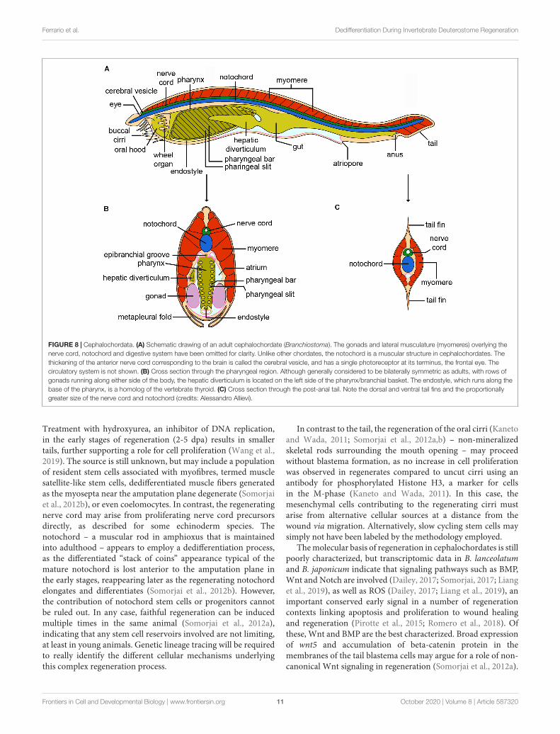
Cephalochordates (Clade Leptocardii; also called “amphioxus”or “lancelets”) are the earliest diverging invertebrate chordates(Figure 1A) and share the most similar body plan to thatof vertebrates (Bourlat et al., 2006; Delsuc et al., 2006,2008; Figure 1B). The three extant genera of cephalochordate(Asymmetron, Branchiostoma and Epigonichthys) include 30 orso species, all of which are considered to belong to a singlefamily, the Branchiostomatidae (Poss and Boschung, 1996).Regeneration has been described in a number of species ofBranchiostoma as well as in Asymmetron lucayanum (Andrews,1893; Probst, 1930; reviewed in Somorjai, 2017), most notably ofthe tail, a key chordate feature consisting of notochord, dorsalnerve cord and segmented musculature (Figure 8). Anteriorregeneration, or posterior regeneration of animals amputatedanterior to the anus, are generally poor (Somorjai et al., 2012b).
Tail regeneration in Branchiostoma lanceolatum andB. japonicum is considered to occur via the formation of a trueblastema (Somorjai et al., 2012b; Liang et al., 2019) consistingof at least superficially undifferentiated proliferating cells.
Frontiers in Cell and Developmental Biology | www.frontiersin.org 10 October 2020 | Volume 8 | Article 587320
fcell-08-587320 October 15, 2020 Time: 15:2 # 11
Ferrario et al. Dedifferentiation During Invertebrate Deuterostome Regeneration
FIGURE 8 | Cephalochordata. (A) Schematic drawing of an adult cephalochordate (Branchiostoma). The gonads and lateral musculature (myomeres) overlying thenerve cord, notochord and digestive system have been omitted for clarity. Unlike other chordates, the notochord is a muscular structure in cephalochordates. Thethickening of the anterior nerve cord corresponding to the brain is called the cerebral vesicle, and has a single photoreceptor at its terminus, the frontal eye. Thecirculatory system is not shown. (B) Cross section through the pharyngeal region. Although generally considered to be bilaterally symmetric as adults, with rows ofgonads running along either side of the body, the hepatic diverticulum is located on the left side of the pharynx/branchial basket. The endostyle, which runs along thebase of the pharynx, is a homolog of the vertebrate thyroid. (C) Cross section through the post-anal tail. Note the dorsal and ventral tail fins and the proportionallygreater size of the nerve cord and notochord (credits: Alessandro Allievi).
Treatment with hydroxyurea, an inhibitor of DNA replication,in the early stages of regeneration (2-5 dpa) results in smallertails, further supporting a role for cell proliferation (Wang et al.,2019). The source is still unknown, but may include a populationof resident stem cells associated with myofibres, termed musclesatellite-like stem cells, dedifferentiated muscle fibers generatedas the myosepta near the amputation plane degenerate (Somorjaiet al., 2012b), or even coelomocytes. In contrast, the regeneratingnerve cord may arise from proliferating nerve cord precursorsdirectly, as described for some echinoderm species. Thenotochord – a muscular rod in amphioxus that is maintainedinto adulthood – appears to employ a dedifferentiation process,as the differentiated “stack of coins” appearance typical of themature notochord is lost anterior to the amputation plane inthe early stages, reappearing later as the regenerating notochordelongates and differentiates (Somorjai et al., 2012b). However,the contribution of notochord stem cells or progenitors cannotbe ruled out. In any case, faithful regeneration can be inducedmultiple times in the same animal (Somorjai et al., 2012a),indicating that any stem cell reservoirs involved are not limiting,at least in young animals. Genetic lineage tracing will be requiredto really identify the different cellular mechanisms underlyingthis complex regeneration process.
In contrast to the tail, the regeneration of the oral cirri (Kanetoand Wada, 2011; Somorjai et al., 2012a,b) – non-mineralizedskeletal rods surrounding the mouth opening – may proceedwithout blastema formation, as no increase in cell proliferationwas observed in regenerates compared to uncut cirri using anantibody for phosphorylated Histone H3, a marker for cellsin the M-phase (Kaneto and Wada, 2011). In this case, themesenchymal cells contributing to the regenerating cirri mustarise from alternative cellular sources at a distance from thewound via migration. Alternatively, slow cycling stem cells maysimply not have been labeled by the methodology employed.
The molecular basis of regeneration in cephalochordates is stillpoorly characterized, but transcriptomic data in B. lanceolatumand B. japonicum indicate that signaling pathways such as BMP,Wnt and Notch are involved (Dailey, 2017; Somorjai, 2017; Lianget al., 2019), as well as ROS (Dailey, 2017; Liang et al., 2019), animportant conserved early signal in a number of regenerationcontexts linking apoptosis and proliferation to wound healingand regeneration (Pirotte et al., 2015; Romero et al., 2018). Ofthese, Wnt and BMP are the best characterized. Broad expressionof wnt5 and accumulation of beta-catenin protein in themembranes of the tail blastema cells may argue for a role of non-canonical Wnt signaling in regeneration (Somorjai et al., 2012a).
Frontiers in Cell and Developmental Biology | www.frontiersin.org 11 October 2020 | Volume 8 | Article 587320

fcell-08-587320 October 15, 2020 Time: 15:2 # 12
Ferrario et al. Dedifferentiation During Invertebrate Deuterostome Regeneration
Conversely, identification in the blastema of transcripts of sp5,a downstream target of beta-catenin-dependent Wnt signalingduring amphioxus development, suggests that canonical Wntfunction also operates during regeneration (Dailey et al., 2017).Msx, a marker for undifferentiated cells as well as a target ofBMP signaling, and chordin, a BMP antagonist, are also expressedin B. lanceolatum regenerates (Somorjai et al., 2012b). Recently,it has also been shown that bmp2/4 is expressed in wounds inB. japonicum, both those that induce regeneration and those thatdo not, suggesting a more general role in the repair process andnot just regeneration per se (Liang et al., 2019). In this context,results showing that the implantation of Noggin-soaked beadsat the amputation site and injection of bmp2/4 morpholinos –both of which should reduce BMP signaling – cause degenerationof tails (Liang et al., 2019) deserve further attention. Othergenes expressed during tail regeneration include soxB2, thecephalochordate ortholog of sox17/21 in vertebrates, and pax3/7(transcripts and protein). Both are expressed in the nerve cord,while pax3/7 is also expressed in blastema cells and in cellsthat might constitute muscle satellite-like stem cells (Somorjaiet al., 2012b). There are in fact two Pax3/7 genes in amphioxus,pax3/7a and pax3/7b, arising from a cephalochordate-specifictandem duplication event, and which were originally identified ina tail regenerate transcriptome in B. lanceolatum (Somorjai, 2017;Barton-Owen et al., 2018). Studies elucidating their differentialroles during regeneration are currently underway.
Cirrus regeneration is much less well characterized thantail regeneration molecularly. Skeletogenesis genes soxE andrunx, as well as extracellular matrix (ECM) genes includingSPARC/SPARCL and the fibrillar collagens fcol1 and fcol2, areexpressed in mesenchyme cells during oral cirrus regenerationin B. japonicum (formerly classed as B. belcheri) (Kaneto andWada, 2011), suggesting a recapitulation of developmental geneprograms, similarly to tail regeneration. However, how themolecular and cellular processes underlying regeneration inamphioxus are integrated remain unknown. Detailed analysesof the expression patterns of more genes identified usingtranscriptomic approaches during regeneration will be invaluablein our understanding of the cellular basis of regenerationin cephalochordates.
TUNICATA
Tunicates or urochordates are invertebrate chordates consideredthe sister group of vertebrates (Bourlat et al., 2006; Delsucet al., 2006, 2008; Figure 1A). They are marine filter-feeders, benthic or pelagic, classically subdivided into Ascidiacea(ascidians), Thaliacea (salps and pyrosomes) and Larvacea(appendicularians), although the internal interrelationshipsamong the various taxa are still controversial (Stach et al., 2010).Tunicates owe their name to the distinctive covering embeddingthe body -the tunic- a cellulose-containing structure unique inthe animal kingdom (Deck et al., 1967; Welsch, 1984; Van Daeleet al., 1992), whereas the name “urochordates” comes from thenotochord, the supporting rod characterizing chordates, herelimited to the larval muscular tail. Almost all tunicate species have
a swimming tadpole-like larva that metamorphoses into a highlyderived and specialized juvenile, with a dramatic change of bodyorganization (Stolfi and Brown, 2015).
Tunicates include both solitary and colonial species(Figure 1B): the latter are unique among chordates as theyare capable of asexual reproduction by budding (Brown andSwalla, 2012). Their particular phylogenetic position hasattracted considerable interest; however, the regenerativecapabilities of the group have only been studied in a handful ofspecies of solitary and colonial ascidians. Regeneration studiesstarted in the late XIX century as investigators/scientists werefascinated by the ability of ascidians -unusual among metazoans-to regenerate a functional brain (Berrill, 1951; Jeffery, 2015a).Today, the availability of genomes and transcriptomes of anincreasing number of tunicate species is leading to new analysesof the regenerative process and a better understanding of themolecules and signaling pathways involved. Below, we providean updated review of the main advances in our knowledge ofregeneration in ascidians.
Solitary AscidiansTunic RegenerationThe tunic can easily be detached from the body wall. Oldexperiments demonstrate that, at least in Ciona intestinalis,Ascidia mentula and Ascidiella aspersa, it is easily and rapidlyreformed by the underlying epidermis (Fol, 1908; Azéma, 1927;Pérès, 1948).
Partial Body RegenerationSolitary ascidians (Figures 1B, 9A) are capable of partial bodyregeneration (Gordon et al., 2019). Jeffery and collaboratorshave studied the process in detail in adults of the speciesCiona robusta, previously referred as Ciona intestinalis type A(Caputi et al., 2007). When animals are bisected, the posterior(proximal) region of the body, containing viscera, can regeneratethe anterior (distal) part, including the brain, provided that itcontains at least a part of the pharynx. Conversely, the anteriorpart of the body cannot regenerate any of the proximal structures(Jeffery, 2015a,b). Even when the animal is cut in three partsalong the proximo-distal axis, the middle section can reformthe distal part (Jeffery, 2015b). This implies that the pharynx isimportant for regeneration, and is crucial for the replacement ofdistal body parts.
Regeneration of the oral siphon in Ciona received considerableinterest in the past (Wermel, 1930; Sutton, 1953; Whittaker,1975). Recently, Jeffery (2015a,b) demonstrated that both short-distance and long-distance processes are involved in the process.Short-distance regeneration occurs when the siphon is amputatedat its tip, and leads to the replacement of the oral pigment organs(OPOs) and of the very distant part of the siphon. This kind ofregeneration does not require cell proliferation; neither labelingwith the cell proliferation maker EdU nor effects of proliferationinhibitors colchicine or nocodazole are observed (Jeffery, 2015b).It relies on small aggregates of stem/progenitor cells alreadypresent in the siphon, activated by the injury (Auger et al., 2010;Jeffery, 2015b).
Frontiers in Cell and Developmental Biology | www.frontiersin.org 12 October 2020 | Volume 8 | Article 587320

fcell-08-587320 October 15, 2020 Time: 15:2 # 13
Ferrario et al. Dedifferentiation During Invertebrate Deuterostome Regeneration
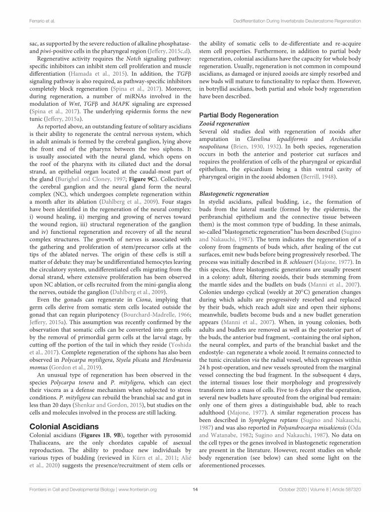
FIGURE 9 | Schematic drawings showing the anatomy of (A) a solitary ascidian, (B) a botryllid colonial ascidian, and (C) the organization of the neural complex of asolitary ascidian (credits: Alessandro Allievi).
Long distance regeneration leads to the formation of newcircular muscle fibers and neurons, and requires the activity ofstem/progenitor cells originating in the pharyngeal region. Thesemigrate distally where they form a blastema, with a well-definedproliferation zone, in the proximal region of the siphon stump(Auger et al., 2010). When the siphon is amputated at its base,only long-distance regeneration occurs, with stem/progenitorcells from the pharyngeal region forming both the blastema andthe OPOs (Jeffery, 2015b).
The stem/progenitor cells originate in the lymph nodes,typical stem cell niches located in the transverse vessels of
the pharynx, where alkaline phosphatase positive, piwi-positiveand EdU-labeled cells reside (Jeffery, 2015b). The lymph nodesare hematopoietic organs, involved in the renewal of thecirculating hemocytes (Ermak, 1976). From the pharynx vessels,EdU positive-cells migrate into the regeneration blastema afterthe amputation of the siphon. This has been confirmed bytransplanting the pharynx from small animals, labeled with EdU,into the pharynx of larger animals: in this case EdU-labeledcells can be found in the regeneration blastema (Jeffery, 2015b).Regenerative abilities decline with age, up to their completedisappearance, due to the depletion of stem cells in the branchial
Frontiers in Cell and Developmental Biology | www.frontiersin.org 13 October 2020 | Volume 8 | Article 587320
fcell-08-587320 October 15, 2020 Time: 15:2 # 14
Ferrario et al. Dedifferentiation During Invertebrate Deuterostome Regeneration
sac, as supported by the severe reduction of alkaline phosphatase-and piwi-positive cells in the pharyngeal region (Jeffery, 2015c,d).
Regenerative activity requires the Notch signaling pathway:specific inhibitors can inhibit stem cell proliferation and muscledifferentiation (Hamada et al., 2015). In addition, the TGFβ
signaling pathway is also required, as pathway-specific inhibitorscompletely block regeneration (Spina et al., 2017). Moreover,during regeneration, a number of miRNAs involved in themodulation of Wnt, TGFβ and MAPK signaling are expressed(Spina et al., 2017). The underlying epidermis forms the newtunic (Jeffery, 2015a).
As reported above, an outstanding feature of solitary ascidiansis their ability to regenerate the central nervous system, whichin adult animals is formed by the cerebral ganglion, lying abovethe front end of the pharynx between the two siphons. Itis usually associated with the neural gland, which opens onthe roof of the pharynx with its ciliated duct and the dorsalstrand, an epithelial organ located at the caudal-most part ofthe gland (Burighel and Cloney, 1997; Figure 9C). Collectively,the cerebral ganglion and the neural gland form the neuralcomplex (NC), which undergoes complete regeneration withina month after its ablation (Dahlberg et al., 2009). Four stageshave been identified in the regeneration of the neural complex:i) wound healing, ii) merging and growing of nerves towardthe wound region, iii) structural regeneration of the ganglionand iv) functional regeneration and recovery of all the neuralcomplex structures. The growth of nerves is associated withthe gathering and proliferation of stem/precursor cells at thetips of the ablated nerves. The origin of these cells is still amatter of debate: they may be undifferentiated hemocytes leavingthe circulatory system, undifferentiated cells migrating from thedorsal strand, where extensive proliferation has been observedupon NC ablation, or cells recruited from the mini-ganglia alongthe nerves, outside the ganglion (Dahlberg et al., 2009).
Even the gonads can regenerate in Ciona, implying thatgerm cells derive from somatic stem cells located outside thegonad that can regain pluripotency (Bourchard-Madrelle, 1966;Jeffery, 2015a). This assumption was recently confirmed by theobservation that somatic cells can be converted into germ cellsby the removal of primordial germ cells at the larval stage, bycutting off the portion of the tail in which they reside (Yoshidaet al., 2017). Complete regeneration of the siphons has also beenobserved in Polycarpa mytiligera, Styela plicata and Herdmaniamomus (Gordon et al., 2019).
An unusual type of regeneration has been observed in thespecies Polycarpa tenera and P. mityligera, which can ejecttheir viscera as a defense mechanism when subjected to stressconditions. P. mityligera can rebuild the branchial sac and gut inless than 20 days (Shenkar and Gordon, 2015), but studies on thecells and molecules involved in the process are still lacking.
Colonial AscidiansColonial ascidians (Figures 1B, 9B), together with pyrosomidThaliaceans, are the only chordates capable of asexualreproduction. The ability to produce new individuals byvarious types of budding (reviewed in Kürn et al., 2011; Aliéet al., 2020) suggests the presence/recruitment of stem cells or
the ability of somatic cells to de-differentiate and re-acquirestem cell properties. Furthermore, in addition to partial bodyregeneration, colonial ascidians have the capacity for whole bodyregeneration. Usually, regeneration is not common in compoundascidians, as damaged or injured zooids are simply resorbed andnew buds will mature to functionality to replace them. However,in botryllid ascidians, both partial and whole body regenerationhave been described.
Partial Body RegenerationZooid regenerationSeveral old studies deal with regeneration of zooids afteramputation in Clavelina lepadiformis and Archiascidianeapolitana (Brien, 1930, 1932). In both species, regenerationoccurs in both the anterior and posterior cut surfaces andrequires the proliferation of cells of the pharyngeal or epicardialepithelium, the epicardium being a thin ventral cavity ofpharyngeal origin in the zooid abdomen (Berrill, 1948).
Blastogenetic regenerationIn styelid ascidians, palleal budding, i.e., the formation ofbuds from the lateral mantle (formed by the epidermis, theperibranchial epithelium and the connective tissue betweenthem) is the most common type of budding. In these animals,so-called “blastogenetic regeneration” has been described (Suginoand Nakauchi, 1987). The term indicates the regeneration of acolony from fragments of buds which, after healing of the cutsurfaces, emit new buds before being progressively resorbed. Theprocess was initially described in B. schlosseri (Majone, 1977). Inthis species, three blastogenetic generations are usually presentin a colony: adult, filtering zooids, their buds stemming fromthe mantle sides and the budlets on buds (Manni et al., 2007).Colonies undergo cyclical (weekly at 20◦C) generation changesduring which adults are progressively resorbed and replacedby their buds, which reach adult size and open their siphons;meanwhile, budlets become buds and a new budlet generationappears (Manni et al., 2007). When, in young colonies, bothadults and budlets are removed as well as the posterior part ofthe buds, the anterior bud fragment, -containing the oral siphon,the neural complex, and parts of the branchial basket and theendostyle- can regenerate a whole zooid. It remains connected tothe tunic circulation via the radial vessel, which regresses within24 h post-operation, and new vessels sprouted from the marginalvessel connecting the bud fragment. In the subsequent 4 days,the internal tissues lose their morphology and progressivelytransform into a mass of cells. Five to 6 days after the operation,several new budlets have sprouted from the original bud remain:only one of them gives a distinguishable bud, able to reachadulthood (Majone, 1977). A similar regeneration process hasbeen described in Symplegma reptans (Sugino and Nakauchi,1987) and was also reported in Polyandrocarpa misakiensis (Odaand Watanabe, 1982; Sugino and Nakauchi, 1987). No data onthe cell types or the genes involved in blastogenetic regenerationare present in the literature. However, recent studies on wholebody regeneration (see below) can shed some light on theaforementioned processes.
Frontiers in Cell and Developmental Biology | www.frontiersin.org 14 October 2020 | Volume 8 | Article 587320

fcell-08-587320 October 15, 2020 Time: 15:2 # 15
Ferrario et al. Dedifferentiation During Invertebrate Deuterostome Regeneration
Colonial circulatory system regenerationThe colonial ascidian B. schlosseri is able to reform the tunicand the colonial vasculature within 24–48 h of experimentalremoval (Zaniolo and Trentin, 1987; Gasparini et al., 2008,2014; Tiozzo et al., 2008). CCS regeneration is preceded bythe proliferation of epidermal cells, as revealed by stainingwith anti-PCNA antibodies, and the formation of new tunicin the damaged region (Gasparini et al., 2008). Both cellsdetaching from the epidermis and hemocytes entering the tuniccontribute to reform the normal tunic cell endowment. Vesselregeneration occurs by sprouting from the vessel remnantsand is stimulated by vertebrate vascular endothelial growthfactor (VEGF) and epidermal growth factor (EGF) injected intothe circulatory system. In addition, antibodies raised againstvertebrate fibroblast growth factor-2 (FGF-2), VEGF, EGF andthe receptors VEGFR1 VEGFR2 and EGFR recognize the apexof the tubular sprouts (Gasparini et al., 2008, 2014). Theinvolvement of the VEGF pathway has been confirmed bythe observation that both knock-down of the Botryllus VEGFreceptor (VEGFR) gene and chemical inhibition of VEGFR blockvascular regeneration (Tiozzo et al., 2008). Cell tracing methodssuggest that regeneration is supported by the proliferationof vascular resident cells without the contribution of mobileprogenitors (Braden et al., 2014).
Whole body regenerationIn this type of regeneration, fragments of a colony containingonly the colonial matrix (i.e., the tunic and part of thecolonial vasculature) can form new buds (and therefore newzooids) from aggregates of circulating cells. These possesscharacteristic features of stem cells, such as small size andhigh nucleus/cytoplasm ratio, and are in contact with theepidermis lining the vasculature (Rinkevich et al., 1995, 2007a,b;Voskoboynik et al., 2007; Brown et al., 2009).
One of the first reports of WBR is that of Berrill and Cohen(1936) in Clavelina lepadiformis. In this species, experimentalfragmentation of the stolon leads to the formation of new zooids,provided that the stolon is of adequate size. Circulating cells ofthe stolon fragment aggregate and reorganize to form an emptyvesicle lined by the stolon epidermis, a situation similar to thedouble vesicle stage of botryllid ascidians (see below). WBR hasalso been reported in Clavelina moluccensis (Davis, 1988).
In B. schlosseri, WBR occurs only after the extirpation of allzooids and buds from the colonial matrix in colonies approachingor undergoing the generation change (Milkman, 1967; Sabbadinet al., 1975; Voskoboynik et al., 2007; Kürn et al., 2011; Ricciet al., 2016). Buds maintain the asymmetry of the parental colony,suggesting a role for the colonial matrix in the transmissionof bilateral asymmetry to the newly formed vascular buds(Sabbadin et al., 1975).
WBR closely resembles vascular budding, a spontaneousformation of new buds from the vessels of the vascular system,first described in botryllid ascidians more than 200 yearsago (Savigny, 1816) and observed and described again byGiard (1872); Bancroft (1903) and Herdman (1925). Vascularbudding of botryllid ascidians is frequently associated with theprocess of estivation or hibernation (e.g., in Botrylloides leachii),
during which colonies resorb their zooids to overcome adverseperiods and reform their zooids from the tunic vessels whenenvironmental conditions turn milder (Bancroft, 1903; Oka andWatanabe, 1959; Burighel et al., 1976; Atsumi and Saito, 2011).In Botryllus primigenus, Botrylloides leni and Botryllus delicates,vascular budding occurs continuously near the leading edge ofthe colony, at the bases of the ampullae (the blind endings ofthe tunic vessels), ensuring a quick increase in the size of thecolony itself (Oka and Watanabe, 1957; Saito and Watanabe,1985; Okuyama and Saito, 2001). Vascular budding has also beenreported in the stolidobranch styelid Symplegma brakenhielmi(Gutierrez and Brown, 2017) and the phlebobranch Perophoraviridis (Freeman, 1964).
In both WBR and vascular budding, hemocytes adheringto the vessel epithelium show the characteristics of stem cells,such as small size and large, round, euchromatic nuclei (Okaand Watanabe, 1957; Freeman, 1964; Rinkevich et al., 2007a,2008), and are able to generate both the soma and germ line(Sunanaga et al., 2006). In the course of bud development, thesecell aggregates grow and organize themselves to form the doublevesicle stage, critical for bud organogenesis (Rinkevich et al.,1995; Oka and Watanabe, 1957; Voskoboynik et al., 2007). Thischaracteristic stage is considered a triploblastic vesicle of thegastrula type (Brien, 1968), based on its organogenetic capacities:the outer vesicle is formed by the epidermis and will giverise to the zooid epidermis, whereas the inner vesicle and theintermediate mesenchyme cells will form all the internal tissuesof the zooid (Manni and Burighel, 2006; Manni et al., 2007;Ricci et al., 2016).
WBR has been particularly well studied in B. leachii. In thisspecies, the process occurs in five stages (Zondag et al., 2016;Blanchoud et al., 2017). In the first, lasting 15 h, wound healingis followed by a restructuring of the vessel architecture and of theampullae, leading to the formation of small regeneration niches(stage 2). The contraction of the tissues marks stage 3, whilehoming of stem cells to the regeneration niches characterizesstage 4. Finally, competition among the various stem cellaggregates (stage 5) leads to the maturation of a single bud perexperimental fragment (Rinkevich et al., 2007a,b, 2008; Zondaget al., 2016; Blanchoud et al., 2017).
The process of zooid formation from buds separated fromthe parental zooid in Polyandrocarpa misakiensis is consideredanalogous to WBR of botryllid ascidians (Kawamura et al.,2018). Here, buds are formed by the epidermis, the peribranchialepithelium and the mesenchyme cells between them. Thesituation resembles the double vesicle stage of botryllid ascidiansand requires transdifferentiation of the peribranchial epithelium(Kawamura and Fujiwara, 1994, 1995).
As regards “stemness” markers, hemocyte aggregates donot express piwi in B. primigenus vascular buds (Sunanagaet al., 2010). However, hemocytes lining the vessel epitheliumwith the capacity to proliferate and expressing piwi have beenpostulated to play a role in the formation of the bud primordiain Botrylloides violaceus (Brown et al., 2009) and B. leachii(Rinkevich et al., 2010) WBR, as well as in B. schlosseri vascularbudding. In B. violaceus WBR, piwi-positive hemocytes aroundthe regenerating mass of cells are frequently immunolabeled
Frontiers in Cell and Developmental Biology | www.frontiersin.org 15 October 2020 | Volume 8 | Article 587320

fcell-08-587320 October 15, 2020 Time: 15:2 # 16
Ferrario et al. Dedifferentiation During Invertebrate Deuterostome Regeneration
TABLE 1 | Main undifferentiated and differentiated cytotypes involved in invertebrate deuterostome regeneration.
Phylum Clade Phenomenon Progenitor cells Cells undergoing dedifferentiation
Echinodermata Crinoidea Arm and visceral mass regeneration Amebocytes, coelomocytes Muscle cells, coelomic epithelium,neurosecretory cells
Echinoidea Spine and test regeneration - - Muscle cells, sclerocytes
Asteroidea Arm regeneration Coelomocytes Muscle cells, coelomic epithelium
Ophiuroidea - - Muscle cells, coelomic epithelium
Holothuroidea - - Muscle cells, coelomic epithelium, glialcells
Hemichordata Enteropneusta Anterior and posterior regeneration Circulating stem cells associated withblood vessels (?), mesenchymal cells (?)
Unclear
Cephalochordata Leptocardii Tail regeneration Muscle satellite-like cells (?) in the tail;coelomocytes (?), mesenchymal cells(?) in oral cirri
Muscle cells (?), nerve cord cells (?),notochord cells (?) in the tail; skeletalrod cells in oral cirri (?)
Tunicata Ascidiacea Short distance partial bodyregenerationLong distance partial body regenerationBlastogenetic regenerationCCS regenerationWBR
ASCs in the oral siphon of Ciona,Cells from the peripharyngeal stem cellnichesBud tissuesVascular epitheliumHemoblasts
Bud tissuesVascularEpitheliumHemoblasts
Terms and Definitions: Blastema, Localized pool of cells, undifferentiated/pluripotent as well as retaining their tissue origin, usually of mesenchymal origin and envelopedby an epithelial layer, able to massively proliferate and differentiate into different cytotypes; Dedifferentiation, Process of cellular reprogramming by which differentiated(mature) somatic cells lose their specialization (oligo/unipotency) and revert to a less differentiated state (pluri/multipotency); Deuterostomes, Eumetazoans that duringembryonic development show radial indeterminate cleavage and enterocoely, with the blastopore becoming the anus; Epithelial-Mesenchymal Transition, Dynamicand finely regulated process during which epithelial cells lose their epithelial features, disrupt their underlying basement membrane and assume features typical ofmesenchymal cells, such as migratory capacity and production of extracellular matrix components. It is typical of embryonic development, tissue regeneration andmorphogenesis, cancer invasion, wound healing, immune response, etc; Epimorphosis, Regeneration model mainly based on the proliferation and differentiation ofcells composing the localized blastema; Morphallaxis, Regeneration model mainly based on the remodeling of pre-existing tissues without the formation of a localizedblastema through significant local cell proliferation. It may involve the dedifferentiation of localized tissues or migration of cells from other distant locations; ProgenitorCells, Stem cell-derived cells lacking unlimited self-renewal capacity, and that differentiate into limited specialized cytotypes; Transdifferentiation, Process of cellularreprograming by which differentiated (mature) somatic cells convert directly into another type of differentiated (mature) somatic cell without passing through an intermediatepluripotent/multipotent state.
by anti-PCNA antibodies: they have been hypothesized to beprecursor cells that will be integrated into the developing bud asthey start to differentiate (Brown et al., 2009).
Budding in Botrylloides WBR requires the presence of retinoidacid (RA), as inhibitors of RA synthesis block the process,whereas RA agonists accelerate bud formation and increasethe number of buds per experimental fragment (Rinkevichet al., 2007b). Serine protease inhibitors alter the developmentof regeneration buds in Botrylloides (Rinkevich et al., 2007b),probably due to the role of serine proteases in remodeling theECM, which is required for proper cell-cell communicationduring regeneration (Rinkevich et al., 2007a). This agreeswith the observed increase in transcription of a trypsin-likeserine protease upon RA treatment in the budding ascidianP. misakiensis (Ohashi et al., 1999). In B. leachii, the transcriptsfor aldehyde dehydrogenase, the enzyme involved in RAsynthesis, and a serine protease similar to the mammalianurokinase-type plasminogen activator, are located in circulatingphagocytes (Rinkevich et al., 2007a,b). This suggests a key roleof these cells in the control of vascular budding and WBR, inaddition to their ascertained role in palleal budding. B. schlosseriphagocytes are, in fact, required for proper clearance of apoptoticcells and corpses from the tissues of old zooids during thegeneration change. The recycling of nutrients derived from theirdigestion is required to support bud growth, as colonies areunable to feed during this period. This important phagocytic role
is further supported by the observation that blocking phagocyteactivity results in the arrest of blastogenesis (Voskoboynik et al.,2004). This also implies the involvement of innate immuneresponses, since phagocytes are key players in morphogeneticevents of compound ascidians (Franchi and Ballarin, 2017).In accordance with this, B. leachii WBR is associated withthe differential transcription of various immune-related genes(Rinkevich et al., 2007a, 2008).
CONCLUDING REMARKS ANDPERSPECTIVES
Reviewing regeneration with a focus only on the contribution ofcell proliferation, blastema formation, or totipotent ASCs leavesa large number of unanswered questions on the cellular andmolecular underpinnings of this complex process. The impressivevariety of regenerative mechanisms displayed within the animalkingdom makes it clear that adopting a comparative approachis as valuable as investigating emerging models. Importantly, thestudy of this fundamental biological phenomenon in invertebratemodels can improve our understanding of core events inboth regeneration-competent animals and those with reducedregenerative ability, such as humans. The vertebrate species mosteasily reared in captivity and used for regeneration studies -suchas rodents, chickens, frogs or zebrafish- are costly to maintain,
Frontiers in Cell and Developmental Biology | www.frontiersin.org 16 October 2020 | Volume 8 | Article 587320

fcell-08-587320 October 15, 2020 Time: 15:2 # 17
Ferrario et al. Dedifferentiation During Invertebrate Deuterostome Regeneration
may possess quite limited regenerative abilities in adulthood,and their management is often problematic for ethical reasons.Many invertebrate deuterostomes, instead, show extensive adultregeneration, are easy to maintain in laboratory conditions,and, except in cases where they have protected status, theiruse for experimentation generally faces fewer restrictions. Pastlimitations, such as the availability of -omics data and techniquesfor genetic manipulation, are also rapidly disappearing. However,despite being reliable research organisms, they are still largelyneglected as models in regeneration research.
Adult regeneration involves not only stem cell recruitment,but also dedifferentiation phenomena, which implies remarkablecellular plasticity. In deuterostomes, a large diversity of processesand cytotypes is often detectable even within the samephylum. Nevertheless, recruitment of cells deriving from thededifferentiation of adult cells, rather than the use of residentstem cells, appears to predominate: indeed, in most clades,localized (tissue-specific) “recycling” of specialized cells is likelyto occur. These cells generally originate from nearby tissues,which are locally remodeled and become the source of newcellular material. In most cases, such progenitors give rise torestricted types of cells, i.e., cells of each tissue regenerateelements of that tissue. There are, however, some exceptions, suchas the multipotent epithelia of tunicates (which originate almostall tissues), and the coelomic epithelia of echinoderms (whichgenerate the coelomocytes, the muscles and, likely, the skeleton).More such examples are likely to be discovered as research in thisarea intensifies with broader taxon sampling.
The involvement of resident undifferentiated cells duringregeneration is generally limited, with the exception ofundifferentiated amebocytes and coelomocytes in crinoidsand hemoblasts in tunicates and, likely, of hemichordates andamphioxus. When present, these cells show a wide range ofpotency and, usually, are multi- or pluripotent stem cells orprogenitor cells (Table 1).
The use of dedifferentiation as a major mechanism for tissuerepair underscores the idea that cell plasticity in invertebratedeuterostomes is higher compared to vertebrates (Table 1).Among chordates, amphioxus and solitary ascidians, whichshow considerable but still relatively limited regenerationcompared to other invertebrate deuterostome groups, can beconsidered good “transition” species between ambulacrariansand vertebrates. Conversely, the high regenerative abilityof colonial ascidians has probably appeared secondarily, inassociation with asexual reproduction. This strict connectionbetween regeneration and asexual reproduction is also truefor echinoderms and hemichordates and is supported by thecommon molecular events associated with the two processes(Sánchez Alvarado, 2000). Although each tissue generallyregenerates independently, it nevertheless has to do so insynchrony with the others to successfully restore a preciseand accurate body plan. In this sense, regeneration is avery complex process that implies a precise and integratedresponse requiring the coordination of a plethora of differenttissues, molecules and signals. Circulating cells can facilitateregeneration, especially in the first phases after injury, since theyare overcoming tissue separation and allow both cell and signal
spreading (Table 1). EMT and cell migration contribute to thisgeneral coordination.
Overall, it is difficult to strictly define the “stemness”properties of the cells involved in adult regeneration ofinvertebrate deuterostomes. In vertebrates, stem cells areusually characterized by precise undifferentiated ultrastructuralfeatures and specific molecular markers for pluri/multipotency,and may be present in well-defined niches. In contrast, inthe invertebrate models reviewed here, cells without obviouslocalization in defined niches [with the possible exception ofthe lymph nodes (Ermak, 1976) and the endostyle in tunicates(Voskoboynik et al., 2008)] and with both undifferentiated anddifferentiated ultrastructural characters effectively act as stemcells, irrespective of their tissue of origin or the mechanisms bywhich they are recruited to the regeneration zone. Moleculardata also underscore the important contribution of bothundifferentiated and differentiated cells, although labeling anddetailed cellular tracking are still required to pinpoint theprecise origin and fate of these cells. Therefore, we propose awider interpretation of the stem cell concept, not necessarilyand strictly related to the classic idea of stem cells asundifferentiated cells, but including also cells deriving fromdedifferentiation phenomena. This is in line with the definitionof stem cells recently proposed by Post and Clevers (2019),which is based on their function rather than any morphologicalor molecular criteria: the ability to replace lost cells throughcell division.
From an evolutionary viewpoint, the extensive cell plasticitydescribed in adult invertebrate deuterostomes -both in termsof cell-lineage restriction and cell potency- may be one ofthe key elements of their successful regenerative responses.However, the relatively limited and patchy informationavailable in these animal models renders difficult theidentification of shared molecular pathways underpinningcell dedifferentiation, if indeed any exist. The observationthat conserved developmental regulators -such as Wnt, Notchand TGFβ/BMP, to name just a few- are often re-deployedduring regeneration may be less consistent with their rolesas causative agents in the dedifferentiation/cell fate process,and more indicative of researcher prejudices for “favoritegenes” and their ease of identification in homology-basedsearches. The question of which molecular switch mightfacilitate dedifferentiation in many invertebrates upon injury,and how this machinery may differ or be inactivated innon-regenerating organisms, is far more difficult to tackleand may lie rather in how epigenetic states and chromatinare regulated (Merrell and Stanger, 2016; Hayashi et al.,2020; Lee et al., 2020), or even in physical properties ofthe cellular microenvironment (Vining and Mooney, 2017).Further study of the various mechanisms regulating cellrecruitment, dedifferentiation, and specification will helpelucidate whether any are conserved between invertebrateand vertebrate deuterostomes, or rather represent adiverse and plastic repertoire of solutions to a commonevolutionary problem.
Another intriguing aspect of regeneration is itsrelationship with immunity (Abnave and Ghigo, 2019;
Frontiers in Cell and Developmental Biology | www.frontiersin.org 17 October 2020 | Volume 8 | Article 587320

fcell-08-587320 October 15, 2020 Time: 15:2 # 18
Ferrario et al. Dedifferentiation During Invertebrate Deuterostome Regeneration
Arenas Gómez et al., 2020): various results indicate that theevolution of the adaptive immune system in vertebrates iscorrelated with a progressive loss of regenerative capabilityfrom fish to mammals (Godwin, 2014; Godwin and Rosenthal,2014). This phenomenon appears related to the persistenceof the inflammatory response, which impairs or limits theregeneration process, and scarring at the site of injury (Merscherand Neff, 2005; Eming et al., 2009; Godwin, 2014; Godwinand Rosenthal, 2014; Peiris et al., 2014; Fumagalli et al.,2018). In contrast, many invertebrates -including the highlyregenerative models described here- show scar-free woundhealing and yet have spectacularly complex and diverse innateimmune repertoires (Rinkevich et al., 2007a; Huang et al., 2008;Ramírez-Gómez and García-Arrarás, 2010; Smith et al., 2010;Dishaw et al., 2012; Tassia et al., 2017; Ferrario et al., 2018;Oren et al., 2019), which have rapidly and often independentlyevolved to deal with pathogen exposure in a sort of hostdefense/pathogen arms race (Ghosh et al., 2011). However,the building blocks of RAG V(D)J recombinase activity onwhich vertebrate adaptive immunity relies have been foundin invertebrate deuterostomes (Morales-Poole et al., 2017).Evidence is even emerging of immune system “memory” aspart of a continuum between innate and acquired immunity(Milutinovic and Kurtz, 2016; Netea et al., 2019). Therefore,the lack of adaptive immunity in invertebrate deuterostomescan partly explain their high regenerative potential, but is notentirely satisfactory when attempting to define causality in theface of such complexity. Our understanding of the impact of theimmune response on healing and regeneration in most systemsis at best rudimentary, and detailed studies of the consequencesof the appearance of adaptive immunity for regeneration abilitiesare still lacking.
A continuing challenge in stem cell research is unequivocallydistinguishing resident undifferentiated stem cells from thecellular products of dedifferentiation. In most non-model andemerging systems, including the invertebrate deuterostomesdiscussed here, differentiation state is inferred by mostlystatic snapshots of cells, accompanied by descriptions oflimited molecular signatures or ultrastructural characteristics.Nevertheless, without such careful and detailed observationsit is impossible to begin to elucidate the cellular basisof regeneration. The mechanisms regulating dedifferentiationare largely unknown, although research is beginning toidentify dedifferentiation as a key player in a number ofregenerative processes in genetically tractable vertebrate systems(Pesaresi et al., 2019; Aires et al., 2020). Therefore, whilewe believe that the available body of evidence indicates anoverarching central role for dedifferentiation in invertebratedeuterostomes, caution still must be exercised in interpretingfindings. Likewise, evidence for cell migration in regenerativeprocesses is mostly indirect, although widely assumed to occur.Exceptions include a few studies in solitary and colonialascidians as well as echinoderms (Voskoboynik et al., 2008;Jeffery, 2015b; Mashanov V. et al., 2017), where cells werelabeled with lipophilic dyes to track cell movements. BrdUand EdU pulse/chase labeling of proliferating cells also giveinsight into not only division dynamics but also lineage
and cell fate, particularly if fine control in labeling only afew cells is exercised. Comprehensive experiments are stilllacking in many regenerating animals (but see Mashanovet al., 2013; Jeffery, 2015b; Cary et al., 2019), and aresomewhat limited in scope, but do offer the advantage of notrequiring extensive resources, and labeling is not affected bythe changes in transcriptional or epigenetic state that occurduring reprograming. Stable transgenic animals, or geneticlabeling with inducible systems, permit by far the best controland resolution, and will be required to truly show changes indifferentiation state and potency. Currently, transgenesis hasbeen developed in some sea urchins (Buckley et al., 2018),amphioxus (Kozmikova and Kozmik, 2015), and the directdeveloping hemichordate Saccoglossus kowalevskii (Minor et al.,2019). A considerable molecular and genetic toolkit is alsoavailable for solitary ascidians, predominantly C. intestinalis butalso Phallusia mammillata (Cota, 2018). However, transgenicshave only been used to a limited degree, in Ciona, tostudy regeneration of the nervous system (Dahlberg et al.,2009), but hold great promise for elucidating the geneticbasis of dedifferentiation, for instance during myocardiumregeneration (Evans Anderson and Christiaen, 2016). Thereasons for this lacuna are manifold. First, many invertebratedeuterostomes have long generation times, and rearing thelarvae through metamorphosis to breeding age can be difficult.In addition, unlike models such as Drosophila melanogasteror Danio rerio, there are currently no repositories of geneticlines for any of the regenerating systems. This is in partdue to the cost to maintain such centralized resources, butalso to barriers to transport and importation of unusual(often marine) organisms. This is compounded by the sheerdiversity of models being used for regeneration studies andthe difficulty in acquiring funding for basic research on “weirdanimals”. However, sharing of knowledge and resources acrossthe stem cell community, facilitated by initiatives such asthe EU Horizon 2020 COST Action 16203 “MARISTEM”(Ballarin et al., 2018) will pave the way for more rapiduptake and development of the tools required to answerfundamental questions about stem cell biology in our closestliving relatives.
In conclusion, the greatest challenge for the regeneration field,from the perspective of future human medical applications, is tocompare invertebrate and vertebrate deuterostomes effectively,in terms of cells and mechanisms involved in the regenerativeprocess. The identification of potential commonalities as wellas differences will be crucial to the goal to improve therather limited regenerative capabilities in humans. Cell tracking,coupled with molecular and microscopy approaches, will becritical to address some of the main issues in animal regeneration,i.e., the understanding of the origin and fate of recruited cells. Assuch, it is first necessary to characterize different cytotypes andidentify cell-specific molecular markers in order to visualize andrecognize the cells involved in regeneration.
Next generation technologies, from single cell to ChIP-sequencing and proteomics, in combination with novelbioinformatic platforms and statistical analyses, will beinstrumental in achieving this objective. The development of
Frontiers in Cell and Developmental Biology | www.frontiersin.org 18 October 2020 | Volume 8 | Article 587320

fcell-08-587320 October 15, 2020 Time: 15:2 # 19
Ferrario et al. Dedifferentiation During Invertebrate Deuterostome Regeneration
in vitro systems, still very difficult in invertebrate deuterostomes(Di Benedetto et al., 2014b; Mercurio et al., 2014), will provide afurther tool to investigate these problems. Ultimately, researchersneed to ask the right questions and identify the model animalsmost appropriate for the study of regeneration. Only witha solid investment in understanding the diversity of cellularmechanisms underlying the remarkable regenerative ability seenin invertebrate models can we hope to unlock the dormantpotential of vertebrate systems.
AUTHOR CONTRIBUTIONS
CF, MS, IMLS, and LB conceived the work and wrote themanuscript. All authors contributed to the article and approvedthe submitted version.
FUNDING
This work was partly supported by a grant from the Italian MIUR.We are also indebted to the COST Action 16203 MARISTEM,which facilitated author discussions over the course of its periodicmeetings. Research in the Somorjai lab is supported by theWellcome Trust ISSF. Research in the Sugni lab is supported by agrant from the University of Milan (Linea 2).
ACKNOWLEDGMENTS
The authors are deeply grateful to Alessandro Allievi for theschematic drawings used for the figures. We also thank the twoanonymous reviewers for their comments, which improved themanuscript. The authors apologise to those whose work could notbe cited due to space restrictions.
REFERENCESAbnave, P., and Ghigo, E. (2019). Role of the immune system in regeneration and
its dynamic interplay with adult stem cells. Semin. Cell Dev. Biol. 87, 160–168.doi: 10.1016/j.semcdb.2018.04.002
Agata, K., Saito, Y., and Nakajima, E. (2007). Unifying principles of regeneration I:epimorphosis versus morphallaxis. Dev. Growth Differ. 49, 73–78. doi: 10.1111/j.1440-169x.2007.00919.x
Aires, R., Keeley, S. D., and Sandoval-Guzmán, T. (2020). “Basics of self-regeneration,” in Cell Engineering and Regeneration, eds J. Gimble, D. MaroltPresen, R. Oreffo, S. Wolbank, and H. Redl (Cham: Springer), 691–734. doi:10.1007/978-3-319-08831-0_66
Alié, A., Hiebert, L. S., Scelzo, M., and Tiozzo, S. (2020). The eventful historyof nonembryonic development in tunicates. J. Exp. Zool. B doi: 10.1002/jez.b.22940 [Epub ahead of print].
Andrews, E. A. (1893). An undescribed acraniate, Asymmetron lucayanum. Stud.Biol. Lab. Johns Hopkins Univ. 5, 213–247.
Annona, G., Holland, N. D., and D’Aniello, S. (2015). Evolution of the notochord.Evodevo 6:30.
Arenas Gómez, C. M., Sabin, K. Z., and Echeverri, K. (2020). Wound healingacross the animal kingdom: crosstalk between the immune system and theextracellular matrix. Dev. Dyn. 249, 834–846. doi: 10.1002/dvdy.178
Arimoto, A., and Tagawa, K. (2015). Hedgehog expression during developmentand regeneration in the hemichordate Ptychodera flava. Zoolog. Sci. 32, 33–37.doi: 10.2108/zs140157
Arimoto, A., and Tagawa, K. (2018). Regeneration in the enteropneusthemichordate, Ptychodera flava, and its evolutionary implications. Dev. GrowthDiffer. 60, 400–408. doi: 10.1111/dgd.12557
Arnone, M. I., Byrne, M., and Martinez, P. (2015). “Echinodermata,” inEvolutionary Developmental Biology of Invertebrates 6 (Deuterostomia), ed. A.Wanninger (Wien: Springer Verlag).
Atsumi, M. O., and Saito, Y. (2011). Studies on Japanese botryllid ascidians. V.A new species of the genus Botrylloides very similar to Botrylloides simodensisin morphology. Zool. Sci. 28, 532–542. doi: 10.2108/zsj.28.532
Auger, H., Sasakura, Y., Joly, J. S., and Jeffery, W. R. (2010). Regeneration oforal siphon pigment organs in the ascidian Ciona intestinalis. Dev. Biol. 339,374–389. doi: 10.1016/j.ydbio.2009.12.040
Azéma, M. (1927). Sur le rôle des prolongements vasculaires au cours de larégénération de la tunique chez Ascidiella aspersa (Müller). Bull. Soc. Zool. Fr.52, 549–557.
Aztekin, C., Hiscock, T. W., Marioni, J. C., Gurdon, J. B., Simons, B. D., and Jullien,J. (2019). Identification of a regeneration-organizing cell in the Xenopus tail.Science 364, 653–658. doi: 10.1126/science.aav9996
Ballarin, L., and Cima, F. (2005). Cytochemical properties of Botryllus schlosserihaemocytes: indications for morpho-functional characterisation. Eur. J.Histochem. 49, 255–264.
Ballarin, L., Rinkevich, B., Bartscherer, K., Burzynski, A., Cambier, S., Cammarata,M., et al. (2018). Maristem-stem cells of marine/aquatic invertebrates: frombasic research to innovative applications. Sustainability 10:526. doi: 10.3390/su10020526
Bancroft, F. W. (1903). Aestivation of Botrylloides Gascoi Della Valle. MarkAnniversary Volume. New York, NY: Henry Holt and Co, 147–166.
Bannister, R., McGonnell, I. M., Graham, A., Thorndyke, M. C., and Beesley, P. W.(2005). Afuni, a novel transforming growth factor-beta gene is involved inarm regeneration by the brittle star Amphiura filiformis. Dev. Genes Evol. 215,393–401. doi: 10.1007/s00427-005-0487-8
Bannister, R., McGonnell, I. M., Graham, A., Thorndyke, M. C., and Beesley,P. W. (2008). Coelomic expression of a novel bone morphogenetic protein inregenerating arms of the brittle star Amphiura filiformis. Dev. Genes Evol. 218,33–38. doi: 10.1007/s00427-007-0193-9
Barton-Owen, T. B., Ferrier, D. E. K., and Somorjai, I. M. L. (2018). Pax3/7duplicated and diverged independently in amphioxus, the basal chordatelineage. Sci. Rep. 8:9414.
Bely, A. E. (2010). Evolutionary loss of animal regeneration: pattern and process.Integr. Comp. Biol. 50, 515–527. doi: 10.1093/icb/icq118
Bely, A. E., and Nyberg, K. G. (2010). Evolution of animal regeneration: re-emergence of a field. Trends Ecol. Evol. 25, 161–170. doi: 10.1016/j.tree.2009.08.005
Ben Khadra, Y., Dimassi, N., and Martinez, P. (2018a). Expression of Wnt genes inEchinaster sepositus arm regeneration. Int. J. Biochem. Physiol. 3:000119.
Ben Khadra, Y., Ferrario, C., Di Benedetto, C., Said, K., Bonasoro, F., CandiaCarnevali, M. D., et al. (2015a). Re-growth, morphogenesis, and differentiationduring starfish arm regeneration. Wound Repair Regen. 23, 623–634. doi:10.1111/wrr.12336
Ben Khadra, Y., Ferrario, C., Di Benedetto, C., Said, K., Bonasoro, F., CandiaCarnevali, M. D., et al. (2015b). Wound repair during arm regeneration inthe red starfish Echinaster sepositus. Wound Repair Regen. 23, 611–622. doi:10.1111/wrr.12333
Ben Khadra, Y., Sugni, M., Ferrario, C., Bonasoro, F., Oliveri, P., Martinez, P.,et al. (2018b). Regeneration in stellate echinoderms: Crinoidea, Asteroidea andOphiuroidea. Results Probl. Cell Differ. 65, 285–320. doi: 10.1007/978-3-319-92486-1_14
Ben Khadra, Y., Sugni, M., Ferrario, C., Bonasoro, F., Varela Coelho, A., Martinez,P., et al. (2017). An integrated view of asteroid regeneration: tissues, cells andmolecules. Cell Tissue Res. 370, 13–28. doi: 10.1007/s00441-017-2589-9
Berrill, N. J. (1948). Structure, tadpole and bud formation in the ascidianArchidistoma. J. Mar. Biol. Ass. U.K. 27, 380–388. doi: 10.1017/s0025315400025431
Berrill, N. J. (1951). Regeneration and budding in tunicates. Biol. Rev. 26, 456–475.doi: 10.1111/j.1469-185x.1951.tb01207.x
Berrill, N. J., and Cohen, A. (1936). Regeneration in Clavelina lepadiformis. J. Exp.Biol. 13, 352–362.
Frontiers in Cell and Developmental Biology | www.frontiersin.org 19 October 2020 | Volume 8 | Article 587320
fcell-08-587320 October 15, 2020 Time: 15:2 # 20
Ferrario et al. Dedifferentiation During Invertebrate Deuterostome Regeneration
Biressi, A., Ting, Z., Dupont, S., Dahlberg, C., Di Benedetto, C., Bonasoro,F., et al. (2010). Wound healing and arm regeneration in Ophiodermalongicaudum and Amphiura filiformis (Ophiuroidea, Echinodermata):comparative morphogenesis and histogenesis. Zoomorphology 129, 1–19.doi: 10.1007/s00435-009-0095-7
Blanchoud, S., Zondag, L., Lamare, M. D., and Wilson, M. J. (2017). Hematologicalanalysis of the ascidian Botrylloides leachii (Savigny, 1816) during whole-bodyregeneration. Biol. Bull. 232, 143–157. doi: 10.1086/692841
Bodnar, A. G., and Coffman, J. A. (2016). Maintenance of somatic tissueregeneration with age in short- and long-lived species of sea urchins. Aging Cell15, 778–787. doi: 10.1111/acel.12487
Bonasoro, F., Ferro, P., Di Benedetto, C., Sugni, M., Mozzi, D., and CandiaCarnevali, M. D. (2004). “Regenerative potential of echinoid test,” inEchinoderms: München, eds T. Heinzeller and J. H. Nebelsick (London: Taylorand Francis Group).
Bosch, T. C. G. (2007). Why polyps regenerate and we don’t: towards a cellularand molecular framework for Hydra regeneration. Dev. Biol. 303, 421–433.doi: 10.1016/j.ydbio.2006.12.012
Bourchard-Madrelle, C. (1966). Régéneration de l’ovaire chez une ascidia simpleCiona intestinalis L. (Protochordé). Bull. Soc. Zool. Fr. 91, 107–114.
Bourlat, S. J., Juliusdottir, T., Lowe, C. J., Freeman, R., Aronowicz, J., Kirschner, M.,et al. (2006). Deuterostome phylogeny reveals monophyletic chordates and thenew phylum Xenoturbellida. Nature 444, 85–88. doi: 10.1038/nature05241
Braden, B. P., Taketa, D. A., Pierce, J. D., Kassmer, S., Lewis, D. D., and De Tomaso,A. W. (2014). Vascular regeneration in a basal chordate is due to the presenceof immobile, bi-functional cells. PLoS One 9:e95460. doi: 10.1371/journal.pone.0095460
Brien, P. (1930). Contribution a l’étude de la régénération naturelle etexpérimentelle chez les Clavelinides. Ann. Soc. R. Zool. Belg. 60, 33–46.
Brien, P. (1932). L’hétéromorphose chez les Tuniciers. La régénérationbithoracique et monothoracique des fragments oesophagiens de Clavelinalepadiformis (Müller). Bull. Acad. Belg. Cl. Sci. 18, 975–1005.
Brien, P. (1968). Blastogenesis and morphogenesis. Adv. Morphog. 7, 151–203.doi: 10.1016/b978-1-4831-9954-2.50009-4
Brito, G. M. (2018). Molecular Mechanisms of Salamander Limb Regeneration.Ph.D. thesis, Karolinska Institute, Solna.
Brown, F. D., Keeling, E. L., Le, A. D., and Swalla, B. J. (2009). Whole bodyregeneration in a colonial ascidian, Botrylloides violaceus. J. Exp. Zool. B 312,885–900. doi: 10.1002/jez.b.21303
Brown, F. D., and Swalla, B. J. (2012). Evolution and development of buddingby stem cells: ascidian coloniality as a case study. Dev. Biol. 369, 151–162.doi: 10.1016/j.ydbio.2012.05.038
Buckley, K. M., Dong, P., Cameron, R. A., and Rast, J. P. (2018). Bacterialartificial chromosomes as recombinant reporter constructs to investigate geneexpression and regulation in echinoderms. Brief. Funct. Genomics 17, 362–371.doi: 10.1093/bfgp/elx031
Burighel, P., Brunetti, R., and Zaniolo, G. (1976). Hibernation of the colonialascidian Botrylloides leachi (Savigny): histological observations. Boll. Zool. 43,293–301. doi: 10.1080/11250007609430146
Burighel, P., and Cloney, R. A. (1997). “Urochordata: ascidiacea,” in MicroscopicAnatomy of Invertebrates, Vol. 15, eds F. W. Harrison and E. E. Ruppert(New York, NY: Wiley-Liss, Inc), 221–347.
Burns, G., Ortega-Martinez, O., Thorndyke, M. C., Peck, L. S., Dupont, S., andClark, M. S. (2011). Dynamic gene expression profiles during arm regenerationin the brittle star Amphiura filiformis. J. Exp. Mar. Biol. Ecol. 407, 315–322.doi: 10.1016/j.jembe.2011.06.032
Canavesi, M. (2018). Analisi Morfologica e Molecolare Della Rigenerazione delBraccio di Amphipholis squamata (Echinodermata: Ophiuroidea). Master thesis,University of Milan, Milan.
Candelaria, A. G., Murray, G., File, S. K., and García-Arrarás, J. E. (2006).Contribution of mesenterial muscle dedifferentiation to intestine regenerationin the sea cucumber Holothuria glaberrima. Cell Tissue Res. 325, 55–65. doi:10.1007/s00441-006-0170-z
Candia Carnevali, M. D. (2006). Regeneration in echinoderms: repair, regrowthand cloning. Invertebr. Surviv. J. 3, 64–76.
Candia Carnevali, M. D., and Bonasoro, F. (2001). A microscopic overview ofcrinoid regeneration. Microsc. Res. Tech. 55, 1–24. doi: 10.1016/b978-0-12-814879-2.00001-7
Candia Carnevali, M. D., Bonasoro, F., and Biale, A. (1997). Pattern ofbromodeoxyuridine incorporation in the advanced stages of arm regenerationin the feather star Antedon mediterranea. Cell Tissue Res. 289, 363–374. doi:10.1007/s004410050883
Candia Carnevali, M. D., Bonasoro, F., Lucca, E., and Thorndyke, M. C. (1995).Pattern of cell proliferation in the early stages of arm regeneration in thefeather star Antedon mediterranea. J. Exp. Zool. 272, 464–474. doi: 10.1002/jez.1402720608
Candia Carnevali, M. D., Bonasoro, F., Welsch, U., and Thorndyke, M. C. (1998).“Arm regeneration and growth factors in crinoids,” in Echinoderms, eds R. Mooiand M. Telford (San Francisco, CA: Balkema), 145–150.
Caputi, L., Andreakis, N., Mastrototaro, F., Cirino, P., Vassillo, M., and Sordino, P.(2007). Cryptic speciation in a model invertebrate chordate. Proc. Natl. Acad.Sci. U.S.A. 104, 9364–9369. doi: 10.1073/pnas.0610158104
Cary, G. A., Wolff, A., Zueva, O., Pattinato, J., and Hinman, V. F. (2019). Analysisof sea star larval regeneration reveals conserved processes of whole-bodyregeneration across the Metazoa. BMC Biol. 17:16. doi: 10.1186/s12915-019-0633-9
Cortés Rivera, Y., Hernández, R. I., San Martin del Angel, P., Zarza Meza, E.,and Cuervo González, R. (2016). Regenerative potential of the sea star Linckiaguildinguii. Hidrobiológica 26, 95–100.
Cota, C. D. (2018). “Transgenic techniques for investigating cell biology duringdevelopment,” in Transgenic Ascidians. Advances in Experimental Medicine andBiology, Vol. 1029, ed. Y. Sasakura (Singapore: Springer).
Czarkwiani, A., Ferrario, C., Dylus, D. V., Sugni, M., and Oliveri, P. (2016).Skeletal regeneration in the brittle star Amphiura filiformis. Front. Zool. 13:18.doi: 10.1186/s12983-016-0149-x
Dahlberg, C., Auger, H., Dupont, S., Sasakura, Y., Thorndyke, M., and Joly,J. S. (2009). Refining the Ciona intestinalis model of central nervous systemregeneration. PLoS One 4:e4458. doi: 10.1371/journal.pone.0004458
Dailey, S. C. (2017). Evolutionary Developmental and Genomic Insights from a TailRegeneration Transcriptome of the Cephalochordate Branchiostoma lanceolatum.Ph.D. thesis, University of St Andrews, Scotland.
Dailey, S. C., Kozmikova, I., and Somorjai, I. M. L. (2017). Amphioxus Sp5 isa member of a conserved specificity protein complement and is modulatedby Wnt/β-catenin signalling. Int. J. Dev. Biol. 61, 723–732. doi: 10.1387/ijdb.170205is
Davis, A. R. (1988). Colony regeneration following damage and size-dependent mortality in the Australian ascidian Podoclavella moluccensissluiter. J. Exp. Mar. Biol. Ecol. 123, 269–285. doi: 10.1016/0022-0981(88)90047-0
Dawydoff, C. (1901). Beiträge zur kenntnis der regenerationsersecheinungen beiden ophiuren. Z. Wiss. Zool. 69, 202–234.
Dawydoff, C. (1909). Beobachtungen über den regenerationsprozess bei denenteropneusten. Z. Wiss. Zool. 93, 237–305.
Dawydoff, C. (1948). “Embranchement des stomocordés,” in Traite de Zoologie.Echinodermes, Stomocordés, Protochordés, Vol. 11, ed. P. P. Grassé (Paris:Masson et Cie), 367–449.
De Mulder, K., Kuales, G., Pfister, D., Willems, M., Egger, B., Salvenmoser, W.,et al. (2009). Characterization of the stem cell system of the acoel Isodiametrapulchra. BMC Dev. Biol. 9:69. doi: 10.1186/1471-213X-9-69
Deck, J. D., Hay, E. D., and Revel, J.-P. (1967). Fine structure and origin of thetunic of Perophora viridis. J. Morphol. 120, 267–280. doi: 10.1002/jmor.1051200304
Delsuc, F., Brinkmann, H., Chourrout, D., and Philippe, H. (2006). Tunicates andnot cephalochordates are the closest living relatives of vertebrates. Nature 439,965–968. doi: 10.1038/nature04336
Delsuc, F., Tsagkogeorga, G., Lartillot, N., and Hervé, P. (2008). Additionalmolecular support for the new chordate phylogeny. Genesis 46, 592–604. doi:10.1002/dvg.20450
Di Benedetto, C., Barbaglio, A., Martinello, T., Alongi, V., Fassini, D., Cullorà,E., et al. (2014a). Production, characterization and biocompatibility of marinecollagen matrices from an alternative and sustainable source: the sea urchinParacentrotus lividus. Mar. Drugs 12, 4912–4933. doi: 10.3390/md12094912
Di Benedetto, C., Parma, L., Barbaglio, A., Sugni, M., Bonasoro, F., and CandiaCarnevali, M. D. (2014b). Echinoderm regeneration: an in vitro approach usingthe crinoid Antedon mediterranea. Cell Tissue Res. 358, 189–201. doi: 10.1007/s00441-014-1915-8
Frontiers in Cell and Developmental Biology | www.frontiersin.org 20 October 2020 | Volume 8 | Article 587320

fcell-08-587320 October 15, 2020 Time: 15:2 # 21
Ferrario et al. Dedifferentiation During Invertebrate Deuterostome Regeneration
Dishaw, L. J., Haire, R. N., and Litman, G. W. (2012). The amphioxus genomeprovides unique insight into the evolution of immunity. Brief. Funct. Genomics11, 167–176. doi: 10.1093/bfgp/els007
Dolmatov, I. Y. (2014). Asexual reproduction in holothurians. Sci. World J.2014:527234. doi: 10.1155/2014/527234
Dubois, P., and Ameye, L. (2001). Regeneration of spines and pedicellariae inechinoderms: a review. Microsc. Res. Tech. 55, 427–437. doi: 10.1002/jemt.1188
Dupont, S., and Thorndyke, M. C. (2006). Growth or differentiation? Adaptiveregeneration in the brittle star Amphiura filiformis. J. Exp. Biol. 209, 3873–3881.doi: 10.1242/jeb.02445
Duque-Alarcon, A. P. (2015). Cellular and Molecular Aspects of Arm Regenerationin Brittle Stars. Ph.D. thesis, University of Milan, Milan.
Eming, S. A., Hammerschmidt, M., Jriegac, T., and Roersd, A. (2009). Interrelationof immunity and tissue repair or regeneration. Semin. Cell Dev. Biol. 20,517–527. doi: 10.1016/j.semcdb.2009.04.009
Emson, R. H., and Wilkie, I. C. (1980). Fission and autotomy in echinoderms.Oceanogr. Mar. Biol. Ann. Rev. 18, 155–250.
Ermak, T. H. (1976). “The hematogenic tissues of tunicates,” in Phylogeny ofThymus and Bone Marrow-Bursa Cells, eds R. H. Wright and E. L. Cooper(Amsterdam: Elsevier), 45–56.
Evans Anderson, H., and Christiaen, L. (2016). Ciona as a simple chordate modelfor heart development and regeneration. J. Cardiovasc. Dev. Dis. 3:25. doi:10.3390/jcdd3030025
Ferrario, C., Ben Khadra, Y., Czarkwiani, A., Zakrzewski, A., Martinez, P.,Colombo, G., et al. (2018). Fundamental aspects of arm repair phase in twoechinoderm models. Dev. Biol. 433, 297–309. doi: 10.1016/j.ydbio.2017.09.035
Ferrario, C., Czarkwiani, A., Dylus, D. V., Piovani, L., Candia Carnevali, M. D.,Sugni, M., et al. (2020). Extracellular matrix gene expression during armregeneration in Amphiura filiformis. Cell Tissue Res. 381, 411–426. doi: 10.1007/s00441-020-03201-0
Ferrario, C., Leggio, L., Leone, R., Di Benedetto, C., Guidetti, L., Coccè, V., et al.(2017). Marine-derived collagen biomaterials from echinoderm connectivetissues. Mar. Environ. Res. Spec. Issue Blue Growth 128, 46–57. doi: 10.1016/j.marenvres.2016.03.007
Ferretti, P., and Géraudie, J. (1998). Cellular and Molecular Basis of Regeneration:from Invertebrates to Humans. Chichester: John Wiley & Sons Ltd.
Fol, A. (1908). Note sur la régénération de la tunique chez les Tuniciers. Bull. Soc.Zool. Fr. 33, 79–81.
Forbes, S. J., and Newsome, P. N. (2016). Liver regeneration - mechanisms andmodels to clinical application. Nat. Rev. Gastroenterol. Hepatol. 13, 473–485.doi: 10.1038/nrgastro.2016.97
Franchi, N., and Ballarin, L. (2017). Immunity in protochordates: thetunicate perspective. Front. Immunol. 8:674. doi: 10.3389/fimmu.2017.00674
Frank, U., Plickert, G., and Müller, W. A. (2009). “Cnidarian interstitial cells: thedawn of stem cell research,” in Stem Cells in Marine Organisms, eds B. Rinkevichand V. Matranga (Dordrecht: Springer). doi: 10.1007/978-90-481-2767-2_3
Freeman, G. (1964). The role of blood cells in the process of asexual reproductionin the tunicate Perophora viridis. J. Exp. Zool. 156, 157–184. doi: 10.1002/jez.1401560204
Fumagalli, M. R., Zapperi, S., and La Porta, C. A. M. (2018). Regeneration indistantly related species: common strategies and pathways. NPJ Syst. Biol. Appl.4:5. doi: 10.1038/s41540-017-0042-z
Funayama, N. (2018). The cellular and molecular bases of the sponge stem cellsystems underlying reproduction, homeostasis and regeneration. Int. J. Dev.Biol. 62, 513–525. doi: 10.1387/ijdb.180016nf
García-Arrarás, J. E. (2018). “Dedifferentiation as a cell source for organregeneration,” in Regenerative Engineering and Developmental Biology:Principles and Applications, Vol. 17, ed. D. M. Gardiner (Boca Raton, FL: CRCPress), 373–394. doi: 10.1201/9781315120188-20
García-Arrarás, J. E., and Dolmatov, I. Y. (2010). Echinoderms: potential modelsystems for studies on muscle regeneration. Curr. Pharm. Des. 16, 942–955.doi: 10.2174/138161210790883426
García-Arrarás, J. E., Estrada-Rodgers, L., Santiago, R., Torres, I. I., Díaz-Miranda, L., and Torres-Avillán, I. (1998). Cellular mechanisms ofintestine regeneration in the sea cucumber, Holothuria glaberrimaSelenka (Holothuroidea: Echinodermata). J. Exp. Zool. 281, 288–304.doi: 10.1002/(SICI)1097-010X(19980701)281:4<288::AID-JEZ5>3.0.CO;2-K
García-Arrarás, J. E., Lázaro-Peña, M. I., and Díaz-Balzac, C. A. (2018).“Holothurians as a model system to study regeneration,” in Marine Organismsas Model Systems in Biology and Medicine, Vol. 65, eds M. Kloc and J. Kubiak(Cham: Springer), 255–283. doi: 10.1007/978-3-319-92486-1_13
García-Arrarás, J. E., Valentín-Tirado, G., Flores, J. E., Rosa, R. J., Rivera-Cruz,A., San Miguel-Ruiz, J. E., et al. (2011). Cell dedifferentiation and epithelial tomesenchymal transitions during intestinal regeneration in H. glaberrima. BMCDev. Biol. 11:61. doi: 10.1186/1471-213X-11-61
Gasparini, F., Burighel, P., Manni, L., and Zaniolo, G. (2008). Vascular regenerationand angiogenic-like sprouting mechanism in a compound ascidian is similar tovertebrates. Evol. Dev. 10, 591–605. doi: 10.1111/j.1525-142X.2008.00274.x
Gasparini, F., Caicci, F., Rigon, F., Zaniolo, G., and Manni, L. (2014). Testingan unusual in vivo vessel network model: a method to study angiogenesis inthe colonial tunicate Botryllus schlosseri. Sci. Rep. 4:6460. doi: 10.1038/srep06460
Gawronska-Kozak, B., and Bukowska, J. (2017). “Animal models of skinregeneration,” in Animal Models for the Study of Human Disease, 2nd Edn,Vol. 14, ed. P. Michael Conn (Cambridge, MA: Academic Press), 343–356.doi: 10.1016/B978-0-12-809468-6.00014-0
Gazave, E., Behague, J., Laplane, L., Guillou, A., Demilly, A., Balavoine, G., et al.(2013). Posterior elongation in the annelid Platynereis dumerilii involves stemcells molecularly related to primordial germ cells. Dev. Biol. 382, 246–267.doi: 10.1016/j.ydbio.2013.07.013
Gentile, L., Cebrià, F., and Bartscherer, K. (2011). The planarian flatworm: anin vivo model for stem cell biology and nervous system regeneration. Dis.Models Mech. 4, 12–19. doi: 10.1242/dmm.006692
Ghosh, J., Lun, C. M., Majeske, A. J., Sacchi, S., Schrankel, C. S., and Smith,L. C. (2011). Invertebrate immune diversity. Dev. Comp. Immunol. 35, 959–974.doi: 10.1016/j.dci.2010.12.009
Giard, A. M. (1872). Recherches sur les ascidies composées ou synascidies. Arch.Zool. Exp. Gén. 1, 501–704.
Gibson, A. W., and Burke, R. D. (1983). Gut regeneration by morphallaxis in thesea cucumber Leptosynapta clarki (Heding, 1928). Can. J. Zool. 61, 2720–2732.doi: 10.1139/z83-358
Godwin, J. (2014). The promise of perfect adult tissue repair and regenerationin mammals: learning from regenerative amphibians and fish. Bioessays 36,861–871. doi: 10.1002/bies.201300144
Godwin, J. W., and Rosenthal, N. (2014). Scar-free wound healingand regeneration in amphibians: immunological influences onregenerative success. Differentiation 87, 66–75. doi: 10.1016/j.diff.2014.02.002
Gordon, T., Manni, L., and Shenkar, N. (2019). Regeneration ability in fourstolidobranch ascidians: ecological and evolutionary implications. J. Exp. Mar.Biol. Ecol. 519:151184. doi: 10.1016/j.jembe.2019.151184
Gross, M. (2018). Regeneration lessons from the axolotl. Curr. Biol. 28, R187–R207.doi: 10.1016/j.cub.2018.02.033
Gutierrez, S., and Brown, F. D. (2017). Vascular budding in Symplegmabrakenhielmi and the evolution of coloniality in styelid ascidians. Dev. Biol. 423,152–169. doi: 10.1016/j.ydbio.2017.01.012
Hamada, M., Goricki, S., Byerly, M. S., Satoh, N., and Jeffery, W. R. (2015).Evolution of the chordate regeneration blastema: differential gene expressionand conserved role of notch signaling during siphon regeneration inthe ascidian Ciona. Dev. Biol. 405, 304–315. doi: 10.1016/j.ydbio.2015.07.017
Hayashi, S., Tamura, K., and Yokoyama, H. (2020). Chromatin dynamicsunderlying the precise regeneration of a vertebrate limb–epigenetic regulationand cellular memory. Semin. Cell Dev. Biol. 97, 16–25. doi: 10.1016/j.semcdb.2019.04.006
Herdman, E. C. (1925). Botryllus. Trans. Liverp. Biol. Soc. Proc. 39, 201–236.Hernroth, B., Farahani, F., Brunborg, G., Dupont, S., Dejmek, A., and Skold, H.
(2010). Possibility of mixed progenitor cells in sea star arm regeneration. J. Exp.Zool. 6, 457–468. doi: 10.1002/jez.b.21352
Hobson, A. (1930). Regeneration of the spines in sea-urchins. Nature 125:168.doi: 10.1038/125168c0
Huang, S., Yuan, S., Guo, L., Yu, Y., Li, J., Wu, T., et al. (2008). Genomic analysisof the immune gene repertoire of amphioxus reveals extraordinary innatecomplexity and diversity. Genome Res. 18, 1112–1126. doi: 10.1101/gr.069674.107
Frontiers in Cell and Developmental Biology | www.frontiersin.org 21 October 2020 | Volume 8 | Article 587320

fcell-08-587320 October 15, 2020 Time: 15:2 # 22
Ferrario et al. Dedifferentiation During Invertebrate Deuterostome Regeneration
Humphreys, T., Sasaki, A., Uenishi, G., Taparra, K., Arimoto, A., and Tagawa,K. (2010). Regeneration in the hemichordate Ptychodera flava. Zoolog. Sci. 27,91–95. doi: 10.2108/zsj.27.91
Hyman, L. H. (1955). The Invertebrates: Echinodermata, Vol. IV. New York, NY:McGraw-Hill Book Company.
Jeffery, W. R. (2015a). Closing the wounds: one hundred and twenty-five yearsof regenerative biology in the ascidian Ciona intestinalis. Genesis 53, 48–65.doi: 10.1002/dvg.22799
Jeffery, W. R. (2015b). Distal regeneration involves the age dependent activity ofbranchial sac stem cells in the ascidian Ciona intestinalis. Regeneration 2, 1–18.doi: 10.1002/reg2.26
Jeffery, W. R. (2015c). Regeneration, stem cells, and aging in the tunicate Ciona:insights from the oral siphon. Int. Rev. Cell Mol. Biol. 319, 255–282. doi:10.1016/bs.ircmb.2015.06.005
Jeffery, W. R. (2015d). The tunicate Ciona: a model system for understanding therelationship between regeneration and aging. Invertebr. Reprod. Dev. 59, 17–22.doi: 10.1080/07924259.2014.925515
Kalacheva, N. V., Eliseikina, M. G., Frolova, L. T., and Dolmatov, I. Y.(2017). Regeneration of the digestive system in the crinoid Himerometrarobustipinna occurs by transdifferentiation of neurosecretory-like cells. PLoSOne 12:e0182001. doi: 10.1371/journal.pone.0182001
Kamenev, Y. O., and Dolmatov, I. Y. (2017). Anterior regeneration afterfission in the holothurian Cladolabes schmeltzii (Dendrochirotida:Holothuroidea). Microsc. Res. Tech. 30, 183–194. doi: 10.1002/jemt.22786
Kaneto, S., and Wada, H. (2011). Regeneration of amphioxus oral cirri and itsskeletal rods: implications for the origin of the vertebrate skeleton. J. Exp. Zool.B 316, 409–417. doi: 10.1002/jez.b.21411
Kawamura, K., and Fujiwara, S. (1994). Transdifferentiation of pigmentedmultipotent epithelium during morphallactic development of buddingtunicates. Int. J. Dev. Biol. 38, 369–377.
Kawamura, K., and Fujiwara, S. (1995). Cellular and molecular characterization oftransdifferentiation in the process of morphallaxis of budding tunicates. Semin.Cell Biol. 6, 117–126. doi: 10.1006/scel.1995.0017
Kawamura, K., Yoshida, T., and Sekida, S. (2018). Autophagic dedifferentiationinduced by cooperation between TOR inhibitor and retinoic acid signalsin budding tunicates. Dev. Biol. 433, 384–393. doi: 10.1016/j.ydbio.2017.08.023
Kondo, M., and Akasaka, K. (2010). Regeneration in crinoids. Dev. Growth Differ.52, 57–68.
Kozmikova, I., and Kozmik, Z. (2015). Gene regulation in amphioxus: An insightfrom transgenic studies in amphioxus and vertebrates. Mar. Genomics 24,159–166. doi: 10.1016/j.margen.2015.06.003
Kürn, U., Rendulic, S., Tiozzo, S., and Lauzon, R. J. (2011). Asexual propagationand regeneration in colonial ascidians. Biol. Bull. 221, 43–61. doi:10.1086/BBLv221n1p43
Lai, A. G., and Aboobaker, A. A. (2018). EvoRegen in animals: time touncover deep conservation or convergence of adult stem cell evolution andregenerative processes. Dev. Biol. 433, 118–131. doi: 10.1016/j.ydbio.2017.10.010
Lee, H. J., Hou, Y., Chen, Y., Dailey, Z. Z., Riddihough, A., Jang, H. S., et al. (2020).Regenerating zebrafish fin epigenome is characterized by stable lineage-specificDNA methylation and dynamic chromatin accessibility. Genome Biol. 21:52.doi: 10.1186/s13059-020-1948-0
Li, X., Sun, L., Yang, H., Zhang, L., Miao, T., Xing, L., et al. (2017). Identificationand expression characterization of WntA during intestinal regeneration in thesea cucumber Apostichopus japonicus. Comp. Biochem. Physiol. 210B, 55–63.doi: 10.1016/j.cbpb.2017.06.005
Liang, Y., Rathnayake, D., Huang, S., Pathirana, A., Xu, Q., and Zhang, S.(2019). BMP signaling is required for amphioxus tail regeneration. Development146:dev166017. doi: 10.1242/dev.166017
Luttrell, S. M., Gotting, K., Ross, E., Alvarado, A. S., and Swalla, B. J.(2016). Head regeneration in hemichordates is not a strict recapitulationof development. Dev. Dyn. 245, 1159–1175. doi: 10.1002/dvdy.24457
Luttrell, S. M., Su, Y. H., and Swalla, B. J. (2018). Getting a head withPtychodera flava larval regeneration. Biol. Bull. 234, 152–164. doi: 10.1086/698510
Maienschein, J. (2011). Regenerative medicine’s historical roots in regeneration,transplantation, and translation. Dev. Biol. 358, 278–284. doi: 10.1016/j.ydbio.2010.06.014
Majone, F. (1977). Regeneration of isolated bud fragments of Botryllus schlosseri.Acta Embryol. Exp. 1, 11–19.
Manni, L., and Burighel, P. (2006). Common and divergent pathways in alternativedevelopmental processes of ascidians. Bioessays 28, 902–912. doi: 10.1002/bies.20462
Manni, L., Zaniolo, G., Cima, F., Burighel, P., and Ballarin, L. (2007). Botryllusschlosseri: a model ascidian for the study of asexual reproduction. Dev. Dyn.236, 335–352. doi: 10.1002/dvdy.21037
Marques, I., Lupi, E., and Mercader, N. (2019). Model systems for regeneration:zebrafish. Development 146:dev167692. doi: 10.1242/dev.167692
Mashanov, V., Akiona, J., Khoury, M., Ferrier, J., Reid, R., Jacob Machado, D., et al.(2020). Active notch signaling is required for arm regeneration in a brittle star.PLoS One 15:e0232981. doi: 10.1371/journal.pone.0232981
Mashanov, V., Zueva, O., Mashanova, D., and García-Arrarás, J. E. (2017).Expression of stem cell factors in the adult sea cucumber digestive tube. CellTissue Res. 370, 427–440. doi: 10.1007/s00441-017-2692-y
Mashanov, V. S., and Zueva, O. R. (2019). Radial glial cells in echinoderms. Dev.Neurobiol. 79, 396–405. doi: 10.1002/dneu.22659
Mashanov, V. S., Zueva, O. R., and García-Arrarás, J. E. (2012). Expression of Wnt9,TCTP, and Bmp1/Tllin sea cucumber visceral regeneration. Gene Expr. Patterns12, 24–35. doi: 10.1016/j.gep.2011.10.003
Mashanov, V. S., Zueva, O. R., and García-Arrarás, J. E. (2013). Radial glial cellsplay a key role in echinoderm neural regeneration. BMC Biol. 11:49. doi: 10.1186/1741-7007-11-49
Mashanov, V. S., Zueva, O. R., and García-Arrarás, J. E. (2014). Transcriptomicchanges during regeneration of the central nervous system in an echinoderm.BMC Genomics 15:357. doi: 10.1186/1471-2164-15-357
Mashanov, V. S., Zueva, O. R., and García-Arrarás, J. E. (2015). Expression ofpluripotency factors in echinoderm regeneration. Cell Tissue Res. 359, 521–536.doi: 10.1007/s00441-014-2040-4
Mashanov, V. V., Zueva, O. R., and García-Arrarás, J. E. (2017). Inhibition of cellproliferation does not slow down echinoderm neural regeneration. Front. Zool.14:12. doi: 10.1186/s12983-017-0196-y
Mashanov, V. S., Zueva, O. R., and Heinzeller, T. (2008). Regeneration of theradial nerve cord in a holothurian: a promising new model system for studyingpost traumatic recovery in the adult nervous system. Tissue Cell 40, 351–372.doi: 10.1016/j.tice.2008.03.004
McGovern, T. M. (2002). Patterns of sexual and asexual reproduction in the brittlestar Ophiactis savignyi in the Florida Keys. Mar. Ecol. Prog. Ser. 230, 119–126.doi: 10.3354/meps230119
Mercurio, S., Di Benedetto, C., Sugni, M., and Candia Carnevali, M. D. (2014).Primary cell cultures from sea urchin ovaries: a new experimental tool. In VitroCell. Dev. Biol. Anim. 50, 139–145. doi: 10.1007/s11626-013-9686-1
Merrell, A. J., and Stanger, B. Z. (2016). Adult cell plasticity in vivo: de-differentiation and transdifferentiation are back in style. Nat. Rev. Mol. Cell Biol.17, 413–425. doi: 10.1038/nrm.2016.24
Merscher, A. L., and Neff, A. W. (2005). “Regenerative capacity and the developingimmune system,” in Regenerative Medicine I. Advances in BiochemicalEngineering, Vol. 93, ed. I. V. Yannas (Berlin: Springer). doi: 10.1007/b99966
Milkman, R. (1967). Genetic and developmental studies on Botryllus schlosseri.Biol. Bull. 132, 229–243. doi: 10.2307/1539891
Milutinovic, B., and Kurtz, J. (2016). Immune memory in invertebrates. Semin.Immunol. 28, 328–342. doi: 10.1016/j.smim.2016.05.004
Minor, P. J., Clarke, D. N., López, J. M., Fritzenwanker, J. H., Gray, J., Lowe, C. J.,et al. (2019). I-SceI Meganuclease-mediated transgenesis in the acorn worm,Saccoglossus kowalevskii. Dev. Biol. 445, 8–15. doi: 10.1016/j.ydbio.2018.10.022
Mitchell, C. E., Melchin, M. J., Cameron, C. B., and Maletz, J. (2013). Phylogeneticanalysis reveals that Rhabdopleura is an extant graptolite. Lethaia 46, 34–56.doi: 10.1111/j.1502-3931.2012.00319.x
Miyamoto, N., and Saito, Y. (2010). Morphological characterization of the asexualreproduction in the acorn worm Balanoglossus simodensis. Dev. Growth Differ.52, 615–627. doi: 10.1111/j.1440-169X.2010.01197.x
Mladenov, P. V., Bisgrove, B., Asotra, S., and Burke, R. D. (1989). Mechanisms ofarm-tip regeneration in the sea star Leptasterias hexactis. Rouxs Arch. Dev. Biol.189, 19–28. doi: 10.1007/BF00376366
Frontiers in Cell and Developmental Biology | www.frontiersin.org 22 October 2020 | Volume 8 | Article 587320

fcell-08-587320 October 15, 2020 Time: 15:2 # 23
Ferrario et al. Dedifferentiation During Invertebrate Deuterostome Regeneration
Mladenov, P. V., and Burke, R. D. (1994). “Echinodermata: asexual propagation,”in Reproductive Biology of Invertebrates Vol. VI Part B. Asexual Propagation andReproductive Strategies, eds K. G. Adiyodi and R. G. Adyodi (New Delhi: Oxfordand Hill), 339–383.
Morales-Poole, J. R., Huang, S. F., Xu, A., Bayet, J., and Pontarotti, P. (2017).The RAG transposon is active through the deuterostome evolution anddomesticated in jawed vertebrates. Immunogenetics 69, 391–400. doi: 10.1007/s00251-017-0979-5
Morgan, T. H. (1901). Regeneration. New York, NY: Macmillan. doi: 10.5962/bhl.title.87895
Morgulis, S. (1909). Regeneration in the brittle star Ophiocoma pumila withreference to the influence of the nervous system. Proc. Am. Acad. Arts Sci. 44,655–659. doi: 10.2307/20022487
Moss, C., Hunter, J., and Thorndyke, M. C. (1998). Pattern of bromodeoxyuridineincorporation and neuropeptide immunoreactivity during arm regeneration inthe starfish Asterias rubens. Philos. Trans. R Soc. Lond. B Biol. Sci. 353, 421–436.doi: 10.1098/rstb.1998.0220
Mozzi, D., Dolmatov, I. Y., Bonasoro, F., and Candia Carnevali, M. D. (2006).Visceral regeneration in the crinoid Antedon mediterranea: basic mechanisms,tissues and cells involved in gut regrowth. Cent. Eur. J. Biol. 1, 609–635. doi:10.2478/s11535-006-0042-2
Nakano, N., Hibino, T., Hara, Y., Tatsuo, O., and Amemiya, S. (2004). Regrowthof the stalk of the sea lily, Metacrinus rotundus (Echinodermata: Crinoidea).J. Exp. Zool. A 301, 464–471. doi: 10.1002/jez.a.77
Netea, M. G., Schlitzer, A., Placek, K., Joosten, L. A., and Schultze, J. L. (2019).Innate and adaptive immune memory: an evolutionary continuum in the host’sresponse to pathogens. Cell Host Microbe 25, 13–26. doi: 10.1016/j.chom.2018.12.006
Nielsen, C., and Hay-Schmidt, A. (2007). Development of the enteropneustPtychodera flava: Ciliary bands and nervous system. J. Morphol. 268, 551–570.doi: 10.1002/jmor.10533
Nishikawa, T. (1977). Preliminary report on the biology of the enteropneust,Ptychodera flava Eschscholtz, in the vicinity of Kushimoto, Japan. Publ. SetoMar. Biol. Lab. 23, 393–419. doi: 10.5134/175947
Oda, T., and Watanabe, H. (1982). Induction of malformed zooids anddetermination of polarity in palleal buds of the polystyelid ascidian,Polyandrocarpa misakiensis. J. Exp. Zool. 220, 21–31. doi: 10.1002/jez.1402200104
Ohashi, M., Kawamura, K., Fujii, N., Yubisui, T., and Fujiwara, S. (1999). A retinoicacid-inducible modular protease in budding ascidians. Dev. Biol. 214, 38–45.doi: 10.1006/dbio.1999.9400
Oji, T. (2001). Fossil record of echinoderm regeneration with special regard tocrinoids. Microsc. Res. Tech. 55, 397–402. doi: 10.1002/jemt.1186
Oji, T. (2015). Regeneration, predatory-prey interaction, and evolutionary historyof articulate crinoids. Palaeoworld 24, 389–392. doi: 10.1016/j.palwor.2015.06.001
Oka, H., and Watanabe, H. (1957). Vascular budding, a new type of budding inBotryllus. Biol. Bull. 112, 225–240. doi: 10.2307/1539200
Oka, H., and Watanabe, H. (1959). Vascular budding in Botrylloides. Biol. Bull. 117,340–346. doi: 10.2307/1538913
Okada, A., and Kondo, M. (2019). Regeneration of the digestive tract of an anterior-eviscerating sea cucumber, Eupentacta quinquesemita, and the involvement ofmesenchymal-epithelial transition in digestive tube formation. Zool. Lett. 5:21.doi: 10.1186/s40851-019-0133-3
Okuyama, M., and Saito, Y. (2001). Studies on Japanese botryllid ascidians. I. A newspecies of the genus Botryllus from the Izu Islands. Zool. Sci. 18, 261–267.doi: 10.2108/zsj.18.261
Oren, M., Rosental, B., Hawley, T. S., Kim, G. Y., Agronin, J., Reynolds, C. R., et al.(2019). Individual sea urchin coelomocytes undergo somatic immune geneıdiversification. Front. Immunol. 10:1298. doi: 10.3389/fimmu.2019.01298
Ortiz-Pineda, P. A., Ramírez-Gómez, F., Pérez-Ortiz, J., González-Díaz, S.,Santiago-De Jesús, F., Hernández-Pasos, J., et al. (2009). Gene expressionprofiling of intestinal regeneration in the sea cucumber. BMC Genomics 10:262.doi: 10.1186/1471-2164-10-262
Oulhen, N., Heyland, A., Carrier, T. J., Zazueta-Novoa, V., Fresques, T., Laird, J.,et al. (2016). Regeneration in bipinnaria larvae of the bat star Patiria miniatainduces rapid and broad new gene expression. Mech. Dev. 142, 10–21. doi:10.1016/j.mod.2016.08.003
Patruno, M., McGonnel, I., Graham, A., Beesley, P., Candia Carnevali, M. D.,and Thorndyke, M. C. (2003). AnBMP2/4 is a new member of the TGF-βsuperfamily isolated from a crinoid and involved in regeneration. Proc. Biol.Sci. 270, 1341–1347. doi: 10.1098/rspb.2003.2367
Patruno, M., Thorndyke, M. C., Candia Carnevali, M. D., Bonasoro, F., andBeesley, P. W. (2001). Growth factors, heat-shock proteins and regeneration inechinoderms. J. Exp. Biol. 204, 843–848.
Peiris, T. H., Hoyer, K. K., and Oviedo, N. J. (2014). Innate immune system andtissue regeneration in planarians: an area ripe for exploration. Semin. Immunol.26, 295–302. doi: 10.1016/j.smim.2014.06.005
Pellettieri, J. (2019). Regenerative tissue remodeling in planarians - the mysteriesof morphallaxis. Semin. Cell Dev. Biol. 87, 13–21. doi: 10.1016/j.semcdb.2018.04.004
Pérès, J. M. (1948). Recherche sur la genése et la régéneration de la tunique chezCiona intestinalis L. Bull. Inst. Océanogr. 936, 1–12.
Pesaresi, M., Sebastian-Perez, R., and Cosma, M. P. (2019). Dedifferentiation,transdifferentiation and cell fusion: in vivo reprogramming strategies forregenerative medicine. FEBS J. 286, 1074–1093. doi: 10.1111/febs.14633
Pfefferli, C., and Jazwinska, A. (2015). The art of fin regeneration in zebrafish.Regeneration 2, 72–83. doi: 10.1002/reg2.33
Piovani, L. (2015). Skeletogenic Processes in the Regenerating Arm of the Brittle StarAmphiura filiformis (O.F Müller, 1776). Master’s thesis, University of Milan.
Pirotte, N., Stevens, A. S., Fraguas, S., Plusquin, M., Van Roten, A., Van Belleghem,F., et al. (2015). Reactive oxygen species in planarian regeneration: an upstreamnecessity for correct patterning and brain formation. Oxid. Med. Cell. Longev.2015:392476. doi: 10.1155/2015/392476
Poss, S. G., and Boschung, H. B. (1996). Lancelets (Cephalochordata:Branchiostomatidae): how many species are valid? Isr. J. Zool. 42,S13–S66.
Post, Y., and Clevers, H. (2019). Defining adult stem cell function at its simplest:the ability to replace lost cells through mitosis. Cell Stem Cell 25, 174–183.doi: 10.1016/j.stem.2019.07.002
Probst, G. (1930). Regenerationsstudien an Anneliden und Branchiostomalanceolatum (Pallas). Rev. Suisse Zool. 37, 343–352.
Purushothaman, S., Saxena, S., Meghah, V., Swamy, C. V. B., Ortega-Martinez, O.,Dupont, S., et al. (2015). Transcriptomic and proteomic analyses of Amphiurafiliformis arm tissue-undergoing regeneration. J. Proteomics 112, 113–124. doi:10.1016/j.jprot.2014.08.011
Ramírez-Gómez, F., and García-Arrarás, J. E. (2010). Echinoderm immunity.Invertebrate Surviv. J. 7, 211–220.
Rao, K. P. (1955). Morphogenesis during regeneration in an enteropneust. J. Anim.Morphol. Physiol. 1, 1–7.
Reinardy, H. C., Emerson, C. E., Manley, J. M., and Bodnar, A. G. (2015). Tissueregeneration and biomineralization in sea urchins: role of Notch signaling andpresence of stem cell markers. PLoS One 10:e0133860. doi: 10.1371/journal.pone.0133860
Ricci, L., Cabrera, F., Lotito, S., and Tiozzo, S. (2016). Redeployment of germlayer related TFs shows regionalized expression during two non-embryonicdevelopments. Dev. Biol. 416, 235–248. doi: 10.1016/j.ydbio.2016.05.016
Rigby, S. (1994). Hemichordate skeletal growth: shared patterns in Rhabdopleuraand graptoloids. Lethaia 27, 317–324. doi: 10.1111/j.1502-3931.1994.tb01581.x
Rinkevich, B., Shlemberg, Z., and Fishelson, L. (1995). Whole-body protochordateregeneration from totipotent blood cells. Proc. Natl. Acad. Sci. U.S.A. 92,7695–7699. doi: 10.1073/pnas.92.17.7695
Rinkevich, Y., Douek, J., Haber, O., Rinkevich, B., and Reshef, R. (2007a).Urochordate whole body regeneration inaugurates a diverse innate immuneprofile. Dev. Biol. 312, 131–146. doi: 10.1016/j.ydbio.2007.09.005
Rinkevich, Y., Paz, G., Rinkevich, B., and Reshef, R. (2007b). Systemic budinduction and retinoic acid signaling underlie whole body regeneration inthe urochordate Botrylloides leachi. PLoS Biol. 5:e71. doi: 10.1371/journal.pbio.0050071
Rinkevich, Y., Rinkevich, B., and Resheaf, R. (2008). Cell signaling andtranscription factor genes expressed during whole body regeneration ina colonial chordate. BMC Dev. Biol. 8:100. doi: 10.1186/1471-213X-8-100
Rinkevich, Y., Rosner, A., Rabinowitz, C., Lapidot, Z., Moiseeva, E., and Rinkevich,B. (2010). Piwi positive cells that line the vascular epithelium, underlie whole
Frontiers in Cell and Developmental Biology | www.frontiersin.org 23 October 2020 | Volume 8 | Article 587320

fcell-08-587320 October 15, 2020 Time: 15:2 # 24
Ferrario et al. Dedifferentiation During Invertebrate Deuterostome Regeneration
body regeneration in a basal chordate. Dev. Biol. 345, 94–104. doi: 10.1016/j.ydbio.2010.05.500
Rojas-Cartagena, C., Ortíz-Pineda, P., Ramírez-Gómez, F., Suárez-Castillo, E. C.,Matos-Cruz, V., Rodríguez, C., et al. (2007). Distinct profiles of expressedsequence tags during intestinal regeneration in the sea cucumber Holothuriaglaberrima. Physiol. Genomics 31, 203–215. doi: 10.1152/physiolgenomics.00228.2006
Romero, M. M. G., McCathie, G., Jankun, P., and Roehl, H. H. (2018).Damage-induced reactive oxygen species enable zebrafish tail regeneration byrepositioning of Hedgehog expressing cells. Nat. Commun. 9:4010. doi: 10.1038/s41467-018-06460-2
Rossi, L., and Salvetti, A. (2019). Planarian stem cell niche, the challenge forunderstanding tissue regeneration. Semin. Cell Dev. Biol. 87, 30–36. doi: 10.1016/j.semcdb.2018.03.005
Röttinger, E., and Lowe, C. J. (2012). Evolutionary crossroads in developmentalbiology: hemichordates. Development 139, 2463–2475. doi: 10.1242/dev.066712
Rychel, A. L., and Swalla, B. J. (2008). Anterior regeneration in the hemichordatePtychodera flava. Dev. Dyn. 237, 3222–3232. doi: 10.1002/dvdy.21747
Rychel, A. L., and Swalla, B. J. (2009). “Regeneration in hemichordates andechinoderms,” in Stem Cells in Marine Organisms, eds B. Rinkevich and V.Matranga (Dordrecht: Springer). doi: 10.1007/978-90-481-2767-2_10
Sabbadin, A., Zaniolo, G., and Majone, F. (1975). Determination of polarity andbilateral asymmetry in palleal and vascular buds of the ascidian Botryllusschlosseri. Dev. Biol. 46, 79–87. doi: 10.1016/0012-1606(75)90088-3
Saito, Y., and Watanabe, H. (1985). Studies on Japanese compound styelidascidians. IV. Three new species of the genus Botrylloides from the vicinity ofShimoda. Publ. Seto Mar. Biol. Lab. 30, 227–240. doi: 10.5134/176112
Salvetti, A., and Rossi, L. (2019). “Planarian stem cell heterogeneity,” in Stem CellsHeterogeneity - Novel Concepts. Advances in Experimental Medicine and Biology,Vol. 1123, ed. A. Birbrair (Cham: Springer).
Sánchez Alvarado, A. (2000). Regeneration in the metazoans: why does it happen?Bioessays 22, 578–590. doi: 10.1002/(SICI)1521-1878(200006)22:6<578::AID-BIES11>3.0.CO;2-\#
Sánchez Alvarado, A., and Tsonis, P. A. (2006). Bridging the regeneration gap:genetic insights from diverse animal models. Nat. Rev. Genet. 7, 873–884. doi:10.1038/nrg1923
Savigny, J. C. (1816). Mémoires Sur Les Animaux Sans Vertèbres, Vol. 2. Paris:Doufour. doi: 10.5962/bhl.title.125538
Sharlaimova, N. S., and Petukhova, O. A. (2012). Characteristics of populationsof the coelomic fluid and coelomic epithelium cells from the starfish Asteriasrubens L. able to attach to and spread on various substrates. Cell Tissue Biol. 6,176–188. doi: 10.1134/S1990519X12020113
Sharlaimova, N. S., Pinaev, G. P., and Petukhova, O. A. (2010). Comparativeanalysis of behavior and proliferative activity in culture of cells of coelomicfluid and of cells of various tissues of the sea star Asterias rubens L. isolatedfrom normal and injured animals. Cell Tissue Biol. 4, 280–288. doi: 10.1134/S1990519X10030107
Sharlaimova, N. S., Shabelnikov, S., and Petukhova, O. A. (2014). Small coelomicepithelial cells of the starfish Asterias rubens L. that are able to proliferate in vivoand in vitro. Cell Tissue Res. 356, 83–95. doi: 10.1007/s00441-013-1766-8
Shenkar, N., and Gordon, T. (2015). Gut-spilling in chordates: evisceration in thetropical ascidian Polycarpa mytiligera. Sci. Rep. 5:9614. doi: 10.1038/srep09614
Shibata, H. Y., and Komatsu, M. (2011). Life cycle of the multiarmed sea starCoscinasterias acutispina (Stimpson, 1862) in laboratory culture: sexual andasexual reproductive pathways. Zool. Sci. 28, 313–317. doi: 10.2108/zsj.28.313
Shibata, T. F., Oji, T., Akasaka, K., and Agata, K. (2010). Staging of regenerationprocess of an arm of the feather star Oxycomanthus japonicus focusing on theoral-aboral boundary. Dev. Dyn. 239, 2947–2961. doi: 10.1002/dvdy.22429
Simakov, O., Kawashima, T., Marlétaz, F., Jenkins, J., Koyanagi, R., Mitros, T.,et al. (2015). Hemichordate genomes and deuterostome origins. Nature 527,459–465. doi: 10.1038/nature16150
Sköld, H. N., Obst, M., Sköld, M., and Åkesson, B. (2009). “Stem cells in asexualreproduction of marine invertebrates,” in Stem Cells in Marine Organisms, edsB. Rinkevich and V. Matranga (London: Springer), 105–137. doi: 10.1007/978-90-481-2767-2_5
Smith, L. C., Ghosh, C., Buckley, K. M., Clow, L. A., Dheilly, N. M., et al. (2010).“Echinoderm immunity,” in Invertebrate Immunity, ed. K. Söderhäll (Boston,MA: Springer), 260–301. doi: 10.1007/978-1-4419-8059-5_14
Somorjai, I. M., Escrivà, H., and Garcia-Fernàndez, J. (2012a). Amphioxus makesthe cut-Again. Commun. Integr. Biol. 5, 499–502. doi: 10.4161/cib.21075
Somorjai, I. M., Somorjai, R. L., Garcia-Fernàndez, J., and Escrivà, H. (2012b).Vertebrate-like regeneration in the invertebrate chordate amphioxus. Proc.Natl. Acad. Sci. U.S.A. 109, 517–522. doi: 10.1073/pnas.1100045109
Somorjai, I. M. L. (2017). Amphioxus regeneration: evolutionary and biomedicalimplications. Int. J. Dev. Biol. 61, 689–696. doi: 10.1387/ijdb.170219is
Spallanzani, L. (1768). Prodromo di Un’opera da Imprimersi Sopra le RiproduzioniAnimali Dato in Luce Dall’abate Spallanzani. Modena: Nella stamperia diGiovanni Montanari. doi: 10.5962/bhl.title.158642
Spina, E. J., Guzman, E., Zhou, H., Kosik, K. S., and Smith, W. C. (2017).A microRNA-mRNA expression network during oral siphon regeneration inCiona. Development 144, 1787–1797. doi: 10.1242/dev.144097
Stach, T., Braband, A., and Podsiadlowski, L. (2010). Erosion of phylogeneticsignal in tunicate mitochondrial genomes on different levels ofanalysis. Mol. Phylogenet. Evol. 55, 860–870. doi: 10.1016/j.ympev.2010.03.011
Stocum, D. L. (2019). “Mechanisms of blastema formation and growth inregenerating urodele limbs,” in Principles of Regenerative Medicine, 3rd Edn,Vol. 3, eds A. Atala, R. Lanza, A. G. Mikos, and R. Nerem (Cambridge, MA:Academic Press), 37–48. doi: 10.1016/B978-0-12-809880-6.00003-5
Stolfi, A., and Brown, F. D. (2015). “Tunicata,” in Evolutionary DevelopmentalBiology of Invertebrates 6, ed. A. Wanninger (Vienna: Springer), 135–204. doi:10.1007/978-3-7091-1856-6_4
Sugino, Y. M., and Nakauchi, M. (1987). Budding, life pan, regeneration andcolonial regulation in the ascidian, Symplegma reptans. J. Exp. Zool. 244,117–124. doi: 10.1002/jez.1402440114
Sugio, M., Yoshida-Noro, C., Ozawa, K., and Tochinai, S. (2012). Stem cellsin asexual reproduction of Enchytraeus japonensis (Oligochaeta, Annelid):proliferation and migration of neoblasts. Dev. Growth Differ. 54, 439–450. doi:10.1111/j.1440-169X.2012.01328.x
Sugni, M., Mozzi, D., Barbaglio, A., Bonasoro, F., and Candia Carnevali,M. D. (2007). Endocrine disrupting compounds and echinoderms: newecotoxicological sentinels for the marine ecosystem. Ecotoxicology 16, 95–108.doi: 10.1007/s10646-006-0119-8
Sun, L., Chen, M., Yang, H., Wang, T., Liu, B., Shu, C., et al. (2011). Large scalegene expression profiling during intestine and body wall regeneration in the seacucumber Apostichopus japonicus. Comp. Biochem. Physiol. Part D GenomicsProteomics 6, 195–205. doi: 10.1016/j.cbd.2011.03.002
Sunanaga, T., Inubushi, H., and Kawamura, K. (2010). Piwi-expressing hemoblastsserve as germline stem cells during postembryonic germ cell specification incolonial ascidian, Botryllus primigenus. Dev. Growth Differ. 52, 603–614. doi:10.1111/j.1440-169X.2010.01196.x
Sunanaga, T., Saito, Y., and Kawamura, K. (2006). Postembryonic epigenesis ofVasa-positive germ cells from aggregated hemoblasts in the colonial ascidian,Botryllus primigenus. Dev. Growth Differ. 48, 87–100. doi: 10.1111/j.1440-169X.2006.00849.x
Sutton, M. F. (1953). The regeneration of the siphons of Ciona intestinalis L. J. Mar.Biol. Ass. U.K. 32, 249–268. doi: 10.1017/S0025315400011541
Tassia, M. G., Whelan, N. V., and Halanych, K. M. (2017). Toll-like receptorpathway evolution in deuterostomes. Proc. Nat. Acad. Sci. U.S.A. 114, 7055–7060. doi: 10.1073/pnas.1617722114
Thorndyke, M. C., Chen, W. C., Moss, C., Candia Carnevali, M. D., and Bonasoro,F. (1999). “Regeneration in echinoderms: cellular and molecular aspects,” inEchinoderm Research, eds M. D. Candia Carnevali and F. Bonasoro (Rotterdam:Balkema), 159–164.
Thorndyke, M. C., Patruno, M., Dewael, Y., Dupont, S., and Mallefet, J. (2003).“Regeneration in the ophiuroid Amphiura filiformis: cell biology, physiologyand luminescence,” in Echinoderm Research 2001, eds J. P. Féral and B. David(Lisse: Swets and Zeitlinger), 193–199.
Tiozzo, S., and Copley, R. R. (2015). Reconsidering regeneration in metazoans:an evo-devo approach. Front. Ecol. Evol. 3:67. doi: 10.3389/fevo.2015.00067
Frontiers in Cell and Developmental Biology | www.frontiersin.org 24 October 2020 | Volume 8 | Article 587320
fcell-08-587320 October 15, 2020 Time: 15:2 # 25
Ferrario et al. Dedifferentiation During Invertebrate Deuterostome Regeneration
Tiozzo, S., Voskoboynik, A., Brown, F. D., and De Tomaso, A. W. (2008). Aconserved role of the VEGF pathway in angiogenesis of an ectodermally-derived vasculature. Dev. Biol. 315, 243–255. doi: 10.1016/j.ydbio.2007.12.035
Topper, T. P., Guo, J., Clausen, S., Skovsted, C. B., and Zhang, Z. (2019). Astem group echinoderm from the basal Cambrian of China and the origins ofAmbulacraria. Nat. Commun. 10:1366. doi: 10.1038/s41467-019-09059-3
Tweedell, K. S. (1961). Regeneration of the enteropneust Saccoglossus kowalevskii.Biol. Bull. 120, 118–127. doi: 10.2307/1539342
Urata, M., Iwasaki, S., and Ohtsuka, S. (2012). Biology of the swimming acornworm Glandiceps hacksi from the Seto Inland Sea of Japan. Zool. Sci. 29,305–310. doi: 10.2108/zsj.29.305
Urbanek, A. (1963). On generation and regeneration of Cladia in some UpperSilurian monograptids. Acta Palaeontol. Pol. 8, 135–254.
Van Daele, Y., Revol, J.-F., Gaill, F., and Goffinet, G. (1992). Characterization andsupramolecular architecture of the cellulose-protein fibrils in the tunic of thesea peach (Halocynthia papillosa, Ascidiacea, Urochordata). Biol. Cell 76, 87–96.doi: 10.1016/0248-4900(92)90198-A
Vergara, M. N., Tsissios, G., and Del Rio-Tsonis, K. (2018). Lens regeneration: ahistorical perspective. Int. J. Dev. Biol. 62, 351–361. doi: 10.1387/ijdb.180084nv
Vining, K., and Mooney, D. (2017). Mechanical forces direct stem cell behaviourin development and regeneration. Nat. Rev. Mol. Cell Biol. 18, 728–742. doi:10.1038/nrm.2017.108
Voskoboynik, A., Rinkevich, B., Weiss, A., Moiseeva, E., and Reznick, A. Z. (2004).Macrophage involvement for successful degeneration of apoptotic organs inthe colonial urochordate Botryllus schlosseri. J. Exp. Biol. 207, 2409–2416. doi:10.1242/jeb.01045
Voskoboynik, A., Simon-Blecher, N., Soen, Y., Rinkevich, B., De Tomaso, A. W.,Ishizuka, K., et al. (2007). Striving for normality: whole body regenerationthrough a series of abnormal generations. FASEB J. 7, 1335–1344. doi: 10.1096/fj.06-7337com
Voskoboynik, A., Soen, Y., Rinkevich, Y., Rosner, A., Ueno, H., Reshef, R., et al.(2008). Identification of the endostyle as a stem cell niche in a colonial chordate.Cell Stem Cell 3, 456–464. doi: 10.1016/j.stem.2008.07.023
Wang, Y. T., Tseng, T. L., Kuo, Y. C., Yu, J. K., Su, Y. H., Poss, K. D., et al. (2019).Genetic reprogramming of positional memory in a regenerating appendage.Curr. Biol. 29, 4193–4207. doi: 10.1016/j.cub.2019.10.038
Welsch, U. (1984). “Urochordata,” in Biology of the Integument, 1: Invertebrates, edsJ. Bereiter-Hahn, A. G. Matoltsy, and K. S. Richards (Berlin: Springer-Verlag),800–816. doi: 10.1007/978-3-642-51593-4_44
Wermel, J. (1930). Über den einfluss der regeneration und überernährung aufdie siphonenlänge bei Ciona intestinalis L. Roux Arch. Dev. Biol. 122, 22–47.doi: 10.1007/BF00576964
Whittaker, J. R. (1975). Siphon regeneration in Ciona. Nature 255, 224–225. doi:10.1038/255224a0
Willey, A. (1899). Enteropneusta from the South Pacific, with notes on theWest Indian species. Willeys Zool. Res. 3, 223–331. doi: 10.5962/bhl.title.82497
Worsaae, K., Sterrer, W., Kaul-Strehlow, S., Hay-Schmidt, A., and Giribet,G. (2012). An anatomical description of a miniaturized acorn worm(Hemichordata, Enteropneusta) with asexual reproduction by paratomy. PLoSOne 7:e48529. doi: 10.1371/journal.pone.0048529
Yoshida, K., Hozumi, A., Treen, N., Sakuma, T., Yamamoto, T., Shirae-Kurabayashi, M., et al. (2017). Germ cell regeneration-mediated, enhancedmutagenesis in the ascidian Ciona intestinalis reveals flexible germ cellformation from different somatic cells. Dev. Biol. 423, 111–125. doi: 10.1016/j.ydbio.2017.01.022
Yoshimura, K., Morino, Y., and Wada, H. (2019). Regeneration of the acornworm pygochord with the implication for its convergent evolution with thenotochord. Dev. Growth Differ. 61, 158–165. doi: 10.1111/dgd.12581
Zaniolo, G., and Trentin, P. (1987). Regeneration of the tunic in thecolonial ascidian Botryllus schlosseri. Acta Embryol. Morphol. 8,173–180.
Zeleny, C. (1903). A study of the rate of regeneration of the arms in the brittle starophioglypha lacertosa. Biol. Bull. 6, 12–17. doi: 10.2307/1535809
Zondag, L. E., Rutherford, K., Gemmell, N. J., and Wilson, M. J. (2016). Uncoveringthe pathways underlying whole body regeneration in a chordate model,Botrylloides leachi using de novo transcriptome analysis. BMC Genomics 17:114.doi: 10.1186/s12864-016-2435-6
Conflict of Interest: The authors declare that the research was conducted in theabsence of any commercial or financial relationships that could be construed as apotential conflict of interest.
Copyright © 2020 Ferrario, Sugni, Somorjai and Ballarin. This is an open-accessarticle distributed under the terms of the Creative Commons Attribution License(CC BY). The use, distribution or reproduction in other forums is permitted, providedthe original author(s) and the copyright owner(s) are credited and that the originalpublication in this journal is cited, in accordance with accepted academic practice. Nouse, distribution or reproduction is permitted which does not comply with these terms.
Frontiers in Cell and Developmental Biology | www.frontiersin.org 25 October 2020 | Volume 8 | Article 587320