BEST PAPER_12249_2009_Article_9342
10
Research Article Preparation of Intravenous Stealthy Acyclovir Nanoparticles with Increased Mean Residence Time Amany O. Kamel, 1,2 Gehanne A. S. Awad, 1 Ahmed S. Geneidi, 1 and Nahed D. Mortada 1 Received 15 October 2008; accepted 7 November 2009; published online 1 December 2009 Abstract. A major cause of thromboplebitis, during acyclovir (ACV) parenteral administration is the high pH of its reconstituted solution (pH 11). Its plasma half life is 2.5 h, requiring repeated administration which may result in excess of drug solubility leading to possible renal damage and acute renal failure. The present study reports the efficiency of stealthy ACV nanoparticles (NPs) to increase the mean residence time of the drug 29 times. It caused a marked decrease in thrombophlebitis when injected into rabbit’s ear vein. The polymers used were (Poly lactic acid, polylactic-co-glycolic (PLGA) 85/15, PLGA 75/25, PLGA 50/50). Particles were evaluated for their encapsulation efficiency, morphology, particle size and size distribution, zeta potential, and in vitro drug release. Small NPs (280–300 nm) with 60% drug release after 48 h were obtained. Among the block copolymer used, poloxamer 407 was of superior coating properties with a coat thickness in the range of 1.5–8.3 nm and a decreased surface charge. KEY WORDS: acyclovir; nanoparticles; parenteral; pharmacokinetics, thrombophlebitis; poly (lactide- co-glycolide). INTRODUCTION Homo- and copolymers of lactic acid and glycolic acid are being extensively used as controlled release carriers. Being biodegradable, these polyesters do not accumulate in the body even in case of repeated injections, which makes them very attractive materials for preparing nanocarriers. Polylactic acid (PLA) has an excellent loading capacity, while polylactic co-glycolic (PLGA) has higher degradation kinetics than PLA. Once injected, drug delivery nanocarriers are rapidly removed from the bloodstream as a result of interaction with the mononuclear phagocyte system or with the complement system (1,2). The opsonization of nanoparticulate drug carriers from the body by the reticuloendothelial system (RES) is a major obstacle that affects the efficiency of the nanoparticulate drug delivery systems. In this respect, the properties of the nanocarriers are important. Charged polymers are recognized by the plasmatic proteins and oriented towards the macrophages of the RES (3). Whenever the plasma proteins are not deposited on their surface, the nanocarriers are not recognized by the RES, opsonization is delayed and they belong to the sterically stabilized nano- carriers. Their lifetime in the bloodstream may be longer. Poly(ethylene oxide) (PEO), and block copolymer are commonly used to modify the surface of carriers for making them “stealthy”. PEG derivatives (4) and block copolymers are used in many drug delivery systems, because their physico-chemical properties, such an amphiphilicity and degradation rate, can be changed by the choice, content, and molecular weight of the constitutive blocks. Acyclovir (ACV) is available for parenteral administra- tion as acyclovir sodium for intravenous (IV) infusion only, with a pH 11 for its reconstituted solution. Its plasma half life is 2.5 h (5). The most frequent adverse reactions reported during administration of acyclovir sodium were phlebitis at the injection site and transient elevations of serum creatinine with higher incidence following rapid intravenous infusion (less than 10 min). Precipitation of ACV crystals in renal tubules can occur if the maximum solubility of free acyclovir (2.5 mg/mL at 37°C in water) is exceeded or if the drug is administered by bolus injection. Ensuing renal tubular damage can produce acute renal failure. Aiming at extending its plasma half- life, hence reducing the daily dose and the accompanied side effects (including thrombophlebitis), ACV NPS were fabricated. Meanwhile the influence of the polymer type, the lactide to glycolide (L/G) ratio of PLGA on the encapsulation efficiency (EE) and particle size were investigated. To prevent opsonization, block copoly- mer with hydrophobe molecular weight 4000 and 70% ethylene oxide, Poloxamer 407 was used during NPs preparation. NPs were also prepared using Tween 80 which were then protected from opsonization by external surface adsorption with different block copolymers with different ethylene oxide/propylene oxide chain length. The blood circulation time and pharmacokinetics parameters, after IV administration, of ACV NPs prepared by incorporating the block copolymers during fabrication or their 1 Department of Pharmaceutics, Faculty of Pharmacy, Ain Shams University, Cairo, Egypt. 2 To whom correspondence should be addressed. (e-mail: Amany. [email protected]) AAPS PharmSciTech, Vol. 10, No. 4, December 2009 ( # 2009) DOI: 10.1208/s12249-009-9342-y 1427 1530-9932/09/0400-1427/0 # 2009 American Association of Pharmaceutical Scientists
description
NEW
Transcript of BEST PAPER_12249_2009_Article_9342
Research Article
Preparation of Intravenous Stealthy Acyclovir Nanoparticles with IncreasedMean Residence Time
Amany O. Kamel,1,2 Gehanne A. S. Awad,1 Ahmed S. Geneidi,1 and Nahed D. Mortada1
Received 15 October 2008; accepted 7 November 2009; published online 1 December 2009
Abstract. A major cause of thromboplebitis, during acyclovir (ACV) parenteral administration is thehigh pH of its reconstituted solution (pH 11). Its plasma half life is 2.5 h, requiring repeatedadministration which may result in excess of drug solubility leading to possible renal damage and acuterenal failure. The present study reports the efficiency of stealthy ACV nanoparticles (NPs) to increasethe mean residence time of the drug 29 times. It caused a marked decrease in thrombophlebitis wheninjected into rabbit’s ear vein. The polymers used were (Poly lactic acid, polylactic-co-glycolic (PLGA)85/15, PLGA 75/25, PLGA 50/50). Particles were evaluated for their encapsulation efficiency,morphology, particle size and size distribution, zeta potential, and in vitro drug release. Small NPs(280–300 nm) with 60% drug release after 48 h were obtained. Among the block copolymer used,poloxamer 407 was of superior coating properties with a coat thickness in the range of 1.5–8.3 nm and adecreased surface charge.
KEY WORDS: acyclovir; nanoparticles; parenteral; pharmacokinetics, thrombophlebitis; poly (lactide-co-glycolide).
INTRODUCTION
Homo- and copolymers of lactic acid and glycolic acidare being extensively used as controlled release carriers.Being biodegradable, these polyesters do not accumulate inthe body even in case of repeated injections, which makesthem very attractive materials for preparing nanocarriers.Polylactic acid (PLA) has an excellent loading capacity, whilepolylactic co-glycolic (PLGA) has higher degradation kineticsthan PLA.
Once injected, drug delivery nanocarriers are rapidlyremoved from the bloodstream as a result of interaction withthe mononuclear phagocyte system or with the complementsystem (1,2). The opsonization of nanoparticulate drugcarriers from the body by the reticuloendothelial system(RES) is a major obstacle that affects the efficiency of thenanoparticulate drug delivery systems. In this respect, theproperties of the nanocarriers are important. Chargedpolymers are recognized by the plasmatic proteins andoriented towards the macrophages of the RES (3). Wheneverthe plasma proteins are not deposited on their surface, thenanocarriers are not recognized by the RES, opsonization isdelayed and they belong to the sterically stabilized nano-carriers. Their lifetime in the bloodstream may be longer.Poly(ethylene oxide) (PEO), and block copolymer arecommonly used to modify the surface of carriers for making
them “stealthy”. PEG derivatives (4) and block copolymersare used in many drug delivery systems, because theirphysico-chemical properties, such an amphiphilicity anddegradation rate, can be changed by the choice, content,and molecular weight of the constitutive blocks.
Acyclovir (ACV) is available for parenteral administra-tion as acyclovir sodium for intravenous (IV) infusion only,with a pH 11 for its reconstituted solution. Its plasma half lifeis 2.5 h (5). The most frequent adverse reactions reportedduring administration of acyclovir sodium were phlebitis atthe injection site and transient elevations of serum creatininewith higher incidence following rapid intravenous infusion(less than 10 min). Precipitation of ACV crystals in renaltubules can occur if the maximum solubility of free acyclovir(2.5 mg/mL at 37°C in water) is exceeded or if the drug isadministered by bolus injection. Ensuing renal tubulardamage can produce acute renal failure.
Aiming at extending its plasma half- life, hence reducingthe daily dose and the accompanied side effects (includingthrombophlebitis), ACV NPS were fabricated. Meanwhile theinfluence of the polymer type, the lactide to glycolide (L/G)ratio of PLGAon the encapsulation efficiency (EE) and particlesize were investigated. To prevent opsonization, block copoly-mer with hydrophobe molecular weight 4000 and 70% ethyleneoxide, Poloxamer 407 was used during NPs preparation. NPswere also prepared using Tween 80 which were then protectedfrom opsonization by external surface adsorption with differentblock copolymers with different ethylene oxide/propylene oxidechain length. The blood circulation time and pharmacokineticsparameters, after IV administration, of ACV NPs prepared byincorporating the block copolymers during fabrication or their
1 Department of Pharmaceutics, Faculty of Pharmacy, Ain ShamsUniversity, Cairo, Egypt.
2 To whom correspondence should be addressed. (e-mail: [email protected])
AAPS PharmSciTech, Vol. 10, No. 4, December 2009 (# 2009)DOI: 10.1208/s12249-009-9342-y
1427 1530-9932/09/0400-1427/0 # 2009 American Association of Pharmaceutical Scientists

surface adsorption after fabrication were studied in rabbits. Theresults were compared with those of equivalent doses ofcommercial ACV sodium injections. As far as we know, stealthyACV PLGA NPs for IV administration have not yet beenreported. However, self assembled lipid conjugate ACV NPswere prepared (6–8), but were cleared from the bloodcirculation very rapidly (90% within 5 min) after IV admin-istration to rabbits. ACV loaded PLA nanospheres, with verylow EE, for ophthalmic drug delivery were also prepared (9).Valaciclovir NPs bovine serum albumin were used for livertargeting (10). PLGA-ACV-loaded microparticles were provento be promising carriers for treatment of HSV-1 infections (11).Furthermore they have been broadly used in ocular (12–17) andtopical (18) delivery.
MATERIALS AND METHODS
Materials
Acyclovir was a gift fromGlaxo–smithkline (Cairo, Egypt).Poly (D, L-lactide) (DL-PLA) inherent visc.: 0.67 dL/gmolecularweight (MW) 93,200 Da, 85/15 poly DL-lactide-co-glycolide(85/15 DL-PLGA) inherent viscosity:0.64 dL/g, MW 74,000–94,000 Da, 75/25 poly DL-lactide-co- glycolide (75/25 DL-PLGA) inherent viscosity: 0.67 dL/g MW 97,000–105,000 Da,50/50 poly DL-lactide-co- glycolide (50/50 DL-PLGA) inherentviscosity: 0.58 dL/gMW 75,000–80,000 Da were purchased fromBirmingham polymers, Inc. USA. Poly vinylalchol (PVA) MW14000, Elgomhoria company (Cairo, Egypt). Tween 80: Atlaschemical Industries, USA. Poloxamer 188 (F-68), Poloxamer338 (F-108), Poloxamer 407 (F-127) were purchased fromBASFCorporation, USA.
Preparation of Nanoparticles
Aqueous PLA NP dispersions were prepared accordingto the SESD method developed by Niwa et al. (19) with slightmodification. Briefly 200 mg of PLA and 100 mg of ACVwere dissolved and suspended respectively in 25 ml acetone.This organic phase was then poured slowly into 50 ml ofaqueous surfactant solution under stirring at 15,000 rpm usinga homogenizer (Heidolph Diax 900, Germany) for a fewminutes at room temperature. The prepared system was thenstirred using a magnetic stirrer. NPs were immediatelyformed, and acetone was then removed from the colloidalsuspension by roto evaporation (Bibby sterilin LTD,RE200B, U.K) under reduced pressure. The resulting particlesuspension was filtered through a 1.2 µm membrane filter(Micro filtration systems, California, USA) to ensure theabsence of microparticles. The raw suspension was centri-fuged at 15,000 rpm at 4°C for 30 min. (Cooling centrifuge,Heraeus, Germany). The supernatant was discarded and theNP precipitate was then washed with distilled water and againcentrifuged three times. The washed NPs were then freezedried. A full factorial design was built to study the effect ofchanging in the polymer, surfactant types and surfactantconcentration. Polymer type was studied at four levels namely(DL-PLA, 85/15 DL-PLGA, 75/25 DL-PLGA and 50/50 DL-PLGA), surfactant type at three levels (PVA, Tween 80 andPoloxamer 407) and surfactant concentration at three levelsviz (0.5, 1, and 2% w/v). A typical design of the full factorial
experiment with the composition of different formulas isshown in Table I. Triplicates were prepared for eachformulation.
Evaluation of the Prepared Nanoparticles
Particle Morphology
The morphological examination of nanospheres wasperformed using transmission electron microscopy (Jeol Jem1230, Tokyo, Japan with Semafore program ver. 4.01 forparticle size detection).The dried NP samples were suspendedin distilled water before examination.
Particle Size
Particle diameter was determined using the particle sizeanalyzer (Malvern Mastersizer, Malvern Instruments Ltd.,Malvern, UK). Accordingly, the dried NP samples weresuspended in distilled water. The obtained homogenoussuspensions were examined to determine the mean diameterand polydispersity index.
Determination of Drug Encapsulation Efficiency
The amount of drug incorporated in the NPs wasdetermined using a UV spectrophotometer (Unicam, HeliosAlpha, England). Freeze dried NPs were dissolved in 5 mlmethylene chloride. ACV was then extracted from themethylene chloride by three portions of 20 ml 1 M sodiumhydroxide solution, and measured at 254 nm. The individualvalues are reported according to the following equation andthe results expressed are the mean of three determinations:
Encapsulation efficiency % EEð Þ ¼ mass of drug in nanoparticles� 100mass of drug used in formulation
ð1Þ
Zeta Potential
The zeta potential of the NPs was measured using thezetasizer 2000 (Malvern instruments Ltd. Malvern UK). Thefreeze dried NPs were suspended in bidistilled water.Measurements were carried out in triplicate at 25°C in10−3 M NaCl solution to keep the ionic strength constant.
In Vitro Release Study
The in vitro drug release studies were performed using adialysis membrane with 12000 MWCO (Spectra Por, Sigma,USA) retaining NPs and allowing free drug into the releasemedia. Briefly, 2 mg of ACV and amount equivalent to 2 mgACV in drug-loaded NPs were transferred to a glass cylinderhaving the length of 10 cm and diameter of 2.5 cm fitted witha membrane presoaked in distilled water. The cylinder wasplaced in a receiving compartment containing 50 ml phos-phate buffer saline (pH 7.4). This volume provided completesink conditions for the drug. The entire system was kept at37±0.5°C with continuous magnetic stirring at 100 rpm. Atpredetermined time intervals (1, 2, 3, 4, 5, 6, 8, 10, 12, 24, and
1428 Kamel, Awad, Geneidi and Mortada
48 h), aliquots of the release media were withdrawn andassayed spectrophotometrically at 254 nm. Withdrawn sam-ples were replaced by fresh buffer.
Coating of ACV NPs
The dispersion of the prepared NPs of formulas 4, 14, 20,22, and 23 was prepared, divided into three portions and thencoated with different copolymers. The first portion was mixedwith appropriate amount of poloxamer 188, the second withpoloxamer 338, and the third with poloxamer 407 to producea final surfactant concentration of 1% w/v. The samples wereincubated over night at room temperature and then freezedried. For each poloxamer, the thickness of the coating layerwith each poloxamer was determined by subtracting thediameter of the uncoated particles from that of the coatedones and then dividing the obtained figure by two (20). Dueto batch-to-batch variation, all comparative studies were doneon the same NPs batch. The in vitro drug release of the mostefficiently coated NPs was studied as well as their zetapotential.
Sterilization by Gamma Radiation
Different batches of the selected formulas were sterilizedusing gamma radiation. They were first packed in dry iceinside polyurethane container and then sterilized. Theirradiation facility used was cobalt-60 Gamma Chamber4000-A. According to European pharmacopoeia, the receiveddose was 25 KGy. This was considered as adequate forsterilizing pharmaceutical products when the bioburden is notknown. After sterilization, the NPs were evaluated for theirEE, particle size, and ACV release.
Differential Scanning Calorimetry
The thermal properties of ACV, selected polymer, blankNPs and selected ACV-loaded NPs were investigated using(differential scanning calorimetry (DSC)60 Shimadzu, Japan).Samples (3–5 mg) were put in hermetically sealed aluminumpans, heated, and scanned, first, from room temperature to80°C at a heating rate of 10°C /min. After cooling the samplebelow zero, using liquid nitrogen, a second scan wasperformed from 0° to 300°C at a heating rate of 4°C/min.
Pharmacokinetic Study
Intravenous Administration of ACV to Rabbits
Twelve white male albino rabbits were divided into fourgroups (three for each group) and used for the pharmacoki-netic study. The study protocol was reviewed and approvedby the Experiments and Advanced Pharmaceutical ResearchUnit (EAPRU), Faculty of Pharmacy; Ain shams Universityon the use of the animals. The treatment was as follows:
Group 1: received commercial ACV sodium injection.Group 2: received uncoated ACV NPs prepared with 75/
25 PLGA and 1% Tween 80 (formula 20).Group 3: received formula 20 coated with poloxamer
407 by surface adsorption (formula 20C).Group 4: received the formula prepared with 75/25 PLGA
using 1% poloxamer 407 by incorporationduring fabrication (formula 26).
The rabbits were anesthetized using an intramuscularinjection of 8.9 mg/kg of xylazine hydrochloride. A dose of10 mg/kg (21) of ACV solution and an equivalent amount ofACV in NPs were given through the marginal vein of therabbit’s ear as a bolus injection. Samples of 2 ml blood werethen withdrawn from the other ear at different time intervalsof 0.083, 0.25, 0.5, 1, 2, 3, 4, and 6 h for commercial productand 0.083, 0.25, 0.5, 1, 2, 3, 6, 12, 24, and 48 h for ACV NPs.Samples were collected in heparinized tubes and centrifuged at3,000 rpm for 15min. The separated plasma was transferred intotubes, sealed, and stored at −20°C until assayed.
Sample Preparations and Analysis
At time of analysis 0.5 ml of ethyl paraben solution inmethanol (1 µg/ml) and 0.5 ml acetonitrile were added to 0.5 mlof each plasma sample. The samples were vortexed, centrifugedat 3,000 rpm for 10 min, then filtered through a 0.45 µmmembrane filter. The filtrate was evaporated to dryness usingvacuum concentrator. The obtained residue was reconstituted in100 µl of mobile phase. Samples of 20 µl were injected into thehigh-performance liquid chromatography (HPLC) apparatusfollowing experimental conditions: HPLC apparatus (modelLC–10 AS, Shimadzu, Japan) connected to an ultra-violetvariable wavelength detector (Model SPD-10 A, Shimadzu,
Table I. Composition of Different ACV-NPs Prepared by SESD According to the Factorial Design
Surfactant typeSurfactantconcentration (%)
Polymer type
DL-PLA 85/15 DL-PLGA 75/25 DL-PLGA 50/50 DL-PLGA
Tween 80 0.5 1 10 19 281 2 11 20 292 3 12 21 30
PVA 0.5 4 13 22 311 5 14 23 322 6 15 24 33
F-127 0.5 7 16 25 341 8 17 26 352 9 18 27 36
1429Intravenous Stealthy ACV NPs with Increased Mean Residence Time

Japan). A C18 reversed-phase column (Nova-Pak®, 250×4.6 mm), Water, MA, USA. An isocratic pump (model LC-10AS, Shimadzu, Japan). The mobile phase consisted of 0.03 Mphospate buffer/methanol/acetonitrile (35:40:25) adjusted topH 4 with phosphoric acid. The system was operated at a flowrate of 1.5 ml/min and the detection wavelength was 230 nm.
Data Analysis
Plasma concentrations versus time data for commercialACV sodium and ACVNPs in individual rabbits were analyzedby non-compartmental estimations using Kinetica software(version 4.4.1). The area under the plasma concentration-timecurve AUC 0�1ð Þ , area under the moment concentration-time curve AUMC 0�1ð Þ
� �and mean plasma residence time
MRT 0�1ð Þ , expressed by the following equations were calcu-lated by the trapezoidal rule plus monoexponential extrapola-tion to infinity using the same pharmacokinetic program.
AUC 0�1ð Þ ¼Z 1
0Cpdt ð2Þ
AUMC 0�1ð Þ ¼Z 1
0Cp� tð Þdt ð3Þ
MRT 0�1ð Þ ¼ AUMC 0�1ð Þ=AUC 0�1ð Þ ð4Þ
Statistical Analysis
All statistical analysis was undertaken using ANOVAtest followed by Fisher’s PLSD (pairwise least significantdifference) for multiple comparisons at p≤0.0001 with Statview statistical software program.
RESULTS AND DISCUSSION
All NPs prepared showed discrete spherical appearance(Fig. 1) with mean diameters in the range of 190 to 765 nm(Table II). The polydispersity index (PI) varied from 0.1–0.13,indicating a narrow particle size distribution. ANOVA showed that for ACV NPs particle size, the
main effects of polymer and surfactant types were significant(p≤0.0001) while that of surfactant concentration was not.
Figure 2a shows that the particle size of the nanospheresprepared with different polymers can be arranged in ascendingmanner: 288<323<381<405 for the respective polymers 50/50PLGA<75/25 PLGA<85/15 PLGA<DL-PLA. The smallestparticle size given with the lowest MW polymer (50/50PLGA)can be due to the influence of the copolymer MW on theviscosity of the organic polymer solution.
Figure 2b shows that PVA gave higher particle size(528 nm) compared to with Tween 80 (275 nm) andpoloxamer 407 (244 nm). The relatively lower value obtainedwith poloxamer 407 can be attributed to its relatively higherHLB value (HLB=18). The surfactants with high HLB arebetter o/w emulsifier thus stabilizing the aqueous phase andreducing the size of the particles (22).
The EEs of various formulas shown in Table II rangedfrom 14.02% to 48.38%. ANOVA of the EE values of all thetested formulas showed that the polymer and surfactant typeand surfactant concentration were significant (p≤0.0001), but
Fig. 1. TEM photomicrographs of ACV-loaded NPs prepared bySESD (×150,000)
Table II. EE, Particle Diameter, and Zeta Potential of the DifferentFormulas Prepared by SESD
Formulation EE%±SDParticle diameter(nm)±SD
Zetapotential±SD
1 15.71±1.66 290±14.1 −16.6±2.42 20.36±1.05 350±28.2 −16.6±5.43 26.14±2.86 270±42.4 −16.5±0.94 40.32±2.52 620±14.2 −14.8±1.55 33.71±2.34 655±49.5 −14.7±2.46 34.95±0.43 765±12.0 −14.7±1.47 32.92±1.45 285±21.2 −9.3±1.88 28.97±6.76 220±28.3 −9.2±2.59 36.77±1.05 190±14.13 −9.1±2.710 18.36±1.90 265±35.4 −18.4±1.111 21.64±0.12 230±84.9 −18.2±0.912 23.57±3.43 250±70.7 −17.8±0.513 40.39±0.60 715±17.7 −15.5±1.414 43.61±1.87 655±21.2 −15.5±2.215 34.86±2.33 625±35.4 −15.4±1.216 19.33±0.03 310±14 −12.2±2.917 26.60±1.88 190±14.4 −11.9±1.418 27.98±2.42 190±14.2 −11±3.219 16.96±0.23 260±28.3 −17.6±0.520 32.83±6.44 305±35.4 −17.1±0.921 24.27±5.85 220±28 −16.8±0.622 39.56±3.13 560±56.6 −15.4±3.323 35.91±0.71 290±14.1 −15.3±0.524 25.12±0.67 455±30.4 −14.9±5.125 14.72±1.11 270±42.4 −10.1±0.226 14.02±1.19 285±12 −10.1±1.327 14.14±2.90 265±35.3 −9.7±1.228 30.27±2.49 290±14.13 −22.1±3.529 33.98±1.84 305±35.3 −21.4±7.530 24.00±1.89 270±42.4 −19.2±2.531 48.38±4.37 430±14 −16.1±1.932 36.13±3.60 325±35 −16.1±1.833 32.68±2.18 245±63.6 −15.5±434 32.17±3.57 265±21.2 −13.3±1.635 36.52±1.82 190±14.5 −12.7±1.736 30.67±1.46 270±42.4 −12.5±2.4
1430 Kamel, Awad, Geneidi and Mortada
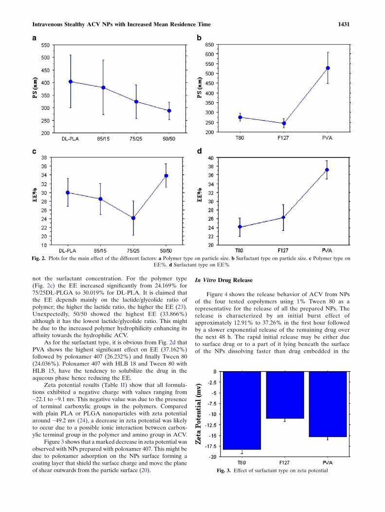
not the surfactant concentration. For the polymer type(Fig. 2c) the EE increased significantly from 24.169% for75/25DL-PLGA to 30.019% for DL-PLA. It is claimed thatthe EE depends mainly on the lactide/glycolide ratio ofpolymer; the higher the lactide ratio, the higher the EE (23).Unexpectedly, 50/50 showed the highest EE (33.866%)although it has the lowest lactide/glycolide ratio. This mightbe due to the increased polymer hydrophilicity enhancing itsaffinity towards the hydrophilic ACV.
As for the surfactant type, it is obvious from Fig. 2d thatPVA shows the highest significant effect on EE (37.162%)followed by poloxamer 407 (26.232%) and finally Tween 80(24.036%). Poloxamer 407 with HLB 18 and Tween 80 withHLB 15, have the tendency to solubilize the drug in theaqueous phase hence reducing the EE.
Zeta potential results (Table II) show that all formula-tions exhibited a negative charge with values ranging from−22.1 to −9.1 mv. This negative value was due to the presenceof terminal carboxylic groups in the polymers. Comparedwith plain PLA or PLGA nanoparticles with zeta potentialaround −49.2 mv (24), a decrease in zeta potential was likelyto occur due to a possible ionic interaction between carbox-ylic terminal group in the polymer and amino group in ACV.
Figure 3 shows that amarked decrease in zeta potential wasobserved with NPs prepared with poloxamer 407. This might bedue to poloxamer adsorption on the NPs surface forming acoating layer that shield the surface charge and move the planeof shear outwards from the particle surface (20).
In Vitro Drug Release
Figure 4 shows the release behavior of ACV from NPsof the four tested copolymers using 1% Tween 80 as arepresentative for the release of all the prepared NPs. Therelease is characterized by an initial burst effect ofapproximately 12.91% to 37.26% in the first hour followedby a slower exponential release of the remaining drug overthe next 48 h. The rapid initial release may be either dueto surface drug or to a part of it lying beneath the surfaceof the NPs dissolving faster than drug embedded in the
Fig. 2. Plots for the main effect of the different factors: a Polymer type on particle size. b Surfactant type on particle size. c Polymer type onEE%. d Surfactant type on EE%
Fig. 3. Effect of surfactant type on zeta potential
1431Intravenous Stealthy ACV NPs with Increased Mean Residence Time

center of the spheres. The dissolution medium enters thesphere through pores, dissolving the drug. A saturateddrug solution will be formed from which drug diffusesslowly into the surrounding medium. Further drug willdiffuse through the polymer into the water-filled channelsuntil drug depletion from the NPs. The small size of theNPs is also a major factor which influences the burstrelease effect.
Figure 4 shows also that the presence of poloxamer 407in the NPs enhanced the release of ACV compared to thoseprepared using Tween 80 and PVA, where a higher burst effectwas always noticed in all tested polymers. As an example, 75/25PLGA released 30.30% in the first hour using 1% poloxamer407 compared to 21.23% and 21.41% in case PVA and Tween80, respectively, with the same concentration.
The polymer composition (ratio of lactic to glycolic acidmoiety) and its molecular weight influenced the pattern ofACV release from nanospheres. ACV was released fasterfrom nanospheres prepared using 50/50 PLGA polymercontaining higher fraction of glycolic acid moiety and is oflower molecular weight compared with other polymers. Rateof hydration of polymeric materials depend on water uptakeof the polymer. Lactic acid polymer because of the methylgroup is more hydrophobic than glycolide polymer leading to
lower water uptake. The water uptake increases as theglycolide ratio in copolymer increases (25) and hence PLGA50/50 showed faster release compared with PLA, PLGA 85/15, and PLGA 75/25.
Fig. 4. Release profile of ACV-NPs prepared using 1% Tween 80 and 1% poloxamer 407
Fig. 5. TEM photomicrographs of 75/25 PLGA ACV NPs using 1%tween 80 and coated with poloxamer 407
1432 Kamel, Awad, Geneidi and Mortada

Effect of Coating on ACV NPs
Figure 5 shows the TEMphotomicrographs of formula 20C(75/25 PLGA with 1% Tween 80 and coated with poloxamer407) as a representative of the coated NPs. NPs appearedbrighter than the uncoated ones (26).
Coating with the different block copolymers viz poloxamer188, 338, and 407 caused nonsignificant change in the EE of alltried formulas at p≤0.001 (results not shown).
Table III shows the coating layer thickness of thedifferent poloxamers used. All NPs with large particlediameter showed undetectable coat layers. However, thethickness of the poloxamers layers in smaller particles (280and 300 nm) was in the range 1.5–8.3 nm. These values weresomewhat lower than those reported for the same blockcopolymers adsorbed to polystyrene particles (10–15 nm)(27). The difference can be attributed to the different natureof the NP surfaces. In general, during coating, the block(PPO) of the polymer will be attached to the polymer surfacewhile (PEO) blocks will extend into the dispersion medium.A block copolymer such as poloxamer adsorbed to a NPsurface will exist in different conformations that will dependon the packing of the polymer at the surface. A close packingof the polymer molecules at the surface will result in anextended polymer “brush” arrangement which will provide agreater adsorbed hydrodynamic thickness than less closelypacked polymer chains. Accordingly, the more hydrophobicthe NP polymer is, the more it attracts PPO and is bettercoated than less hydrophobic one. Hence polystyrene par-ticles being hydrophobic in nature had a thicker coat thanPLGA particles (more polar) (27).
Both copolymers poloxamer 338 and 407 gave thickerlayers compared with 188, this might be due to the highermolecular weights hence higher content of their poly(oxy-ethylene) chain compared with poloxamer 188 providing
Table IV. Effect of Coating with Different Poloxamers on ZetaPotential of NPs
Formula
Zeta potential (mv)±SD
Uncoated
Coating substance (Poloxamer)
133 338 407
4 −14.6±3 −14.5±1.6 −14.4±1.6 −14.4±1.614 −15.2±1.6 −15.6±1.6 −15.7±1.4 −15.9±1.620 −17.6±1.8 −7.6±0.8 −6.6±1.4 −6.5±1.622 −14.2±1.7 −14.2±4 −14.3±1.6 −14.6±1.223 −15.1±3.5 −7.7±0.6 −6.9±4.8 −6.8±0.4
Fig. 6. DSC thermograms during heating from room temperature to80°C in the first run and during heating from 0°C to 300°C in thesecond run. a ACV. b Polymer. c Unloaded NPs. d Loaded NPs
Table III. Effect of Different Poloxamers on the Coating LayerThickness of ACV NPs
Formula
Diameter of uncoatedNPs (nm)±SD(polydispersity index)
Adsorbed layer thickness (nm)
Coating substance(Poloxamer)
188 338 407
4 625±65 (0.8) ND ND ND14 640±60 (0.3) ND ND ND20 300±10 (0.6) 1.65 5 8.322 570±15 (0.3) ND ND ND23 280±10 (0.5) 1.5 6.1 6.65
ND not detected
1433Intravenous Stealthy ACV NPs with Increased Mean Residence Time
greater adsorbed hydrodynamic thickness and expectedreduction in the opsonization effect. Gref et al. (28) optimizedthe thickness and density of a PEO coating at the surface ofbiodegradable PLA nanoparticles, in order to reduce simul-taneously surface charge, plasma protein adsorption, andinteraction with phagocytic cells. They observed a sharpdecrease in the protein adsorption upon increasing themolecular weight of the polyether chains.
The presence of adsorbed coating layers of poloxamer188, 338 and 407 on both formulas 20 and 23 was reflected intheir zeta potential results (Table IV) where a reduction wasseen in the zeta potentials after coating. This might bebecause of a higher concentration of surfactant adsorbed onthe particle surfaces with the formation of a denser surfactantfilm on the NP surface, thus, eliciting a reduced electro-phoretic mobility, and a higher absolute potential valueensures a high-energy barrier that stabilizes the nanosuspen-sion (29). The unchanged zeta potential of the large particles(formulas 4, 14, 22) confirmed the undetectable coat thicknessof these formulas.
Coating with any of the three block copolymersenhanced the drug release especially after the first hour(results not shown). This might be due to the greaterhydrophilic character of the coated surface leading to an
easier penetration of the release medium to the particle andhence faster drug release.
From the previous results it is obvious that the uncoatedFormula 20 made from 75/25 PLGA with the use of 1%Tween 80 and formula 20C (75/25 PLGA with 1% Tween 80and coated with poloxamer 407) had suitable diameters(300 and 316.6 nm, respectively), and optimum drug release( 64.26 and 68.56 after 48 h, respectively). Besides the blockcopolymer, poloxamer 407 gave thicker coat layer (8.3 nm)compared to other block copolymers. Hence they werechosen for further studies. For comparison purpose, for-mula 26 which was prepared from the same polymer (75/25PLGA) and 1% poloxamer 407 internally incorporatedduring fabrication was also chosen in order to comparebetween the effect of coating by adsorption and by incorpo-ration methods.
DSC Studies
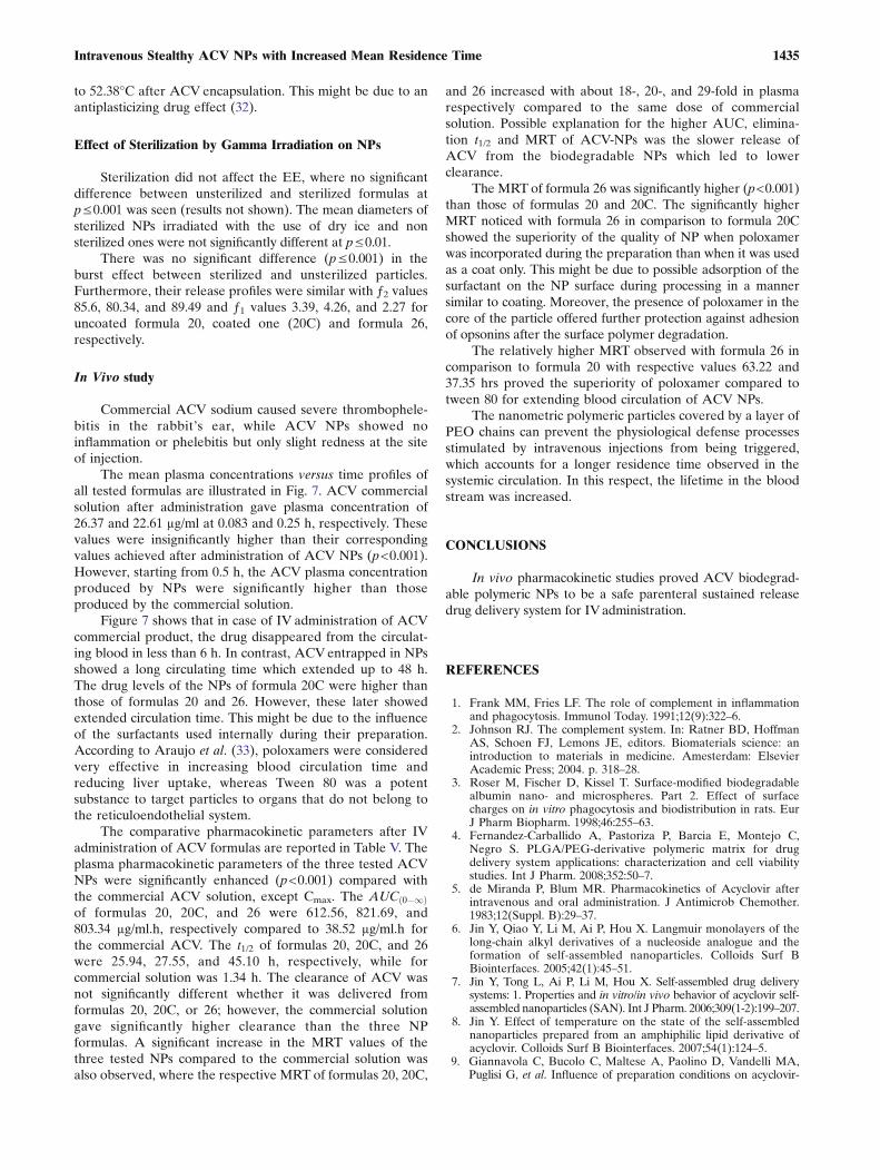
Figure 6 shows the DSC thermograms of 75/25 PLGA,unloaded NPs and ACV-loaded NPs (formula 20) during thefirst and the second heating runs. The DSC thermogram ofACV showed a small endothermic peak at 171.77°C, a broadendotherm at 98.44°C and a sharp narrow melting endothermat 236.78°C. The last two endotherms corresponds to loss ofresidual water and melting temperature of the drug, respec-tively. So according to Sohn and Kim, the used drug might bethe hydrate form (30). In the thermogram of ACV NPs, theendothermic peak of ACV disappeared indicating theabsence of any crystalline drug materials in the NPs. Thismight be due to the polymer inhibiting crystallization of ACVduring NP formation. So, it can be concluded that ACV in theNPs was in an amorphous or a solid solution state in thepolymer matrix after the fabrication. The Tg of the polymerwas shifted from 49.15°C in case of raw polymer to 51.28°Cafter fabrication of unloaded NPs. This might be due to thereduction in molecular mobility of the polymer. One possibleexplanation for this reduction may be the restriction ofPLGA molecular mobility at the surface of particles magni-fied by the higher surface to volume ratio of the nanometerscale particles (31). The Tg of the polymer was further shifted
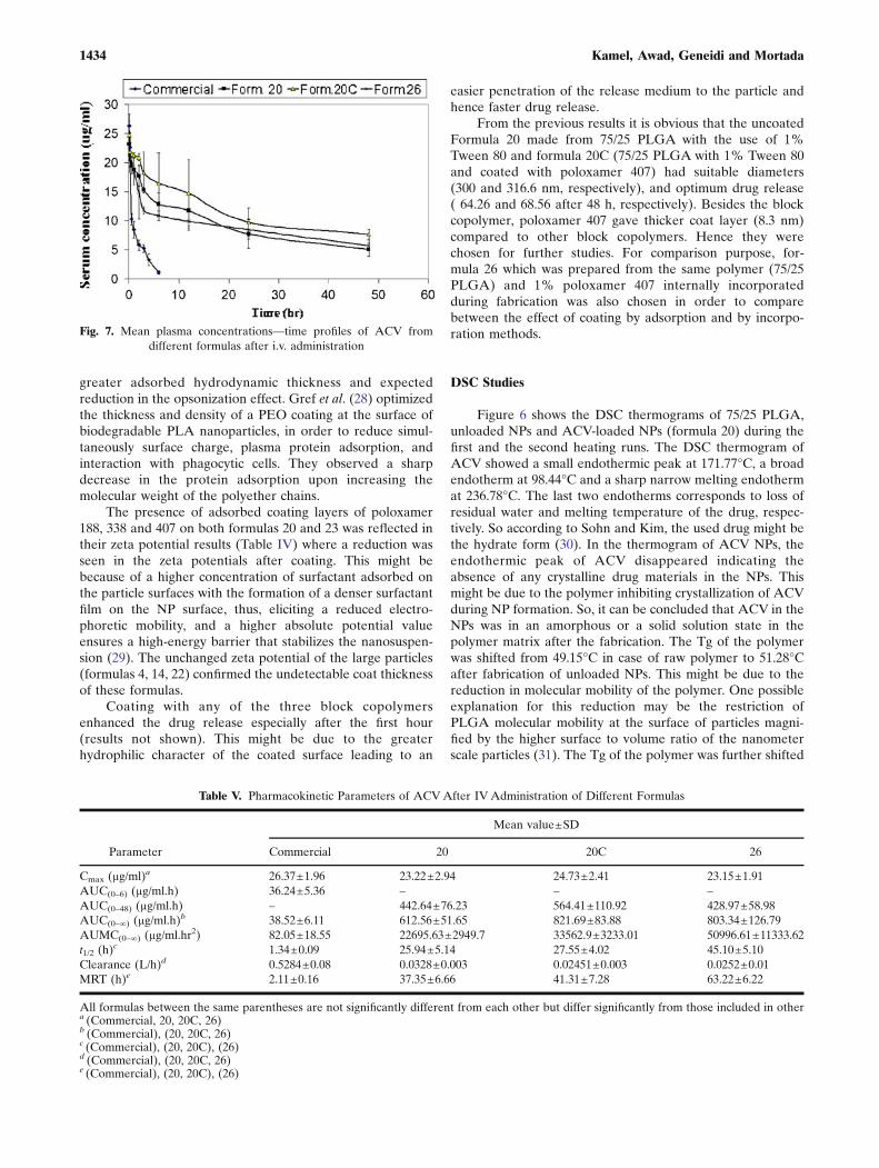
Fig. 7. Mean plasma concentrations—time profiles of ACV fromdifferent formulas after i.v. administration
Table V. Pharmacokinetic Parameters of ACVAfter IVAdministration of Different Formulas
Parameter
Mean value±SD
Commercial 20 20C 26
Cmax (µg/ml)a 26.37±1.96 23.22±2.94 24.73±2.41 23.15±1.91AUC(0–6) (µg/ml.h) 36.24±5.36 – – –AUC(0–48) (µg/ml.h) – 442.64±76.23 564.41±110.92 428.97±58.98AUC(0–∞) (µg/ml.h)b 38.52±6.11 612.56±51.65 821.69±83.88 803.34±126.79AUMC(0–∞) (µg/ml.hr2) 82.05±18.55 22695.63±2949.7 33562.9±3233.01 50996.61±11333.62t1/2 (h)
c 1.34±0.09 25.94±5.14 27.55±4.02 45.10±5.10Clearance (L/h)d 0.5284±0.08 0.0328±0.003 0.02451±0.003 0.0252±0.01MRT (h)e 2.11±0.16 37.35±6.66 41.31±7.28 63.22±6.22
All formulas between the same parentheses are not significantly different from each other but differ significantly from those included in othera (Commercial, 20, 20C, 26)b (Commercial), (20, 20C, 26)c (Commercial), (20, 20C), (26)d (Commercial), (20, 20C, 26)e (Commercial), (20, 20C), (26)
1434 Kamel, Awad, Geneidi and Mortada
to 52.38°C after ACV encapsulation. This might be due to anantiplasticizing drug effect (32).
Effect of Sterilization by Gamma Irradiation on NPs
Sterilization did not affect the EE, where no significantdifference between unsterilized and sterilized formulas atp≤0.001 was seen (results not shown). The mean diameters ofsterilized NPs irradiated with the use of dry ice and nonsterilized ones were not significantly different at p≤0.01.
There was no significant difference (p≤0.001) in theburst effect between sterilized and unsterilized particles.Furthermore, their release profiles were similar with ƒ2 values85.6, 80.34, and 89.49 and ƒ1 values 3.39, 4.26, and 2.27 foruncoated formula 20, coated one (20C) and formula 26,respectively.
In Vivo study
Commercial ACV sodium caused severe thrombophele-bitis in the rabbit’s ear, while ACV NPs showed noinflammation or phelebitis but only slight redness at the siteof injection.
The mean plasma concentrations versus time profiles ofall tested formulas are illustrated in Fig. 7. ACV commercialsolution after administration gave plasma concentration of26.37 and 22.61 µg/ml at 0.083 and 0.25 h, respectively. Thesevalues were insignificantly higher than their correspondingvalues achieved after administration of ACV NPs (p<0.001).However, starting from 0.5 h, the ACV plasma concentrationproduced by NPs were significantly higher than thoseproduced by the commercial solution.
Figure 7 shows that in case of IV administration of ACVcommercial product, the drug disappeared from the circulat-ing blood in less than 6 h. In contrast, ACVentrapped in NPsshowed a long circulating time which extended up to 48 h.The drug levels of the NPs of formula 20C were higher thanthose of formulas 20 and 26. However, these later showedextended circulation time. This might be due to the influenceof the surfactants used internally during their preparation.According to Araujo et al. (33), poloxamers were consideredvery effective in increasing blood circulation time andreducing liver uptake, whereas Tween 80 was a potentsubstance to target particles to organs that do not belong tothe reticuloendothelial system.
The comparative pharmacokinetic parameters after IVadministration of ACV formulas are reported in Table V. Theplasma pharmacokinetic parameters of the three tested ACVNPs were significantly enhanced (p<0.001) compared withthe commercial ACV solution, except Cmax. The AUC 0�1ð Þof formulas 20, 20C, and 26 were 612.56, 821.69, and803.34 µg/ml.h, respectively compared to 38.52 µg/ml.h forthe commercial ACV. The t1/2 of formulas 20, 20C, and 26were 25.94, 27.55, and 45.10 h, respectively, while forcommercial solution was 1.34 h. The clearance of ACV wasnot significantly different whether it was delivered fromformulas 20, 20C, or 26; however, the commercial solutiongave significantly higher clearance than the three NPformulas. A significant increase in the MRT values of thethree tested NPs compared to the commercial solution wasalso observed, where the respective MRT of formulas 20, 20C,
and 26 increased with about 18-, 20-, and 29-fold in plasmarespectively compared to the same dose of commercialsolution. Possible explanation for the higher AUC, elimina-tion t1/2 and MRT of ACV-NPs was the slower release ofACV from the biodegradable NPs which led to lowerclearance.
The MRTof formula 26 was significantly higher (p<0.001)than those of formulas 20 and 20C. The significantly higherMRT noticed with formula 26 in comparison to formula 20Cshowed the superiority of the quality of NP when poloxamerwas incorporated during the preparation than when it was usedas a coat only. This might be due to possible adsorption of thesurfactant on the NP surface during processing in a mannersimilar to coating. Moreover, the presence of poloxamer in thecore of the particle offered further protection against adhesionof opsonins after the surface polymer degradation.
The relatively higher MRT observed with formula 26 incomparison to formula 20 with respective values 63.22 and37.35 hrs proved the superiority of poloxamer compared totween 80 for extending blood circulation of ACV NPs.
The nanometric polymeric particles covered by a layer ofPEO chains can prevent the physiological defense processesstimulated by intravenous injections from being triggered,which accounts for a longer residence time observed in thesystemic circulation. In this respect, the lifetime in the bloodstream was increased.
CONCLUSIONS
In vivo pharmacokinetic studies proved ACV biodegrad-able polymeric NPs to be a safe parenteral sustained releasedrug delivery system for IVadministration.
REFERENCES
1. Frank MM, Fries LF. The role of complement in inflammationand phagocytosis. Immunol Today. 1991;12(9):322–6.
2. Johnson RJ. The complement system. In: Ratner BD, HoffmanAS, Schoen FJ, Lemons JE, editors. Biomaterials science: anintroduction to materials in medicine. Amesterdam: ElsevierAcademic Press; 2004. p. 318–28.
3. Roser M, Fischer D, Kissel T. Surface-modified biodegradablealbumin nano- and microspheres. Part 2. Effect of surfacecharges on in vitro phagocytosis and biodistribution in rats. EurJ Pharm Biopharm. 1998;46:255–63.
4. Fernandez-Carballido A, Pastoriza P, Barcia E, Montejo C,Negro S. PLGA/PEG-derivative polymeric matrix for drugdelivery system applications: characterization and cell viabilitystudies. Int J Pharm. 2008;352:50–7.
5. de Miranda P, Blum MR. Pharmacokinetics of Acyclovir afterintravenous and oral administration. J Antimicrob Chemother.1983;12(Suppl. B):29–37.
6. Jin Y, Qiao Y, Li M, Ai P, Hou X. Langmuir monolayers of thelong-chain alkyl derivatives of a nucleoside analogue and theformation of self-assembled nanoparticles. Colloids Surf BBiointerfaces. 2005;42(1):45–51.
7. Jin Y, Tong L, Ai P, Li M, Hou X. Self-assembled drug deliverysystems: 1. Properties and in vitro/in vivo behavior of acyclovir self-assembled nanoparticles (SAN). Int J Pharm. 2006;309(1-2):199–207.
8. Jin Y. Effect of temperature on the state of the self-assemblednanoparticles prepared from an amphiphilic lipid derivative ofacyclovir. Colloids Surf B Biointerfaces. 2007;54(1):124–5.
9. Giannavola C, Bucolo C, Maltese A, Paolino D, Vandelli MA,Puglisi G, et al. Influence of preparation conditions on acyclovir-
1435Intravenous Stealthy ACV NPs with Increased Mean Residence Time
loaded poly-d, l-lactic acid nanospheres and effect of PEG coating onocular drug bioavailability. Pharm Res. 2003;20(4):584–90.
10. Mao S, Hou S, Zhang L, Wei D, Zhang J, Qiao X, et al.Preparation of valaciclovir loaded bovine serum albumin nano-particles surface-modified with glycyrrhizin and its characteristicsof targeting to liver. Sheng wu yi xue gong cheng xue za zhi (JBiomed Eng). 2004;21(4):570–4.
11. de Jalon EG, Blanco-Prıeto MJ, Ygartua P, Santoyo S. Increasedefficacy of acyclovir-loaded microparticles against herpes simplexvirus type 1 in cell culture. Eur J Pharm Biopharm. 2003;56:183–7.
12. Choonara YE, Pillay V, Carmichael T, Danckwerts MP. Studies ona novel doughnut-shaped minitablet for intraocular drug delivery.AAPS PharmSciTech. 2007;8(4):E1–7.
13. Martınez-Sancho C, Herrero-Vanrell R, Negro S. Vitamin Apalmitate and aciclovir biodegradable microspheres for intra-ocular sustained release. Int J Pharm. 2006;326:100–6.
14. Martınez-Sancho C, Herrero-Vanrell R, Negro S. Study of gamma-irradiation effects on acyclovir poly(D, L-lactic-co-glycolic) acidmicrospheres for intravitreal administration. J Cont Release.2004;99:41–52.
15. Martınez-Sancho C, Herrero-Vanrell R, Negro S. Optimisation ofaciclovir poly(d, l-lactide-co-glycolide) microspheres for intravitrealadministration using a factorial design study. Int J Pharm.2004;273:45–56.
16. Martınez-Sancho C, Herrero-Vanrell R, Negro S. Poly(D, L-lactide-co-glycolide) microspheres for long-term intravitreal deliv-ery of aciclovir: influence of fatty and non-fatty additives. JMicroencapsulation. 2003;20(6):799–810.
17. Chowdhury DK, Mitra AK. Kinetics of a model nucleoside(guanosine) release from biodegradable poly(DL-lactide-co-glycolide) microspheres: a delivery system for long-term intraoculardelivery. Pharm Dev Technol. 2000;5(2):279–85.
18. de Jalon E, Blanco-Prıeto MJ, Ygartua P, Santoyo S. Topicalapplication of acyclovir-loaded microparticles:quantification of thedrug in porcine skin layers. J Contr Release. 2001;75:191–7.
19. Niwa T, Takeuchi H, Hino T, Kunou N, Kawashima Y. Prepara-tion of biodegradable nanospheres of water soluble and insolubledrugs with D, L-lactide / glycolide copolymer by a novel sponta-neous emulsification solvent diffusion method and the drugrelease behavior. J Contr Release. 1993;25:89–98.
20. Redhead HM, Davis SS, Illum L. Drug delivery in poly (lactide-co-glycolide) nanoparticles surface modified with poloxmer 407and poloxamine 908: in vitro characterization and in vivoevaluation. J Contr Release. 2001;70:353–63.
21. Stagni G, Ali ME, Weng D. Pharmacokinetics of acyclovir inrabbit skin after i.v.-bolus, ointment, and iontophoretic admin-istrations. Int J Pharm. 2004;274(1-2):201–11.
22. Lin WJ, Haung LI. Influence of pluronics on protein loaded poly(epsilon-caprolactone) microparticles. J Microencaps. 2001;18:191–7.
23. Jeon HJ, Jeong YI, Jang MK, Park YH, Nah JW. Effect ofsolvent on the preparation of surfactant free poly (DL-lactide-co-glycolide) nanoparticle and norfloxacin release characteristics.Int J Pharm. 2000;207:99–108.
24. Govender T, Stolnik S, Garnett MC, Illum L, Davis SS. PLGAnanoparticles prepared by nanoprecipitation drug loading andrelease studies of water sol. Drug J Contr Release. 1999;57:171–85.
25. Lewis DH. Controlled release of bioactive agents from lactideglycolide polymers. In: Chasin M, Langer R, editors. Biodegrad-able polymers as drug delivery systems. New York: MarcelDekker; 1990. p. 1–40.
26. Hu Y, Jiang X, Ding Y, Zhang L, Yang C, Zhang J, et al.Preparation and drug release behaviors of nimodipine-loadedpoly(caprolactone)-poly(ethylene oxide)-polylactide amphiphiliccopolymer nanoparticles. Biomaterials. 2003;24(13):2395–404.
27. Muller RH, Wallis KH. Surface modification of IV injectablebiodegradable nanoparticles with poloxamer polymers andpoloxamine 908. Int J Pharm. 1993;89:25–31.
28. Gref R, Luck M, Quellec P, Marchand M, Dellacherie E,Harnisch S, et al. ‘Stealth’ corona-core nanoparticles surfacemodified by polyethylene glycol (PEG): influences of the corona(PEG chain length and surface density) and of the corecomposition on phagocytic uptake and plasma protein adsorp-tion. Colloids Surf B Biointerfaces. 2000;18(3-4):301–13.
29. Muller RH. Charge determinations. In: Muller RH, editor.Colloidal carriers for controlled drug delivery and targetingmodification, characterization and in vivo distribution. BocaRaton: CRC Press; 1991. p. 57–97.
30. Sohn YT, Kim SH. Polymorphism and pseudopolymorphism ofacyclovir. Arch Pharm Res. 2008;31(2):231–4.
31. Dean Allison S. Effect of structural relaxation on the preparationand drug release behavior of poly(lactic-co-glycolic) acid micro-particle drug delivery systems. J Pharm Sci. 2008;97(6):2022–35.
32. Okada H, Doken Y, Ogawa Y, Toguchi H. Preparation of three-month injectable microspheres of leuprorelin acetate usingbiodegradable polymers. Pharm Res. 1994;11:1143–7.
33. Araujo L, Sheppard M, Lobenberg R, Kreuter J. Uptake ofPMMa nanoparticles from the gastrointestinal tract after oraladministration to rats: modification of the body distribution aftersuspension in surfactant solutions and in oil vehicles. Int. J.Pharm. 1999;176:209–24.
1436 Kamel, Awad, Geneidi and Mortada