Benign Tumors of the Liver
81
BENIGN TUMORS BENIGN TUMORS OF THE LIVER OF THE LIVER MARIA THERESA M. NAVARRO, MD MARIA THERESA M. NAVARRO, MD 4 4 th th Year Radiology Resident Year Radiology Resident Department of Medical Imaging Department of Medical Imaging Quirino Memorial Medical Center Quirino Memorial Medical Center
-
Upload
thessnavarro -
Category
Documents
-
view
2.292 -
download
11
description
Powerpoint presentation on benign tumors of the liver, with MRI and CT illustrations
Transcript of Benign Tumors of the Liver

BENIGN BENIGN TUMORS OF TUMORS OF THE LIVERTHE LIVER
MARIA THERESA M. NAVARRO, MDMARIA THERESA M. NAVARRO, MD44thth Year Radiology Resident Year Radiology ResidentDepartment of Medical ImagingDepartment of Medical ImagingQuirino Memorial Medical CenterQuirino Memorial Medical Center

Hepatic Hepatic HemangiomasHemangiomas
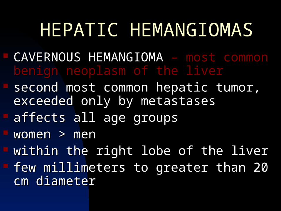
HEPATIC HEMANGIOMASHEPATIC HEMANGIOMAS CAVERNOUS HEMANGIOMA CAVERNOUS HEMANGIOMA – most common – most common
benign neoplasm of the liverbenign neoplasm of the liver second most common hepatic tumor, exceeded second most common hepatic tumor, exceeded
only by metastasesonly by metastases affects all age groupsaffects all age groups women > menwomen > men within the right lobe of the liverwithin the right lobe of the liver few millimeters to greater than 20 cm diameterfew millimeters to greater than 20 cm diameter

HEPATIC HEMANGIOMASHEPATIC HEMANGIOMAS
mesodermal in originmesodermal in origin histologically : blood-filled cavernous histologically : blood-filled cavernous
vascular spaces of variable size and vascular spaces of variable size and shape lined single layer of flat endotheliumshape lined single layer of flat endothelium
stable lesions, rarely increase or decrease stable lesions, rarely increase or decrease in sizein size

IMAGING FEATURES OF IMAGING FEATURES OF CAVERNOUS HEMANGIOMASCAVERNOUS HEMANGIOMAS
PLAIN FILMSPLAIN FILMS mostly, too small to be identified on plain films of mostly, too small to be identified on plain films of
the abdomenthe abdomen very large lesions, nonspecific findings : very large lesions, nonspecific findings :
hepatomegaly or a large upper abdominal masshepatomegaly or a large upper abdominal mass

IMAGING FEATURES OF CAVERNOUS IMAGING FEATURES OF CAVERNOUS HEMANGIOMASHEMANGIOMAS
CHARACTERISTIC (but rarely seen)CHARACTERISTIC (but rarely seen) presence of multiple calcified phleboliths presence of multiple calcified phleboliths numerous calcified “trabeculations and spicules” numerous calcified “trabeculations and spicules”
that arise from a central point and radiate out that arise from a central point and radiate out toward the periphery of the lesiontoward the periphery of the lesion

IMAGING FEATURES OF CAVERNOUS IMAGING FEATURES OF CAVERNOUS HEMANGIOMASHEMANGIOMAS
ULTRASOUNDULTRASOUND well-circumscribed, homogenous, densely well-circumscribed, homogenous, densely
echogenic masses echogenic masses with posterior acoustic with posterior acoustic enhancement enhancement
differential diagnosis:differential diagnosis: hepatocellular cahepatocellular ca metastatic diseasemetastatic disease hepatic adenomahepatic adenoma focal nodular hyperplasiafocal nodular hyperplasia

IMAGING FEATURES OF CAVERNOUS IMAGING FEATURES OF CAVERNOUS HEMANGIOMASHEMANGIOMAS
ULTRASOUNDULTRASOUND demonstrate varying internal complexity, demonstrate varying internal complexity,
depending on the degree of thrombosis, fibrosis, depending on the degree of thrombosis, fibrosis, hemorrhagic necrosis, or calcificationhemorrhagic necrosis, or calcification
sonographic heterogeneity – more common in sonographic heterogeneity – more common in larger lesions.larger lesions.
hyperechoic pattern (67% to 70%) – presence of hyperechoic pattern (67% to 70%) – presence of multiple vascular interfaces between the walls of multiple vascular interfaces between the walls of the cavernous sinuses and the blood within the cavernous sinuses and the blood within themthem

COMPUTED TOMOGRAPHYCOMPUTED TOMOGRAPHY CT diagnosis is based on the understanding of CT diagnosis is based on the understanding of
the neoplasm’s vascular hemodynamicsthe neoplasm’s vascular hemodynamics blood typically circulates slowly within the the blood typically circulates slowly within the the
dilated cavernous spaces of this tumor and dilated cavernous spaces of this tumor and tends to flow tends to flow from peripherally located sinusoids from peripherally located sinusoids toward those at the lesion center.toward those at the lesion center.
IMAGING FEATURES OF CAVERNOUS IMAGING FEATURES OF CAVERNOUS HEMANGIOMASHEMANGIOMAS

IMAGING FEATURES OF CAVERNOUS IMAGING FEATURES OF CAVERNOUS HEMANGIOMASHEMANGIOMAS
COMPUTED TOMOGRAPHYCOMPUTED TOMOGRAPHY series of noncontrast-enhanced CT scans to localize series of noncontrast-enhanced CT scans to localize
suspected hemangiomasuspected hemangioma rapid-sequence, single-level dynamic CT scan are rapid-sequence, single-level dynamic CT scan are
obtained to the largest segment of the lesion during an obtained to the largest segment of the lesion during an IV bolus injection of IV bolus injection of
150 ml of 60% contrast agent (42g of iodine)150 ml of 60% contrast agent (42g of iodine) sequential delayed scans acquired for up to 30 minutes sequential delayed scans acquired for up to 30 minutes
after injectionafter injection or until lesion is isodense with normal hepatic or until lesion is isodense with normal hepatic
parenchyma so that the lesion’s contrast enhancement parenchyma so that the lesion’s contrast enhancement profile can be evaluated.profile can be evaluated.

IMAGING FEATURES OF CAVERNOUS IMAGING FEATURES OF CAVERNOUS HEMANGIOMASHEMANGIOMAS
DYNAMIC PHASE OF BOLUS CONTRAST INJECTIONDYNAMIC PHASE OF BOLUS CONTRAST INJECTION show peripheral enhancement at the margins of the show peripheral enhancement at the margins of the
hemangiomahemangioma initial enhancement need not encircle the entire lesion initial enhancement need not encircle the entire lesion
but may appear as : but may appear as : well defined focal regions of intense nodular well defined focal regions of intense nodular
enhancement at or near the lesion peripheryenhancement at or near the lesion periphery during the during the arterial phase of hepatic enhancementarterial phase of hepatic enhancement (10 to (10 to
30 seconds after bolus initiation) - mural nodules 30 seconds after bolus initiation) - mural nodules represent as feeding vascular nidusrepresent as feeding vascular nidus


IMAGING FEATURES OF CAVERNOUS IMAGING FEATURES OF CAVERNOUS HEMANGIOMASHEMANGIOMAS
DYNAMIC PHASE OF BOLUS CONTRAST DYNAMIC PHASE OF BOLUS CONTRAST INJECTIONINJECTION centripetal pattern of hemangioma with fill-in centripetal pattern of hemangioma with fill-in
occuring from the periphery toward the lesion occuring from the periphery toward the lesion centercenter
the most intense enhancement is seen early and the most intense enhancement is seen early and involves those vascular spaces that received the involves those vascular spaces that received the iodinated contrast firstiodinated contrast first
the degree of lesion enhancement then the degree of lesion enhancement then gradually decreases as the concentration of gradually decreases as the concentration of iodine in the bloodstream fallsiodine in the bloodstream falls

(Left) Axial NECT shows large mass in lateral segment, most of which is isodense to blood except for hypodense foci of scar. (Right) Axial CECTin venous parenchymal phase shows cloud-like peripheral enhancement that is isodense to vessels

(Left) Sagittal sonogram shows uniformly hyperechoic lesion in peripheral right lobe. (Right) Axial CECT in venous phase shows typical large hemangioma with nodular peripheral enhancement and nonenhancing scar (arrow).Capillary hemangioma (curved arrow) isodense to vessels in all phases.

““Even though CT is highly accurate Even though CT is highly accurate in the diagnosis of hemangiomas, in the diagnosis of hemangiomas, it does not consistently yield it does not consistently yield characteristic enhancement characteristic enhancement patterns, thus limiting is clinical patterns, thus limiting is clinical usefulness.”usefulness.”

MAGNETIC RESONANCE IMAGINGMAGNETIC RESONANCE IMAGING Most sensitive modality for detecting hepatic Most sensitive modality for detecting hepatic
cavernous hemangiomacavernous hemangioma high signal characterics similar to those of fluidshigh signal characterics similar to those of fluids Hypointense on T1 images and significantly Hypointense on T1 images and significantly
hyperintense on T2 imageshyperintense on T2 images Retain their marked signal intensity on heavily Retain their marked signal intensity on heavily
T2-weighted multiecho images T2-weighted multiecho images (“light bulb sign”)(“light bulb sign”)
IMAGING FEATURES OF CAVERNOUS IMAGING FEATURES OF CAVERNOUS HEMANGIOMASHEMANGIOMAS
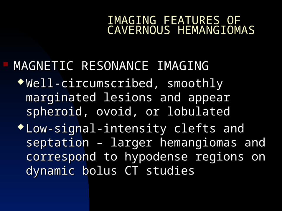
MAGNETIC RESONANCE IMAGINGMAGNETIC RESONANCE IMAGING Well-circumscribed, smoothly marginated Well-circumscribed, smoothly marginated
lesions and appear spheroid, ovoid, or lobulatedlesions and appear spheroid, ovoid, or lobulated Low-signal-intensity clefts and septation – larger Low-signal-intensity clefts and septation – larger
hemangiomas and correspond to hypodense hemangiomas and correspond to hypodense regions on dynamic bolus CT studiesregions on dynamic bolus CT studies
IMAGING FEATURES OF CAVERNOUS IMAGING FEATURES OF CAVERNOUS HEMANGIOMASHEMANGIOMAS

IMAGING FEATURES OF CAVERNOUS IMAGING FEATURES OF CAVERNOUS HEMANGIOMASHEMANGIOMAS
MAGNETIC RESONANCE IMAGINGMAGNETIC RESONANCE IMAGING
HEMANGIOMASHEMANGIOMAS METASTASESMETASTASES
Homogenous hyperintense Homogenous hyperintense patternpattern
Heterogenous amorphous Heterogenous amorphous appearance appearance
Well-circumscribedWell-circumscribed Ringed morphologyRinged morphology
Smoothly marginatedSmoothly marginated Indistinct marginationIndistinct margination
Surrounding peritumoral Surrounding peritumoral edemaedema

IMAGING FEATURES OF CAVERNOUS IMAGING FEATURES OF CAVERNOUS HEMANGIOMASHEMANGIOMAS
RED BLOOD CELL RED BLOOD CELL SCINTIGRAPHYSCINTIGRAPHY
typically display a “hot spot” typically display a “hot spot” appearance on delayed appearance on delayed labeled-RBC scans, reflecting labeled-RBC scans, reflecting the circulatory characteristics the circulatory characteristics of these neoplasmsof these neoplasms

IMAGING FEATURES OF CAVERNOUS IMAGING FEATURES OF CAVERNOUS HEMANGIOMASHEMANGIOMAS
ANGIOGRAPHYANGIOGRAPHY Has long been considered gold standard in the Has long been considered gold standard in the
diagnosis of hepatic hemagiomadiagnosis of hepatic hemagioma Classic Finding : Classic Finding :
normal main and feeding hepatic arterynormal main and feeding hepatic artery early contrast accumulation within the lesion early contrast accumulation within the lesion
during the late arterial phaseduring the late arterial phase Prolonged, delayed stain that persists throughout Prolonged, delayed stain that persists throughout
the capillary phase and well into the late venous the capillary phase and well into the late venous phasephase

IMAGING FEATURES OF CAVERNOUS IMAGING FEATURES OF CAVERNOUS HEMANGIOMASHEMANGIOMAS
ANGIOGRAPHYANGIOGRAPHY Atypical angiographic features:Atypical angiographic features:
Hypovascular massHypovascular mass Or a dense, homogeneous hypervascular Or a dense, homogeneous hypervascular
massmass Arterial-portal venous shuntingArterial-portal venous shunting

Identification of a persistent pool of Identification of a persistent pool of contrast puddling within such contrast puddling within such lesions may be the only means of lesions may be the only means of diagnosing these complicated diagnosing these complicated hemangiomas short of performing hemangiomas short of performing a biopsy or surgical resectiona biopsy or surgical resection

APPROACH TO IMAGING OF SUSPECTED APPROACH TO IMAGING OF SUSPECTED HEMANGIOMASHEMANGIOMAS
(Modification scheme by Freeny)(Modification scheme by Freeny) GROUP IGROUP I Patients who have well-circumscribed, homogeneous, Patients who have well-circumscribed, homogeneous,
hyperechoic lesionshyperechoic lesions No clinical symptoms referable to the liverNo clinical symptoms referable to the liver Normal liver function resultsNormal liver function results No known primary neoplasmNo known primary neoplasm Patients who have lesions with atypical sonographic Patients who have lesions with atypical sonographic
patternspatterns Abnormal clinical findingsAbnormal clinical findings Known primary neoplasmKnown primary neoplasm

Group I Patients Labeled-RBC scintigraphy with SPECT
tomographic imaging is the preferred procedure of definitive lesion characterization due to its relative low cost and near 100% specificity and positive predictive value.
(1.5 cm to 2.0 cm lesions)

Group I Patients Magnetic Resonance Imaging reserved for lesions smaller than1.5 to 2.0 cm lesions 2.5 cm or smaller located adjacent to the
heart or major intrahepatic vessels Both modalities – useful in patients with multiple
suspected hemangiomas

Group I Patients
MRI is not advocated if the patient has a known primary hypervascular endocrine neoplasm
If these studies yield undeterminate findings for hemangioma
angiography or percutaneous needle
biopsy

APPROACH TO IMAGING OF SUSPECTED APPROACH TO IMAGING OF SUSPECTED HEMANGIOMASHEMANGIOMAS
(Modification scheme by Freeny)(Modification scheme by Freeny) GROUP IIGROUP II Patients who have focal hepatic lesions detected Patients who have focal hepatic lesions detected
on routine dynamic-incremental bolus CT on routine dynamic-incremental bolus CT studiesstudies
Peripheral contrast
enhancement
Delayed Images
Show Isodense Fill-
in
HEMANGIOMA

APPROACH TO IMAGING OF SUSPECTED APPROACH TO IMAGING OF SUSPECTED HEMANGIOMASHEMANGIOMAS
GROUP IIGROUP II
ATYPICAL CT
FEATURES
KNOWN PRIMARY TUMOR
ABNORMAL LIVER
FINDINGS
ADDITIONAL IMAGING WITH LABELED-RBC
SPECT
MRI using the size and specific criteria
already outlined

APPROACH TO IMAGING OF APPROACH TO IMAGING OF SUSPECTED HEMANGIOMASSUSPECTED HEMANGIOMAS
GROUP IIIGROUP III Whose hepatic lesions are first detected on MRIWhose hepatic lesions are first detected on MRI
MRI APPEARANCE IS CLASSIC
NO KNOWN PRIMARY NEOPLASM
HEMANGIOMA
APPROACH TO IMAGING OF SUSPECTED APPROACH TO IMAGING OF SUSPECTED HEMANGIOMASHEMANGIOMAS
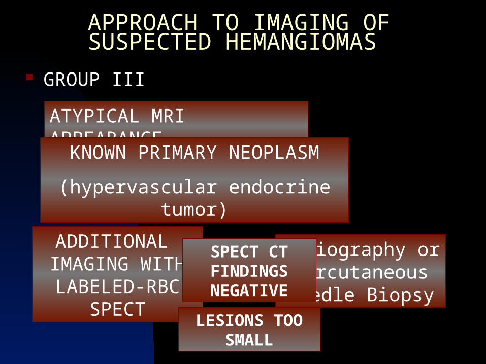
GROUP IIIGROUP III
ATYPICAL MRI APPEARANCE KNOWN PRIMARY NEOPLASM
(hypervascular endocrine tumor)
ADDITIONAL IMAGING WITH LABELED-RBC
SPECT
Angiography or Percutaneous Needle Biopsy
SPECT CT FINDINGS NEGATIVE
LESIONS TOO SMALL
APPROACH TO IMAGING OF SUSPECTED HEMANGIOMASAPPROACH TO IMAGING OF SUSPECTED HEMANGIOMAS
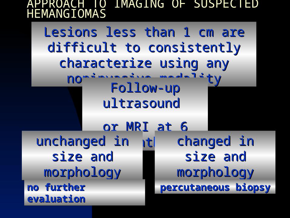
Lesions less than 1 cm are Lesions less than 1 cm are difficult to consistently difficult to consistently characterize using any characterize using any noninvasive modalitynoninvasive modality
Follow-up Follow-up ultrasound ultrasound
or MRI at 6 or MRI at 6 monthsmonthsunchanged in unchanged in
size and size and morphologymorphology
no further no further evaluationevaluation
changed in size changed in size and morphologyand morphology
percutaneous biopsypercutaneous biopsy

Hepatocellular Hepatocellular Adenoma,Adenoma,Focal Nodular Focal Nodular Hyperplasia, and Hyperplasia, and OthersOthers
Hepatocellular Adenoma,Hepatocellular Adenoma,
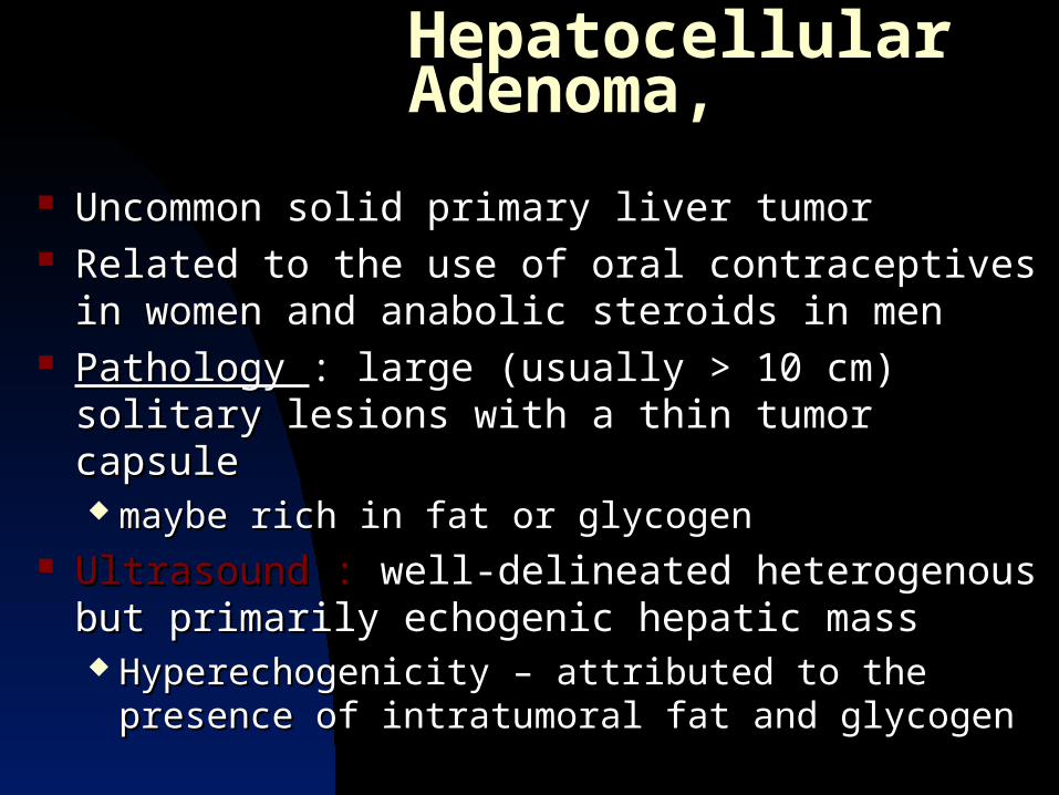
Uncommon solid primary liver tumorUncommon solid primary liver tumor Related to the use of oral contraceptives in women Related to the use of oral contraceptives in women
and anabolic steroids in menand anabolic steroids in men Pathology Pathology : large (usually > 10 cm) solitary lesions : large (usually > 10 cm) solitary lesions
with a thin tumor capsulewith a thin tumor capsule maybe rich in fat or glycogenmaybe rich in fat or glycogen
Ultrasound : Ultrasound : well-delineated heterogenous but well-delineated heterogenous but primarily echogenic hepatic mass primarily echogenic hepatic mass Hyperechogenicity – attributed to the presence of Hyperechogenicity – attributed to the presence of
intratumoral fat and glycogenintratumoral fat and glycogen

Hepatocellular Adenoma,Hepatocellular Adenoma, Non-contrast-enhanced CTNon-contrast-enhanced CT Predominantly isodense with liver but may appear Predominantly isodense with liver but may appear
either uniformly hypodense due to extensive either uniformly hypodense due to extensive steatosissteatosis
Or heterogeneous with foci of hypodensity caused by Or heterogeneous with foci of hypodensity caused by tumor necrosis and areas of hyperdensity secondary tumor necrosis and areas of hyperdensity secondary to recent intratumoral hemorrhageto recent intratumoral hemorrhage


Hepatocellular Adenoma,Hepatocellular Adenoma,
Distinctive perfusion characteristic of HADistinctive perfusion characteristic of HA arterial phase of contrast infusionarterial phase of contrast infusion (15 to 25 seconds (15 to 25 seconds
after bolus injection of contrast media) - consists of after bolus injection of contrast media) - consists of dense enhancement dense enhancement maybe homogenous in small tumors or maybe homogenous in small tumors or
heterogeneous in a larger massheterogeneous in a larger mass The lesion become hyperdense relative to normal The lesion become hyperdense relative to normal
liver, which has enhanced minimally at this time.liver, which has enhanced minimally at this time.

Hepatocellular Adenoma,Hepatocellular Adenoma,
portal venous phaseportal venous phase (45 to 180 seconds after the injection of (45 to 180 seconds after the injection of contrast media)contrast media) tumoral enhancement diminishes rapidly and the tumoral enhancement diminishes rapidly and the
tumors become isodense to normal liver during thetumors become isodense to normal liver during the
Radionuclide ScintigraphyRadionuclide Scintigraphy Most HAs are devoid of Kupffer cells, they Most HAs are devoid of Kupffer cells, they appear cold on sulfur colloid scintigraphyappear cold on sulfur colloid scintigraphyShow uptake of the tracerShow uptake of the tracerBecause of the lack of bile ductules, the tracer Because of the lack of bile ductules, the tracer is not excreted, and delayed scans therefore is not excreted, and delayed scans therefore depict HAs as areas of markedly increased depict HAs as areas of markedly increased activityactivity

Hepatocellular Adenoma,Hepatocellular Adenoma,

Hepatocellular Adenoma,Hepatocellular Adenoma,
Magnetic Resonance ImagingMagnetic Resonance Imaging Mimic malignant liver tumors, with the non-necrotic, Mimic malignant liver tumors, with the non-necrotic,
nonhemorrhagic solid component showingnonhemorrhagic solid component showing Hypointense appearance on T1 images and slight Hypointense appearance on T1 images and slight
hyperintense on T2 imageshyperintense on T2 images However, the high fat or glycogen content, or both, of these However, the high fat or glycogen content, or both, of these
tumors can render them isointense or even hyperintense on tumors can render them isointense or even hyperintense on T1 images T1 images
Hypointense tumor capsule best seen on T1 imagesHypointense tumor capsule best seen on T1 images Intratumoral bleeding – hyperintense on T1 images and Intratumoral bleeding – hyperintense on T1 images and
hypointense on T2 imageshypointense on T2 images

(Left) Axial T1WI MR shows hypointense encapsulated mass with hyperintense foci (hemorrhage or fat). (Right) Axial T2WI MR shows mass nearly isointense to liver withcentral focus of hyperintensity (hemorrhage).

Hepatocellular Adenoma,Hepatocellular Adenoma,
AngiogramsAngiograms Hypervascular tumors with large peripheral vesselsHypervascular tumors with large peripheral vessels Centripetal flowCentripetal flow Usually there is no arteriovenous shunting or Usually there is no arteriovenous shunting or
vascular invasion, as is often seen in hepatocellular vascular invasion, as is often seen in hepatocellular carcinomacarcinoma
No specific diagnostic specificity can be gainedNo specific diagnostic specificity can be gained

Hepatocellular Adenoma,Hepatocellular Adenoma,
CONCLUSIONCONCLUSION Hypervascular tumors with large peripheral vesselsHypervascular tumors with large peripheral vessels Centripetal flowCentripetal flow Usually there is no arteriovenous shunting or Usually there is no arteriovenous shunting or
vascular invasion, as is often seen in hepatocellular vascular invasion, as is often seen in hepatocellular carcinomacarcinoma
No specific diagnostic specificity can be gainedNo specific diagnostic specificity can be gained

FOCAL NODULAR HYPERPLASIAFOCAL NODULAR HYPERPLASIA
Clinical BackgroundClinical Background most common solid benign tumor of the livermost common solid benign tumor of the liver usually found incidentallyusually found incidentally etiology unknown, but postulated that a congenital etiology unknown, but postulated that a congenital
vascular malformation may trigger the development vascular malformation may trigger the development of hepatocyte hyperplasiaof hepatocyte hyperplasia
hormonal influence hormonal influence more common in women on more common in women on their 3their 3rdrd to 5 to 5thth decades decades
controversy regarding its association with the use of controversy regarding its association with the use of oral contraceptives oral contraceptives

FOCAL NODULAR HYPERPLASIAFOCAL NODULAR HYPERPLASIA PathologyPathology well-circumscribed, usually solitary mass, no capsulewell-circumscribed, usually solitary mass, no capsule with centrally located scar tissue surrounded by nodules of with centrally located scar tissue surrounded by nodules of
hyperplastic hepatocyteshyperplastic hepatocytes nodules divided by thin septae that radiate from the central nodules divided by thin septae that radiate from the central
scar, when there is onescar, when there is one no normal portal venous structuresno normal portal venous structures usually located at the liver surface usually located at the liver surface bulge in the liver contour bulge in the liver contour or pedunculated massor pedunculated mass 1 cm to > 15 cm1 cm to > 15 cm hemorrhage, necrosis, and calcification are rarehemorrhage, necrosis, and calcification are rare

FOCAL NODULAR HYPERPLASIAFOCAL NODULAR HYPERPLASIA
RadiologyRadiology
Diagnostic : Diagnostic : presence of hepatocellular presence of hepatocellular reticuloendothelial function in liver tumors that reticuloendothelial function in liver tumors that possess a cental scar containing vessels and bile possess a cental scar containing vessels and bile ducts ducts
FOCAL NODULAR HYPERPLASIAFOCAL NODULAR HYPERPLASIA
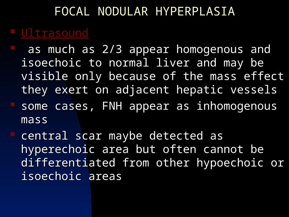
UltrasoundUltrasound as much as 2/3 appear homogenous and isoechoic as much as 2/3 appear homogenous and isoechoic
to normal liver and may be visible only because of to normal liver and may be visible only because of the mass effect they exert on adjacent hepatic the mass effect they exert on adjacent hepatic vesselsvessels
some cases, FNH appear as inhomogenous masssome cases, FNH appear as inhomogenous mass central scar maybe detected as hyperechoic area but central scar maybe detected as hyperechoic area but
often cannot be differentiated from other hypoechoic often cannot be differentiated from other hypoechoic or isoechoic areasor isoechoic areas

FOCAL NODULAR HYPERPLASIAFOCAL NODULAR HYPERPLASIA
Color-coded Doppler UltrasoundColor-coded Doppler Ultrasound the hypervascular nature of FNH take on the the hypervascular nature of FNH take on the
appearance of numerous scattered arterial and appearance of numerous scattered arterial and venous Doppler signals exhibiting a “comet tail” venous Doppler signals exhibiting a “comet tail” appearance throughout the tumorappearance throughout the tumor

FOCAL NODULAR HYPERPLASIAFOCAL NODULAR HYPERPLASIA Computed TomographyComputed Tomography with the exception of a central scar, the with the exception of a central scar, the
appearance of FNH on CT scans is usually appearance of FNH on CT scans is usually indistinguishable from that of HAindistinguishable from that of HA
generally isodense to normal liver on generally isodense to normal liver on noncontrast-enhanced CT studiesnoncontrast-enhanced CT studies
can be identified only if they can be identified only if they deform the liver deform the liver contours or possess a prominent stellate-contours or possess a prominent stellate-shapedshaped central scar central scar

FOCAL NODULAR HYPERPLASIAFOCAL NODULAR HYPERPLASIA Dynamic CT scanDynamic CT scan arterial phasearterial phase (approximately the first 30 (approximately the first 30
seconds) - rapid enhancement, appearing seconds) - rapid enhancement, appearing hyperdense relative to liverhyperdense relative to liver
portal phase portal phase – steady decrease in attenuation, – steady decrease in attenuation, so they appear relatively isodense or so they appear relatively isodense or hypodense to uninvolved liver tissuehypodense to uninvolved liver tissue
delayed CT scan – vascular scar tissue delayed CT scan – vascular scar tissue appear hyperdenseappear hyperdense
perfusion profile also seen in both perfusion profile also seen in both hepatocellular carcinoma and adenomahepatocellular carcinoma and adenoma

(Left) Axial CECTin arterial phase shows intense homogeneous enhancementof mass with central scar with thin radiating septa. Typical FNH appearanceresembles cross section of an orange. (Right) Axial CECT in venous phase shows mass (arrow) isodense to liverwith delayed enhancement of central scar.

FOCAL NODULAR HYPERPLASIAFOCAL NODULAR HYPERPLASIA
Nuclear ScintigraphyNuclear Scintigraphy technetium sulfur colloid scans, which portray technetium sulfur colloid scans, which portray
reticuloendothelial cellular function, the uptake of reticuloendothelial cellular function, the uptake of tracer is normal approximately 50% of FNHs and tracer is normal approximately 50% of FNHs and
uptake is increased in approximately 10%uptake is increased in approximately 10% as many of 40% of the FNHs do not possess as many of 40% of the FNHs do not possess
reticuloendothelial cellular function, the presence of reticuloendothelial cellular function, the presence of a “cold lesion” does not exclude the possibility of a “cold lesion” does not exclude the possibility of FNH.FNH.

FOCAL NODULAR HYPERPLASIAFOCAL NODULAR HYPERPLASIA
MRI ScanMRI Scan suggested if the liver tumor appears isointense on suggested if the liver tumor appears isointense on
both T1 and T2 images and has a central scarboth T1 and T2 images and has a central scar some cases, detected only by their mass effect on some cases, detected only by their mass effect on
normal hepatic vasculature plus the presence of normal hepatic vasculature plus the presence of central scar (hypointense on T1 and hyperintense on central scar (hypointense on T1 and hyperintense on T2 images)T2 images)
uncommonly FNH may mimic other solid tumors by uncommonly FNH may mimic other solid tumors by being hypointense on T1 and hyperintense on T2 being hypointense on T1 and hyperintense on T2 images.images.

(Left) Axial T1 C+ MR in arterial phase shows intense homogeneous enhancement of mass with central hypointense scar. (Right) Axial T2WI MR shows mass is minimally hyperintense, scar (arrow) is hyperintense.

FOCAL NODULAR HYPERPLASIAFOCAL NODULAR HYPERPLASIA
Hepatic scars are also not specific for FNHHepatic scars are also not specific for FNH
““Thus differentiation of FNH from other Thus differentiation of FNH from other malignant or benign (solid) tumors is malignant or benign (solid) tumors is frequently not possible with MRI”frequently not possible with MRI”

FOCAL NODULAR HYPERPLASIAFOCAL NODULAR HYPERPLASIA
Dynamic T1 MRI studies Dynamic T1 MRI studies arterial phase (first 30 seconds)arterial phase (first 30 seconds) – dense – dense
enhancementenhancement 60 seconds after injection 60 seconds after injection – rapid washout – rapid washout
of the agent leading to isointensity of the of the agent leading to isointensity of the FNH to normal liverFNH to normal liver
similar tumoral enhancement in similar tumoral enhancement in differentiated hepatocellular carcinomadifferentiated hepatocellular carcinoma

FOCAL NODULAR HYPERPLASIAFOCAL NODULAR HYPERPLASIA
AngiographAngiographyy seldom performed for the tissue seldom performed for the tissue
characterization of focal liver tumorscharacterization of focal liver tumors hypervascular mass possessing a hypervascular mass possessing a
centrifugal or “spoke wheel” pattern of centrifugal or “spoke wheel” pattern of vascular supplyvascular supply

MACROREGENERATIVE NODULE AND MACROREGENERATIVE NODULE AND NODULE REGENERATIVE HYPERPLASIANODULE REGENERATIVE HYPERPLASIA
Clinical BackgroundClinical Background MACROREGENERATIVE NODULE (MRN)MACROREGENERATIVE NODULE (MRN) 10%-14% of the patients with chronic liver 10%-14% of the patients with chronic liver
disease, such as advanced cirrhosisdisease, such as advanced cirrhosis Severe hepatic injury such as aftermath of Severe hepatic injury such as aftermath of
massive hepatic necrosismassive hepatic necrosis

MACROREGENERATIVE NODULE AND MACROREGENERATIVE NODULE AND NODULE REGENERATIVE HYPERPLASIANODULE REGENERATIVE HYPERPLASIA
Clinical BackgroundClinical Background NODULAR REGENERATIVE HYPERPLASIA NODULAR REGENERATIVE HYPERPLASIA
(NRH)(NRH) much rarer conditionmuch rarer condition arises without any hepatic injury or fibrosis arises without any hepatic injury or fibrosis but is associated with a variety of systemic but is associated with a variety of systemic
diseases such as rheumatoid arthritis and diseases such as rheumatoid arthritis and polyarteritis nodosa.polyarteritis nodosa.


MACROREGENERATIVE NODULE (MRNs)MACROREGENERATIVE NODULE (MRNs)
PathologyPathology Well-circumscribed nodules composed of Well-circumscribed nodules composed of
hepatocytes that are arranged in normal hepatocytes that are arranged in normal cords and contain portal areascords and contain portal areas
Fibrosis is absent or slightFibrosis is absent or slight MultipleMultiple 1 to 6 cm in diameter1 to 6 cm in diameter Necrosis and hemorrhage (rare)Necrosis and hemorrhage (rare)

NODULAR REGENERATIVE HYPERPLASIA (NRH)NODULAR REGENERATIVE HYPERPLASIA (NRH)
PathologyPathology Not knownNot known May stem from occlusion of the intrahepatic May stem from occlusion of the intrahepatic
branches of the portal veinbranches of the portal vein Few millimeter to 1 cmFew millimeter to 1 cm Scattered diffusely throughout the liverScattered diffusely throughout the liver

MACROREGENERATIVE NODULE AND NODULE MACROREGENERATIVE NODULE AND NODULE REGENERATIVE HYPERPLASIAREGENERATIVE HYPERPLASIA
Cross sectional imaging – Cross sectional imaging – difficult because difficult because of the small size of NRHof the small size of NRH altered gross hepatic morphology that accompanies altered gross hepatic morphology that accompanies
cirrhosis and MRNscirrhosis and MRNs alteration in the echogenicity and MR signal intensity alteration in the echogenicity and MR signal intensity
of uninvolved hepatic tissue can interfere with lesion of uninvolved hepatic tissue can interfere with lesion detectiondetection
No specific features on UTZ and CT, except for No specific features on UTZ and CT, except for round liver lesions up to several centimeters in round liver lesions up to several centimeters in diameter in the setting of MRNsdiameter in the setting of MRNs

MACROREGENERATIVE NODULE AND NODULE MACROREGENERATIVE NODULE AND NODULE REGENERATIVE HYPERPLASIAREGENERATIVE HYPERPLASIA
however on T2 MR images, MRNs may appear as however on T2 MR images, MRNs may appear as lowlow-signal-intensity nodules-signal-intensity nodules

(Left) Axial CECT shows dysmorphic liver with collateral blood vessels on the surface of the liver. Hypervascular lesion with hypodense ring (arrow) represents a focus of nodular regenerative hyperplasia(Right) Axial CECTshows dysmorphic liver; intra and extrahepatic collaterals bypassing occluded portal vein.

(Left) Axial T7 C+ MR shows numerous 2 cm hyperintense lesions in a patient withBudd-Chiari syndrome. (Right) Axial T2WI MR demonstrates inconspicuous hypointense foci of nodularregenerative hyperplasia (arrow) in the right hepatic lobe.

HEPATIC CYSTSHEPATIC CYSTS Clinical Background and PathologyClinical Background and Pathology maybe classified as:maybe classified as:
DevelopmentalDevelopmental InfectiousInfectious TraumaticTraumatic
arise from the bile duct epithelium arise from the bile duct epithelium (cholangiocellular)(cholangiocellular)
unilocular unilocular solitary or multiplesolitary or multiple

HEPATIC CYSTSHEPATIC CYSTS Clinical Background and PathologyClinical Background and Pathology Cyst wall – 1mm or less in thicknessCyst wall – 1mm or less in thickness
lined by a simple layer of cuboidal epitheliumlined by a simple layer of cuboidal epithelium less commonly by squamous or columnar less commonly by squamous or columnar
epitheliumepithelium adjacent liver tissue is normal and free of adjacent liver tissue is normal and free of
fibrosis or inflammationfibrosis or inflammation 5 to 14% of the population5 to 14% of the population slightly more prevalent in womenslightly more prevalent in women

HEPATIC CYSTSHEPATIC CYSTS UltrasoundUltrasound anechoicanechoic posterior wall is posterior wall is
well seenwell seen enhanced through enhanced through
transmissiontransmission

HEPATIC CYSTSHEPATIC CYSTS
Computed TomographyComputed Tomography thin-walledthin-walled sharply marginated lesionssharply marginated lesions water density ( 0 water density ( 0 ++ 15 HU) 15 HU) non-enhancingnon-enhancing versus HYDATID CYSTSversus HYDATID CYSTS possess septations, mural calcifications possess septations, mural calcifications
and daughter cysts within the parent cystand daughter cysts within the parent cyst

HYDATID CYST

HEPATIC CYSTSHEPATIC CYSTS
Magnetic Resonance ImagingMagnetic Resonance Imaging long T1 and T2 relaxationlong T1 and T2 relaxation hypointense on T1 and markedly hypointense on T1 and markedly
hyperintense on T2hyperintense on T2 homogenous, sharply marginatedhomogenous, sharply marginated signal intensity is indistinguishable from signal intensity is indistinguishable from
hepatic hemangiomahepatic hemangioma comparable to signal intensity of bile and comparable to signal intensity of bile and
CSFCSF

POLYCYSTIC LIVER DISEASEPOLYCYSTIC LIVER DISEASE
Clinical Background and PathologyClinical Background and Pathology numerous simple hepatic cystsnumerous simple hepatic cysts adjacent liver tissue contains fibrotic adjacent liver tissue contains fibrotic
areas areas
((von Meyenberg complexes)von Meyenberg complexes)
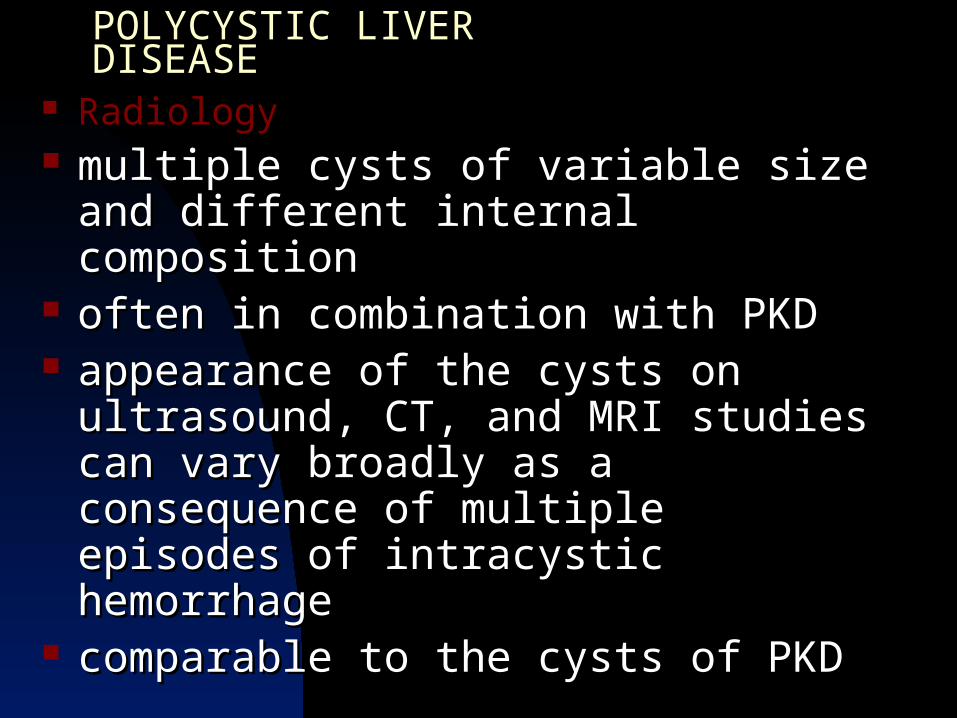
POLYCYSTIC LIVER DISEASEPOLYCYSTIC LIVER DISEASE RadiologyRadiology multiple cysts of variable size and different multiple cysts of variable size and different
internal compositioninternal composition often in combination with PKDoften in combination with PKD appearance of the cysts on ultrasound, appearance of the cysts on ultrasound,
CT, and MRI studies can vary broadly as a CT, and MRI studies can vary broadly as a consequence of multiple episodes of consequence of multiple episodes of intracystic hemorrhageintracystic hemorrhage
comparable to the cysts of PKDcomparable to the cysts of PKD

MESENCHYMAL TUMORS AND MESENCHYMAL TUMORS AND HETEROTOPIC RESTSHETEROTOPIC RESTS
Clinical Background and PathologyClinical Background and Pathology Hemangioma – Hemangioma – most commonmost common prevalence of remaining mesenchymal benign prevalence of remaining mesenchymal benign
liver tumor is lowliver tumor is low no unique radiographic featuresno unique radiographic features exception are tumors that contain fatexception are tumors that contain fat
lipomas, angiomyolipomas, myelolipomaslipomas, angiomyolipomas, myelolipomas no malignant potentialno malignant potential

MESENCHYMAL TUMORS AND HETEROTOPIC RESTSMESENCHYMAL TUMORS AND HETEROTOPIC RESTS
UltrasoundUltrasound fat-containing tumors are echogenicfat-containing tumors are echogenic posterior acoustic enhancement in homogenous appearing posterior acoustic enhancement in homogenous appearing
tumors (specifically lipomas)tumors (specifically lipomas) Computed TomographyComputed Tomography low density = low density = >> 20 HU 20 HU pure lipomas show no enhancementpure lipomas show no enhancement partial and inhomogenous contrast enhancement should partial and inhomogenous contrast enhancement should
suggest angiomatous (angiomyolipoma) or myeloid suggest angiomatous (angiomyolipoma) or myeloid (myelolipoma)(myelolipoma)
MESENCHYMAL TUMORS AND HETEROTOPIC RESTSMESENCHYMAL TUMORS AND HETEROTOPIC RESTS
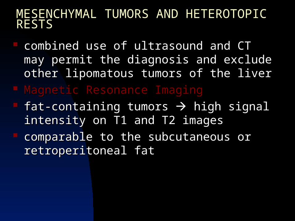
combined use of ultrasound and CT may permit combined use of ultrasound and CT may permit the diagnosis and exclude other lipomatous the diagnosis and exclude other lipomatous tumors of the livertumors of the liver
Magnetic Resonance ImagingMagnetic Resonance Imaging fat-containing tumors fat-containing tumors high signal intensity on high signal intensity on
T1 and T2 imagesT1 and T2 images comparable to the subcutaneous or comparable to the subcutaneous or
retroperitoneal fatretroperitoneal fat

HEPATIC HEMANGIOMA

FOCAL NODULAR HYPERPLASIA

FOCAL NODULAR HYPERPLASIA