SAFIYA GHORI-AHMAD vs. UNITED STATES COMMISSION ON INTERNATIONAL RELIGIOUS FREEDOM
Adoption of in vitro systems and zebrafish embryos as alternative models for reducing rodent use in assessments of immunological and oxidative stress responses to nanomaterialsHelinor J Johnston a*, Rachel Verdon a, Suzanne Gillies a, David M Brown a, Teresa F Fernandes a, Theodore B Henry a, Adriano G Rossi b, Lang Tran c, Carl Tucker b, Charles R Tyler d, Vicki Stone a
a Nano Safety Research Group, Heriot-Watt University,Edinburgh b Medical Research Council (MRC) Centre for Inflammation Research, The Queen's Medical Research Institute, University of Edinburgh, Edinburghc Institute of Occupational Medicine, Edinburghd Biosciences, College of Life and Environmental Sciences, University of Exeter
*Corresponding author: Helinor Johnston, [email protected], +44(0)131 451 3303
1
Abstract:
Assessing the safety of engineered nanomaterials (NMs) is paramount to the responsible and sustainable development of nanotechnology, which provides huge societal benefits. Currently, there is no evidence that engineered NMs cause detrimental health effects in humans. However, investigation of NM toxicity using in vivo, in vitro, in chemico and in silico models has demonstrated that some NMs stimulate oxidative stress and inflammation, which may lead to adverse health effects. Accordingly, investigation of these responses currently dominates NM safety assessments. There is a need to reduce reliance on rodent testing in nanotoxicology for ethical, financial and legislative reasons, and due to evidence that rodent models do not always predict the human response. We advocate that in vitro models and zebrafish embryos should have greater prominence in screening for NM safety, to better align nanotoxicology with the 3Rs principles. Zebrafish are accepted for use by regulatory agencies in chemical safety assessments (e.g. developmental biology) and there is growing acceptance of their use in biomedical research, providing strong foundations for their use in nanotoxicology. We suggest that investigation of the response of phagocytic cells (e.g. neutrophils, macrophages) in vitro should also form a key part of NM safety assessments, due to their prominent role in the first line of defence. The development of a tiered testing strategy for NM hazard assessment that promotes the more widespread adoption of non-rodent, alternative models and focuses on investigation of inflammation and oxidative stress could make nanotoxicology testing more ethical, relevant, and cost and time efficient.
Keywords: nanomaterial, nanoparticle, nanotoxicology, zebrafish, in vitro, 3Rs, inflammation, oxidative stress, neutrophil, macrophage
2
Contents I. Introduction:
1) Aligning Nanomaterial Safety Assessments with the 3Rs Principles
II. NM Mechanism of Toxicity
1) NMs and inflammation
1a) Neutrophils
1b) Macrophages
2) NMs and oxidative stress
2a)
III. Alternative models to investigate NM mediated inflammogenicity and oxidative stress
1) Zebrafish
1a) Zebrafish and the innate immune response to NMs
1b) Zebrafish embryos and oxidative stress
1c) Zebrafish: Recommendations for a testing strategy
1ci) Life stage
1cii).Route of administration
2) In vitro models
3) In Vitro to In Vivo Extrapolation (IVIVE)
IV. Conclusions
V Acknowledgements
VI. Declaration of Interest
VII References
3
I. Introduction:Nanotechnology is a fast growing sector and a key enabling technology of the 21st Century. In particular, the exploitation of engineered nanomaterials to develop ‘nano-enabled’ products has already been proven to have many benefits for society and the global economy (Schmidt, 2009). Nanomaterials (NMs) are defined as having at least one dimension that is 1-100nm in diameter (European Commission, 2011). At the nanoscale, novel properties emerge in materials that are not observed in their bulk counterparts, resulting in, for example, changes in reactivity, optical properties, and conductivity. This has been exploited in an array of consumer products, with NMs now used across diverse sectors including pharmaceuticals, cosmetics, textiles, food, electronics, automotive, agriculture, and pigments/inks, to name a few. NMs first entered the marketplace in consumer products in the mid-1990s (Fairbrother and Fairbrother, 2009) and their exploitation has since grown enormously, and continues to do so today. In 2016, the global market for Nanotechnology was worth $39.2 billion and it is estimated that this will rise to $90.5 billion by 2021 (McWilliams, 2016). To illustrate this point further, in 2005 a Nanotechnology Consumer Product Inventory (CPI) identified 54 products that had incorporated nanotechnology, and by 2014 this had increased to 1814 products (Vance et al., 2015). There is now a drive to minimise any risks posed by NMs, to maximize the benefits of this relatively new technology (Maynard et al., 2006). Epidemiological evidence for respirable particles (e.g. particulate air pollution, asbestos, quartz), some of which have a size comparable to that of NMs, demonstrates that they can have detrimental impacts on human health (e.g. Dockery et al., 1993, Borm and Tran 2002 Driscoll et al., 2005, IARC 1997, Wagner et al., 1960), prompting concern regarding the safety of NMs.
Despite the increased prevalence of NMs in the marketplace, there are still uncertainties surrounding their potential impact on humans and the environment. Currently, there is no evidence that engineered NMs are harmful to humans, however, a range of in vivo (e.g. rodent), in vitro, in chemico and in silico models have demonstrated that some NMs may be toxic. As safety assessments have not kept pace with the desire to exploit NMs, concerns have been raised about their potential to elicit adverse health effects in humans. There is also evidence for toxic effects of certain NMs, most notably metal and metal oxide NMs, from laboratory studies on species that are used to inform environmental protection (e.g. vertebrates, such as the fish species Danio rerio, Pimephales promelas, Oncorhynchus mykiss, Micropterus salmoides, Oryzias latipes; invertebrates, such as Daphnia magna, Lumbriculus variegatus, Lymnaea stagnalis, Mytilus edulis, Eisenia fetida; and a range of other taxa such as algae, bacteria and plants (e.g. reviewed in Selck et al., 2016). Investigation of impacts on environmental organisms has most commonly encompassed assessment of mortality, reproduction, and accumulation (e.g. body or organ burden), with some studies investigating the mechanism of toxicity, and most prominently oxidative stress. Importantly, existing studies suggest that there are commonalities in the mechanism of NM toxicity across different species (i.e. models which assess impacts on human health and the environment). With few exceptions, however, most of the effects seen for NMs only occur at exposures that far exceed estimated environmental concentrations although it is accepted that hotspots (i.e. areas where concentrations may be higher due to discharges or land application of sludge) do exist and that long term exposures at low concentrations, which would closely reflect reality, cannot always be replicated in the laboratory (Nowack, 2017).
NMs are extremely diverse, and vary with respect to their physicochemical properties including; composition (e.g. metals, metal oxides, carbon, polymers), size (and surface area), morphology, solubility, charge, and surface chemistry. Identification of the properties of NMs that confer toxicity is a core component of toxicity studies, and requires a comprehensive characterization of NM physico-chemical properties in parallel with hazard studies. Human exposure to NMs may occur in occupational, environmental and consumer settings, and it has been established that the properties of NMs may change throughout their life cycle, which can influence their toxicity (Mitrano and Nowack, 2016). The great diversity of NMs, and desire to test NM hazard throughout the life cycle/value chain would require a huge number of safety assessments to evaluate their toxicity. For example, for single walled carbon nanotubes (SWCNTs) alone it has been estimated that up to 50,000 varieties could be produced (Schmidt, 2007), and as the physico-chemical properties of each SWCNT will be different, they are likely to vary with respect to their toxicity. In 2009, it was estimated that the use of traditional approaches to thoroughly evaluate the toxicity of NMs already on the market (i.e. not including future generations of NMs) would cost up to $1.2 billion, take up to 53 years to complete, and require a very
4

large number of animals (Choi et al., 2009). Many would argue that this is unacceptable, and that whilst a comprehensive assessment of NM safety is required to ensure their safe use, there is also a need to minimise rodent testing when evaluating NM toxicity. The enhanced used of alternative models has the additional benefit of potentially simultaneously reducing the cost and time of testing, which are major advantages associated with their use.
1) Aligning Nanomaterial Safety Assessments with the 3Rs Principles:Currently, chemical safety assessments require a systematic evaluation of toxicity using internationally harmonized protocols, which often rely on animal testing (Hartung, 2009). There are concerns regarding the numbers of animals required for chemical safety assessments and so there is a desire to minimise animal use (Rovida and Hartung, 2009). Whilst rodent testing is often the preferred testing system for human health NM hazard assessment, it is not feasible to rely solely on such rodent testing due to ethical, resource, and legislative considerations (Hartung and Sabbioni 2011, Stone et al., 2014, Burden et al., 2017). In addition, although toxicology testing has remained relatively unchanged for the last 50 years, it is known that rodent models are not always able to accurately predict toxic effects in humans (Olsen et al 2000, Mestas and Hughes 2004, Hartung, 2009). Toxicology for the 21st Century aspires to make testing more efficient and more ethical, and questions the extent to which the use of rodent models should dominate testing due to their inability to always accurately predict effects in humans (Hartung, 2009). Instead it encourages identification of the mechanism of toxicity to inform evidence based testing strategies to assess toxicity, and promotes the use of alternative, high-throughput (HTP) systems which use models of human origin (Hartung, 2009).
The 3Rs principles of scientific research, which aim to replace, reduce and refine animal testing (Russell and Burch, 1959), are widely accepted, and have been implemented internationally. There is a desire to accelerate the development and application of alternative (non-animal) testing methods when assessing chemical toxicity to human health and the environment in order to enhance implementation of the 3Rs principles, to reduce the number of vertebrates used for toxicology testing (Tornqvist et al., 2014, Burden et al., 2015, 2016). Recently, the need to better align nanotoxicology testing with the 3Rs principles has been raised (Burden et al., 2017). The EU regulation on the protection of animals used for scientific purposes (Directive 2010/63/EU; European Parliament, 2010 (Article 47)) encourages the implementation of alternative approaches when conducting scientific research. Thus the application of the 3Rs principles is embedded in regulations that are relevant to the safety assessment of NMs (e.g. chemicals, cosmetics, food, pharmaceuticals, medical devices, and biocides) (Rauscher et al., 2017).
The European Chemicals Agency (ECHA) is responsible for implementing REACH, the European chemical safety regulation and came into force in 2007). REACH aims to protect human health and the environment from chemical risks, including NMs. ECHA acknowledges that the safe manufacture and use of chemicals will require significantly more information on potential hazard, and with current testing approaches this is likely to lead to a corresponding increase in animal testing (Rovida and Hartung, 2009, ECHA, 2016a). However, REACH supports strategies to avoid unnecessary animal testing, such as promoting better data sharing to avoid duplication of research and to support read-across between species, and the use of alternative (non-animal) methods (ECHA, 2011, 2014, 2016b). The European Commission regularly publishes statistics on animal use for experimental procedures, and a reduction in animal use by half a million occurred between 2008 and 2011 (ECHA, 2014). Evidence that illustrates registrants actively employ non-animal approaches in fulfilment of the obligations of REACH is illustrated for the 7939 new chemical safety experimental studies logged in the ECHA database for the period between 2009-2013, where 62% of the assessments were conducted in vivo, and 38% in vitro (ECHA, 2014). Demonstration that legislators and regulators are becoming more receptive to, and confident in, the use of data from alternative testing systems is also exemplified by the EU ban on the use of animals for cosmetic safety testing. Alternative models to assess skin irritation and sensitization have been developed and validated and are used widely. However, for other endpoints alternative models are not yet available. Therefore, whilst the adoption of alternatives to animal testing is advocated by regulators, currently animal testing still accounts for the majority of research assessing the toxicity of chemicals. Nevertheless, it is expected that the use of alternative models will increase in the future as more alternative approaches are validated.
Transforming the approach for toxicology testing through adoption of alternative systems, provides major opportunities for reducing animal use in chemical safety assessments. This is particularly
5
appealing for NMs, given their diversity and the vast number of these materials that will require safety assessment. This paper explores the potential to enhance the use of zebrafish embryo and cell (in vitro) models as alternatives to rodent testing when assessing NM safety, with a focus placed on investigation of immunological function and oxidative stress; two of the main effector mechanisms for NM toxicity established to date. It is hoped that the identification of alternative (non-rodent) models, which have the potential to be adopted internationally, will support the implementation of an intelligent testing strategy for NM hazard assessment which reduces the reliance placed on rodent testing and better aligns nanotoxicology research with the 3Rs principles. Of interest is that existing reviews have encouraged greater use of zebrafish for assessing NM toxicity (e.g. Lin et al., 2013, Chakraborty et al., 2016). We build on these reviews and extend our analysis on the state of the art to provide recommendations for the experimental design of future studies by, for example, identifying the biological responses that should be prioritized for assessment of NM effects, based on current knowledge on the mechanism of action of NMs. The approach we advocate will help shape future research activities in nanotoxicolog to improve establishment of common biological responses across different models (rodents, zebrafish and cell (in vitro) to facilitate read-across and inform decisions about the suitability of using alternative models.
II. NM Mechanism of ToxicityKnowledge of the mechanism of toxicity of NMs is essential when designing intelligent and evidence based testing strategies to screen their safety, and to help understand the potential impact of NMs on human health and the environment (Stone et al., 2014). NMs, depending on their physico-chemical properties, have been shown to stimulate a range of biological responses spanning inflammation, fibrosis, oxidative stress, cytotoxicity, mitochondrial perturbations, genotoxicity, and carcinogenicity, at various target sites (e.g. lung, liver, kidneys, cardiovascular system, intestine, CNS). It is established that oxidative stress and inflammation are key drivers of the adverse health effects associated with human exposure to pathogenic particles (e.g. PM10) (reviewed in Stone et al., 2016) and this information has provided the foundations for nanotoxicology research (Oberdorster, Donaldson and Stone, 2007). More recently, omics technologies have been used in vivo and in vitro to generate hypotheses regarding the molecular mechanism of action of NMs (e.g. Labib et al., 2016, vanAerle et al., 2013, Boyles et al., 2015, reviewed in Costa and Fadeel 2016). The relatively high financial cost associated with these approaches is restrictive for their routine use. Therefore, biochemical assays for detecting oxidative stress and inflammation as indicators of NM toxicity have dominated published studies (see below).
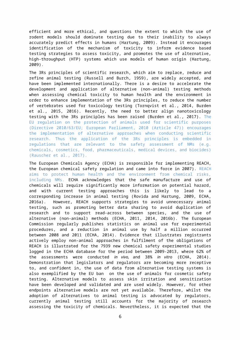
1) NMs and inflammation Inflammation is a normal, protective physiological response to foreign material and injury. However, if inflammatory responses are persistent (chronic) due to an exaggerated or dysregulated response, including a failure of normal resolution mechanisms, a pathological response can emerge (Poon et al.,2014, Robb et al., 2016, Soehnlein et al., 2017) (Figure 1). Therefore, it is essential that inflammation resolves in a timely manner to prevent the manifestation of detrimental health effects (Poon et al.,2014, Robb et al., 2016, Soehnlein et al., 2017). Disease inducing particles and fibres such as particulate air pollution (PM10), silica and asbestos cause a range of adverse health effects in humans (e.g. initiation/exacerbation of respiratory and cardiovascular disease, cancer) via mechanisms involving the stimulation of inflammation (reviewed in e.g. Donaldson and Seaton, 2012, Oberdorster et al, 2007). Macrophages and neutrophils play key roles in the body’s first (innate) response to foreign particles and here we first explore knowledge on their roles in the inflammation response associated with NM toxicity.
6
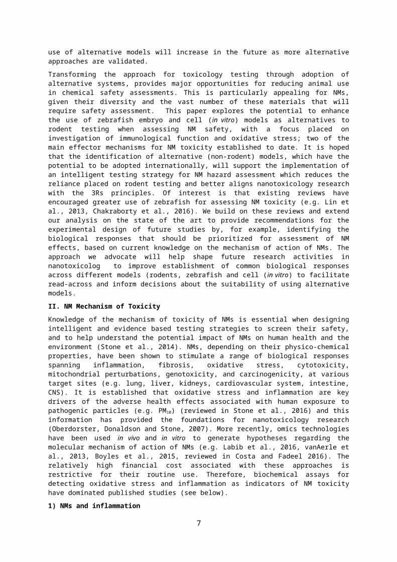
Figure 1 The activation of inflammation by NMs: physiological vs pathological responses. NMs can activate acute inflammatory responses that are dominated by neutrophils and macrophages. Apoptosis of immune cells or stimulation of their emigration promotes the resolution of acute inflammatory responses, and activates repair. Failure of inflammation to resolve in a timely manner may stimulate chronic inflammation which can lead to tissue damage and contribute to disease pathogenesis.
1a) Neutrophils
Neutrophils are the most abundant circulating leukocytes in humans, and are recruited to sites of injury or infection to protect the host (e.g. Nathan, 2006, Filep and Kebir, 2009, Wang and Arase, 2014). Neutrophils are a key first line of immune cell defense in the inflammatory process, and possess several strategies to clear and destroy pathogens (Wang and Arase, 2014, Mayadas et al., 2014) (Figure 2). The resolution of inflammation requires that neutrophils undergo apoptosis and are cleared by phagocytic cells (e.g. macrophages), a process known as efferocytosis (Poon et al., 2011, Robb et al., 2016, Jones et al., 2016). Failure of neutrophillic inflammation to resolve itself can cause tissue damage and contribute to disease pathogenesis due to the release of toxic mediators (e.g. proteases and reactive oxygen species (ROS)) (Poon et al., 2011, 2014, Robb et al., 2016, Filep and Kebir, 2009, Soehnlein et al., 2017). Indeed, activation of neutrophil responses has been linked to genotoxicity and carcinogenicity (reviewed in Knaapen et al., 2006). Knowledge of the cellular and molecular events underlying the activation and resolution of neutrophil responses can inform the design of laboratory studies which assess the ability of NMs to stimulate inflammatory responses (Figure 2).
7
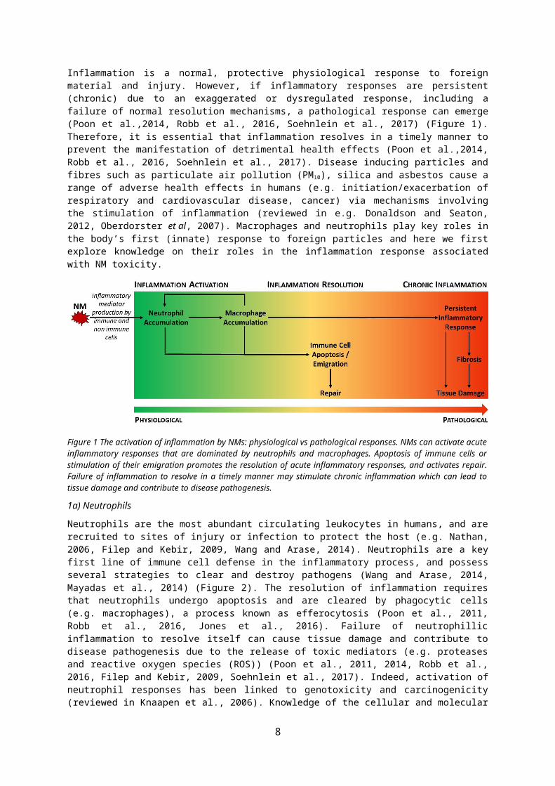
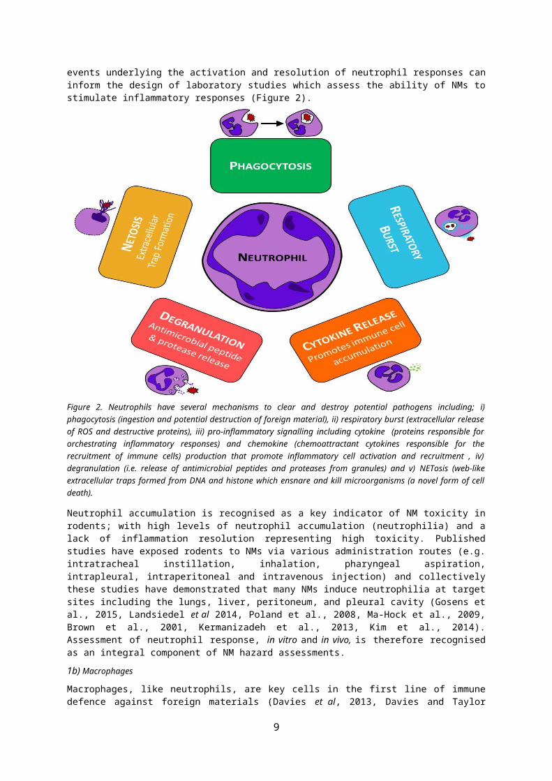
Figure 2. Neutrophils have several mechanisms to clear and destroy potential pathogens including; i) phagocytosis (ingestion and potential destruction of foreign material), ii) respiratory burst (extracellular release of ROS and destructive proteins), iii) pro-inflammatory signalling including cytokine (proteins responsible for orchestrating inflammatory responses) and chemokine (chemoattractant cytokines responsible for the recruitment of immune cells) production that promote inflammatory cell activation and recruitment , iv) degranulation (i.e. release of antimicrobial peptides and proteases from granules) and v) NETosis (web-like extracellular traps formed from DNA and histone which ensnare and kill microorganisms (a novel form of cell death).
Neutrophil accumulation is recognised as a key indicator of NM toxicity in rodents; with high levels of neutrophil accumulation (neutrophilia) and a lack of inflammation resolution representing high toxicity. Published studies have exposed rodents to NMs via various administration routes (e.g. intratracheal instillation, inhalation, pharyngeal aspiration, intrapleural, intraperitoneal and intravenous injection) and collectively these studies have demonstrated that many NMs induce neutrophilia at target sites including the lungs, liver, peritoneum, and pleural cavity (Gosens et al., 2015, Landsiedel et al 2014, Poland et al., 2008, Ma-Hock et al., 2009, Brown et al., 2001, Kermanizadeh et al., 2013, Kim et al., 2014). Assessment of neutrophil response, in vitro and in vivo, is therefore recognised as an integral component of NM hazard assessments.
1b) Macrophages
Macrophages, like neutrophils, are key cells in the first line of immune defence against foreign materials (Davies et al, 2013, Davies and Taylor 2015). Macrophages are found in numerous locations throughout the body (e.g. lungs, liver, peritoneal cavity) and are key to the clearance of NMs from various target sites (e.g. Halpern et al., 1953, Ogawara et al., 1999, Sadauskas et al., 2007, Semmler-Behnke et al., 2007 and 2008, Geiser et al., 2008, Roberts et al., 2013). The defensive strategies activated by pathogens and particles discussed above for neutrophils (Figure 2) are also relevant to macrophages, although there are also cell specific responses that operate in each type of immune cell.
In vivo, assessment of immune cell accumulation, and cytokine production over time are often prioritised to monitor the inflammatory response to NMs (particularly within the lung, but also other target sites). In vitro, the following responses are commonly assessed in macrophages and neutrophils as indicators of cell activation and toxicity; cytotoxicity (e.g. apoptosis), cytokine production, phagocytic function, NM internalisation, and ROS production (intracellular and respiratory
8
burst). Studies which have investigated the response of macrophages and neutrophils to NMs, are discussed in detail below.
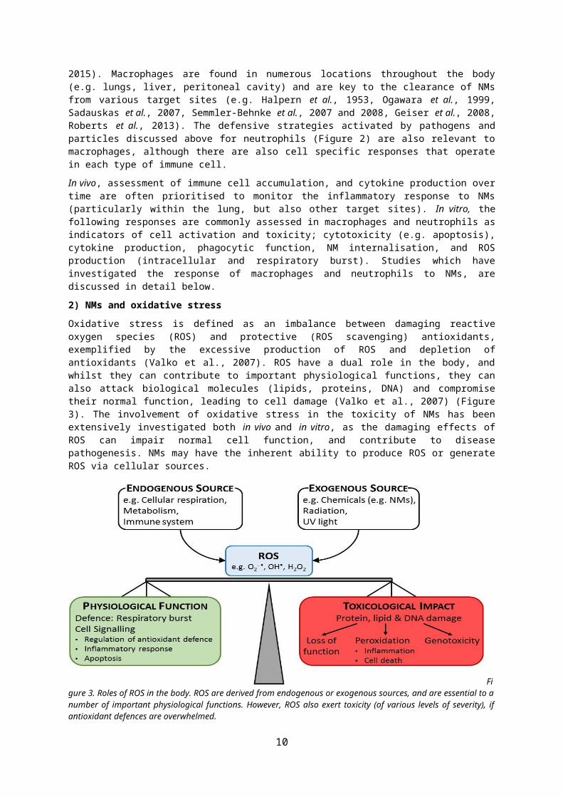
2) NMs and oxidative stressOxidative stress is defined as an imbalance between damaging reactive oxygen species (ROS) and protective (ROS scavenging) antioxidants, exemplified by the excessive production of ROS and depletion of antioxidants (Valko et al., 2007). ROS have a dual role in the body, and whilst they can contribute to important physiological functions, they can also attack biological molecules (lipids, proteins, DNA) and compromise their normal function, leading to cell damage (Valko et al., 2007) (Figure 3). The involvement of oxidative stress in the toxicity of NMs has been extensively investigated both in vivo and in vitro, as the damaging effects of ROS can impair normal cell function, and contribute to disease pathogenesis. NMs may have the inherent ability to produce ROS or generate ROS via cellular sources.
Figure 3. Roles of ROS in the body. ROS are derived from endogenous or exogenous sources, and are essential to a number of important physiological functions. However, ROS also exert toxicity (of various levels of severity), if antioxidant defences are overwhelmed.
The capacity of NMs to generate ROS is frequently used as an in chemico assay to measure their reactivity (e.g. Wilson et al., 2007, Shi et al., 2012). For the cellular response, it is hypothesised that NMs can elicit three different tiers of oxidative stress, which each dictate a different biological response (e.g. Donaldson et al, 2003, Brown et al, 2004, Nel et al, 2006, Li et al, 2008). At low levels (tier 1), enhanced ROS production can stimulate a protective response in cells via the activation of redox sensitive transcription factors (e.g. nuclear factor erythroid related factor (Nrf)2) which control the expression of cytoprotective genes (e.g. antioxidants). As ROS levels increase (tier 2), and protective antioxidant defences become overwhelmed, inflammatory responses can be activated (e.g. via the activation of oxidant and calcium sensitive transcription factors, such as nuclear factor kappa B (NF-κB) and/or activator protein (AP)-1). If ROS levels continue to increase (tier 3) then genotoxicity and cytotoxicity can be stimulated. This hierarchical model of the cellular response to NMs has been used to identify their potential biological effects and can inform the experimental design of studies which investigate NM toxicity.
The respiratory burst is a defensive response that is activated by immune cells (e.g. macrophages, neutrophils), and involves the extracellular release of ROS to destroy invading pathogens. There is evidence that some NMs (e.g. TiO2, Ag, CNTs, SiO2) can stimulate a respiratory burst in neutrophils and macrophages (e.g. Brown et al., 2007a, Gonçalves et al., 2009, Park and Park, 2009, Scherbart et al., 2011, Boyles et al., 2015. Johnston et al., 2015). Whilst this is intended to be a protective response, it can stimulate oxidative stress, and thereby contribute to NM toxicity, and thus should be considered when assessing NM hazard in vitro and in vivo.
For in vitro and in vivo models (e.g. rodent, cell and zebrafish) a wide range of endpoints and assays can be used to probe the involvement of oxidative stress in the toxicity of NMs (Figure 4). Regardless
9
of the experimental approach used, studies have identified that oxidative stress is a key event in the cellular response to many NMs (see below).
Figure 4. Commonly used approaches to assess the contribution of oxidative stress to NM toxicity across models (e.g. rodent, cell and zebrafish). Investigations have typically encompassed assessment of the following; ROS production (in acellular and cellular conditions), the activity or levels of antioxidants (e.g. catalase (CAT), superoxide dismutase (SOD), heme oxygenase (HO)-1, glutathione peroxidase (GPx), glutathione (GSH), nitric oxide synthase (NOX)), markers of oxidative damage (e.g. malondialdehyde (MDA) which is indicative of lipid peroxidation), and identification of whether pre-treatment of cells/animals with antioxidants (such as n-acetyl cysteine (NAC), or trolox) protects against NM induced toxicity. When assessing whether NMs can stimulate oxidative stress a combination of approaches are normally used. The different approaches used to assess the contribution of oxidative stress to NM toxicity are presented in green, and the assays/indicators of toxicity provided in red. EPR = electron paramagnetic resonance, DCFH = 2’7’-dicholorofluorescein diacetate.
Oxidative stress can stimulate inflammation, and vice versa, and thus these processes are intimately linked, and known to contribute to the pathogenesis of many diseases. For example, ROS act as secondary messengers to activate redox responsive signalling pathways and transcription factors (e.g. NFκB) which stimulates the expression of pro-inflammatory proteins (e.g. cytokines, adhesion molecules) that promote inflammation (e.g. reviewed by Torres and Forman, 2003, Mittal et al., 2014). Indeed, there is evidence that NMs can stimulate pro-inflammatory cytokine production from macrophages in vitro via an oxidant mechanism (Brown et al., 2004, Lee et al., 2009). In addition, as discussed above, immune cells can generate ROS as a protective response to destroy pathogens, which can promote oxidative stress. A summary of what we know on the relationship between oxidative stress and inflammation in the toxicity of NMs is outlined in Figure 5.
10

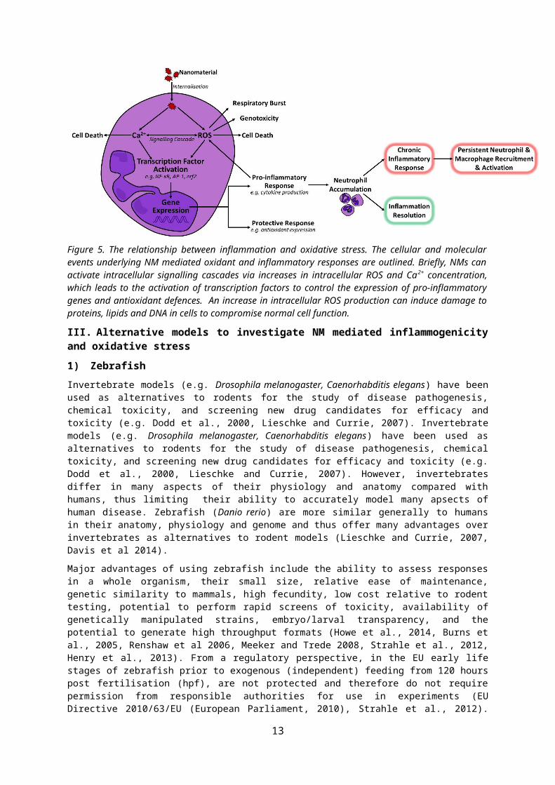
Figure 5. The relationship between inflammation and oxidative stress. The cellular and molecular events underlying NM mediated oxidant and inflammatory responses are outlined. Briefly, NMs can activate intracellular signalling cascades via increases in intracellular ROS and Ca 2+ concentration, which leads to the activation of transcription factors to control the expression of pro-inflammatory genes and antioxidant defences. An increase in intracellular ROS production can induce damage to proteins, lipids and DNA in cells to compromise normal cell function.
III. Alternative models to investigate NM mediated inflammogenicity and oxidative stress 1) Zebrafish Invertebrate models (e.g. Drosophila melanogaster, Caenorhabditis elegans) have been used as alternatives to rodents for the study of disease pathogenesis, chemical toxicity, and screening new drug candidates for efficacy and toxicity (e.g. Dodd et al., 2000, Lieschke and Currie, 2007). Invertebrate models (e.g. Drosophila melanogaster, Caenorhabditis elegans) have been used as alternatives to rodents for the study of disease pathogenesis, chemical toxicity, and screening new drug candidates for efficacy and toxicity (e.g. Dodd et al., 2000, Lieschke and Currie, 2007). However, invertebrates differ in many aspects of their physiology and anatomy compared with humans, thus limiting their ability to accurately model many apsects of human disease. Zebrafish (Danio rerio) are more similar generally to humans in their anatomy, physiology and genome and thus offer many advantages over invertebrates as alternatives to rodent models (Lieschke and Currie, 2007, Davis et al 2014).
Major advantages of using zebrafish include the ability to assess responses in a whole organism, their small size, relative ease of maintenance, genetic similarity to mammals, high fecundity, low cost relative to rodent testing, potential to perform rapid screens of toxicity, availability of genetically manipulated strains, embryo/larval transparency, and the potential to generate high throughput formats (Howe et al., 2014, Burns et al., 2005, Renshaw et al 2006, Meeker and Trede 2008, Strahle et al., 2012, Henry et al., 2013). From a regulatory perspective, in the EU early life stages of zebrafish prior to exogenous (independent) feeding from 120 hours post fertilisation (hpf), are not protected and therefore do not require permission from responsible authorities for use in experiments (EU Directive 2010/63/EU (European Parliament, 2010), Strahle et al., 2012). Embryo transparency is particularly appealing as it facilitates both the direct visualization of developmental processes including organ/tissue structure and function, and enables evaluation of responses at specific proteins or gene loci within target tissues (e.g. via whole mount in situ hybridization and immunohistochemistry). Furthermore, the availability of transgenic zebrafish which express fluorescent proteins in specific cell types enables the visualization and quantification of cellular processes. This is extremely valuable when investigating the cell and molecular basis of disease or the mechanism underlying the toxicity of chemicals (such as NMs). A diverse array of transgenic zebrafish lines are now available (reviewed in Lee et al., 2015), and those particularly relevant to the assessment of NM toxicity are discussed in more detail below. Therefore, zebrafish are arguably relevant as a test species for NM toxicity testing for read across to mammals, including humans with, existing evidence suggesting a good concordance observed between zebrafish and human and rodent studies (see section III.3)
11

Zebrafish are already used widely for biomedical and toxicological research. For example, there are internationally agreed protocols, accepted by regulators, for their use in developmental biology and toxicity studies and as an alternative to rodents (Dodd et al., 2000, OECD Fish Embryo Acute Toxicity Test No 236, reviewed in He et al., 2014). Zebrafish have been used to unravel the molecular basis of human disease (e.g. cancer, diabetes, cardiovascular diseases, infectious disease (e.g. microbe-host interactions), and neurodegenerative disease), as well as for drug screening (reviewed in Goldsmith and Jobin 2012, Lieschke and Currie, 2007, Mathias et al., 2012, MacRae and Peterson, 2015). The nanotoxicology community have widely adopted the use of zebrafish embryo and larval stages up to 120hpf (e.g. Osborne et al., 2013, 2016; Zhu, Tian and Cai., 2012, Massarsky et al., 2013) and adults (e.g. Bilberg et al., 2012, Zhang et al., 2015) to investigate the aquatic toxicity of NMs, typically via assessment of morphological development and mortality (e.g. Lee et al., 2007, 2012, Ganesan et al 2016, Duan et al., 2013, Lin et al., 2011). Zebrafish embryos have also been used to investigate the bioavailability of NMs when exposed via water (Lee et al., 2007, 2012, Goodhead et al., 2015). Therefore, whilst zebrafish have received little attention for evaluating possible impacts of NMs on human health, however they could be a powerful tool to investigate oxidative effects and inflammogenicity, as an alternative to rodents.
1a) Zebrafish and the innate immune response to NMs The immune system of zebrafish has strong similarities to that of humans with the major cell lineages evident, including cells analagous to monocytes, tissue macrophages, granulocytes (e.g. neutrophils, eosinophils) and lymphocytes (reviewed in Traver et al., 2003, Meeker and Trede 2008, Novoa and Figueras, 2012). A functional innate immune system exists in zebrafish within 48 hpf, with the adaptive system evident 4 weeks after fertilization (Renshaw et al., 2006, Trede et al., 2004, Novoa and Figueras, 2012). Here we focus on the use of zebrafish embryos to investigate innate, acute immune cell responses, which are dominated by neutrophils and macrophages.
Neutrophils are distributed throughout the zebrafish embryo via the circulation (e.g. Yang et al., 2014, Duan et al., 2016), whilst macrophages are present in the blood circulation and are resident in tissues (reviewed in Torraca et al., 2014, Herbomel et al., 1999). Several studies have used wildtype zebrafish to investigate inflammatory responses to a range of stimuli (e.g. injury, micro-organisms, chemicals). To quantify the inflammatory response, such studies have identified immune cells based on their morphology, or via staining using immunofluorescence and immunohistochemical techniques. For example, myeloperoxidase (MPO) is a neutrophil-specific enzyme commonly used to identify neutrophils Herbomel et al. (1999), Mathias et al. (2009), Renshaw et al. (2006), and Levraud et al. (2009). Alternatively, the generation of transgenic zebrafish strains with fluorescent, endogenously-labelled immune cells allows inflammatory responses to be visualised and quantified using fluorescent microscopy. The use of such strains presents a major advantage over rodent models – and over traditional histochemical approaches in wildtype fish – owing to the ability to monitor inflammatory responses in real time, in vivo and obtain results more quickly. For example, it is possible to track immune cell movement in zebrafish, which allows monitoring of the recruitment of cells and their reverse migration in order to investigate the activation and resolution of inflammatory responses (Feng et al., 2010, Brown et al., 2007b, Ellett et al., 2015). Of further benefit is that automated HTP in vivo screening of inflammogenicity can also be performed in transgenic zebrafish (e.g. Hall et al., 2014, Lin et al., 2011). Transgenic strains that are most commonly used to study inflammation are those with fluorescently labelled (e.g. with green fluorescent protein (GFP) or mCherry) neutrophils (e.g. Tg(mpx:GFP)i114, and lysC:EGFP)), macrophages (e.g. macrophage expressed gene (mpeg)1:GFP)) and double-transgenics with both fluorescently labelled neutrophils and macrophages (e.g. Tg(mpx:GFP)i114/Tg (mpeg1:mCherry)gl23 (Renshaw et al., 2006, Ellett et al., 2011, Kim et al., 2016). To date, transgenic embryos (< 120 hpf) have been predominately used to investigate inflammatory responses owing to their transparency, which makes them amenable to microscopic approaches. However, casper strains of zebrafish are now available which lack pigment and the transparency of these strains enables immune cell movement to be tracked in adult zebrafish (e.g. White et al., 2008, Kim et al., 2016).
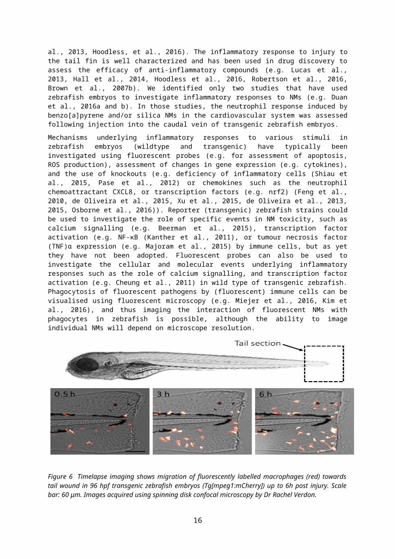
As leukocyte behaviour can be visualised and quantified in transparent zebrafish embryos, they are an extremely useful tool to evaluate fluxes in immune cell accumulation during inflammation (Ellett et al., 2011, Hoodless et al., 2016) (Figure 6). Accordingly, zebrafish have been used to investigate the innate immune response to various stimuli including; lipopolysaccharide (LPS) (Yang et al., 2014), injury (tail fin wound, which involves transection of the zebrafish embryo’s tail; e.g. Renshaw et al., 2006, de Oliveira et al., 2013, Lucas et al., 2013, Hoodless et al., 2016), and micro-organisms (e.g Renshaw et al., 2006, Li et al., 2012, Nguyen-Chi et al 2014, reviewed in Meeker and Trede, 2008).
12
Existing studies have shown that reproducible and quantifiable neutrophillic inflammatory responses are stimulated in zebrafish embryos (e.g. following tailfin injury) and resolve in a time frame similar to mammalian models (e.g. Renshaw et al., 2007, Loynes et al.,2010, Robertson et al., 2017, Lucas et al., 2013, Hoodless, et al., 2016). The inflammatory response to injury to the tail fin is well characterized and has been used in drug discovery to assess the efficacy of anti-inflammatory compounds (e.g. Lucas et al., 2013, Hall et al., 2014, Hoodless et al., 2016, Robertson et al., 2016, Brown et al., 2007b). We identified only two studies that have used zebrafish embryos to investigate inflammatory responses to NMs (e.g. Duan et al., 2016a and b). In those studies, the neutrophil response induced by benzo[a]pyrene and/or silica NMs in the cardiovascular system was assessed following injection into the caudal vein of transgenic zebrafish embryos.
Mechanisms underlying inflammatory responses to various stimuli in zebrafish embryos (wildtype and transgenic) have typically been investigated using fluorescent probes (e.g. for assessment of apoptosis, ROS production), assessment of changes in gene expression (e.g. cytokines), and the use of knockouts (e.g. deficiency of inflammatory cells (Shiau et al., 2015, Pase et al., 2012) or chemokines such as the neutrophil chemoattractant CXCL8, or transcription factors (e.g. nrf2) (Feng et al., 2010, de Oliveira et al., 2015, Xu et al., 2015, de Oliveira et al., 2013, 2015, Osborne et al., 2016)). Reporter (transgenic) zebrafish strains could be used to investigate the role of specific events in NM toxicity, such as calcium signalling (e.g. Beerman et al., 2015), transcription factor activation (e.g. NF-κB (Kanther et al., 2011), or tumour necrosis factor (TNF)α expression (e.g. Majoram et al., 2015) by immune cells, but as yet they have not been adopted. Fluorescent probes can also be used to investigate the cellular and molecular events underlying inflammatory responses such as the role of calcium signalling, and transcription factor activation (e.g. Cheung et al., 2011) in wild type of transgenic zebrafish. Phagocytosis of fluorescent pathogens by (fluorescent) immune cells can be visualised using fluorescent microscopy (e.g. Miejer et al., 2016, Kim et al., 2016), and thus imaging the interaction of fluorescent NMs with phagocytes in zebrafish is possible, although the ability to image individual NMs will depend on microscope resolution.
Figure 6 Timelapse imaging shows migration of fluorescently labelled macrophages (red) towards tail wound in 96 hpf transgenic zebrafish embryos (Tg[mpeg1:mCherry]) up to 6h post injury. Scale bar: 60 μm. Images acquired using spinning disk confocal microscopy by Dr Rachel Verdon.
1b) Zebrafish embryos and oxidative stress Wildtype and transgenic zebrafish have been used as a test organism to investigate the involvement of oxidative stress in disease pathogenesis, and the toxicity of a variety of chemicals, and including NMs (e.g. Ag, ZnO, TiO2, C60, CuO). A wide range of responses have been used to assess the contribution of oxidative stress to NM toxicity in zebrafish including; depletion of antioxidants (e.g. glutathione, or catalase (e.g. Usenko et al., 2008, Massarsky et al., 2013, Faria et al., 2014, Choi et al., 2010)), activity of antioxidant enzymes (e.g. SOD, CAT and GPx (Faria et al., 2014, Zhao et al., 2013, Xiong et al., 2011, Ganesan et al 2016), oxidative damage (e.g. malondialdehyde (MDA)) (Zhao et al., 2013, Choi et al., 2010, Xiong et al., 2011, Massarsky et al., 2013, Ganesan et al 2016), and the ability of antioxidant supplementation to reduce NM mediated toxicity (Usenko et al., 2008).
13
Furthermore, the mechanism of toxicity of Ag NMs has also been investigated via whole mount in situ hybridization for genes forming part of the oxidative response cascade (Osborne et al., 2016). ROS production has also been monitored in zebrafish in vivo, in real time, using fluorescent probes (e.g. Mugoni et al., 2014, Hall et al., 2013). As an example, 2’,7’ dichlorofluorescin diacetate (DCFH) has been used successfully in whole zebrafish to monitor respiratory burst activation in response to phorbol myristate acetate (PMA) (Hermann et al., 2004), and ROS production upon exposure to toxicants (e.g. H2O2), using a high-throughput technique (Walker et al., 2012). At this time, the DCFH assay has not been used in zebrafish to assess NM mediated ROS production in vivo. Therefore, this assay has the potential to be applied to the study of NM toxicity in future. Of importance, is that existing studies which have investigated the involvement of oxidative stress in the zebrafish response to NMs have focused on assessment of the aquatic toxicity of NMs, however the results obtained are also relevant to human health. Interestingly, the majority of existing studies have used adult zebrafish, which has the benefit of allowing oxidative stress to be investigated in specific tissues (e.g. gills, liver).
Transgenic zebrafish with fluorescence reporters have been developed for real time monitoring of biological responses to oxidative stressors in whole organisms. Reporter strains developed include those which enable H2O2 production (Niethammer et al., 2009) and lipid peroxidation (Fang et al., 2011) to be investigated. Another transgenic zebrafish line known to be in development includes the electrophile response element (EpRE) reporter for oxidative stress, which shows tissue specificity for different environmental contaminants including nanomaterials (pers comm; Mourabit et al., 2017) . With the widespread uptake of Clustered Regularly Interspaced Short Palindromic Repeats (CRISPR) cas, a technique that allows for rapid and specific gene manipulation, it is expected that more transgenic model fish systems will become available in the near future for studying oxidative stress mechanisms.
1c) Zebrafish: Recommendations for a testing strategy As detailed above there are well established zebrafish models and tools that can be immediately applied to study NM toxicity. Whilst the use of zebrafish provides exciting opportunities to investigate the activation and resolution of inflammatory and oxidant driven responses by NMs, careful consideration of the experimental design is required to ensure the models are fit for purpose.
1ci) Life stage
We recommend the use of non-protected early life stages (<120hpf) of zebrafish to assess NM toxicity should be prioritized to encourage the more widespread implementation of the 3Rs principles in nanotoxicology. Of benefit is that the innate immune system and many organs (e.g. cardiovascular system, liver, kidney) are functional in zebrafish within this time frame. However, we acknowledge that under certain circumstances it may be more appropriate (and even necessary) to use adult life stages, for example when i) investigating impacts on the adaptive immune response, which is not functionally mature until ~4-6 weeks (Trede et al., 2004, Novoa and Figueras 2012), ii) assessing responses in organs that are not fully developed in early life stages, iii) assessing the chronic toxicity of NMs, and iv) investigating impacts following the onset of ingestion beyond 120 hpf at which point the zebrafish are protected.
When investigating the inflammatory response in early, non-protected life stages of zebrafish, embryos have typically been used at 3 days post fertilisation (dpf), which allows inflammatory and oxidative responses to be monitored up to 48h post exposure (i.e. up until 120hpf). Existing studies have demonstrated that this time frame is sufficient to capture the activation and resolution of neutrophil and macrophage driven responses following a tail wound injury (e.g. Hoodless et al., 2016). Renshaw et al. (2006, 2007) have demonstrated that the kinetics of the inflammatory response are similar between zebrafish and rodents and typically characterised by an infiltration of neutrophils which peaks between 6-24h, followed by an influx of macrophages. The expression of genes related to oxidative stress, and detoxification following NM exposure have been shown to be dependent on the target site and developmental stage, as well as the time point under investigation (Osborne et al., 2016). Furthermore, different approaches used to investigate oxidative stress vary in their sensitivity (e.g. biochemical assay assessing protein levels/activity vs gene expression). These findings should be considered when designing experiments to investigate oxidative stress in zebrafish, following exposure to NMs. Of benefit is that zebrafish could potentially be used to assess toxicity following single or repeated NM exposures, and therefore help address a knowledge gap in the area of nanotoxicology (Stone et al., 2016).
1cii) Route of administration
14
When investigating NM toxicity, it is advisable to assess both local (i.e. at the exposure site) and systemic effects. In rodents, assessment of toxicity has been assessed following pulmonary exposure, ingestion, dermal application and intravenous injection, and it is relevant to consider what the equivalent exposure routes are in zebrafish. Exposure of zebrafish via water has been the most commonly used technique by studies investigating the aquatic toxicity of NMs, however in order to use zebrafish to assess potential impacts of NMs on human health other administration routes need consideration. The choice of administration route will typically be influenced by the hypotheses under investigation (e.g. the pathway of human NM exposure intended to be represented) and the developmental stage of the zebrafish.
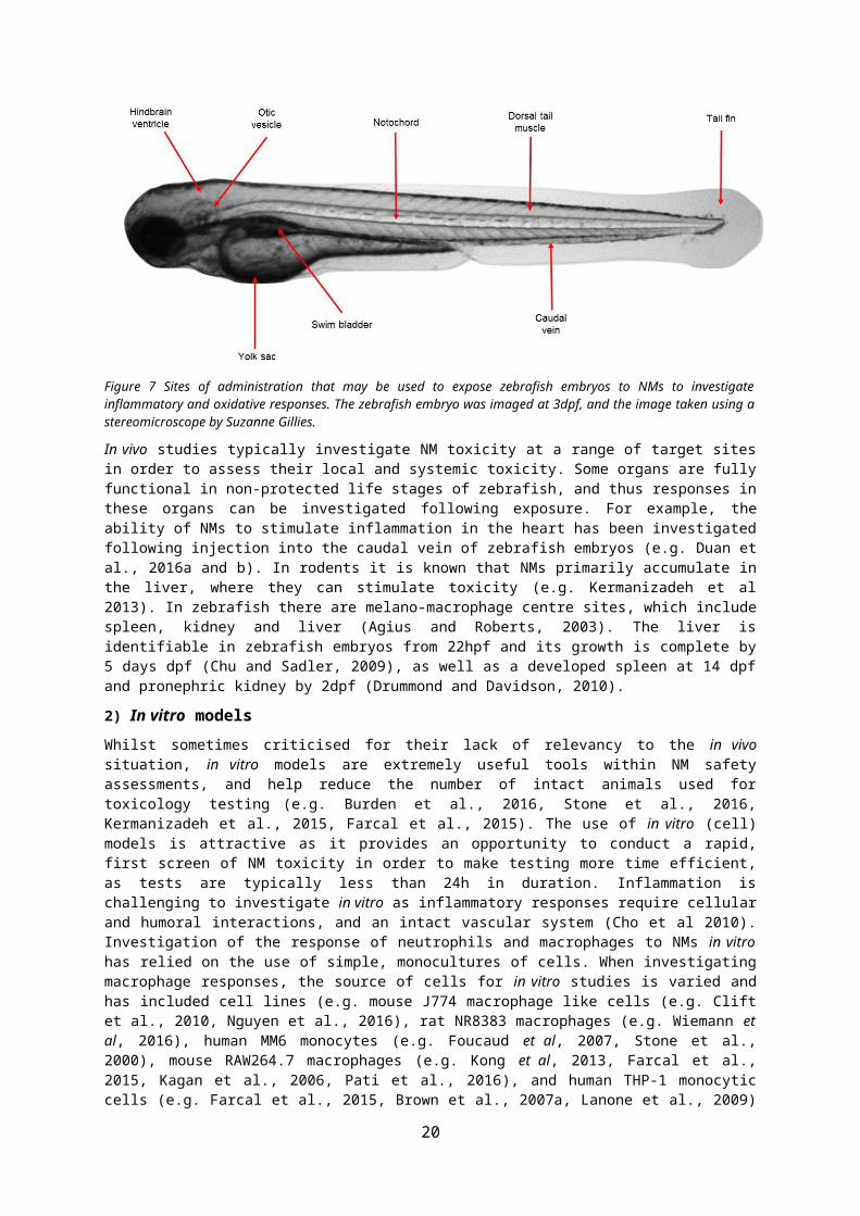
There are a variety of administration routes available for exposure of zebrafish to NMs (reviewed in Benard et al., 2012) (Figure 7). Existing studies have exposed zebrafish via water (e.g. Xu et al., 2015, Wang et al., 2014), or injection into the yolk sac (Yang et al., 2014), caudal vein (Kim et al., 2016), dorsal tail muscle (Lin et al., 2009), hindbrain ventricle (Davis et al., 2002), otic vesicle (ear, Deng et al., 2013), swim bladder (Gratacap et al., 2017) and notochord (Nguyen-Chi et al., 2014) to investigate local or systemic responses to pathogens/toxicants (reviewed in Harvie and Huttenlocher 2015). The majority of rodent studies have assessed the pulmonary response to NMs. Although zebrafish do not have lungs, respiratory tissues on the gills have structural similarities with the gas exchange regions of mammalian lungs, and share common pathways of inflammation upon stimulation (Progatzky et al., 2016). Thus, exposure of zebrafish to NMs via water could be used for investigating effects of substances on human lung tissues (e.g. McLeish et al., 2010, Progatzky et al 2016). However, the gills are not fully developed and functional until relatively late in the development of a zebrafish (>14dpf) (Rombough, 2002). As an alternative, injection of NMs into other locations (e.g. tail muscle, swim bladder or otic vesicle) could be used to investigate a localised response to NMs as a ‘surrogate’ for the pulmonary response in embryos (or adults). For example, the tail fin wound model, has been used to identify new anti-inflammatory compounds to treat respiratory diseases as this injury model stimulates a neutrophilic response, which if unresolved is the basis of many respiratory diseases (Robertson et al., 2016, Martin and Renshaw 2009). If investigating impacts of NMs following ingestion, injection into the yolk sac of non-protected life stages of zebrafish could be performed. Alternatively, exposure of adult zebrafish to NMs via food or water can be used to investigate impacts following ingestion (e.g. Merrifield et al., 2013, Osborne et al., 2015, Xiong et al., 2011). Exposure to NMs via water could allow for impacts on the skin to be investigated (e.g. McLeish et al., 2010). Injection into the caudal vein could mimic exposure of rodents/humans via intravenous injection, which is particularly relevant to NMs used in biomedical applications (e.g. nanomedicines). Injection of NMs into the hindbrain ventricle, and notochord could be used to investigate impacts on the CNS.
Figure 7 Sites of administration that may be used to expose zebrafish embryos to NMs to investigate inflammatory and oxidative responses. The zebrafish embryo was imaged at 3dpf, and the image taken using a stereomicroscope by Suzanne Gillies.
In vivo studies typically investigate NM toxicity at a range of target sites in order to assess their local and systemic toxicity. Some organs are fully functional in non-protected life stages of zebrafish, and thus responses in these organs can be investigated following exposure. For example, the ability of NMs to stimulate inflammation in the heart has been investigated following injection into the caudal
15
vein of zebrafish embryos (e.g. Duan et al., 2016a and b). In rodents it is known that NMs primarily accumulate in the liver, where they can stimulate toxicity (e.g. Kermanizadeh et al 2013). In zebrafish there are melano-macrophage centre sites, which include spleen, kidney and liver (Agius and Roberts, 2003). The liver is identifiable in zebrafish embryos from 22hpf and its growth is complete by 5 days dpf (Chu and Sadler, 2009), as well as a developed spleen at 14 dpf and pronephric kidney by 2dpf (Drummond and Davidson, 2010).
2) In vitro models
Whilst sometimes criticised for their lack of relevancy to the in vivo situation, in vitro models are extremely useful tools within NM safety assessments, and help reduce the number of intact animals used for toxicology testing (e.g. Burden et al., 2016, Stone et al., 2016, Kermanizadeh et al., 2015, Farcal et al., 2015). The use of in vitro (cell) models is attractive as it provides an opportunity to conduct a rapid, first screen of NM toxicity in order to make testing more time efficient, as tests are typically less than 24h in duration. Inflammation is challenging to investigate in vitro as inflammatory responses require cellular and humoral interactions, and an intact vascular system (Cho et al 2010). Investigation of the response of neutrophils and macrophages to NMs in vitro has relied on the use of simple, monocultures of cells. When investigating macrophage responses, the source of cells for in vitro studies is varied and has included cell lines (e.g. mouse J774 macrophage like cells (e.g. Clift et al., 2010, Nguyen et al., 2016), rat NR8383 macrophages (e.g. Wiemann et al, 2016), human MM6 monocytes (e.g. Foucaud et al, 2007, Stone et al., 2000), mouse RAW264.7 macrophages (e.g. Kong et al, 2013, Farcal et al., 2015, Kagan et al., 2006, Pati et al., 2016), and human THP-1 monocytic cells (e.g. Farcal et al., 2015, Brown et al., 2007a, Lanone et al., 2009) and primary cells (e.g. peripheral blood monocytes (PBMCs) isolated from human blood (e.g. Farcal et al., 2015, Brown et al., 2007a, Lee et al., 2009, Witsap et al., 2009, Tuomela et al., 2013), or rat/mouse alveolar or peritoneal macrophages (e.g. Warheit et al., 2009)). For neutrophils, cells are typically isolated from human blood (e.g. Goncalves et al., 2010, Babin et al., 2013), although cell lines (e.g. HL60) have been used to a more limited extent (Johnston et al., 2015, Brown et al., 2010). The use of cell lines can be preferred due to their availability, lower cost and lack of requirement for ethical approval. However, there are a lack of studies which have compared the response of primary neutrophils and neutrophil cell lines, and this needs to be addressed to identify if cell lines are an appropriate model to routinely use when assessing NM toxicity.
Assessment of the response of macrophages and neutrophils to NMs in vitro has centred around investigation of: phagocytosis, respiratory burst, cytokine release, cell viability and apoptosis. For example, the response of macrophages to NMs has been extensively investigated in vitro, using the following indicators of cell activation and toxicity; cytotoxicity, NM uptake by cells, ROS production (intracellular and respiratory burst), cytokine production (e.g. TNFα, interleukin (IL)-8, IL-1), intracellular Ca2+ concentration, and phagocytic function (e.g. Brown et al., 2004, 2007a, Foucaud et al, 2007, Clift et al., 2008, 2010, Witsap et al., 2009, Lee et al., 2009, Farcal et al., 2015, Nguyen et al., 2016, Pati et al., 2016, Weimann et al., 2016). Global gene expression profiling has also been performed to investigate the response of human monocyte-derived macrophages to NMs (e.g. Tuomela et al., 2013). NMs, depending on their physico-chemical properties can also activate neutrophils in vitro, as indicated by an increase in cytokine production (e.g. IL-8, GRO, MCP-1), activation of a respiratory burst, stimulation of neutrophil degranulation, increase in intracellular Ca2+
concentration, activation of NETosis and apoptosis and changes in cell morphology (Abrikossova et al., 2012, Couto et al., 2014, Johnston et al., 2015, Babin et al., 2013, Goncalves et al., 2010, Jovanovic et al., 2010, Farrera et al., 2014, Haase et al., 2014, Liz et al., 2015, Soares et al., 2016). Of interest is that whilst the uptake of NMs by macrophages has been commonly investigated, there are a lack of studies have investigated the uptake of NMs by neutrophils, despite their phagocytic function, and this knowledge gap needs to be addressed given the key role of neutrophils in the acute inflammatory response to NMs in vivo. More in depth in vitro mechanistic studies are required to assess the molecular mechanism underlying the activation of neutrophils and macrophages by NMs, with existing data suggesting that oxidant and calcium signaling are important (e.g. Brown et al., 2004, 2010, Lee et al., 2009, Johnston et al., 2015).
The contribution of non-immune cells (e.g. epithelial cells) is important to inflammatory responses in vivo and their role in NM toxicity can be investigated in vitro. The release of neutrophil and macrophage chemoattractants (e.g. IL-8, GROα, MCP-1, MIP-1) from a variety of cell types including hepatocytes (e.g. Kermanidazeh et al., 2013c), alveolar epithelial cells (e.g. Baktur et al., 2011, Brown et al., 2001, Duffin et al., 2007, Monteiller et al., 2007), intestinal epithelial cells (e.g. Gerloff et al., 2013), keratinocytes (e.g. Monteiro-Riviere et al., 2005,), and renal cells (Kermanizadeh et al., 2013a)
16
has been observed in vitro. Furthermore, more complex in vitro models which incorporate >2 cell types (e.g. immune and non-immune cells) in static or fluidic conditions, for various target sites such as the lung, and gut (e.g. Lehmann et al., 2011, Kim et al., 2015, Susewind et al., 2015), can be used to better mimic in vivo situations when evaluating NM toxicity, and in order to investigate responses which require communication between different cell types (e.g. immune cell chemotaxis).
Of benefit is that the assays and approaches described previously to investigate oxidative stress (figure 4) can be applied to both zebrafish and in vitro cell models. The contribution of oxidative stress to the toxicity of NMs is frequently investigated in vitro in immune, and non-immune cells. There is evidence that NMs (e.g. CNTs, Ag, ZnO), depending on their physico-chemical properties, can stimulate; intracellular ROS production, a depletion in antioxidants (e.g. glutathione), changes in antioxidant enzyme activity (e.g. SOD, catalase) and oxidative damage (e.g. lipid peroxidation, DNA damage) and reduction of toxicity with pre-treatment of antioxidants in macrophages (e.g. Kagan et al., 2006, Nguyen et al., 2016, Brown et al., 2004, Pati et al., 2016, Lee et al., 2009, Wilson et al., 2002, Stone et al., 1999) and neutrophils (e.g. Liz et al., 2015). As fewer studies have investigated oxidative stress in neutrophils following NM exposure, this area could be prioritised in the future. When investigating the response of neutrophils and macrophages to NM exposure, activation of a respiratory burst is commonly assessed as a marker of cell activation (e.g. Kagan et al., 2006, Johnston et al., 2015, Lee et al., 2009), with the release of ROS from cells following NM exposure likely to contribute to their toxicity (e.g. van Berlo et al., 2010). Oxidant signalling is important to controlling the function of immune cells, and an increase in intracellular ROS production in macrophages and neutrophils can lead to cell activation (e.g. Brown et al., 2004, Johnston et al., 2015). Increased ROS can also increase intracellular Ca2+ in phagocytes to activate cells (Ermak and Davies, 2002). Understanding the mechanism by which immune cells are activated by NMs enables evidence based endpoints to screen their toxicity to be identified.
3) In Vitro to In Vivo Extrapolation (IVIVE)A general concern regarding the use of alternative systems (e.g. zebrafish and cell models) is that they cannot always predict the rodent and human response. Therefore, when applying alternative models to assess NM toxicity it is prudent to consider whether they are capable of predicting the findings of in vivo (rodent and human) models. This will support validation of their use so that they may gain regulatory acceptance and are adopted more widely. It is noteworthy that despite their widespread use, it is accepted that rodents will not always correctly predict human responses. Therefore, it is likely that no testing system will provide a perfect prediction of impacts of NMs on human health.
The relevance of using in vitro (macrophage or neutrophil) models to investigate NM toxicity has been explored in the published literature. For example, a good correlation between in vitro (primary human neutrophils) and in vivo (mouse) findings was reported when the response of neutrophils to TiO2 NMs was assessed (Gonçalves et al 2010, Gonçalves and Girard 2011). Furthermore, macrophage responses in vitro can predict the pulmonary toxicity of NMs in rodents. For example, Weimann et al., (2016) demonstrated that macrophage responses in vitro (e.g. LDH release, TNF production) provided a comparable ranking of NM toxicity to that observed in vivo following inhalation (rats; elevation of neutrophils), when particle concentration was quantified on a surface area basis. These findings are supported by several other studies (e.g. Rushton et al., 2010, Han et al., 2012 Kim et al., 2014, Teeguarden et al., 2014), which used a range of in vitro/ex vivo models (e.g. macrophages, lung epithelial cells, mouse lung slices) to demonstrate that a good prediction of the in vivo pulmonary response could be achieved, if NM dose was quantified on a surface area basis. Therefore, whilst in vitro models are often criticised for not mimicking the in vivo situation, there is evidence that the findings from simple, monocultures of cells which are used to screen the toxicity of NMs can provide a good correlation to the toxicity exhibited by NMs in vivo, but this depends on the dose metric used. Conversely, some studies have concluded that in vitro models are not always capable of predicting the in vivo response (Sayes et al., 2007, Warheit et al., 2009, Cho et al., 2010). This may derive from the cell type(s) investigated in the in vitro study, the NMs tested, the dose metric used to quantify exposure, and the lack of compatibility of responses investigated between models (e.g. cytotoxicity in vitro vs inflammation in vivo). Of interest is that the ability of in vitro models to predict the in vivo response has also been explored for a wider range of cell types. For example, as part of the EU funded project ENPRA, a panel of NMs were tested at a range of target sites (e.g. immune system, liver, cardiovascular system, lung, kidney), and a similar ranking of NM toxicity observed across in vitro and in vivo models (Kermanizadeh et al., 2016).
17
The use of zebrafish has been instrumental to toxicity assessments (and in particular cardiotoxicity, neurotoxicity, teratogenicity and hepatotoxicity), in understanding the pathogenesis of disease, and the development of new therapeutics (i.e. drug discovery) (Hill et al., 2005, Zon and Peterson, 2005, Rubinstein 2006, Lieschke and Currie, 2007, Goldsmith and Jobin 2012, Mathias et al., 2012, MacRae and Peterson, 2015). Of benefit, is that the findings from zebrafish studies often are in good concordance with those from human studies, and rodent models (e.g. Hill et al., 2005, Renshaw et al., 2007, Barros et al., 2008, Eimon and Rubinstein, 2009, Martin and Renshaw 2009, Vojtech et al., 2009, Sipes et al., 2011, Sukardi et al., 2011, Afrikanova et al., 2013, Mesens et al., 2015). Overall, the good level of predictability of zebrafish models suggests that they could help bridge the gap between in vitro, and in vivo models, when conducting toxicity testing (Sukardi et al., 2011). For example, Driessen et al., (2013 and 2015) compared the response of in vivo (zebrafish and mouse) and in vitro (human, mouse and rat hepatocytes) models to known hepatotoxins using transcriptomics and histopathology. Although there were model specific changes in gene expression in each model, overall zebrafish were able to provide similar findings (at a pathway level) to the more traditionally used cell and rodent models, suggesting a good concordance between models. Of note is that there were life stage specific effects observed in zebrafish, which is likely to be related to incomplete development of some systems in embryos. Despite increasing use of zebrafish in biomedical research (e.g. for drug discovery and toxicity testing) the application of zebrafish to assess NM impacts on human health is in its infancy, and few studies have evaluated NM toxicity across rodent, cell and zebrafish models. Rizzo et al. (2013) compared the toxicity of gold, iron oxide and polymeric NMs across zebrafish embryos (via assessment of developmental toxicity) and 3 cell lines; HeLa (human cervical carcinoma), HUVEC (human umbilical vein endothelial) and SMC (ovine smooth muscle), using cytotoxicity as an indicator of toxicity. In this work they found there was not always a good correlation in NM toxicity between the zebrafish and in vitro models. However, this may derive from the choice of cells for the in vitro experiments, and the selection of endpoints they compared between models. More work is certainly needed to determine the suitability of using zebrafish to screen for inflammatory and oxidative responses to NMs. We suggest that there are common markers for inflammatory and oxidative responses across rodent, zebrafish and cell models which can be adopted to do this.
Of benefit is that a number of existing in vivo (rodent) studies have assessed the toxicity of NMs from the JRC nanomaterial repository (which has a supply of ‘representative’ NMs available to the scientific community), (e.g. Landsiedel et al., 2014a, Gosens et al., 2015). Therefore, we recommend that these NMs should be prioritised when assessing NM toxicity using zebrafish and in vitro immune cell models, as the existing in vivo data can be used to perform in vitro in vivo extrapolations to determine whether these alternative models can predict the in vivo response. Indeed, the predictive nature of in vitro and in vivo models for a selection of these NMs has already been performed (Landsiedel et al., 2014b, Wiemann et al., 2016).
18

Figure 8. Tiered Testing Strategy for assessment of NM toxicity. A tiered testing strategy focusing on assessment of immunological and oxidative stress responses as primary effect mechanisms would improve testing efficiency for many NMs, and alignment of testing with the 3R principles and toxicology for the 21st Century. In such a strategy we suggest that toxicity of NMs is first screened in vitro using cell (macrophage and neutrophil) lines (preferably of human origin), and where possible using HTP systems. Next, focused studies would be performed using primary cells to increase confidence in the data obtained from cell lines. The use of zebrafish would then be used to bridge the gap between in vitro and the potential for effects in rodents. If significant potential hazard is identified in these test systems then this would direct the need for rodent toxicity testing. The use of a tiered testing strategy also helps to inform on dose selection for focused in vivo studies.
Table 1. A summary of the key characteristics of rodent, zebrafish and in vitro (human cell) models for assessing NM toxicity. Key +++: excellent, ++: very good, +: good, -: poor, N/A: not applicable
19
Rodent Zebrafish Cell (in vitro) Biological Relevance
Anatomical Similarity ++ + N/A Genetic similarity to humans
++
>80%
++
~70%
+++ *If using cells of human
origin Husbandry & Maintenance Considerations
Ease of maintenance + ++ +++ Lifespan <3 years 2-3 years N/A Access to facilities ++ ++ +++ Number of offspring + +++ N/A Ethical Implications +
*Approval required for
all life stages
++
*Approval required once organism can feed
independently
+++
*Approval required for use of primary animal or
human cells Practical Considerations
Ease of introducing genetic changes
+ +++ +++
Amenable to HTP - ++ +++ Model transparency (e.g. for imaging)
- +++ *non-protected life
stages or casper stains of adult fish
+++
Multiple Dosing +++ +++ + Assessment of chronic toxicity
+++ +++ +
IV. Conclusions
Assessment of the toxicity of chemicals has traditionally been performed using rodent in vivo models. Whilst these models have been invaluable in the assessment of chemical safety, the use of rodent models is not sustainable in the long term, particularly given the number of NMs that will require testing. Alternative approaches to investigate the activation and resolution of inflammatory and oxidant responses by NMs could improve implementation of the 3Rs principles and align studies with the principles of Toxicology for the 21st Century, whilst simultaneously reducing the cost and time associated with testing NM hazard. We suggest that immune cell based in vitro models, and transgenic zebrafish embryos are sensitive and relevant alternative (non-rodent) approaches that should be prioritized when assessing NM safety in the future as part of a tiered testing strategy (Figure 8). More specifically, it is suggested that a tiered testing strategy is introduced which uses immune cell (macrophage and neutrophil) based in vitro models (preferably using human cell lines) in the first instance to screen NM toxicity, with primary cells used for comparative purposes (if deemed appropriate). Next, (transgenic) zebrafish embryos should be used to assess inflammatory and oxidant responses in a whole organism following single or repeated exposure to NMs. Use of zebrafish in biomedical research is well established, and acceptance of their use by regulatory agencies (e.g. for developmental biology) is a strong foundation for development of the zebrafish model for assessment of more diverse markers of toxicity (e.g. inflammation). Our approach to screening NM toxicity would facilitate prioritising NM selection and dose setting for in vivo testing, and may inform the need for performing in vivo studies for NMs and thereby reduce or replace rodent testing. Of benefit is that these alternative models are amenable to HTP testing. Model systems (e.g. rodents, cell and zebrafish) used for toxicology testing have their own advantages and limitations and these are presented in Table 1. The choice of model is influenced by a range of factors including access to facilities, biological relevance of model organism, cost and speed of testing, ethical implications of performing the proposed research, amenability to HTP screening. Oxidative stress and inflammation are common to many diseases, and the mechanism of toxicity of chemicals. Thus, the
20
models discussed are not only relevant to nanotoxicology studies but to other chemicals and pathogens, and thus the models could be widely adopted by toxicologists. This review has focused on assessment of inflammatory and oxidant responses, however, we recognise that NMs may stimulate other biological effects that are not captured in the models identified. Thus, use of the zebrafish and in vitro models described in this review alone are unlikely to be sufficient to provide a comprehensive assessment of NM hazard. What we illustrate, however, is that the use of alternative models can be enhanced in nanotoxicology testing, and suggest how the integration of these alternative models will support the development of a tiered testing strategy that improves the implementation of the 3Rs principles. Adoption of these models will provide an invaluable insight into the toxicity of NMs and have the potential to reduce the burden placed on animal testing, but further analyses are required to assess fully their capacity to reduce and replace rodent testing.
V. AcknowledgementsThe authors would like to thank Dr Nikki Gellaty at the UK National Centre for the Replacement, Refinement and Reduction of Animals in Research (NC3Rs) for providing useful feedback on the content of the manuscript. The authors would also like to thank the reviewers (selected by the editor of the journal) for their anonymous and very constructive feedback on our review.
VI. Declaration of InterestThe authors would like to acknowledge funding from the UK National Centre for the Replacement, Refinement and Reduction of Animals in Research (NC3Rs), for an ongoing research project which is applying in vitro (neutrophil) models and transgenic zebrafish to investigate the neutrophil response to nanomaterials (Grant number: NC/P001149/1).
The authors’ affiliations are as shown on the cover page. The review was conducted during the normal course of the authors’ employment, and the authors had sole responsibility for the writing and content of the review. The review, and its conclusions, do not necessarily represent the views of the authors’ employees or their funders. The authors have not engaged in legal or regulatory proceedings during the past five years which are related to the content of this review. This review was not commissioned and the authors have not received additional funding from any sources to write this review.
21

VII. References
van Aerle R, Lange A, Moorhouse A, Paszkiewicz K, Ball K, Johnston BD, de-Bastos E, Booth T, Tyler CR, Santos EM. (2013). Molecular mechanisms of toxicity of silver nanoparticles in zebrafish embryos. Environ Sci Technol. ;47(14):8005-14.
Abrikossova N, Skoglund C, Ahrén M, Bengtsson T, Uvdal K. (2012). Effects of gadolinium oxide nanoparticles on the oxidative burst from human neutrophil granulocytes. Nanotechnology. 23(27):275101.
Afrikanova T, Serruys AS, Buenafe OE, Clinckers R, Smolders I, de Witte PA, Crawford AD, Esguerra CV. Validation of the zebrafish pentylenetetrazol seizure model: locomotor versuselectrographic responses to antiepileptic drugs. PLoS One. 2013;8(1):e54166.
Agius C, Roberts RJ. (2003). Melano-macrophage centres and their role in fish pathology. J Fish Dis. 26(9):499-509.
Babin K, Antoine F, Goncalves DM, Girard D. (2013). TiO2, CeO2 and ZnO nanoparticles and modulation of the degranulation process in human neutrophils. Toxicol Lett.; 221(1):57-63.
Barros TP, Alderton WK, Reynolds HM, Roach AG, Berghmans S. (2008). Zebrafish: an emerging technology for in vivo pharmacological assessment to identify potential safety liabilities in early drug discovery. Br J Pharmacol. 154(7):1400-13
BCC Report (2016). The Maturing Nanotechnology Market: Products and Applications https://www.bccresearch.com/market-research/nanotechnology/nanotechnology-market-products-applications-report-nan031g.html
Beerman RW, Matty MA, Au GG, Looger LL, Choudhury KR, Keller PJ, Tobin DM. (2015). Direct In Vivo Manipulation and Imaging of Calcium Transients in Neutrophils Identify a Critical Role for Leading-Edge Calcium Flux. Cell Rep.13(10):2107-17.
van Berlo D, Wessels A, Boots AW, Wilhelmi V, Scherbart AM, Gerloff K, van Schooten FJ, Albrecht C, Schins RP. (2010). Neutrophil-derived ROS contribute to oxidative DNA damage induction by quartz particles. Free Radic Biol Med. 49(11):1685-93.
Benard EL, van der Sar AM, Ellett F, Lieschke GJ, Spaink HP, Meijer AH. (2012) Infection of zebrafish embryos with intracellular bacterial pathogens. J Vis Exp. 15;(61) 3781.
Bilberg, Katrine, Mads Bruun Hovgaard, Flemming Besenbacher, Erik Baatrup (2012). In Vivo Toxicity of Silver Nanoparticles and Silver Ions in Zebrafish (Danio rerio) J Toxicol.; 293784.
Borm PJ, Tran L. (2002). From quartz hazard to quartz risk: the coal mines revisited. Ann Occup Hyg. 46(1):25-32.
Boyles MS, Ranninger C, Reischl R, Rurik M, Tessadri R, Kohlbacher O, Duschl A, Huber CG. (2016). Copper oxide nanoparticle toxicity profiling using untargeted metabolomics. Part Fibre Toxicol. 13(1):49
Boyles MS, Young L, Brown DM, MacCalman L, Cowie H, Moisala A, Smail F, Smith PJ, Proudfoot L, Windle AH, Stone V. (2015). Multi-walled carbon nanotube induced frustrated phagocytosis, cytotoxicity and pro-inflammatory conditions in macrophages are length dependent and greater than that of asbestos. Toxicol In Vitro. 29(7):1513-28.
Braakhuis HM, Gosens I, Krystek P, Boere JA, Cassee FR, Fokkens PH, Post JA, van Loveren H, Park MV. (2014). Particle size dependent deposition and pulmonary inflammation after short-term inhalation of silver nanoparticles. Part Fibre Toxicol. 11:49.
Brown DM, Donaldson K, Borm PJ, Schins RP, Dehnhardt M, Gilmour P, Jimenez LA, Stone V. (2004). Ca2+ and ROS-mediated activation of transcription factors and TNF-cytokine gene expression in macrophages exposed to ultrafine particles. Am. J. Physiol.: Lung Cell. Mol. Physiol. 286, L344–L353.
22

Brown DM, Kinloch IA, Bangert U, Windle AH, Walter DM, Walker GS, Scotchford CA, Donaldson K, Stone V. (2007a). An in vitro study of the potential of carbon nanotubes and nanofibres to induce inflammatory mediators and frustrated phagocytosis. Carbon 45 (9); 1743-1756
Brown SB, Tucker CS, Ford C, Lee Y, Dunbar DR, Mullins JJ. (2007b). Class III antiarrhythmic methanesulfonanilides inhibit leukocyte recruitment in zebrafish. J Leukoc Biol. 82(1):79-84.
Burden N, Benstead R, Clook M, Doyle I, Edwards P, Maynard SK, Ryder K, Sheahan D, Whale G, van Egmond R, Wheeler JR, Hutchinson TH. (2016). Advancing the 3Rs in regulatory ecotoxicology: A pragmatic cross-sector approach. Integr Environ Assess Manag. 12(3):417-21.
Burden N, Mahony C, Müller BP, Terry C, Westmoreland C, Kimber I. (2015). Aligning the 3Rs with new paradigms in the safety assessment of chemicals. Toxicology. 2015 Apr 1;330:62-6.
Burden N , Aschberger K, Chaudhry Q, Clift MJD, Doak S, Fowler P, Johnston H, Landsiedel R, Rowland J, Stone V. (2017). The 3Rs as a framework to support a 21st century approach for nanosafety assessment. Nano Today 12; 10–13
Burns CG, Milan DJ, Grande EJ, Rottbauer W, MacRae CA, Fishman MC. (2005). High-throughput assay for small molecules that modulate zebrafish embryonic heart rate. Nat Chem Biol. 1(5):263-4.
Chakraborty C, Sharma AR, Sharma G, Lee SS. (2016) Zebrafish: A complete animal model to enumerate the nanoparticle toxicity. J Nanobiotechnology. 14(1):65.
Cho WS, Duffin R, Poland CA, Howie SE, MacNee W, Bradley M, Megson IL, Donaldson K. (2010). Metal oxide nanoparticles induce unique inflammatory footprints in the lung: important implications for nanoparticle testing. Environ Health Perspect. 118(12):1699-706.
Choi JY, Ramachandran G, Kandlikar M. (2009) The impact of toxicity testing costs on nanomaterial regulation. Environ Sci Technol. 43(9):3030-4.
Choi JE, Kim S, Ahn JH, Youn P, Kang JS, Park K, Yi J, Ryu DY. (2010) Induction of oxidative stress and apoptosis by silver nanoparticles in the liver of adult zebrafish. Aquat Toxicol. ;100(2):151-9.
Clift MJ, Boyles MS, Brown DM, Stone V. (2010). An investigation into the potential for different surface-coated quantum dots to cause oxidative stress and affect macrophage cell signalling in vitro. Nanotoxicology. 4(2):139-49.
Clift MJ, Varet J, Hankin SM, Brownlee B, Davidson AM, Brandenberger C, Rothen-Rutishauser B, Brown DM, Stone V. (2011). Quantum dot cytotoxicity in vitro: an investigation into the cytotoxic effects of a series of different surface chemistries and their core/shell materials. Nanotoxicology 5(4):664-74
Cheung CY, Webb SE, Love DR, Miller AL. (2011). Visualization, characterization and modulation of calcium signaling during the development of slow muscle cells in intact zebrafish embryos. Int J Dev Biol. 55(2):153-74.
Chu J, Sadler KC (2009). A New School in Liver Development: Lessons from Zebrafish. Hepatology 50(5): 1656–1663.
Couto D, Freitas M, Vilas-Boas V, Dias I, Porto G, Lopez-Quintela MA, Rivas J, Freitas P, Carvalho F, Fernandes E. (2014). Interaction of polyacrylic acid coated and non-coated iron oxide nanoparticles with human neutrophils. Toxicol Lett. 10;225(1):57-65.
Costa PM, Fadeel B. (2016). Emerging systems biology approaches in nanotoxicology: Towards a mechanism-based understanding of nanomaterial hazard and risk. Toxicol Appl Pharmacol. 299:101-11.
Davis JM, Clay H, Lewis JL, Ghori N, Herbomel P, Ramakrishnan L. (2002). Real-time visualization of mycobacterium-macrophage interactions leading to initiation of granuloma formationin zebrafish embryos. Immunity. 17(6):693-702.
Davis EE, Frangakis S, Katsanis N. (2014) Interpreting human genetic variation with in vivo zebrafish assays. Biochim Biophys Acta. 1842(10):1960-1970
23

de Oliveira S, Reyes-Aldasoro CC, Candel S, Renshaw SA, Mulero V, Calado A. (2013). Cxcl8 (IL-8) mediates neutrophil recruitment and behavior in the zebrafish inflammatory response. J Immunol. 190(8):4349-59.
de Oliveira S, Lopez-Muñoz A, Martínez-Navarro FJ, Galindo-Villegas J, Mulero V, Calado Â. (2015). Cxcl8-l1 and Cxcl8-l2 are required in the zebrafish defense against Salmonella Typhimurium. Dev Comp Immunol.;49(1):44-8.
Deng Q, Sarris M, Bennin DA, Green JM, Herbomel P, Huttenlocher A. (2013). Localized bacterial infection induces systemic activation of neutrophils through Cxcr2signaling in zebrafish. J Leukoc Biol. 93(5):761-9.
Dockery DW, Pope CA 3rd, Xu X, Spengler JD, Ware JH, Fay ME, Ferris BG Jr, Speizer FE. (1993). An association between air pollution and mortality in six U.S. cities. N Engl J Med. 329(24):1753-9.
Dodd A, Curtis PM, Williams LC, Love DR. (2000). Zebrafish: bridging the gap between development and disease. Hum Mol Genet. 9(16):2443-9.
Donaldson, K., Stone, V., Borm, P.J., Jimenez, L.A., Gilmour, P.S., Schins, R.P., Knaapen, A.M., Rahman, I., Faux, S.P., Brown, D.M., MacNee, W., (2003). Oxidative stress and Ca2+ signaling in the adverse effects of environmental particles (PM10). Free Radic. Biol. Med. 34(11), 1369–1382
Driessen M, Kienhuis AS, Pennings JL, Pronk TE, van de Brandhof EJ, Roodbergen M, Spaink HP, van de Water B, van der Ven LT. (2013). Exploring the zebrafish embryo as an alternative model for the evaluation of liver toxicity by histopathology and expression profiling. Arch Toxicol. 87(5):807-23.
Driessen M, Vitins AP, Pennings JL, Kienhuis AS, Water B, van der Ven LT. (2015). A transcriptomics-based hepatotoxicity comparison between the zebrafish embryo and established human and rodent in vitro and in vivo models using cyclosporine A, amiodarone and acetaminophen. Toxicol Lett. 232(2):403-12.
Driscoll T, Nelson DI, Steenland K, Leigh J, Concha-Barrientos M, Fingerhut M, Prüss-Ustün A. (2005). The global burden of non-malignant respiratory disease due to occupational airborne exposures. Am J Ind Med. ;48(6):432-45.
Duan J, Yu Y, Shi H, Tian L, Guo C, Huang P, Zhou X, Peng S, Sun Z. (2013). Toxic effects of silica nanoparticles on zebrafish embryos and larvae. PLoS One. 8(9):e74606.
Duan J, Yu Y, Li Y, Wang Y, Sun Z. (2016). Inflammatory response and blood hypercoagulable state induced by low level co-exposure with silica nanoparticles and benzo[a]pyrene in zebrafish (Danio rerio) embryos. Chemosphere. 151:152-62.
Duan J, Yu Y, Li Y, Li Y, Liu H, Jing L, Yang M, Wang J, Li C, Sun Z. (2016). Low-dose exposure of silica nanoparticles induces cardiac dysfunction via neutrophil-mediated inflammation and cardiac contraction in zebrafish embryos. Nanotoxicology 10(5):575-85.
ECHA (2011). The use of alternatives to testing on animals for the REACH regulation. https://echa.europa.eu/documents/10162/13639/alternatives_test_animals_2011_en.pdf/9b0f7e93-4d61-401d-ba2c-80b3b9faaf66
ECHA (2014). The Use of Alternatives to Testing on Animals for the REACH Regulation. Second report under Article 117(3) of the REACH Regulation https://echa.europa.eu/documents/10162/13639/alternatives_test_animals_2014_en.pdf
ECHA (2015). Read-across assessment framework (RAAF). https://echa.europa.eu/documents/10162/13628/raaf_en.pdf
ECHA (2016a). REACH testing requirements and the ban on animal testing for cosmetics: reply to Peta campaign. http://ec.europa.eu/environment/chemicals/reach/animal_en.htm
ECHA (2016b). Practical Guide: How to use alternatives to animal testing https://echa.europa.eu/documents/10162/13655/practical_guide_how_to_use_alternatives_en.pdf
European Commission (2011). Recommendation on the definition of a nanomaterial. http://ec.europa.eu/environment/chemicals/nanotech/pdf/commission_recommendation.pdf
24

European Parliament (2010). DIRECTIVE 2010/63/EU OF THE EUROPEAN PARLIAMENT AND OF THE COUNCIL of 22 September 2010 on the protection of animals used for scientific purposes. http://eur-lex.europa.eu/legal-content/EN/TXT/?uri=CELEX:32010L0063
Ellett F, Pase L, Hayman JW, Andrianopoulos A, Lieschke GJ. (2011). mpeg1 promoter transgenes direct macrophage-lineage expression in zebrafish. Blood 117(4):e49-56.
Ellett F, Elks PM, Robertson AL, Ogryzko NV, Renshaw SA. (2015) Defining the phenotype of neutrophils following reverse migration in zebrafish. J Leukoc Biol. 98(6):975-81.
Ermak, G., Davies, K.J. (2002). Ca2+ and oxidative stress: from cell signaling to cell death. Mol. Immunol. 38 (10), 713–721.
Fairbrother A, Fairbrother JR. (2009). Are environmental regulations keeping up with innovation? A case study of the nanotechnology industry. Ecotoxicol Environ Saf. 72(5):1327-30.
Fang L, Green SR, Baek JS, Lee SH, Ellett F, Deer E, Lieschke GJ, Witztum JL, Tsimikas S, Miller YI. (2011). In vivo visualization and attenuation of oxidized lipid accumulation in hypercholesterolemic zebrafish. J Clin Invest. 121(12):4861-9.
Faria M, Navas JM, Ráldua D, Soares AM, Barata C. (2014). Oxidative stress effects of titanium dioxide nanoparticle aggregates in zebrafish embryos. Sci Total Environ. 470-471:379-89.
Farcal L, Torres Andón F, Di Cristo L, Rotoli BM, Bussolati O, Bergamaschi E, Mech A, Hartmann NB, Rasmussen K, Riego-Sintes J, Ponti J, Kinsner-Ovaskainen A, Rossi F, Oomen A, Bos P, Chen R, Bai R, Chen C, Rocks L, Fulton N, Ross B, Hutchison G, Tran L, Mues S, Ossig R, Schnekenburger J, Campagnolo L, Vecchione L, Pietroiusti A, Fadeel B. (2015). Comprehensive In Vitro Toxicity Testing ofa Panel of Representative Oxide Nanomaterials: First Steps towards an Intelligent Testing Strategy. PLoS One 10(5):e0127174.
Farrera C, Bhattacharya K, Lazzaretto B, Andón FT, Hultenby K, Kotchey GP, Star A, Fadeel B. (2014). Extracellular entrapment and degradation of single-walled carbon nanotubes. Nanoscale. 19.
Feng Y, Santoriello C, Mione M, Hurlstone A, Martin P. (2010). Live imaging of innate immune cell sensing of transformed cells in zebrafish larvae: parallels between tumor initiation and wound inflammation. PLoS Biol. 8(12):e1000562.
Forest V, Leclerc L, Hochepied JF, Trouvé A, Sarry G, Pourchez J. (2017). Impact of cerium oxide nanoparticles shape on their in vitro cellular toxicity. Toxicol In Vitro . 38:136-141.
Ganesan S, Anaimalai Thirumurthi N, Raghunath A, Vijayakumar S, Perumal E. (2016). Acute and sub-lethal exposure to copper oxide nanoparticles causes oxidative stress and teratogenicity in zebrafish embryos. J Appl Toxicol. 36(4):554-67.
Gerloff K, Pereira DI, Faria N, Boots AW, Kolling J, Förster I, Albrecht C, Powell JJ, Schins RP. (2013). Influence of simulated gastrointestinal conditions on particle-induced cytotoxicity and interleukin-8 regulation in differentiated and undifferentiated Caco-2 cells. Nanotoxicology 7(4):353-66.
Goldsmith J. R., Jobin C (2012). Think Small: Zebrafish as a Model System of Human Pathology. J Biomed Biotechnol. 817341
Gonçalves DM, Chiasson S, Girard D Activation of human neutrophils by titanium dioxide (TiO2) nanoparticles. (2010). Toxicol In Vitro 24(3):1002-8.
Gonçalves DM, Girard D. (2011). Titanium dioxide (TiO2) nanoparticles induce neutrophil influx and local production of several pro-inflammatory mediators in vivo. Int Immunopharmacol. 11(8):1109-15.
Gosens I, Kermanizadeh A, Jacobsen NR, Lenz AG, Bokkers B, de Jong WH, Krystek P, Tran L, Stone V, Wallin H, Stoeger T, Cassee FR. (2015). Comparative hazard identification by a single dose lung exposure of zinc oxide and silver nanomaterials in mice. PLoS One. 10(5):e0126934.
Gratacap R, Scherer AK, Seman BG, Wheeler RT. (2017). Control of mucosal candidiasis in the zebrafish swimbladder depends on neutrophils that block filament invasion and drive extracellular trap production Infect Immun [In Press]
25
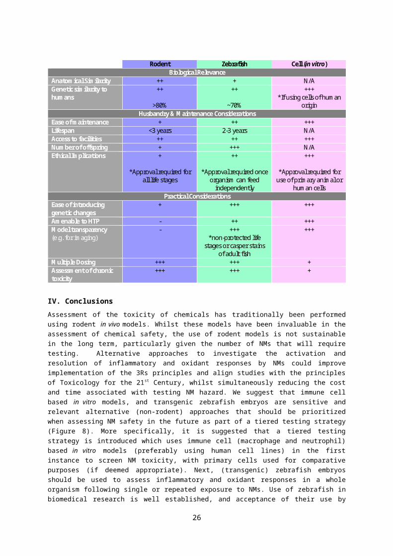
Haase H, Fahmi A, Mahltig B. (2014). Impact of silver nanoparticles and silver ions on innate immune cells. J Biomed Nanotechnol. 10(6):1146-56.
Hall CJ, Boyle RH, Astin JW, Flores MV, Oehlers SH, Sanderson LE, Ellett F, Lieschke GJ, Crosier KE, Crosier PS. (2013). Immunoresponsive gene 1 augments bactericidal activity of macrophage-lineage cells by regulating β-oxidation-dependent mitochondrial ROS production. Cell Metab. 18(2):265-78.
Han X, Corson N, Wade-Mercer P, Gelein R, Jiang J, Sahu M, Biswas P, Finkelstein JN, Elder A, Oberdörster G. (2012). Assessing the relevance of in vitro studies in nanotoxicology by examining correlations between in vitro and in vivo data. Toxicology. 297(1-3):1-9.
Handy RD, Owen R, Valsami-Jones E. (2008). The ecotoxicology of nanoparticles and nanomaterials: current status, knowledge gaps, challenges, and future needs. Ecotoxicology. 17(5):315-25.
Hartung T. (2009). Toxicology for the twenty-first century Nature 460; 208-212
Harvie EA, Huttenlocher A. (2015). Non-invasive Imaging of the Innate Immune Response in a Zebrafish Larval Model of Streptococcus iniae Infection. J Vis Exp. 98
He JH, Gao JM, Huang CJ, Li CQ. (2014). Zebrafish models for assessing developmental and reproductive toxicity. Neurotoxicol Teratol. 42:35-42.
Herbomel P, Thisse B, Thisse C. (1999). Ontogeny and behaviour of early macrophages in the zebrafish embryo. Development. 126(17):3735–3745.
Hermann AC, Millard PJ, Blake SL, Kim CH (2004). Development of a respiratory burst assay using zebrafish kidneys and embryos. J Immunol Methods. 292(1-2):119-29.
Hill AJ, Teraoka H, Heideman W, Peterson RE. (2005). Zebrafish as a model vertebrate for investigating chemical toxicity. Toxicol Sci. 86(1):6-19.
Hoodless LJ, Lucas CD, Duffin R, Denvir MA, Haslett C, Tucker CS, Rossi AG. (2016) Genetic and pharmacological inhibition of CDK9 drives neutrophil apoptosis to resolve inflammation in zebrafish in vivo. Sci Rep. 5:36980.
Hoodless LJ, Lucas CD, Duffin R, Denvir MA, Haslett C, Tucker CS, Rossi AG. (2016). Genetic and pharmacological inhibition of CDK9 drives neutrophil apoptosis to resolve inflammation in zebrafish in vivo. Sci Rep. 5:36980
Howe K, Clark MD, Torroja CF, Torrance J, Berthelot C, Muffato M, Collins JE, Humphray S, McLaren K, Matthews L, McLaren S, Sealy I, Caccamo M, Churcher C, Scott C, Barrett JC, Koch R, Rauch GJ, White S, Chow W, Kilian B, Quintais LT, Guerra-Assunção JA, Zhou Y, Gu Y, Yen J, Vogel JH, Eyre T, Redmond S, Banerjee R, Chi J, Fu B, Langley E, Maguire SF, Laird GK, Lloyd D, Kenyon E, Donaldson S, Sehra H, Almeida-King J, Loveland J, Trevanion S, Jones M, Quail M, Willey D, Hunt A, Burton J, Sims S, McLay K, Plumb B, Davis J, Clee C, Oliver K, Clark R, Riddle C, Elliot D, Threadgold G, Harden G, Ware D, Begum S, Mortimore B, Kerry G, Heath P, Phillimore B, Tracey A, Corby N, Dunn M, Johnson C, Wood J, Clark S, Pelan S, Griffiths G, Smith M, Glithero R, Howden P, Barker N, Lloyd C, Stevens C, Harley J, Holt K, Panagiotidis G, Lovell J, Beasley H, Henderson C, Gordon D, Auger K, Wright D, Collins J, Raisen C, Dyer L, Leung K, Robertson L, Ambridge K, Leongamornlert D, McGuire S, Gilderthorp R, Griffiths C, Manthravadi D, Nichol S, Barker G, Whitehead S, Kay M, Brown J, Murnane C, Gray E, Humphries M, Sycamore N, Barker D, Saunders D, Wallis J, Babbage A, Hammond S, Mashreghi-Mohammadi M, Barr L, Martin S, Wray P, Ellington A, Matthews N, Ellwood M, Woodmansey R, Clark G, Cooper J, Tromans A, Grafham D, Skuce C, Pandian R, Andrews R, Harrison E, Kimberley A, Garnett J, Fosker N, Hall R, Garner P, Kelly D, Bird C, Palmer S, Gehring I, Berger A, Dooley CM, Ersan-Ürün Z, Eser C, Geiger H, Geisler M, Karotki L, Kirn A, Konantz J, Konantz M, Oberländer M, Rudolph-Geiger S, Teucke M, Lanz C, Raddatz G, Osoegawa K, Zhu B, Rapp A, Widaa S, Langford C, Yang F, Schuster SC, Carter NP, Harrow J, Ning Z, Herrero J, Searle SM, Enright A, Geisler R, Plasterk RH, Lee C, Westerfield M, de Jong PJ, Zon LI, Postlethwait JH, Nüsslein-Volhard C, Hubbard TJ, Roest Crollius H, Rogers J, Stemple DL. (2013). The zebrafish reference genome sequence and its relationship to the human genome. Nature. 496(7446):498-503.
26

Hwang TL, Aljuffali IA, Hung CF, Chen CH, Fang JY. (2015). The impact of cationic solid lipid nanoparticles on human neutrophil activation and formation of neutrophil extracellular traps (NETs). Chem Biol Interact. 235:106-14.
International Agency for Research on Cancer (IARC), 1997. Monographs on the evaluation of carcinogenic risks to humans. Vol. 68. Silica, Some Silicates, Coal Dust and Para-Aramid Fibrils. Lyon, France:
International Agency for Research on Cancer (IARC) (1997). Monographs on the evaluation of carcinogenic risks to humans. Vol. 68. Silica, Some Silicates, Coal Dust and Para-Aramid Fibrils. Lyon, France
Johnston BD, Scown TM, Moger J, Cumberland SA, Baalousha M, Linge K, van Aerle R, Jarvis K, Lead JR, Tyler CR. (2010). Bioavailability of nanoscale metal oxides TiO(2), CeO(2), and ZnO to fish. Environ Sci Technol. 44(3):1144-51.
Jones HR, Robb CT, Perretti M, Rossi AG. (2016). The role of neutrophils in inflammation resolution. Semin Immunol. 28(2):137-45
Jovanović B, Anastasova L, Rowe EW, Zhang Y, Clapp AR, Palić D. (2011). Effects of nanosized titanium dioxide on innate immune system of fathead minnow (Pimephales promelas Rafinesque, 1820). Ecotoxicol Environ Saf. 74(4):675-83.
Kagan VE, Tyurina YY, Tyurin VA, Konduru NV, Potapovich AI, Osipov AN, Kisin ER, Schwegler-Berry D, Mercer R, Castranova V, Shvedova AA. (2006). Direct and indirect effects of single walled carbon nanotubes on RAW 264.7 macrophages: role of iron. Toxicol Lett. 165(1):88-100.
Kanther M, Sun X, Mühlbauer M, Mackey LC, Flynn EJ 3rd, Bagnat M, Jobin C, Rawls JF. (2011). Microbial colonization induces dynamic temporal and spatial patterns of NF-κB activationin the zebrafish digestive tract. Gastroenterology. 141(1):197-207.
Kermanizadeh, A., Brown, D.M., Hutchison, G.R., Stone, V. (2013a). Engineered nanomaterial impact in the liver following exposure via an intravenous route – the role of polymorphonuclear leukocytes and gene expression in the organ. J. Nanomed. Nanotechol. 4, 157.
Kermanizadeh A, Pojana G, Gaiser BK, Birkedal R, Bilanicová D, Wallin H, Jensen KA, Sellergren B, Hutchison GR, Marcomini A, Stone V. (2013b). In vitro assessment of engineered nanomaterials using a hepatocyte cell line: cytotoxicity, pro-inflammatory cytokines and functional markers. Nanotoxicology 7(3):301-13.
Kermanizadeh A, Vranic S, Boland S, Moreau K, Baeza-Squiban A, Gaiser BK, Andrzejczuk LA, Stone V. (2013c). An in vitro assessment of panel of engineered nanomaterials using a human renal cell line: cytotoxicity, pro-inflammatory response, oxidative stress and genotoxicity. BMC Nephrol. 14:96.
Kim YH, Boykin E, Stevens T, Lavrich K, Gilmour MI. (2014). Comparative lung toxicity of engineered nanomaterials utilizing in vitro, ex vivo and in vivo approaches. J Nanobiotechnology. 26;12:47.
Jim KK, Engelen-Lee J, van der Sar AM, Bitter W, Brouwer MC, van der Ende A, Veening JW, van de Beek D, Vandenbroucke-Grauls CM. (2016). Infection of zebrafish embryos with live fluorescent Streptococcus pneumoniae as a real-time pneumococcal meningitis model. J Neuroinflammation. 13(1):188.
Jim KK, Engelen-Lee J, van der Sar AM, Bitter W, Brouwer MC, van der Ende A, Veening JW, van de Beek D, Vandenbroucke-Grauls CM. (2016). Infection of zebrafish embryos with live fluorescent Streptococcus pneumoniae as a real-time pneumococcal meningitis model J Neuroinflammation. 13(1):188.
Knaapen AM, Güngör N, Schins RP, Borm PJ, Van Schooten FJ. (2006). Neutrophils and respiratory tract DNA damage and mutagenesis: a review. Mutagenesis. 21(4):225-36.
Labib S, Williams A, Yauk CL, Nikota JK, Wallin H, Vogel U. (2016). Halappanavar S. Nano-risk Science: application of toxicogenomics in an adverse outcome pathway framework for risk assessment of multi-walled carbon nanotubes. Part Fibre Toxicol.;13:15
27

Lanone S, Rogerieux F, Geys J, Dupont A, Maillot-Marechal E, Boczkowski J, Lacroix G, Hoet P. (2009).Comparative toxicity of 24 manufactured nanoparticles in human alveolar epithelial and macrophage cell lines. Part Fibre Toxicol. ;6:14.
Landsiedel R, Ma-Hock L, Hofmann T, Wiemann M, Strauss V, Treumann S, Wohlleben W, Gröters S, Wiench K, van Ravenzwaay B. (2014a). Application of short-term inhalation studies to assess the inhalation toxicity of nanomaterials. Part Fibre Toxicol. 11:16.
Landsiedel R, Sauer UG, Ma-Hock L, Schnekenburger J, Wiemann M. (2014b). Pulmonary toxicity of nanomaterials: a critical comparison of published in vitro assays and in vivo inhalation or instillation studies. Nanomedicine (Lond). 9(16):2557-85
Lee KJ, Nallathamby PD, Browning LM, Osgood CJ, Xu XH. (2007). In vivo imaging of transport and biocompatibility of single silver nanoparticles in early development of zebrafish embryos. ACS Nano. 1(2):133-43.
Lee HM, Shin DM, Song HM, Yuk JM, Lee ZW, Lee SH, Hwang SM, Kim JM, Lee CS, Jo EK. (2009). Nanoparticles up-regulate tumor necrosis factor-alpha and CXCL8 via reactive oxygen species and mitogen-activated protein kinase activation. Toxicol Appl Pharmacol. 238(2):160-9.
Lee, O., Green, J.M., Tyler, C.R., (2015). Transgenic fish systems and their application in ecotoxicology. Critical Reviews in Toxicology 45, 124-141.
Lee KJ, Browning LM, Nallathamby PD, Desai T, Cherukuri PK, Xu XH.(2012). In vivo quantitative study of sized-dependent transport and toxicity of single silvernanoparticles using zebrafish embryos. Chem Res Toxicol. 25(5):1029-46.
Levraud JP, Disson O, Kissa K, Bonne I, Cossart P, Herbomel P, Lecuit M. (2009). Real-time observation of listeria monocytogenes-phagocyte interactions in living zebrafish larvae. Infect Immun. 77(9):3651–3660
Li L, Zhang WQ, Yan B, Shi YQ, Wen ZL. (2012). Live imaging reveals differing roles of macrophages and neutrophils during zebrafish tail fin regeneration. Journal of Biological Chemistry 287(30), 25353-25360.
Lieschke GJ, Currie PD. (2007). Animal models of human disease: zebrafish swim into view. Nat Rev Genet. 8(5):353-67.
Lin S, Zhao Y, Xia T, Meng H, Ji Z, Liu R, George S, Xiong S, Wang X, Zhang H, Pokhrel S, Mädler L, Damoiseaux R, Lin S, Nel AE. (2011). High content screening in zebrafish speeds up hazard ranking of transition metal oxide nanoparticles. ACS Nano. 5(9):7284-95.
Lin A, Loughman JA, Zinselmeyer BH, Miller MJ, Caparon MG (2009). Streptolysin S Inhibits Neutrophil Recruitment during the Early Stages of Streptococcus pyogenes Infection. Infect Immun. Nov; 77(11): 5190–5201.
Lin S, Zhao Y, Nel AE, Lin S. (2013). Zebrafish: an in vivo model for nano EHS studies. Small. 9(9-10):1608-18.
Liz R, Simard JC, Leonardi LB, Girard D. (2015). Silver nanoparticles rapidly induce atypical human neutrophil cell death by a process involving inflammatory caspases and reactive oxygen species and induce neutrophil extracellular traps release upon cell adhesion. Int Immunopharmacol. 28(1):616-25.
Lieschke GJ, Currie PD. (2007). Animal models of human disease: zebrafish swim into view. Nat Rev Genet. 8(5):353-67.
Loynes CA, Martin JS, Robertson A, Trushell DM, Ingham PW, Whyte MK, Renshaw SA. (2010). Pivotal Advance: Pharmacological manipulation of inflammation resolution during spontaneously resolving tissue neutrophilia in the zebrafish. J Leukoc Biol. 87(2):203-12.
Lucas CD, Allen KC, Dorward DA, Hoodless LJ, Melrose LA, Marwick JA, Tucker CS, Haslett C, Duffin R, Rossi AG. (2013). Flavones induce neutrophil apoptosis by down-regulation of Mcl-1 via a proteasomal-dependent pathway. FASEB J. 27(3):1084-94.
28

Ma-Hock L, Treumann S, Strauss V, Brill S, Luizi F, Mertler M, Wiench K, Gamer AO, van Ravenzwaay B, Landsiedel R. (2009). Inhalation toxicity of multiwall carbon nanotubes in rats exposed for 3 months. Toxicol Sci. 112(2):468-81.
MacRae CA, Peterson RT. (2015). Zebrafish as tools for drug discovery. Nat Rev Drug Discov. 14(10):721-31.
Marjoram L, Alvers A, Deerhake ME, Bagwell J, Mankiewicz J, Cocchiaro JL, Beerman RW, Willer J, Sumigray KD, Katsanis N, Tobin DM, Rawls JF, Goll MG, Bagnat M. (2015). Epigenetic control of intestinal barrier function and inflammation in zebrafish. Proc Natl Acad Sci U S A. 112(9):2770-5.
Mayadas TN, Cullere X, Lowell CA. (2014). The multifaceted functions of neutrophils. Annu Rev Pathol. 9:181-218.
Maynard AD, Aitken RJ, Butz T, Colvin V, Donaldson K, Oberdörster G, Philbert MA, Ryan J, Seaton A, Stone V, Tinkle SS, Tran L, Walker NJ, Warheit DB. (2006). Safe handling of nanotechnology. Nature. 444(7117):267-9.
Massarsky A, Dupuis L, Taylor J, Eisa-Beygi S, Strek L, Trudeau VL, Moon TW. (2013). Assessment of nanosilver toxicity during zebrafish (Danio rerio) development. Chemosphere. 92(1):59-66.
Mathias JR, Walters KB, Huttenlocher A. (2009). Neutrophil motility in vivo using zebrafish. Methods Mol Biol. 571:151-66.
Mathias JR, Saxena MT, Mumm JS. (2012). Advances in zebrafish chemical screening technologies. Future Med Chem. 4(14):1811-22.
Meeker ND, Trede NS. (2008). Immunology and zebrafish: spawning new models of human disease. Dev Comp Immunol. 32(7):745-57
McLeish JA, Chico TJ, Taylor HB, Tucker C, Donaldson K, Brown SB. (2010). Skin exposure to micro- and nano-particles can cause haemostasis in zebrafish larvae. Thromb Haemost. 103(4):797-807.
McWilliams, A., The Maturing Nanotechnology Market: Products and Applications, BCCResearch, Editor. 2016.
Meijer AH. (2016).Protection and pathology in TB: learning from the zebrafish model. Semin Immunopathol. 38(2):261-73.
Merrifield DL, Shaw BJ, Harper GM, Saoud IP, Davies SJ, Handy RD, Henry TB. (2013). Ingestion of metal-nanoparticle contaminated food disrupts endogenous microbiota inzebrafish (Danio rerio). Environ Pollut. 174:157-63
Mesens N, Crawford AD, Menke A, Hung PD, Van Goethem F, Nuyts R, Hansen E, Wolterbeek A, Van Gompel J, De Witte P, Esguerra CV. (2015). Are zebrafish larvae suitable for assessing the hepatotoxicity potential of drug candidates? J Appl Toxicol. 35(9):1017-29.
Mestas J, Hughes CC. (2004). Of mice and not men: differences between mouse and human immunology. J Immunol. 172(5):2731-8.
Mittal M, Siddiqui MR, Tran K, Reddy SP, Malik AB. (2014). Reactive oxygen species in inflammation and tissue injury. Antioxid Redox Signal. 20(7):1126-67.
Mitrano and Nowack
Mugoni V, Camporeale A, Santoro MM. (2014). Analysis of Oxidative Stress in Zebrafish Embryos. JoVE. 89:51328.
Murphy FA, Poland CA, Duffin R, Donaldson K. (2013). Length-dependent pleural inflammation and parietal pleural responses after deposition of carbon nanotubes in the pulmonary airspaces of mice. Nanotoxicology. 7(6):1157-67.
Nathan C. (2006).Neutrophils and immunity: challenges and opportunities. Nat Rev Immunol. 6(3):173-82.
Nel A, Xia T, Madler L, Li N. (2006). Toxic potential of materials at the nanolevel. Science 311(5761), 622–7.
Niethammer P, Grabher C, Look AT, Mitchison TJ. (2009). A tissue-scale gradient of hydrogen peroxide mediates rapid wound detection in zebrafish. Nature. 459(7249):996-9.
29
Nguyen-Chi, M., Phan, Q. T., Gonzalez, C., Dubremetz, J.-F., Levraud, J.-P. and Lutfalla, G. (2014). Transient infection of the zebrafish notochord with E. coli induces chronic inflammation. Disease Models & Mechanisms 7(7), 871-882.
Nguyen KC, Richards L, Massarsky A, Moon TW, Tayabali AF. (2016). Toxicological evaluation of representative silver nanoparticles in macrophages and epithelial cells. Toxicol In Vitro 33:163-73.
Novoa B, Figueras A. (2012). Zebrafish: model for the study of inflammation and the innate immune response to infectious diseases. Adv Exp Med Biol. 946:253-75.
Nowack B. (2017). Evaluation of environmental exposure models for engineered nanomaterials in a regulatory context. NanoImpact [In Press]
Oberdörster G, Stone V, Donaldson K (2007). Toxicology of nanoparticles: A historical perspective. Nanotoxicology 1; 2-25
OECD (2015). Test No. 442C. In Chemico Skin Sensitisation Direct Peptide Reactivity Assay (DPRA). OECD Guidelines for the Testing of Chemicals, Section 4: Health Effects. OECD, Paris.
de Oliveira S, Reyes-Aldasoro CC, Candel S, Renshaw SA, Mulero V, Calado A. (2013). Cxcl8 (IL-8) mediates neutrophil recruitment and behavior in the zebrafish inflammatory response. J Immunol. 190(8):4349-59.
de Oliveira S, Lopez-Muñoz A, Martínez-Navarro FJ, Galindo-Villegas J, Mulero V, Calado Â. (2015). Cxcl8-l1 and Cxcl8-l2 are required in the zebrafish defense against Salmonella Typhimurium. Dev Comp Immunol. 49(1):44-8.
Olsen H, Betton G, Robinson D, Thomas K, Monro A, Kolaja G, Lilly P, Sanders J, Sipes G, Bracken W, Dorato M, Van Deun K, Smith P, Berger B, Heller A. (2000) Concordance of the Toxicity of Pharmaceuticals in Humans and in Animals. Regulatory Toxicology and Pharmacology 32, 56–67
Osborne OJ, Johnston BD, Moger J, Balousha M, Lead JR, Kudoh T, Tyler CR. (2013). Effects of particle size and coating on nanoscale Ag and TiO₂ exposure in zebrafish (Danio rerio) embryos. Nanotoxicology. 7(8):1315-24.
Osborne OJ, Lin S, Chang CH, Ji Z, Yu X, Wang X, Lin S, Xia T, Nel AE. (2015). Organ-Specific and Size-Dependent Ag Nanoparticle Toxicity in Gills and Intestines of Adult Zebrafish. ACS Nano. 27;9(10):9573-84.
Osborne OJ, Mukaigasa K, Nakajima H, Stolpe B, Romer I, Philips U, Lynch I, Mourabit S, Hirose S, Lead JR, Kobayashi M, Kudoh T, Tyler CR. (2016). Sensory systems and ionocytes are targets for silver nanoparticle effects in fish. Nanotoxicology. 10(9):1276-86.
Park EJ, Park K. (2009). Oxidative stress and pro-inflammatory responses induced by silica nanoparticles in vivo and in vitro. Toxicol Lett. 184(1):18-25.
Pase L, Layton JE, Wittmann C, Ellett F, Nowell CJ, Reyes-Aldasoro CC, Varma S, Rogers KL, Hall CJ, Keightley MC, Crosier PS, Grabher C, Heath JK, Renshaw SA, Lieschke GJ. (2012). Neutrophil-delivered myeloperoxidase dampens the hydrogen peroxide burst after tissue wounding in zebrafish. Curr Biol. 22(19):1818-24.
Pati R, Das I, Mehta RK, Sahu R, Sonawane A. (2016). Zinc-Oxide Nanoparticles Exhibit Genotoxic, Clastogenic, Cytotoxic and Actin Depolymerization Effects by Inducing Oxidative Stress Responses in Macrophages and Adult Mice. Toxicol Sci. 150(2):454-72
Piccinno F, Gottschalk F, Seeger S, Nowack B (2012). Industrial production quantities and uses of ten engineered nanomaterials in Europe and the world J Nanopart Res 14: 1109.
Poon IK, Lucas CD, Rossi AG, (2014). Ravichandran KS. Apoptotic cell clearance: basic biology and therapeutic potential. Nat Rev Immunol. 14(3):166-80.
Poon IK, Lucas CD, Rossi AG, Ravichandran KS. (2014). Apoptotic cell clearance: basic biology and therapeutic potential. Nat Rev Immunol. 14(3):166-80.
Progatzky F, Cook HT, Lamb JR, Bugeon L, Dallman MJ. (2016). Mucosal inflammation at the respiratory interface: a zebrafish model. Am J Physiol Lung Cell Mol Physiol. 310(6):L551-61.
30
Rauscher, H., Rasmussen, K. and Sokull-Klüttgen, B. (2017), Regulatory Aspects of Nanomaterials in the EU. Chemie Ingenieur Technik, 89: 224–231.
Renshaw SA, Loynes CA, Elworthy S, Ingham PW, Whyte MK. (2007). Modeling inflammation in the zebrafish: how a fish can help us understand lung disease. Exp Lung Res. 33(10):549-54.
Rizzo LY, Golombek SK, Mertens ME, Pan Y, Laaf D, Broda J, Jayapaul J, Möckel D, Subr V, Hennink WE, Storm G, Simon U, Jahnen-Dechent W, Kiessling F, Lammers T. (2013). In Vivo Nanotoxicity Testing using the Zebrafish Embryo Assay. J Mater Chem B Mater Biol Med. 10.1039/C3TB20528B
Robb CT, Regan KH, Dorward DA, Rossi AG. Key mechanisms governing resolution of lung inflammation. Semin Immunopathol. 2016 Jul;38(4):425-48.
Robertson AL, Ogryzko NV, Henry KM, Loynes CA, Foulkes MJ, Meloni MM, Wang X, Ford C, Jackson M, Ingham PW, Wilson HL, Farrow SN, Solari R, Flower RJ, Jones S, Whyte MK, Renshaw SA. (2016). Identification of benzopyrone as a common structural feature in compounds with anti-inflammatory activity in a zebrafish phenotypic screen. Dis Model Mech. 9(6):621-32.
Rombough P. (2002). Gills are needed for ionoregulation before they are needed for O(2) uptake in developing zebrafish, Danio rerio. J Exp Biol. 205(Pt 12):1787-94.
Rovida C, Hartung T. (2009). Re-evaluation of animal numbers and costs for in vivo tests to accomplish REACH legislation requirements for chemicals - a report by the transatlantic think tank for toxicology. ALTEX. 26(3):187-208.
Rushton EK, Jiang J, Leonard SS, Eberly S, Castranova V, Biswas P, Elder A, Han X, Gelein R, Finkelstein J, Oberdörster G. (2010). Concept of assessing nanoparticle hazards considering nanoparticle dosemetric and chemical/biological response metrics. J Toxicol Environ Health A. 73(5):445-61.
Russell WMS, Burch RL. 1959. (as reprinted 1992). The principles of humane experimental technique. Wheathampstead (UK): Universities Federation for Animal Welfare.
Sayes CM, Reed KL, Warheit DB. (2007). Assessing toxicity of fine and nanoparticles: comparing in vitro measurements to in vivo pulmonary toxicity profiles. Toxicol Sci. 97(1):163-80.
Schmidt K. (2007). NanoFrontiers: Visions for the Future of Nanotechnology. Washington, DC: The Project on Emerging Nanotechnologies, Woodrow Wilson International Center for Scholars; 2007.http://www.nanotechproject.org/process/assets/files/2704/181_pen6_nanofrontiers.pdf
Schmidt CW. (2009) Nanotechnology-Related Environment, Health, and Safety Research Examining the National Strategy. Environ Health Persp. 117:A158–A161.
Scherbart AM, Langer J, Bushmelev A, van Berlo D, Haberzettl P, van Schooten FJ, , Schmidt AM, Rose CR, Schins RP, Albrecht C. (2011). Contrasting macrophage activation by fine and ultrafine titanium dioxide particles is associated with different uptake mechanisms. Part Fibre Toxicol. ;8:31
Selck H, Handy RD, Fernandes TF, Klaine SJ, Petersen EJ. (2016). Nanomaterials in the aquatic environment: A European Union-United States perspective on the status of ecotoxicity testing, research priorities, and challenges ahead. Environ Toxicol Chem. 35(5):1055-67.
Shiau CE, Kaufman Z, Meireles AM, Talbot WS. (2015). Differential requirement for irf8 in formation of embryonic and adult macrophages in zebrafish. PLoS One. 10(1):e0117513.
Shi M, Kwon HS, Peng Z, Elder A, Yang H. (2012). Effects of surface chemistry on the generation of reactive oxygen species by coppernanoparticles. ACS Nano. 6(3):2157-64
Shvedova AA, Kisin E, Murray AR, Johnson VJ, Gorelik O, Arepalli S, Hubbs AF, Mercer RR, Keohavong P, Sussman N, Jin J, Yin J, Stone S, Chen BT, Deye G, Maynard A, Castranova V, Baron PA, Kagan VE. (2008). Inhalation vs. aspiration of single-walled carbon nanotubes in C57BL/6 mice: inflammation, fibrosis, oxidative stress, and mutagenesis. Am J Physiol Lung Cell Mol Physiol. 295(4):L552-65.
31
Silva RM, Anderson DS, Franzi LM, Peake JL, Edwards PC, Van Winkle LS, Pinkerton KE. (2015). Pulmonary effects of silver nanoparticle size, coating, and dose over time upon intratracheal instillation. Toxicol Sci. 144(1):151-62.
Soares T, Ribeiro D, Proença C, Chisté RC, Fernandes E, Freitas M. (2016). Size-dependent cytotoxicity of silver nanoparticles in human neutrophils assessed by multiple analytical approaches. Life Sci. 145:247-54.
Soehnlein O, Steffens S, Hidalgo A, Weber C. (2017). Neutrophils as protagonists and targets in chronic inflammation. Nat Rev Immunol. 17(4):248-261.
Strähle U, Scholz S, Geisler R, Greiner P, Hollert H, Rastegar S, Schumacher A, Selderslaghs I, Weiss C, Witters H,Braunbeck T. (2012). Zebrafish embryos as an alternative to animal experiments--a commentary on the definition of the onset of protected life stages in animal welfare regulations. Reprod Toxicol. 33(2):128-32.
Stone V, Miller MR, Clift MJ, Elder A, Mills NL, Møller P, Schins RP, Vogel U, Kreyling WG, Jensen KA, Kuhlbusch TA, Schwarze PE, Hoet P, Pietroiusti A, De Vizcaya-Ruiz A, Baeza-Squiban A, Tran CL, Cassee FR. (2016). Nanomaterials vs Ambient Ultrafine Particles: an Opportunity to Exchange Toxicology Knowledge. Environ Health Perspect. [Epub ahead of print]
Stone V, Tuinman M, Vamvakopoulos JE, Shaw J, Brown D, Petterson S, Faux SP, Borm P, MacNee W, Michaelangeli F, Donaldson K. (2000). Increased calcium influx in a monocytic cell line on exposure to ultrafine carbon black. Eur Respir J. 15(2):297-303.
Susewind J, de Souza Carvalho-Wodarz C, Repnik U, Collnot EM, Schneider-Daum N, Griffiths GW, Lehr CM. (2016). A 3D co-culture of three human cell lines to model the inflamed intestinal mucosa for safety testing of nanomaterials. Nanotoxicology. 10(1):53-62.
Teeguarden JG, Mikheev VB, Minard KR, Forsythe WC, Wang W, Sharma G, Karin N, Tilton SC, Waters KM, Asgharian B, Price OR, Pounds JG, Thrall BD. (2014). Comparative iron oxide nanoparticle cellular dosimetry and response in mice by the inhalation and liquid cell culture exposure routes. Part Fibre Toxicol. 2014 11:46.
Timme-Laragy AR, Karchner SI, Franks DG, Jenny MJ, Harbeitner RC, Goldstone JV, McArthur AG, Hahn ME. (2011). Nrf2b, novel zebrafish paralog of oxidant-responsive transcription factor NF-E2-related factor 2 (NRF2). J Biol Chem. 287(7):4609-27.
Torraca V, Masud S, Spaink HP, Meijer AH. (2014). Macrophage-pathogen interactions in infectious diseases: new therapeutic insights from the zebrafish host model. Dis Model Mech. 7(7):785-97.
Traver D, Herbomel P, Patton EE, Murphey RD, Yoder JA, Litman GW, Catic A, Amemiya CT, Zon LI, Trede NS. (2003). The zebrafish as a model organism to study development of the immune system Adv Immunol, 81; 253–330
Usenko CY, Harper SL, Tanguay RL. (2008). Fullerene C60 exposure elicits an oxidative stress response in embryonic zebrafish Toxicol Appl Pharmacol. 229(1): 44–55.
Törnqvist E, Annas A, Granath B, Jalkesten E, Cotgreave I, Öberg M. (2014). Strategic focus on 3R principles reveals major reductions in the use of animals in pharmaceutical toxicity testing. PLoS One. 9(7):e101638.
Trede NS, Langenau DM, Traver D, Look AT, Zon LI. (2004). The use of zebrafish to understand immunity. Immunity. 20(4):367-79.
Tuomela S, Autio R, Buerki-Thurnherr T, Arslan O, Kunzmann A, Andersson-Willman B, Wick P, Mathur S, Scheynius A, Krug HF, Fadeel B, Lahesmaa R. (2013). Gene expression profiling of immune-competent human cells exposed to engineered zinc oxide or titanium dioxide nanoparticles. PLoS One. 8(7):e68415.
Valko M, Leibfritz D, Moncol J, Cronin MT, Mazur M, Telser J. (2007). Free radicals and antioxidants in normal physiological functions and human disease. Int J Biochem Cell Biol. 39(1):44-84.
Vance ME, Kuiken T, Vejerano EP, McGinnis SP, Hochella MF Jr, Rejeski D, Hull MS. (2015). Nanotechnology in the real world: Redeveloping the nanomaterial consumer productsinventory. J Nanotechnol. 6:1769-80.
32
Vojtech LN, Sanders GE, Conway C, Ostland V, Hansen JD. (2009). Host immune response and acute disease in a zebrafish model of Francisella pathogenesis. Infect Immun. 77(2):914-25.
Wagner JC, Sleggs CA, Marcgand P. (1960). Diffuse pleural mesothelioma and asbestos exposure in the North Western Cape Province. Br J Ind Med. 17:260-71.
Walker SL, Ariga J, Mathias JR, Coothankandaswamy V, Xie X, Distel M, Köster RW, Parsons MJ, Bhalla KN, Saxena MT, Mumm JS. (2012). Automated reporter quantification in vivo: high-throughput screening method for reporter-based assays in zebrafish. PLoS One. 7(1):e29916.
Wang J, Arase H. (2014). Regulation of immune responses by neutrophils. Ann N Y Acad Sci. 1319:66-81.
Wang X, Robertson AL, Li J, Chai RJ, Haishan W, Sadiku P, Ogryzko NV, Everett M, Yoganathan K, Luo HR, Renshaw SA, Ingham PW. (2014). Inhibitors of neutrophil recruitment identified using transgenic zebrafish to screen a natural product library. Dis Model Mech. 7(1):163-9.
Warheit DB, Sayes CM, Reed KL. (2009). Nanoscale and fine zinc oxide particles: can in vitro assays accurately forecast lung hazards following inhalation exposures? Environ Sci Technol. 43(20):7939-45.
White RM, Sessa A, Burke C, Bowman T, LeBlanc J, Ceol C, Bourque C, Dovey M, Goessling W, Burns CE, Zon LI. (2008). Transparent adult zebrafish as a tool for in vivo transplantation analysis. Cell Stem Cell. 2(2):183-9.
Wiemann M, Vennemann A, Sauer UG, Wiench K, Ma-Hock L, Landsiedel R. (2016). An in vitro alveolar macrophage assay for predicting the short-term inhalation toxicity of nanomaterials. J Nanobiotechnology.14:16.
Wilson MR, Lightbody JH, Donaldson K, Sales J, Stone V. (2002). Interactions between ultrafine particles and transition metals in vivo and in vitro. Toxicol Appl Pharmacol.184(3):172-9.
Wilson MR, Foucaud L, Barlow PG, Hutchison GR, Sales J, Simpson RJ, Stone V. (2007). Nanoparticle interactions with zinc and iron: implications for toxicology and inflammation. Toxicol Appl Pharmacol. 225(1):80-9.
Witasp E, Shvedova AA, Kagan VE, Fadeel B. (2009). Single-walled carbon nanotubes impair human macrophage engulfment of apoptotic cell corpses. Inhal Toxicol. 21 Suppl 1:131-6.
Xiong D, Fang T, Yu L, Sima X, Zhu W. (2011). Effects of nano-scale TiO2, ZnO and their bulk counterparts on zebrafish: acute toxicity, oxidative stress and oxidative damage. Sci Total Environ. 409(8):1444-52.
Xu H, Dong X, Zhang Z, Yang M, Wu X, Liu H, Lao Q, Li C. (2015). Assessment of immunotoxicity of dibutyl phthalate using live zebrafish embryos. Fish Shellfish Immunol. 45(2):286-92
Zhang Y, Zhu L, Zhou Y, Chen J. (2015). Accumulation and elimination of iron oxide nanomaterials in zebrafish (Danio rerio) upon chronic aqueous exposure. J Environ Sci (China). 30:223-30.
Zhao X, Wang S, Wu Y, You H, Lv L. (2013). Acute ZnO nanoparticles exposure induces developmental toxicity, oxidative stress and DNA damage in embryo-larval zebrafish. Aquat Toxicol. 136-137:49-59.
Zhu X, Tian S, Cai Z. (2012). Toxicity assessment of iron oxide nanoparticles in zebrafish (Danio rerio) early life stages. PLoS One. 7(9):e46286.
33