BASOSQUAMOUS CARCINOMA: A RARE CASE OF EAR …
10
Supplement No. 1/2016 164 Medicine BASOSQUAMOUS CARCINOMA: A RARE CASE OF EAR METASTATSIS IN A 13 YEARS-OLD Zamfir-Radu IONESCU 1 ABSTRACT: WE PRESENT THE CASE OF A 13-YEARS OLD FEMALE CHILD, WITH A NODULAR TUMOR ON THE EAR LOBE, RESECTED AND INVESTIGATED IN THE PEDIATRIC HOSPITAL OF PITEȘTI, ROMANIA. THE INITIAL CLINICAL DIAGNOSIS WAS OF A SEBACOUS CYST, BUT, AFTER CLOSER INSPECTION AT THE MICROSCOPE, WE DETECTED THE PRESENCE OF SQUAMOID PROLIFERATIONS, WITH KERATINISATION AND BASALOID FEATURES, CONCLUDING THE DIAGNOSIS AS A METASTASIS WITH BASOSQUAMOUS CARCINOMA ORIGIN. BASOSQUAMOUS CARCINOMA REMAINS A RARE FINDING IN CHILDREN AND, IS OFTEN, MISINTERPRETED AS A SUQAMOUS CARCINOMA. THEREFORE, THIS WORK IS INTENDED TO FOCUS THE IMPORTANCE OF THIS DISEASE FOR PATHOLOGISTS WHO INVESTIGATE CHILD DERMATOPATHOLOGY, REGARDING THIS MATTER. KEY WORDS: BAZOCELLULAR CARCINOMA, BASOSQUAMOUS CARCINOMA, METATYPICAL CARCINOMA, COLLISION TUMOR INTRODUCTION Basal cell carcinoma, clinically, may present on sun-exposed skin areas, particularly on face, being more frequent in white adults. Occurrence in young adults, other than in the context of nevus sebaceous, nevoid basal cell carcinoma syndrome, or Rombo syndrome remains exceptional. However, in children, Gorlin syndrome is characterized by basal cell carcinomas, with odontogenic jaw cysts, pitted depression of the hands and feet, osseous anomalies, a broad nasal root and hypertelorism. The indicence of all basosquamous carcinoma (BSQC) remains at 1-2% of all dermatological malignancy cases, having different synonyms: metatypical carcinoma, basaloid squamous carcinoma, or collision tumor, if both components (squamous and basal cell) are even. Futhermore, a BSQC metastasis presents itself as well delimited formation, although its incidence is lower than 5% in children younger than 20 years old 2 . Clinically, BSQC metastasis has no specific features, while the biopsy is the only way to figure out the final diagnosis. Thus, the histopathological diagnosis might be very problematic, because the growth is very slow. There might be encountered areas of basocellular and squamous carcinoma. The cells of basal component are larger, with 1 PhD, MD of Pathology, Head of the Pathology Department of Pediatric Hospital, Pitești, Argeș, , Romania, dr.raduionescu@yahoo. Com, Tel: +40346086086 2 O. Karatas Silistreli, M. Ayhan, Z. Aytug. Periocular metatypical cell carcinoma: Clinicopathologic correlation, management, and follow-up in 35 patients, J Plast Reconstr Aesthet Surg, 2006; 59:1280–1287;
Transcript of BASOSQUAMOUS CARCINOMA: A RARE CASE OF EAR …

Supplement No. 1/2016
164
Medicine
BASOSQUAMOUS CARCINOMA: A RARE CASE OF EAR
METASTATSIS IN A 13 YEARS-OLD
Zamfir-Radu IONESCU1
ABSTRACT: WE PRESENT THE CASE OF A 13-YEARS OLD FEMALE CHILD, WITH A NODULAR TUMOR ON THE
EAR LOBE, RESECTED AND INVESTIGATED IN THE PEDIATRIC HOSPITAL OF PITEȘTI, ROMANIA.
THE INITIAL CLINICAL DIAGNOSIS WAS OF A SEBACOUS CYST, BUT, AFTER CLOSER INSPECTION AT
THE MICROSCOPE, WE DETECTED THE PRESENCE OF SQUAMOID PROLIFERATIONS, WITH
KERATINISATION AND BASALOID FEATURES, CONCLUDING THE DIAGNOSIS AS A METASTASIS
WITH BASOSQUAMOUS CARCINOMA ORIGIN. BASOSQUAMOUS CARCINOMA REMAINS A RARE
FINDING IN CHILDREN AND, IS OFTEN, MISINTERPRETED AS A SUQAMOUS CARCINOMA.
THEREFORE, THIS WORK IS INTENDED TO FOCUS THE IMPORTANCE OF THIS DISEASE FOR
PATHOLOGISTS WHO INVESTIGATE CHILD DERMATOPATHOLOGY, REGARDING THIS MATTER.
KEY WORDS: BAZOCELLULAR CARCINOMA, BASOSQUAMOUS CARCINOMA, METATYPICAL
CARCINOMA, COLLISION TUMOR
INTRODUCTION
Basal cell carcinoma, clinically, may present on sun-exposed skin areas, particularly
on face, being more frequent in white adults. Occurrence in young adults, other than in the
context of nevus sebaceous, nevoid basal cell carcinoma syndrome, or Rombo syndrome
remains exceptional. However, in children, Gorlin syndrome is characterized by basal cell
carcinomas, with odontogenic jaw cysts, pitted depression of the hands and feet, osseous
anomalies, a broad nasal root and hypertelorism. The indicence of all basosquamous
carcinoma (BSQC) remains at 1-2% of all dermatological malignancy cases, having different
synonyms: metatypical carcinoma, basaloid squamous carcinoma, or collision tumor, if both
components (squamous and basal cell) are even. Futhermore, a BSQC metastasis presents
itself as well delimited formation, although its incidence is lower than 5% in children
younger than 20 years old2. Clinically, BSQC metastasis has no specific features, while the
biopsy is the only way to figure out the final diagnosis. Thus, the histopathological diagnosis
might be very problematic, because the growth is very slow. There might be encountered
areas of basocellular and squamous carcinoma. The cells of basal component are larger, with
1 PhD, MD of Pathology, Head of the Pathology Department of Pediatric Hospital, Pitești, Argeș, , Romania,
dr.raduionescu@yahoo. Com, Tel: +40346086086 2 O. Karatas Silistreli, M. Ayhan, Z. Aytug. Periocular metatypical cell carcinoma: Clinicopathologic
correlation, management, and follow-up in 35 patients, J Plast Reconstr Aesthet Surg, 2006; 59:1280–1287;

November 2014
165
pale cytoplasm, with areas of squamoid features that have abundant eosinophilic cytoplasm.
Frequently, it might be visible another area of cells having transitional features intermingled
in between both components. Not rarely, characteristic peripheral pallisading and stromal
retraction remain inconspicuous. The diagnosis must well document the superposition of
basal cell carcinoma with the squamous one3.
CASE REPORT
The differential diagnosis encompassed lymphoepithelioma-like carcinoma,
metastatic ameloblastic carcinoma and Ewing sarcoma. These possible situations were taken
into account as the localization of metastasis was more frequent in head and neck regions.
However, pathological and imagery criteria were not met for any of these tumors. The final
diagnosis was that of a metastatic bazosquamous carcinoma with local invasion of the left ear
lobe.
We present the case of a 13 years old female child, having a good health status, with
no other complaints or symptoms, except for a mass on the left ear lobe, that enlarged within
a 6 weeks duration to 1,5/0,7 cm. The rest of clinical examination and imagistics proved no
obvious or worrisome lesions.
Laboratory routine blood tests
proved negative for all counts. The
ear-nose-throat department in our
hospital referred the case for
resection within 2-4 mm margins
for esthetical reasons. We received
the tissue sample in our Pathology
Department, fixed in 10% buffered
formalin. It presented as a well
delineated, nodular mass, with a
gritty aspect, having intermingled
white to brown areas and a general
non-homogenous aspect (fig nr. 1).
After tissue processing in
successive alcohol concentrations
(70, 80 and 96 degrees), paraffin
embedding and sectioning with haematoxilin and eosin staining, we found definite squamous
areas with ortho- and parakeratin forming pearls with basaloid features, without peripheral
pallisading and with pseudo-stromal retraction (fig. nr. 2). These carcinomatous entities were
detected inside dilated, ectatic blood vessels encircled by a desmoplastic, lymphoplasmocytic
infiltrated fibrous strands, as the metastatic cells already invaded within the local perivascular
connective tissues (fig. nr. 3). In transitional areas, more visible towards the basaloid areas,
squamous morules were detected, thus proving the tendency for squamous differentiation.
Atypical mitoses were found exclusively in basaloid areas of the BSQC, approximately 5 to 6
mitoses per field.
The case was reviewed interdisciplinary – pediatric surgeon, ear-nose-throat senior
specialist, pathologist – for oncological treatment in a regional specialized department for
similar pediatric cases.
3 R.C. Martin, M.J. Edwards, T.G. Cawte. Basosquamous carcinoma: Analysis of prognostic factors influencing
recurrence, Cancer.,2000; 88:1365–1369;
Fig. 1. Resected nodule from left ear in a 13 years old girl.
Gritty aspect with variable brown to white coloured areas is
visible after sectioning (10% buffered formalin).
Supplement No. 1/2016
166
Fig. 2. Basaloid areas toegether with squamous differentiation in
the resected tumor; metatypical morules are visible (center) with
few mitotic figures
DISCUSSION
The differential diagnosis encompassed lymphoepithelioma-like carcinoma, metastatic ameloblastic carcinoma and Ewing sarcoma. These possible situations were taken into account as the localization of metastasis was more frequent in head and neck regions. However, pathological and imagery criteria were not met for any of these tumors. The final diagnosis was that of a metastatic bazosquamous carcinoma with local invasion of the left ear lobe. For definitive
diagnosis,
immunohistochemistry is
necessary, although it has
limited value. Representative
paraffin embedded tissue
samples should reserved for
immunostaining with
AE1/AE3, bcl-2, TGF-A, Ver-
EP4, p53 molecular antibodies.
With this kind of approach, it
might be best visible the
transitional area between the
two components of the
BSQC4. The optimal way to
observe this kind of
transitional cellular effect is
with Ber-EP4, who is detected in all basocellular tumors. This might indicate the presence of
a pluipotential cell capable of differentiation trough a genetical mutation, causing frequent
appearance of metastasis in such a young age5.
Main therapy is represented by resectional surgery with security margins and
chemotherapy, although this kind of tumor remains unresponsive, especially with oncological
radiotherapy and pharmacological approach, in metastastic disease. In chemotherapy, we
might use doxorubicin, cisplatin, fluorouracil in varying cycles, depending of CT volumetric
response of the main tumor6.
4 B. Lennox, A. Wells. Differentiation in the rodent ulcer group of tumors, Br J Cancer, 1951, 5:195–212; 5 M.S. Jones, K.F. Helm, M.E. Maloney; The immunohistochemical characteristic of the basosquamous cell
carcinoma, Dermatol Surg., 1997; 23:181–184; 6 P. Clement, J. Verheezen, S. Nuyts. A single institution retrospective analysis of basosquamous carcinoma of
the head and neck, J Clin Oncol., 2006; 24:15530;
November 2014
167
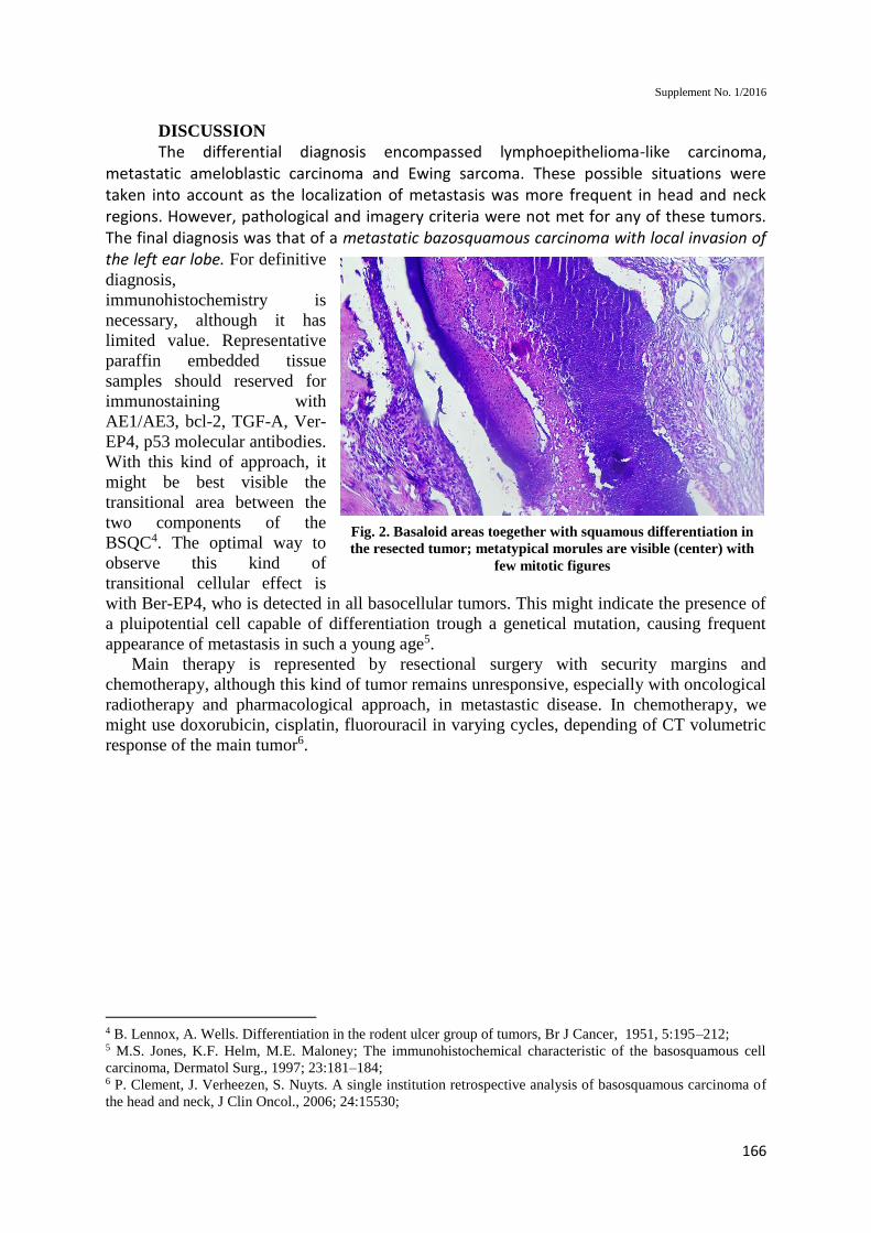
Fig. 3. Squamoid features are intermingled with basaloid
ones. Perivascular invasion is visible toegether with squamou
pearls and atypical mitoses (10x20, HE).
CONCLUSIONS
This collision tumor or BSQC
remains with an overall low
frequency among all cutaneous
malignancies, in children; it has
origins in basal cells of the
epidermis, and a high potential for
metastasing or invasion, especially
because of the squamous features.
Supplement No. 1/2016
168
REFERENCES
1. O. Karatas Silistreli, M. Ayhan, Z. Aytug. Periocular metatypical cell carcinoma: Clinicopathologic
correlation, management, and follow-up in 35 patients, J Plast Reconstr Aesthet Surg, 2006; 59:1280–
1287;
2. R.C. Martin, M.J. Edwards, T.G. Cawte. Basosquamous carcinoma: Analysis of prognostic factors
influencing recurrence, Cancer.,2000; 88:1365–1369;
3. B. Lennox, A. Wells. Differentiation in the rodent ulcer group of tumors, Br J Cancer, 1951, 5:195–
212;
4. M.S. Jones, K.F. Helm, M.E. Maloney; The immunohistochemical characteristic of the
basosquamous cell carcinoma, Dermatol Surg., 1997; 23:181–184;
5. P. Clement, J. Verheezen, S. Nuyts. A single institution retrospective analysis of basosquamous
carcinoma of the head and neck, J Clin Oncol., 2006; 24:15530;

November 2014
169
Medicine
CRUSHING INJURY WITH INFECTED FEMORAL
FRACTURE - CASE PRESENTATION
Alexandru DIMITRIU1
Olivera LUPESCU2
Mihail NAGEA3
Nicolae CIUREA4
Doriana LUPESCU5
ABSTRACT LIMB TRAUMA ARE OFTEN EVALUATED ONLY REGARDING THE BONE INJURY, THUS NEGLECTING
THE IMPORTANCE OF SOFT TISSUE DAMAGE, WHICH MAY LEAD TO SEVERE COMPLICATIONS;
ESPECIALLY IN CLOSED HIGH ENERGY TRAUMA, SUCH AS CRUSHING INJURIES LACK OF PROPER
DEBRIDEMENT OF INJURED TISSUES RESULTS IN SEPTIC COMPLICATIONS, DUE TO BACTERIAL
AFFINITY TO NECROTIC STRUCTURES. WE PRESENT A CASE OF AN INFECTED ISOLATED
CRUSHING INJURY OF THE THIGH (TRAFFIC ACCIDENT), ASSOCIATING A COMMINUTED FEMORAL
FRACTURE AND SEVERE SOFT TISSUE INJURIES, WITH EXTENSIVE MUSCULAR NECROSIS.
COMPLEX SURGICAL TREATMENT INCLUDING REPEATED DEBRIDEMENTS WAS NECESSARY FOR
HEALING THE INFECTION AND SEQUENTIAL METHOD WAS USED FOR STABILIZING THE
FRACTURE. THE OUTCOME OF THE PATIENT WAS FAVORABLE BUT ONLY AFTER SERIAL
DEBRIDEMENTS AND LAVAGES NEUTRALIZED THE INITIAL CONTAMINATION. THIS CASE
DEMONSTRATES THE IMMEDIATE AND SECONDARY COMPLICATIONS OF CRUSHING INJURIES
WHICH MUST BE TAKEN INTO CONSIDERATION WHEN TREATING THIS TYPE OF TRAUMA.
KEYWORDS: HIGH ENERGY TRAUMA, FEMORAL FRACTURE, CRUSHING INJURY, SEQUENTIAL
METHOD
CLINICAL CASE INTRODUCTION: Crushing trauma of the extremities is characterized by a
traumatic agent acting upon a certain segment of a limb which is situated on a tough surface,
thus playing the role of a counteraction. So, the injuries are produced by the sum between: the
energy of the traumatic agent and the counter-resistance, thus generating extensive muscular
necrosis; this post-traumatic rhabdomyolysis which threatens not only the functional outcome
1 "Carol Davila” University of Medicine and Pharmacy Bucharest; Clinical Emergency Hospital; Orthopaedics
and Trauma Clinic 2 "Carol Davila” University of Medicine and Pharmacy Bucharest; Clinical Emergency Hospital; Orthopaedics
and Trauma Clinic. Corresponding author 3 Clinical Emergency Hospital Bucharest; Orthopaedics and Trauma Clinic 4 Clinical Emergency Hospital Bucharest; Orthopaedics and Trauma Clinic 5 Buftea County Hospital; Intensive Care Unit

Supplement No. 1/2016
170
6, but also the vitality of the injured limb, is responsible for the systemic effects of crushing
injuries, called “the crushing syndrome “, which threatens the life of the patient in most
severe cases 7. In order to avoid limb or life loss, early complete treatment in a
multidisciplinary team is mandatory, including complete surgical debridement and fracture
stabilization 8. Due to the evolving character of the crushing injuries, the debridement must
be repeated until all the necrotic tissue is excised, so as the oxygen provided by the vital
muscles to ensure healing of the bone and soft tissues and avoid infection 9.
METHODS: We present a clinical case reflecting the difficulties in treating crushing
trauma once septic complications have installed. The patient, male, 45 years old, without any
other comorbidities, was admitted in the Orthopedics and Trauma Clinic of Clinical
Emergency Hospital Bucharest 4 days after a traffic accident by transfer from a county
hospital, with a crushing injury of the thigh and open femoral fracture. On admission, the
patient presented with a tensioned suture on the thigh, blisters, areas of skin necrosis and a
partially stable external fixator (ExFix), without proper reduction of the fracture.
Fig 1 - Radiologic view of the fracture and initial external fixation
Fig. 2 - Tension sutures, blisters, areas of skin necrosis
Initial aspect suggested increased tension of the subcutaneous structures, determining
the blisters; the sutures were removed and the wound on the anterior aspect of the thigh
became dehiscent, with a considerable residual skin defect; using an external incision,
6 . DJ Malinoski, MS Slater, RJ Mullins; Crush injury and rhabdomyolysis; Critical care clinics, 2004 - Elsevier 7. D Gonzalez; Crush syndrome; Critical care medicine, 2005 8 G.I. Popescu, O. Lupescu, M. Nagea, C. Patru, I. Stoian, C. Vasilache; Problems concerning diagnosis and
treatment of compartment syndromes after lower limb trauma; Chirurgia (2010) 105: 171-176Nr. 2, March -
April 9 . J.G.B. MacLean ∗, 1, D.S. Barrett 2; Rhabdomyolysis: a neglected priority in the early management of severe
limb trauma; Injury, 1993 - Elsevier
November 2014
171
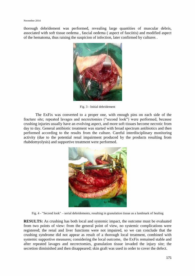
thorough debridement was performed, revealing large quantities of muscular debris,
associated with soft tissue oedema , fascial oedema ( aspect of fasciitis) and modified aspect
of the hematoma, thus raising the suspicion of infection, later confirmed by cultures .
Fig. 3 - Initial debridement
The ExFix was converted to a proper one, with enough pins on each side of the
fracture site; repeated lavages and necrectomies (“second look") were performed, because
crushing injuries usually have an evolving aspect, and more soft tissues become necrotic from
day to day. General antibiotic treatment was started with broad spectrum antibiotics and then
performed according to the results from the culture. Careful interdisciplinary monitoring
activity (due to the potential renal impairment produced by the products resulting from
rhabdomyolysis) and supportive treatment were performed.
Fig. 4 - "Second look" – serial debridements, resulting in granulation tissue as a landmark of healing
RESULTS: As crushing has both local and systemic impact, the outcome must be evaluated
from two points of view: from the general point of view, no systemic complications were
registered; the renal and liver functions were not impaired, so we can conclude that the
crushing syndrome did not appear as result of a thorough local treatment, combined with
systemic supportive measures; considering the local outcome, the ExFix remained stable and
after repeated lavages and necrectomies, granulation tissue invaded the injury site; the
secretion diminished and then disappeared; skin graft was used in order to cover the defect.

Supplement No. 1/2016
172
Fig. 5- Clinical aspect after skin integration
When all signs of inflammation disappeared, the skin flaps were completely
integrated, and the soft tissue injuries seemed to have healed, sequential method was
considered to be suitable for the patient, external fixation being followed by intramedullary
nail. No complications appeared after nailing and several years later, the nail was removed.
Since no local complications appeared, it can be concluded that the local treatment was
complete, providing healing conditions, despite the initial contamination.
Fig. 6 - Radiological aspect after intramedullary nail insertion
Fig. 7 - functional outcome 6 months follow up

November 2014
173
CONCLUSION: Crushing trauma represent one of the most challenging type of modern
traumatology, not only because they are increasingly frequent, but also because they associate
complex injuries, affecting all the structures of a limb; although the bone injury is the most
obvious one, the soft tissue damage is definitely the most important for the prognosis of the
limb and of the patient, especially because necrotic muscles represent a very good
environment for bacterial growth, thus considerably increasing the risk of early and severe
infections. That is why infected fractures are frequent after complex high energy trauma; they
require a prolonged and complicate treatment applied by a multidisciplinary team; the general
measures- antibiotics and supportive treatment- are mandatory, but the most important is the
local treatment which must create a clean environment. Complete debridement, always
requiring several surgical interventions and optimal stabilization of the fracture are crucial
not only for limb preservation, but also for patient’s survival.


![Inflammation and cancer: How hot is the link? · carcinoma [30], colon carcinoma, lung carcinoma, squamous cell carcinoma, pancreatic cancer [31,32], ovarian carcinoma biochemical](https://static.fdocuments.in/doc/165x107/5fcdd6c81c76a34db570e7e6/iniammation-and-cancer-how-hot-is-the-link-carcinoma-30-colon-carcinoma.jpg)