Baclofen facilitates sleep, neuroplasticity, and recovery ... fileafter stroke in rats ... of sleep...
13
RESEARCH ARTICLE Baclofen facilitates sleep, neuroplasticity, and recovery after stroke in rats Aleksandra Hodor 1,,a , Svitlana Palchykova 1 , Francesca Baracchi 1 , Daniela Noain 2 & Claudio L. Bassetti 1 1 Center for Experimental Neurology (ZEN), Department of Neurology, Inselspital, Bern University Hospital, 3010, Bern, Switzerland 2 Department of Neurology, University Hospital Z€ urich, 8091, Z€ urich, Switzerland Correspondence Aleksandra Hodor, Center for Experimental Neurology (ZEN), Department of Neurology, Inselspital, Bern University Hospital, Freiburgstrasse 3, 3010 Bern, Switzerland. Tel: +41 (0)31 63 20441; Fax: +41 (0)31 632 96 79; E-mail: [email protected]. ch Received: 19 May 2014; Revised: 22 July 2014; Accepted: 15 August 2014 doi: 10.1002/acn3.115 a Aleksandra Hodor is a Ph.D. student of the Neuroscience Center Z€ urich (ZNZ), University of Z€ urich, Switzerland. Abstract Objective: Sleep disruption in the acute phase after stroke has detrimental effects on recovery in both humans and animals. Conversely, the effect of sleep promo- tion remains unclear. Baclofen (Bac) is a known non-rapid eye movement (NREM) sleep-promoting drug in both humans and animals. The aim of this study was to investigate the effect of Bac on stroke recovery in a rat model of focal cerebral ischemia (isch). Methods: Rats, assigned to three experimental groups (Bac/isch, saline/isch, or Bac/sham), were injected twice daily for 10 consecutive days with Bac or saline, starting 24 h after induction of stroke. The sleep–wake cycle was assessed by EEG recordings and functional motor recovery by single pellet reaching test (SPR). In order to identify potential neuroplasticity mecha- nisms, axonal sprouting and neurogenesis were evaluated. Brain damage was assessed by Nissl staining. Results: Repeated Bac treatment after ischemia affected sleep, motor function, and neuroplasticity, but not the size of brain damage. NREM sleep amount was increased significantly during the dark phase in Bac/isch compared to the saline/isch group. SPR performance dropped to 0 immediately after stroke and was recovered slowly thereafter in both ischemic groups. How- ever, Bac-treated ischemic rats performed significantly better than saline-treated animals. Axonal sprouting in the ipsilesional motor cortex and striatum, and neu- rogenesis in the peri-infarct region were significantly increased in Bac/isch group. Conclusion: Delayed repeated Bac treatment after stroke increased NREM sleep and promoted both neuroplasticity and functional outcome. These data support the hypothesis of the role of sleep as a modulator of poststroke recovery. Introduction Ischemic stroke is one of the most prevalent neurological conditions and a leading cause of death and long-term disability worldwide. 1,2 Despite progress made in under- standing the mechanisms involved in neuronal damage during ischemia, limited advances have been reached in developing effective treatments for stroke patients. 3,4 Spontaneous partial recovery after stroke is associated with neuronal plasticity mechanisms. Data from both patients and animal models showed a remodeling of neu- ronal networks in the hemisphere affected by stroke as well as recruitment of additional circuits from the contralesional hemisphere. 5–8 Therefore, methods induc- ing or enhancing neuronal plasticity processes in the lesioned brain area may represent a novel effective thera- peutic strategy for stroke. There is growing evidence suggesting an important role of sleep in facilitating brain plasticity. 9,10 Changes in sleep and sleep electroencephalogram (EEG) may not only reflect changes in connectivity within cortical neuronal network but also drive changes in synaptic strength. 11,12 Moreover, it has been shown that plasticity-related genes and proteins display differential expression in sleep, wake- fulness, and following sleep deprivation (SD). 13 Increased slow-wave activity (SWA) in nonrapid eye movement (NREM) sleep, a marker of sleep intensity, was observed in the brain regions stimulated by intense activities during wakefulness both in humans and animals. 14,15 Further- more, specific waking activities can trigger both induction ª 2014 The Authors. Annals of Clinical and Translational Neurology published by Wiley Periodicals, Inc on behalf of American Neurological Association. This is an open access article under the terms of the Creative Commons Attribution-NonCommercial-NoDerivs License, which permits use and distribution in any medium, provided the original work is properly cited, the use is non-commercial and no modifications or adaptations are made. 1 source: https://doi.org/10.7892/boris.63677 | downloaded: 13.3.2017
Transcript of Baclofen facilitates sleep, neuroplasticity, and recovery ... fileafter stroke in rats ... of sleep...
RESEARCH ARTICLE
Baclofen facilitates sleep, neuroplasticity, and recoveryafter stroke in ratsAleksandra Hodor1,,a, Svitlana Palchykova1, Francesca Baracchi1, Daniela Noain2 & Claudio L.Bassetti1
1Center for Experimental Neurology (ZEN), Department of Neurology, Inselspital, Bern University Hospital, 3010, Bern, Switzerland2Department of Neurology, University Hospital Z€urich, 8091, Z€urich, Switzerland
Correspondence
Aleksandra Hodor, Center for Experimental
Neurology (ZEN), Department of Neurology,
Inselspital, Bern University Hospital,
Freiburgstrasse 3, 3010 Bern, Switzerland.
Tel: +41 (0)31 63 20441; Fax: +41 (0)31 632
96 79; E-mail: [email protected].
ch
Received: 19 May 2014; Revised: 22 July
2014; Accepted: 15 August 2014
doi: 10.1002/acn3.115
aAleksandra Hodor is a Ph.D. student of the
Neuroscience Center Z€urich (ZNZ), University
of Z€urich, Switzerland.
Abstract
Objective: Sleep disruption in the acute phase after stroke has detrimental effects
on recovery in both humans and animals. Conversely, the effect of sleep promo-
tion remains unclear. Baclofen (Bac) is a known non-rapid eye movement
(NREM) sleep-promoting drug in both humans and animals. The aim of this
study was to investigate the effect of Bac on stroke recovery in a rat model of focal
cerebral ischemia (isch). Methods: Rats, assigned to three experimental groups
(Bac/isch, saline/isch, or Bac/sham), were injected twice daily for 10 consecutive
days with Bac or saline, starting 24 h after induction of stroke. The sleep–wakecycle was assessed by EEG recordings and functional motor recovery by single
pellet reaching test (SPR). In order to identify potential neuroplasticity mecha-
nisms, axonal sprouting and neurogenesis were evaluated. Brain damage was
assessed by Nissl staining. Results: Repeated Bac treatment after ischemia affected
sleep, motor function, and neuroplasticity, but not the size of brain damage.
NREM sleep amount was increased significantly during the dark phase in Bac/isch
compared to the saline/isch group. SPR performance dropped to 0 immediately
after stroke and was recovered slowly thereafter in both ischemic groups. How-
ever, Bac-treated ischemic rats performed significantly better than saline-treated
animals. Axonal sprouting in the ipsilesional motor cortex and striatum, and neu-
rogenesis in the peri-infarct region were significantly increased in Bac/isch group.
Conclusion: Delayed repeated Bac treatment after stroke increased NREM sleep
and promoted both neuroplasticity and functional outcome. These data support
the hypothesis of the role of sleep as a modulator of poststroke recovery.
Introduction
Ischemic stroke is one of the most prevalent neurological
conditions and a leading cause of death and long-term
disability worldwide.1,2 Despite progress made in under-
standing the mechanisms involved in neuronal damage
during ischemia, limited advances have been reached in
developing effective treatments for stroke patients.3,4
Spontaneous partial recovery after stroke is associated
with neuronal plasticity mechanisms. Data from both
patients and animal models showed a remodeling of neu-
ronal networks in the hemisphere affected by stroke as
well as recruitment of additional circuits from the
contralesional hemisphere.5–8 Therefore, methods induc-
ing or enhancing neuronal plasticity processes in the
lesioned brain area may represent a novel effective thera-
peutic strategy for stroke.
There is growing evidence suggesting an important role
of sleep in facilitating brain plasticity.9,10 Changes in sleep
and sleep electroencephalogram (EEG) may not only
reflect changes in connectivity within cortical neuronal
network but also drive changes in synaptic strength.11,12
Moreover, it has been shown that plasticity-related genes
and proteins display differential expression in sleep, wake-
fulness, and following sleep deprivation (SD).13 Increased
slow-wave activity (SWA) in nonrapid eye movement
(NREM) sleep, a marker of sleep intensity, was observed
in the brain regions stimulated by intense activities during
wakefulness both in humans and animals.14,15 Further-
more, specific waking activities can trigger both induction
ª 2014 The Authors. Annals of Clinical and Translational Neurology published by Wiley Periodicals, Inc on behalf of American Neurological Association.
This is an open access article under the terms of the Creative Commons Attribution-NonCommercial-NoDerivs License, which permits use and
distribution in any medium, provided the original work is properly cited, the use is non-commercial and no modifications or adaptations are made.
1source: https://doi.org/10.7892/boris.63677 | downloaded: 13.3.2017
of plasticity-related genes in the cortex and homeostatic
response during the subsequent sleep episode.16 SD
impaired induction and/or maintenance of long-term
potentiation (LTP),17 a basic mechanism thought to under-
lie neuronal plasticity and memory formation, whereas
sleep oscillations induced LTP in the adult18 and in the
developing cat following monocular deprivation.19 In addi-
tion, recent data have shown that NREM sleep has a key role
in promoting learning-dependent synapse formation and
maintenance on selected dendritic branches.20 Therefore,
interaction between EEG markers of sleep and the events
mediating plasticity at the molecular, cellular, and network
levels provide a link between sleep and brain plasticity.
There is accumulating evidence suggesting an impor-
tant role for sleep in stroke recovery. Manipulation of
sleep after ischemia may in fact affect stroke outcome.
Disruption of sleep during acute and subacute phase of
stroke aggravated brain damage21 and impeded functional
recovery in rats.22 On the contrary, administration of c-hydroxybutyric acid (GHB), considered a sleep-promoting
drug, immediately after reperfusion accelerated motor
function recovery in mice.23 Furthermore, a physiological
enhancement of sleep (following previous sleep depriva-
tion) occurring immediately after stroke induction, was
also associated with a reduction of brain damage.24 Sleep
may, therefore, play a dual beneficial role in brain repair,
fostering neuroprotection in the acute phase and enhanc-
ing neuroplasticity in the delayed phase after stroke.
The aim of the current study was to investigate the effect
of sleep enhancement on brain repair and functional recov-
ery after the acute phase of ischemic stroke. We hypothe-
sized that induction of sleep or synchronized neuronal
activity would facilitate motor function recovery and brain
repair mechanisms in a rat model of focal cerebral ischemia.
Considering our strong interest in translational approaches,
we decided to use baclofen (Bac), a Gamma-aminobutyric
acid (GABA)–B receptor agonist which is known to pro-
mote sleep in humans.25 Our group has recently shown that
Bac also increases NREM sleep duration in rats.26
Methods
Animals
Adult male Sprague–Dawley rats (n = 53; Harlan Labora-
tories, Horst, Netherlands; Charles–River, Sulzfeld, Ger-
many), 318 � 17 g at the time of surgery, were
maintained on a 12–12 h light–dark cycle at 22 � 0.5°Cambient temperature. They were kept individually in
Macrolon cages and provided with food and water ad
libitum, except food restriction during behavioral training.
The experiments were carried out with governmental
approval according to local guidelines for the care and
use of laboratory animals at the University Hospital
Z€urich, Switzerland.
Experimental protocol
Two separate experiments were performed. In Experiment
1 (Fig. 1A), rats were implanted with EEG and electro-
myogram (EMG) electrodes. Animals were then subjected
to focal cerebral ischemia (isch) or sham surgery and
assigned to one of the three experimental groups: Bac/isch
(n = 6), saline/isch (n = 7), or Bac/sham (n = 4). Baclo-
fen (Sigma–Aldrich, Buchs, Switzerland; 10 mg/kg) was
diluted in saline (0.9% NaCl) to obtain 3 mg/mL working
solution. The drug was administered intraperitoneally
(i.p.) 24 h after surgery and then twice daily (1 h after
light onset and offset) for 10 consecutive days. Sleep was
recorded during baseline preceding surgery and on day 2,
6, and 11 after surgery. All animals were decapitated
1 day after the last injection and their brains were col-
lected for histological analysis.
In Experiment 2 (Fig. 1B), rats were trained in a single
pellet reaching task (SPR) for ca. 25 days. During training
the preferred paw was identified for every rat. Cerebral ische-
mia or sham surgery was performed, when animals reached a
stable level of performance. Thereafter, rats were subdivided
into three groups Bac/isch (n = 14), saline/isch (n = 14),
and Bac/sham (n = 8) and subjected to the same pharmaco-
logical protocol as in Experiment 1. Motor function was
assessed 1 day after surgery before the first drug injection
and then weekly starting on the day after the end of the drug
administration (days 12, 19, 26, 33, and 40 after surgery).
Proliferation marker, 5-bromo-20-deoxyuridine (BrdU;
Sigma-Aldrich, St. Louis, MO; 50 mg/kg),27 incorporating
into DNA during cell division, was administered for
10 days (2 h after light onset, i.p.), and followed Bac or sal-
ine injections. BrdU was diluted in saline (concentration
10 mg/mL).
The anterograde tracer, biotinylated dextran amine
(BDA, 10%; MW = 10,000 Da; Molecular Probes, Eugene,
OR; diluted in 0.01 mol/L phosphate buffer),28 used to eval-
uate axonal sprouting,22, 29 was microinjected at two loca-
tions into the motor cortex contralateral to the lesion side
(stereotaxic coordinates: antero-posterior (AP) +/–1 mm ,
medio–lateral (ML) 1 mm, dorso–ventral (DV) 3 mm from
the skull). All rats belonging to the Experiment 2 received a
total volume of 1 lL of tracer (0.5 lL of each injection, over10 min) 6 weeks after surgery. Two weeks later rats were
sacrificed and brains were collected for further evaluations.
EEG implantation and recording
Rats were implanted epidurally with EEG and EMG elec-
trodes under deep anesthesia (2% isoflurane in 30% O2
2 ª 2014 The Authors. Annals of Clinical and Translational Neurology published by Wiley Periodicals, Inc on behalf of American Neurological Association.
Effects of Baclofen on Stroke Recovery A. Hodor et al.
and 70% N2O). Four gold-plated mini-screws were posi-
tioned in the skull over the motor cortex of the right and
left hemispheres (�2 mm to bregma, 2 mm lateral to
midline). Electrodes were connected to stainless steel
wires and fixed to the skull with dental cement. Two gold
wires were inserted bilaterally in the neck muscles for
EMG recording. At least 8–10 days were allowed for
recovery. Before baseline recordings, rats were habituated
to the sleep recording apparatus for 4–5 days.
EEG and EMG were sampled at 200 Hz. Signals were
amplified, filtered, and analog-to-digital converted. Hard-
ware EMBLA and software Somnologica-3 (Medcare
Flaga, Reykjavik, Iceland) were used. Activity in the
50 Hz band was discarded from the analysis because of
power line artifacts. The EEG was subjected to a discrete
Fourier transformation yielding power spectra (range: 1–25 Hz; frequency resolution: 0.25-Hz bins; time resolu-
tion: consecutive 4-sec epochs; window function: ham-
ming).
Three vigilance states – NREM sleep, REM sleep, and
wakefulness – were visually scored according to standard
criteria at 4-sec epochs.30 In addition to these three con-
ventional vigilance states we introduced a new state, dis-
tinct from physiological sleep or wakefulness, which was
observed after Bac administration. This state was charac-
terized by atypical behavior and abnormal hypersynchro-
nous EEG pattern (“drug-induced” state), as described
previously.26 The state lasted 195.5 � 7.6 and
197 � 8.3 min in the Bac/isch group (light and dark
phase, respectively), and 103.5 � 32 and
189.1 � 11.5 min in the Bac/sham group. Epochs were
assigned to a specific vigilance state when more than half
of the epoch fulfilled the criteria for that state. Epochs
containing EEG artifacts were identified and excluded
from subsequent spectral analysis in both derivations
(25% of recording time, most of them [17%] occurred
during wakefulness).
Induction of focal cerebral ischemia
Stroke was induced by the three-vessel occlusion method
(3Vo) with permanent occlusion of the distal middle
cerebral artery (MCA) and the ipsilateral common carotid
artery (CCA), superimposed by temporal occlusion of the
contralateral CCA under general anesthesia with 2% iso-
flurane.22, 31 A small piece of the skull overlying the MCA
was removed and the dura was retracted. The MCA and
its three main branches were occluded by bipolar electro-
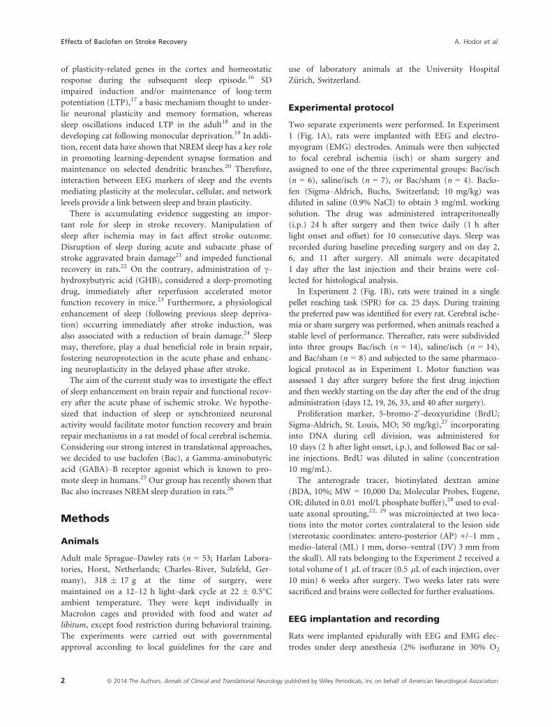
Figure 1. Design of the experiments. (A) In Experiment 1, rats were implanted with EEG/EMG electrodes and 11 days later subjected to ischemia
(isch) or sham surgery. Twenty-four hours after surgery animals were treated with the drug (10 mg/kg baclofen, Bac, or saline, Sal) and then
twice daily for 10 days. EEG and EMG were recorded during a 24-h baseline (BL) day and on days 2, 6, and 11 following isch/sham surgery.
Three treatment groups were designed: Bac/isch (n = 6), Sal/isch (n = 7), and Bac/sham (n = 4). (B) In Experiment 2, rats were trained in the
single pellet reaching (SPR) task for 3–4 weeks preceding isch or sham surgery. Twenty-four hours after surgery rats were treated with Bac or Sal
and then twice daily for 10 days. All rats received also bromodeoxyuridine (BrdU) injection for ten days. SPR performance was assessed over
3 days preceding surgery (baseline) and on days 1, 12, 19, 26, 33, and 40 following surgery. Forty-one days after surgery all rats received
microinjection of biotinylated dextran amine (BDA) and were perfused 2 weeks later. Three treatment groups were designed: Bac/isch (n = 14),
Sal/isch (n = 14) and Bac/sham (n = 8).
ª 2014 The Authors. Annals of Clinical and Translational Neurology published by Wiley Periodicals, Inc on behalf of American Neurological Association. 3
A. Hodor et al. Effects of Baclofen on Stroke Recovery
coagulation. The CCA ipsilateral to the occluded MCA
was ligated permanently with a 4-0 silk suture, whereas
the contralateral CCA was temporarily occluded for
60 min with an aneurysm clip. Rectal temperature was
maintained between 36.5 � 0.5°C by a warm lamp dur-
ing the surgery. Sham-operated rats were subjected to the
same procedure except for occlusion of the MCA and
CCA. Both ischemia and sham surgeries were performed
on the hemisphere contralateral to the preferred forelimb
assessed by SPR task.
SPR task
SPR task was used to assess fine motor skills.32 Rats had
to use their preferred forelimb to retrieve a food pellet
located in a well outside the test chamber.22 Briefly, ani-
mals were placed in a clear Plexiglas box
(41 9 27 9 37 cm) with a vertical slit (1 9 15 cm)
placed in the middle of the front wall, 1 cm above the
floor. A 2-cm wide shelf with small wells was mounted in
the front of the slit, but outside the box wall. Animals
were trained to reach a food pellet (45 mg dustless preci-
sion pellet; Bio-Serv, Frenchtown, NJ) placed in the well
on the shelf. Rats received daily training sessions consist-
ing of 50 pellets for 3–4 weeks. A pellet was placed in the
well on the side contralateral to the preferred paw. A sin-
gle reaching attempt was permitted. Reaching attempts
were classified as successful or failed. In the successful
attempt the rat was expected to make a single reach,
grasp the pellet from the well, bring it to the mouth and
eat it. During the test sessions before and after surgery
rats were given 50 pellets and the session ended when
rats made 50 attempts or when 15 min elapsed. Success
rate was computed as the percentage of successfully
obtained pellets out of 50 possible attempts. The baseline
(BL) performance was computed as the average of the
3 days immediately preceding surgery. Improvement in
poststroke motor performance was computed as a differ-
ence in success rate between days 40 and 1 after ische-
mia.
To increase motivation all animals underwent a food
restriction schedule with 20 g of chow per day during the
training weeks and at the days preceding the test sessions.
During this time rats were maintained at 95% of their
normal body weight.
Tissue collection
Two weeks after BDA administration rats were deeply
anesthetized and perfused transcardially with ice-cold 4%
paraformaldehyde (PFA) in phosphate-buffered saline
(PBS). Brains were removed, postfixed in PFA for 2 h,
and cryoprotected in 15% and 30% sucrose in the
ascending manner. The tissue was stored at �80°C for
further evaluations.
Analysis of lesion volume and corpuscallosum thickness
To determine lesion size and corpus callosum thickness,
40-lm thick coronal sections were cut with a cryostat at
six predefined levels: 2.7 (L1), 1.7 (L2), 0.7 (L3), �0.3
(L4), �1.3 (L5), and �2.3 (L6) mm to bregma.33 Brain
sections were then stained with cresyl violet and digitized.
Measurements were done with ImageJ (NIH, Bethesda,
MD). Brain damage was computed on one section for
each level as a difference between intact hemisphere and
the nonlesioned area of the ischemic hemisphere. Lesion
volume was estimated by multiplying obtained brain
damage values by the size of each level (section thickness
plus distance between levels). The corpus callosum thick-
ness was measured using coronal sections from L6 level.
Immunohistochemistry
For detection of BDA, free-floating brain sections were
incubated overnight with avidin–biotin–peroxidase com-
plex (Elite ABC kit; Vector Laboratories, Burlingame, CA)
and revealed with 3,30-diaminobenzidine (DAB; Sigma-
Aldrich, Buchs, Switzerland).22 The sections were digitized
and the area of BDA-labeled axons was quantified by
determining the number of pixels above the intensity
gray-scale threshold using ImageJ (NIH) as described pre-
viously.29 Briefly, the mean surface area of BDA-labeled
axons was measured in the primary motor cortex and
striatum at the levels L2–L4 of coronal sections. Ratios
between ipsilesional and contralesional cortical surface
areas (corresponding to the corticocortical projections)
and between ipsilesional striatal and contralesional corti-
cal areas (corresponding to the corticostriatal projections)
were used as an index of axonal sprouting. For each rat 2
adjacent sections were averaged on each level (n = 9,
n = 10, n = 7 for corticocortical and n = 7, n = 7, n = 6
for corticostriatal in the saline/isch, Bac/isch and Bac/
sham group, respectively). Ratios near zero indicated a
low BDA penetration from the contra- to the ipsilesional
side and, therefore, limited axonal sprouting.
Double immunofluorescence staining with the antibody
against BrdU together with antibodies against specific
cell-type marker was used to detect cell proliferation and
to assess the type of proliferating cells. Free-floating sec-
tions were incubated in 2 mol/L HCl for 2 h at room
temperature (RT) to denaturate DNA and then washed
four times in PBS pH = 7.4. After preincubation in
blocking solution (0.01 mol/L PBS containing 2% appro-
priate normal sera and 0.3% Triton-X), sections were
4 ª 2014 The Authors. Annals of Clinical and Translational Neurology published by Wiley Periodicals, Inc on behalf of American Neurological Association.
Effects of Baclofen on Stroke Recovery A. Hodor et al.

incubated overnight at +4°C with the rat anti-BrdU anti-
body (1:200; Abcam, Cambridge, UK) and one of the fol-
lowing antibodies for a specific cell-type marker: mouse
anti-NeuN (1:200; Millipore, Billerica, MA; marker for
neurons), rabbit anti-glial fibrillary acidic protein (GFAP,
1:200; Dako, Carpinteria, CA; marker for astrocytes) or
rabbit anti-ionized calcium-binding adapter molecule 1
(Iba1, 1:600; Wako Chemicals, Osaka, Japan; marker for
microglia). Sections were then incubated for 1.5 h at RT
with the fluorophore-conjugated secondary antibodies
Alexa Fluor-488 (green; for detection of BrdU) or Cy3
(red; for detection of other markers) (1:200; Jackson Im-
munoresearch, West Grove, PA) against the appropriate
host species of the primary antibodies. Finally, sections
were rinsed three times with PBS, mounted on gelatin-
subbed slides (Southern Biotechnology Association Inc,
Birmingham, AL, USA) and coverslipped.
Cell counting and microscopy analysis
The number of BrdU-positive (+), NeuN+/BrdU+,GFAP+/BrdU+, and Iba1+/BrdU+ cells were quantified in
the peri-infarct region of ischemic animals and in the cor-
responding cortical region of sham animals using the
optical fractionator probe (Stereo Investigator version 8.2;
MicroBrightField Inc., Williston, VT) at 409 magnifica-
tion on a fluorescence microscope equipped with a
motorized x-y stage (Zeiss Axio Imager Z1, Jena, Ger-
many; 209/0.5 EC Plan-Neofluar objective). The peri-
infarct area was outlined on a 109 magnification using
the tracing function of Stereo Investigator. Several param-
eters were then determined on optical fractionator: count-
ing frame (200 9 200 lm; x–y plane), optical dissector
height (27-36 lm; z plane), distance between sampling
regions (600 lm in x and y direction) and the grid size.
A computer-driven motor stage allowed to analyze sec-
tions at each of the counting frame location under a 409
magnification. This procedure provided unbiased stereol-
ogical quantification, because once the region of interest
was outlined, sampling sites were evenly and randomly
distributed throughout the marked region. Quantification
was performed at levels L2 and L3 and averaged on two
sections per animal (n = 7 per ischemic groups, n = 6
per sham group). The data are presented as the average
of cell number per mm2. All histological and immunohis-
tochemical analyses were determined in a blinded way.
Statistical analysis
Effects of Bac treatment and time on motor performance
in the SPR task were evaluated by a repeated measures
ANOVA (SAS software, SAS Institute, Cary, NC). Effects
of treatment on sleep, neurogenesis, axonal sprouting,
corpus callosum thickness, and brain damage were evalu-
ated by one-way ANOVA. Post hoc paired and unpaired
t-tests, Wilcoxon and Kruskal–Wallis or Tukey–Kramer
test for multiple comparisons were performed if the
results of the ANOVA reached statistical significance
(P < 0.05). All provided values are means � SEM. Pear-
son correlation coefficients were calculated between SPR
performance and BDA or BrdU parameters; P < 0.05 was
considered of statistical significance.
Results
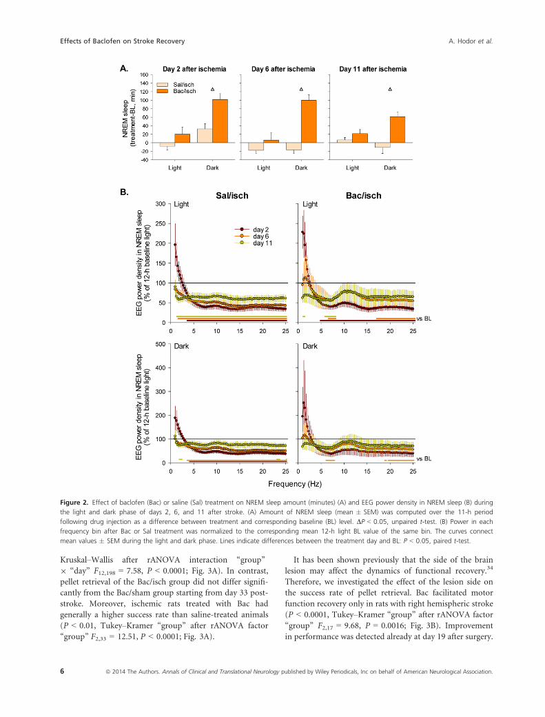
Effects of Bac on vigilance states and EEGpower spectrum
Bac increased the amount of NREM sleep during the dark
period on days 2, 6, and 11 after stroke compared to sal-
ine administration (P < 0.05, unpaired t-test; Fig. 2A).
We found no changes in the amount of REM sleep after
Bac treatment. Amount of NREM sleep was similar
between treatment conditions on the baseline day
(364.5 � 32.7 vs. 361.6 � 36.6 min during the light
phase and 218.8 � 59.7 vs. 207.3 � 55.4 min during the
dark phase in saline/isch vs. Bac/isch group, respectively).
EEG power spectra were affected mostly in the hemi-
sphere ipsilateral to the lesion (Supplementary material S1).
Stroke led to a significant reduction of EEG power density
in NREM sleep throughout most of the frequency range
(Fig. 2B). Thus, in the saline/isch group it was reduced
below BL values in the frequencies >3.75 Hz during both
light and dark phases on days 2 and 6 after stroke and dur-
ing light phase on day 11 (Fig. 2B left). Bac treatment
resulted in a partial recovery of power. Hence, a significant
reduction of power density in the frequencies >4.75 Hz was
observed only during the light phase on day 2 after stroke
(Fig. 2B right). EEG power was below BL values in the fre-
quencies between 5.75 and 7.75 Hz on days 6 and 11 (only
light phase) and above 17 Hz on day 6 (Fig. 2B right).
Effects of Bac on functional recovery
Effects of Bac administration on the recovery of grasping
ability were assessed by SPR task. All rats showed similar
performance in the task prior to ischemia. The success rate
of pellet retrieval was 51.14 � 2.75%, 51.36 � 2.81%, and
45.50 � 2.74% in Bac/isch, saline/isch, and Bac/sham
groups, respectively. Rat performance dropped to zero (rats
were not able to perform the task) immediately after stroke
in both ischemic groups, but remained stable in the sham-
operated animals (Fig. 3A). Slow spontaneous recovery was
observed in the saline/isch group in the course of the fol-
lowing 6 weeks. However, saline/isch rats never reached the
performance level of sham-operated animals (P < 0.005,
ª 2014 The Authors. Annals of Clinical and Translational Neurology published by Wiley Periodicals, Inc on behalf of American Neurological Association. 5
A. Hodor et al. Effects of Baclofen on Stroke Recovery
Kruskal–Wallis after rANOVA interaction “group”
9 “day” F12,198 = 7.58, P < 0.0001; Fig. 3A). In contrast,
pellet retrieval of the Bac/isch group did not differ signifi-
cantly from the Bac/sham group starting from day 33 post-
stroke. Moreover, ischemic rats treated with Bac had
generally a higher success rate than saline-treated animals
(P < 0.01, Tukey–Kramer “group” after rANOVA factor
“group” F2,33 = 12.51, P < 0.0001; Fig. 3A).
It has been shown previously that the side of the brain
lesion may affect the dynamics of functional recovery.34
Therefore, we investigated the effect of the lesion side on
the success rate of pellet retrieval. Bac facilitated motor
function recovery only in rats with right hemispheric stroke
(P < 0.0001, Tukey–Kramer “group” after rANOVA factor
“group” F2,17 = 9.68, P = 0.0016; Fig. 3B). Improvement
in performance was detected already at day 19 after surgery.
Figure 2. Effect of baclofen (Bac) or saline (Sal) treatment on NREM sleep amount (minutes) (A) and EEG power density in NREM sleep (B) during
the light and dark phase of days 2, 6, and 11 after stroke. (A) Amount of NREM sleep (mean � SEM) was computed over the 11-h period
following drug injection as a difference between treatment and corresponding baseline (BL) level. DP < 0.05, unpaired t-test. (B) Power in each
frequency bin after Bac or Sal treatment was normalized to the corresponding mean 12-h light BL value of the same bin. The curves connect
mean values � SEM during the light and dark phase. Lines indicate differences between the treatment day and BL: P < 0.05, paired t-test.
6 ª 2014 The Authors. Annals of Clinical and Translational Neurology published by Wiley Periodicals, Inc on behalf of American Neurological Association.
Effects of Baclofen on Stroke Recovery A. Hodor et al.
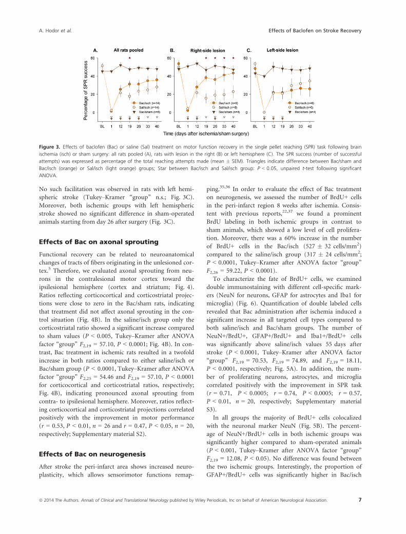
No such facilitation was observed in rats with left hemi-
spheric stroke (Tukey–Kramer “group” n.s.; Fig. 3C).
Moreover, both ischemic groups with left hemispheric
stroke showed no significant difference in sham-operated
animals starting from day 26 after surgery (Fig. 3C).
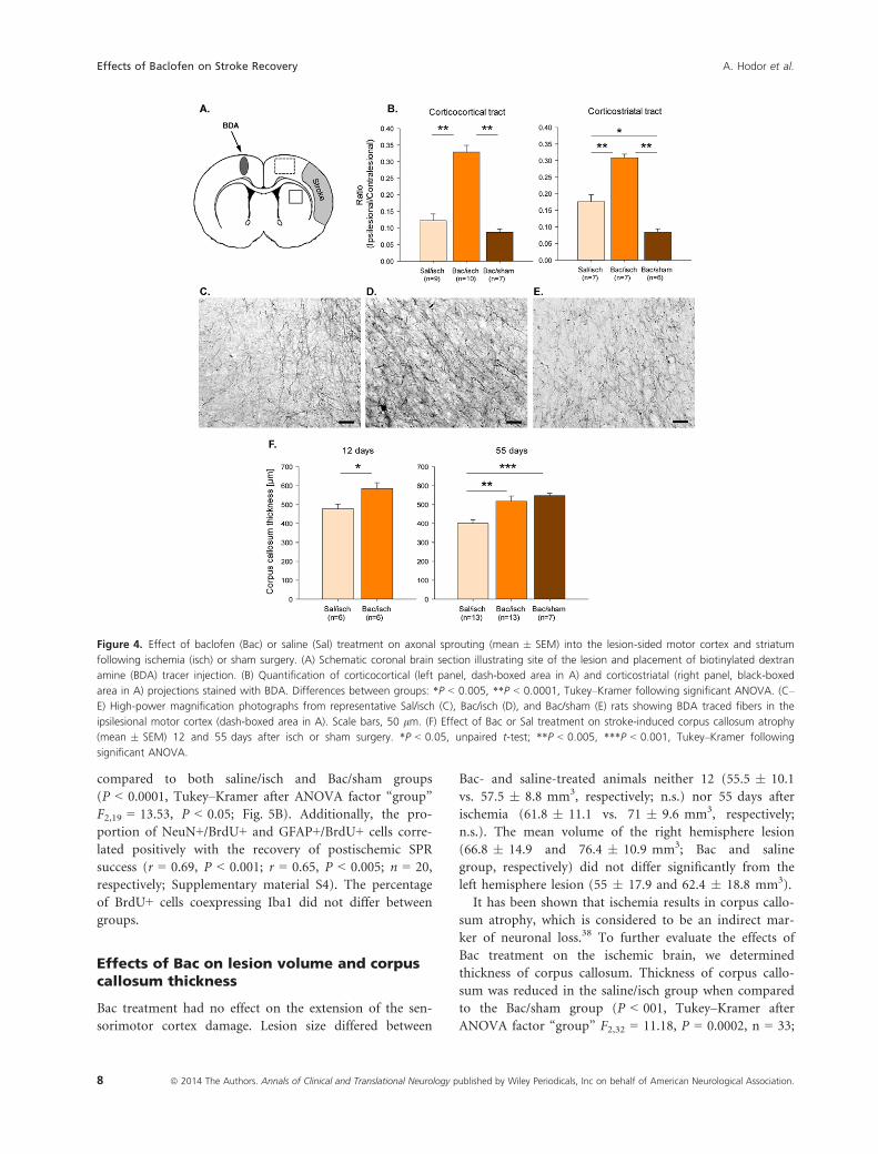
Effects of Bac on axonal sprouting
Functional recovery can be related to neuroanatomical
changes of tracts of fibers originating in the unlesioned cor-
tex.5 Therefore, we evaluated axonal sprouting from neu-
rons in the contralesional motor cortex toward the
ipsilesional hemisphere (cortex and striatum; Fig. 4).
Ratios reflecting corticocortical and corticostriatal projec-
tions were close to zero in the Bac/sham rats, indicating
that treatment did not affect axonal sprouting in the con-
trol situation (Fig. 4B). In the saline/isch group only the
corticostriatal ratio showed a significant increase compared
to sham values (P < 0.005, Tukey–Kramer after ANOVA
factor “group” F2,19 = 57.10, P < 0.0001; Fig. 4B). In con-
trast, Bac treatment in ischemic rats resulted in a twofold
increase in both ratios compared to either saline/isch or
Bac/sham group (P < 0.0001, Tukey–Kramer after ANOVA
factor “group” F2,25 = 54.46 and F2,19 = 57.10, P < 0.0001
for corticocortical and corticostriatal ratios, respectively;
Fig. 4B), indicating pronounced axonal sprouting from
contra- to ipsilesional hemisphere. Moreover, ratios reflect-
ing corticocortical and corticostriatal projections correlated
positively with the improvement in motor performance
(r = 0.53, P < 0.01, n = 26 and r = 0.47, P < 0.05, n = 20,
respectively; Supplementary material S2).
Effects of Bac on neurogenesis
After stroke the peri-infarct area shows increased neuro-
plasticity, which allows sensorimotor functions remap-
ping.35,36 In order to evaluate the effect of Bac treatment
on neurogenesis, we assessed the number of BrdU+ cells
in the peri-infarct region 8 weeks after ischemia. Consis-
tent with previous reports,22,37 we found a prominent
BrdU labeling in both ischemic groups in contrast to
sham animals, which showed a low level of cell prolifera-
tion. Moreover, there was a 60% increase in the number
of BrdU+ cells in the Bac/isch (527 � 32 cells/mm2)
compared to the saline/isch group (317 � 24 cells/mm2;
P < 0.0001, Tukey–Kramer after ANOVA factor “group”
F2,26 = 59.22, P < 0.0001).
To characterize the fate of BrdU+ cells, we examined
double immunostaining with different cell-specific mark-
ers (NeuN for neurons, GFAP for astrocytes and Iba1 for
microglia) (Fig. 6). Quantification of double labeled cells
revealed that Bac administration after ischemia induced a
significant increase in all targeted cell types compared to
both saline/isch and Bac/sham groups. The number of
NeuN+/BrdU+, GFAP+/BrdU+ and Iba1+/BrdU+ cells
was significantly above saline/isch values 55 days after
stroke (P < 0.0001, Tukey–Kramer after ANOVA factor
“group” F2,19 = 70.53, F2,19 = 74.89, and F2,19 = 18.11,
P < 0.0001, respectively; Fig. 5A). In addition, the num-
ber of proliferating neurons, astrocytes, and microglia
correlated positively with the improvement in SPR task
(r = 0.71, P < 0.0005; r = 0.74, P < 0.0005; r = 0.57,
P < 0.01, n = 20, respectively; Supplementary material
S3).
In all groups the majority of BrdU+ cells colocalized
with the neuronal marker NeuN (Fig. 5B). The percent-
age of NeuN+/BrdU+ cells in both ischemic groups was
significantly higher compared to sham-operated animals
(P < 0.001, Tukey–Kramer after ANOVA factor “group”
F2,19 = 12.08, P < 0.05). No difference was found between
the two ischemic groups. Interestingly, the proportion of
GFAP+/BrdU+ cells was significantly higher in Bac/isch
Figure 3. Effects of baclofen (Bac) or saline (Sal) treatment on motor function recovery in the single pellet reaching (SPR) task following brain
ischemia (isch) or sham surgery: all rats pooled (A), rats with lesion in the right (B) or left hemisphere (C). The SPR success (number of successful
attempts) was expressed as percentage of the total reaching attempts made (mean � SEM). Triangles indicate difference between Bac/sham and
Bac/isch (orange) or Sal/isch (light orange) groups; Star between Bac/isch and Sal/isch group: P < 0.05, unpaired t-test following significant
ANOVA.
ª 2014 The Authors. Annals of Clinical and Translational Neurology published by Wiley Periodicals, Inc on behalf of American Neurological Association. 7
A. Hodor et al. Effects of Baclofen on Stroke Recovery
compared to both saline/isch and Bac/sham groups
(P < 0.0001, Tukey–Kramer after ANOVA factor “group”
F2,19 = 13.53, P < 0.05; Fig. 5B). Additionally, the pro-
portion of NeuN+/BrdU+ and GFAP+/BrdU+ cells corre-
lated positively with the recovery of postischemic SPR
success (r = 0.69, P < 0.001; r = 0.65, P < 0.005; n = 20,
respectively; Supplementary material S4). The percentage
of BrdU+ cells coexpressing Iba1 did not differ between
groups.
Effects of Bac on lesion volume and corpuscallosum thickness
Bac treatment had no effect on the extension of the sen-
sorimotor cortex damage. Lesion size differed between
Bac- and saline-treated animals neither 12 (55.5 � 10.1
vs. 57.5 � 8.8 mm3, respectively; n.s.) nor 55 days after
ischemia (61.8 � 11.1 vs. 71 � 9.6 mm3, respectively;
n.s.). The mean volume of the right hemisphere lesion
(66.8 � 14.9 and 76.4 � 10.9 mm3; Bac and saline
group, respectively) did not differ significantly from the
left hemisphere lesion (55 � 17.9 and 62.4 � 18.8 mm3).
It has been shown that ischemia results in corpus callo-
sum atrophy, which is considered to be an indirect mar-
ker of neuronal loss.38 To further evaluate the effects of
Bac treatment on the ischemic brain, we determined
thickness of corpus callosum. Thickness of corpus callo-
sum was reduced in the saline/isch group when compared
to the Bac/sham group (P < 001, Tukey–Kramer after
ANOVA factor “group” F2,32 = 11.18, P = 0.0002, n = 33;
Figure 4. Effect of baclofen (Bac) or saline (Sal) treatment on axonal sprouting (mean � SEM) into the lesion-sided motor cortex and striatum
following ischemia (isch) or sham surgery. (A) Schematic coronal brain section illustrating site of the lesion and placement of biotinylated dextran
amine (BDA) tracer injection. (B) Quantification of corticocortical (left panel, dash-boxed area in A) and corticostriatal (right panel, black-boxed
area in A) projections stained with BDA. Differences between groups: *P < 0.005, **P < 0.0001, Tukey–Kramer following significant ANOVA. (C–
E) High-power magnification photographs from representative Sal/isch (C), Bac/isch (D), and Bac/sham (E) rats showing BDA traced fibers in the
ipsilesional motor cortex (dash-boxed area in A). Scale bars, 50 lm. (F) Effect of Bac or Sal treatment on stroke-induced corpus callosum atrophy
(mean � SEM) 12 and 55 days after isch or sham surgery. *P < 0.05, unpaired t-test; **P < 0.005, ***P < 0.001, Tukey–Kramer following
significant ANOVA.
8 ª 2014 The Authors. Annals of Clinical and Translational Neurology published by Wiley Periodicals, Inc on behalf of American Neurological Association.
Effects of Baclofen on Stroke Recovery A. Hodor et al.
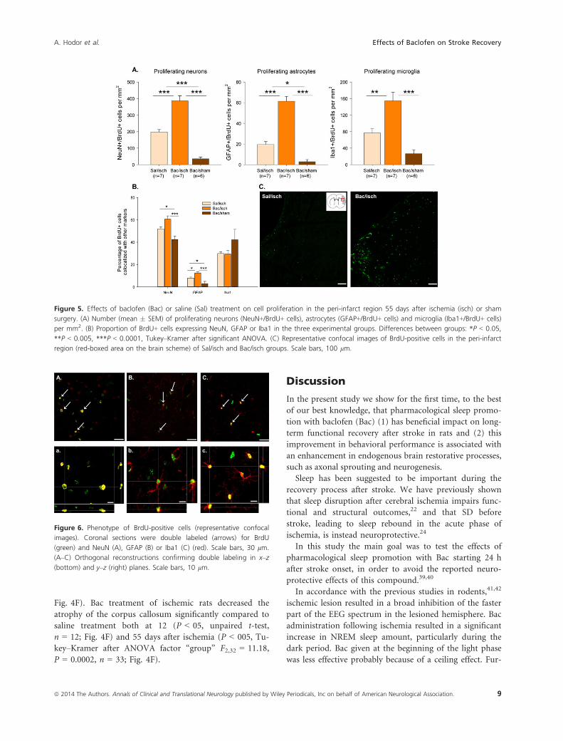
Fig. 4F). Bac treatment of ischemic rats decreased the
atrophy of the corpus callosum significantly compared to
saline treatment both at 12 (P < 05, unpaired t-test,
n = 12; Fig. 4F) and 55 days after ischemia (P < 005, Tu-
key–Kramer after ANOVA factor “group” F2,32 = 11.18,
P = 0.0002, n = 33; Fig. 4F).
Discussion
In the present study we show for the first time, to the best
of our best knowledge, that pharmacological sleep promo-
tion with baclofen (Bac) (1) has beneficial impact on long-
term functional recovery after stroke in rats and (2) this
improvement in behavioral performance is associated with
an enhancement in endogenous brain restorative processes,
such as axonal sprouting and neurogenesis.
Sleep has been suggested to be important during the
recovery process after stroke. We have previously shown
that sleep disruption after cerebral ischemia impairs func-
tional and structural outcomes,22 and that SD before
stroke, leading to sleep rebound in the acute phase of
ischemia, is instead neuroprotective.24
In this study the main goal was to test the effects of
pharmacological sleep promotion with Bac starting 24 h
after stroke onset, in order to avoid the reported neuro-
protective effects of this compound.39,40
In accordance with the previous studies in rodents,41,42
ischemic lesion resulted in a broad inhibition of the faster
part of the EEG spectrum in the lesioned hemisphere. Bac
administration following ischemia resulted in a significant
increase in NREM sleep amount, particularly during the
dark period. Bac given at the beginning of the light phase
was less effective probably because of a ceiling effect. Fur-
Figure 5. Effects of baclofen (Bac) or saline (Sal) treatment on cell proliferation in the peri-infarct region 55 days after ischemia (isch) or sham
surgery. (A) Number (mean � SEM) of proliferating neurons (NeuN+/BrdU+ cells), astrocytes (GFAP+/BrdU+ cells) and microglia (Iba1+/BrdU+ cells)
per mm2. (B) Proportion of BrdU+ cells expressing NeuN, GFAP or Iba1 in the three experimental groups. Differences between groups: *P < 0.05,
**P < 0.005, ***P < 0.0001, Tukey–Kramer after significant ANOVA. (C) Representative confocal images of BrdU-positive cells in the peri-infarct
region (red-boxed area on the brain scheme) of Sal/isch and Bac/isch groups. Scale bars, 100 lm.
Figure 6. Phenotype of BrdU-positive cells (representative confocal
images). Coronal sections were double labeled (arrows) for BrdU
(green) and NeuN (A), GFAP (B) or Iba1 (C) (red). Scale bars, 30 lm.
(A–C) Orthogonal reconstructions confirming double labeling in x–z
(bottom) and y–z (right) planes. Scale bars, 10 lm.
ª 2014 The Authors. Annals of Clinical and Translational Neurology published by Wiley Periodicals, Inc on behalf of American Neurological Association. 9
A. Hodor et al. Effects of Baclofen on Stroke Recovery

thermore, Bac led to a recovery of the power in the
higher frequencies (above 8 Hz). Noteworthy, recent
observations in rats, linked motor function recovery with
EEG frequencies >7 Hz.41,43 SWA during NREM sleep has
been associated with neuroplasticity processes9,17–19 and
therefore could be proposed to play a role in the promo-
tion of endogenous restorative mechanisms and func-
tional improvement during stroke recovery. Low-
frequency synchronous neuronal activity has been sug-
gested to have an important role in the anatomical reor-
ganization and axonal sprouting after brain lesion.29
Therefore, the changes in EEG activity observed immedi-
ately after Bac injections could play a role in improving
the functional outcome.
In the present study cerebral ischemia was induced in
the somatosensory cortex, thus leaving the motor cortical
areas anatomically intact. Nevertheless, we observed a
remarkable drop in the SPR performance after stroke.
This effect was expected since motor cortex function was
severely disrupted by extensive remodeling processes, dur-
ing the recovery period following ischemia even when the
area itself is spared by the lesion.44 Spontaneous gradual
improvement of motor function was present in the stroke
animals injected with saline. However, Bac treatment
accelerated performance improvement after stroke.
After Bac treatment, we found increased BDA labeling
in both cortex and striatum, indicating an enhancement in
the number of axons and/or increased axonal transport.
Axonal sprouting, or the ability of brain to form new con-
nections in areas denervated by the lesion, is a well-known
phenomenon.5,29 Ischemic damage of the sensorimotor
cortex induced sprouting of axons into the perilesion cor-
tex from the homotopic cortex in the contralateral hemi-
sphere29 and into the striatum below the lesion.45 Axonal
sprouting after stroke is regulated by various neuroplastici-
ty-related genes, including growth-promoting and growth-
inhibiting molecules.46 Many of these genes and proteins
associated with neuroplasticity have been shown to be
modulated by sleep.13,47 Previous studies showed that sleep
deprivation after stroke induced an increase in the expres-
sion of neurocan (the main growth-inhibiting molecule to
axonal sprouting),21 while GHB decreased it.23 Therefore,
changes in sleep may affect molecules crucial for post-
stroke axonal sprouting. Furthermore, Carmichael et al.
demonstrated the strong correlation between axonal
sprouting and the periodic synchronized neuronal activ-
ity.29 Bac, besides its effects on sleep, also induced a tran-
sient electrophysiological hypersynchronous pattern
during the subanesthetic state, which could play a role in
axonal growth and functional outcome. In this frame of
reference it is possible to hypothesize that the positive
effects of Bac on axonal sprouting and neurogenesis after
cerebral ischemia are related to sleep-induced mechanisms.
Noteworthy, the corpus callosum atrophy was signifi-
cantly reduced in Bac/isch group, despite the absence of
the beneficial effect of Bac on the extent of brain damage.
Because corpus callosum is the conduit for the interhemi-
spheric communication, its thickness is a sensitive indica-
tor of ischemic neuronal loss and a relevant marker
influencing recovery of function after stroke. Our finding
that Bac administration significantly reduced stroke-
induced corpus callosum atrophy supplements the data
showing a Bac-related boost in axonal sprouting.
Repeated administration of Bac also boosted neurogen-
esis in the ischemic brain. We found an increase in the
number of proliferating cells. Several reports indicated
that ischemic injury induces increased cell prolifera-
tion,37,48 associated with migration of newborn cells to
the lesion sites.49–51 However, only a small fraction of
newborn cells display a long-term survival. Therefore,
enhancement of endogenous neurogenesis, primarily by
improving survival of newborn cells, would be a plausible
strategy for restorative therapies. In our study, extensive
BrdU staining was noted in the peri-infarct region in both
ischemic groups. Peri-infarct area is critical for rehabilita-
tion, it shows intensified neuroplasticity, allowing remap-
ping of sensorimotor function.35,36 Bac-treated rats had
almost twofold increase in BrdU+ cells 55 days after
stroke onset. Hence Bac might not only increase cell pro-
liferation but also prolong survival of newborn cells and,
therefore, enhance endogenous neurogenesis.
Although the majority of newly formed cells found in
the peri-infarct region expressed the neuronal marker
NeuN, we also observed an increase in the number of
proliferating cells expressing glial markers after Bac treat-
ment. Interestingly, the percentage of cells that differenti-
ated into astrocytes was significantly higher in Bac/isch
group, indicating that Bac might also affect differentiation
of newborn cells. Glial cell activation has been demon-
strated to accompany cerebral ischemia. However, there is
a disagreement whether such gliosis is neuroprotective or
harmful.52 Recent experimental evidence indicates that as-
trocytes and microglia play a dual role in tissue repair
and reorganization.53–55 The results of the present study
are in accordance with the emerging view that glial cells
are active participants in the maintenance of a functional
central nervous system and play an important role in the
recovery from the brain lesion.56–58
We observed that the increase in axonal sprouting and
neurogenesis in Bac treated animals was positively corre-
lated with the improvement of functional recovery. Sev-
eral studies have previously suggested an association
between neuronal plasticity and neurological recovery. In
experimental stroke, enhancement of axonal sprouting
improves functional outcome after brain damage.59–61 In
addition, disruption of neurogenesis impedes functional
10 ª 2014 The Authors. Annals of Clinical and Translational Neurology published by Wiley Periodicals, Inc on behalf of American Neurological Association.
Effects of Baclofen on Stroke Recovery A. Hodor et al.

recovery after stroke, whereas treatment strategies aimed
at augmenting neurogenesis are associated with functional
improvement.62,63 We suggest that Bac treatment, most
likely by promoting NREM sleep, enhanced endogenous
mechanisms underlying neuronal plasticity and, therefore,
improved functional recovery.
One intriguing finding of this study was that facilita-
tion of functional recovery by Bac depended on the loca-
tion of the lesion. Right hemisphere lesioned rats treated
with Bac recovered faster, although the stroke extension/
volume was similar on both sides. Brain asymmetry in
motor and other functions was found in humans and
rodents.64,65 There are several mechanisms that could
account for the effects observed in the present study. The
course of spontaneous recovery has been reported to be
worse after lesions in the right, compared to the lesions
in the left hemisphere.34 Hence, the difference between
Bac and saline-treated animals after the right-side injury
could result from the poorer spontaneous recovery, allow-
ing the drug to reveal its efficacy. Another possible expla-
nation could be that this lateralized recovery promoted
by Bac was the consequence of neuroanatomical or chem-
ical asymmetries in the brain. It was shown that only
right cortical or subcortical lesions led to the lateralized
behavioral response and this effect was related to bio-
chemical changes (particularly in dopaminergic (DA) and
noradrenergic (NA) transmission) generated by the
lesion.66–68 Therefore, if DA and NA activity level is chan-
ged only after right-side lesion, and Bac is known to
affect both DA in ventral tegmental area (VTA)69 and NA
in locus coeruleus (LC),70 it might be that the pro-
nounced effect of Bac in right-lesioned rats is caused by
normalization of the balance disturbed by stroke. The
biochemical and anatomical origin of lateralized behav-
ioral responses remains however poorly understood.
In summary, we have shown that an increase in NREM
sleep induced by the delayed administration of baclofen
promotes neuroplasticity and functional outcome in a rat
model of stroke. Further studies are needed to understand
the mechanisms responsible for these sleep-associated
favorable effects.
Acknowledgments
We thank Ertugrul Cam for assistance with surgeries, Bo
Gao for advice, and Susan Leemburg for providing the
microscope. Confocal imaging was performed with the
support of the Microscopy Imaging Center (MIC), Uni-
versity of Bern.
Conflict of Interest
None declared.
References
1. Duncan PW. Stroke recovery and rehabilitation research. J
Rehabil Res Dev. 2002;39:ix–xi.
2. Burns TC, Verfaillie CM, Low WC. Stem cells for ischemic
brain injury: a critical review. J Comp Neurol
2009;515:125–144.
3. Ernst E. A review of stroke rehabilitation and
physiotherapy. Stroke 1990;21:1081–1085.
4. Ottenbacher KJ, Jannell S. The results of clinical trials in
stroke rehabilitation research. Arch Neurol 1993;50:
37–44.
5. Wiessner C, Bareyre FM, Allegrini PR, et al. Anti-Nogo-A
antibody infusion 24 hours after experimental stroke
improved behavioral outcome and corticospinal
plasticity in normotensive and spontaneously
hypertensive rats. J Cereb Blood Flow Metab 2003;23:
154–165.
6. Gerloff C, Bushara K, Sailer A, et al. Multimodal imaging
of brain reorganization in motor areas of the
contralesional hemisphere of well recovered patients after
capsular stroke. Brain 2006;129(Pt 3):791–808.
7. Papadopoulos CM, Tsai SY, Alsbiei T, et al. Functional
recovery and neuroanatomical plasticity following middle
cerebral artery occlusion and IN-1 antibody treatment in
the adult rat. Ann Neurol 2002;51:433–441.
8. Papadopoulos CM, Tsai SY, Cheatwood JL, et al. Dendritic
plasticity in the adult rat following middle cerebral artery
occlusion and Nogo-a neutralization. Cereb Cortex
2006;16:529–536.
9. Vassalli A, Dijk DJ. Sleep function: current questions and
new approaches. Eur J Neurosci 2009;29:1830–1841.
10. Tononi G, Cirelli C. Sleep and synaptic homeostasis: a
hypothesis. Brain Res Bull 2003;62:143–150.
11. Huber R, Ghilardi MF, Massimini M, Tononi G. Local
sleep and learning. Nature 2004 Jul 1;430:78–81.
12. Vyazovskiy VV, Olcese U, Lazimy YM, et al. Cortical firing
and sleep homeostasis. Neuron 2009;63:865–878.
13. Cirelli C, Gutierrez CM, Tononi G. Extensive and
divergent effects of sleep and wakefulness on brain gene
expression. Neuron 2004;41:35–43.
14. Kattler H, Dijk DJ, Borbely AA. Effect of unilateral
somatosensory stimulation prior to sleep on the sleep EEG
in humans. J Sleep Res 1994;3:159–164.
15. Hanlon EC, Faraguna U, Vyazovskiy VV, et al. Effects of
skilled training on sleep slow wave activity and cortical
gene expression in the rat. Sleep 2009;32:719–729.
16. Huber R, Tononi G, Cirelli C. Exploratory behavior,
cortical BDNF expression, and sleep homeostasis. Sleep
2007;30:129–139.
17. McDermott CM, LaHoste GJ, Chen C, et al. Sleep
deprivation causes behavioral, synaptic, and membrane
excitability alterations in hippocampal neurons. J Neurosci
2003;22:9687–9695.
ª 2014 The Authors. Annals of Clinical and Translational Neurology published by Wiley Periodicals, Inc on behalf of American Neurological Association. 11
A. Hodor et al. Effects of Baclofen on Stroke Recovery
18. Chauvette S, Seigneur J, Timofeev I. Sleep oscillations in
the thalamocortical system induce long-term neuronal
plasticity. Neuron 2012;75:1105–1113.
19. Aton SJ, Seibt J, Dumoulin M, et al. Mechanisms of
sleep-dependent consolidation of cortical plasticity.
Neuron 2009;61:454–466.
20. Yang G, Lai CS, Cichon J, et al. Sleep promotes
branch-specific formation of dendritic spines after
learning. Science 2014;344:1173–1178.
21. Gao B, Cam E, Jaeger H, et al. Sleep disruption aggravates
focal cerebral ischemia in the rat. Sleep 2010;33:879–887.
22. Zunzunegui C, Gao B, Cam E, et al. Sleep disturbance
impairs stroke recovery in the rat. Sleep 2011;34:1261–
1269.
23. Gao B, Kilic E, Baumann CR, et al.
Gamma-hydroxybutyrate accelerates functional recovery
after focal cerebral ischemia. Cerebrovasc Dis.
2008;26:413–419.
24. Cam E, Gao B, Imbach L, et al. Sleep deprivation
before stroke is neuroprotective: a pre-ischemic
conditioning related to sleep rebound. Exp Neurol 2013;
247:673–679.
25. Darbari FP, Melvin JJ, Piatt JH Jr, et al. Intrathecal
baclofen overdose followed by withdrawal: clinical and
EEG features. Pediatr Neurol 2005;33:373–377.
26. Hodor A, Palchykova S, Gao B, Bassetti CL. Baclofen and
gamma-hydroxybutyrate differentially altered behaviour,
EEG activity and sleep in rats. Neuroscience 2014; in press.
27. Wojtowicz JM, Kee N. BrdU assay for neurogenesis in
rodents. Nat Protoc 2006;1:1399–1405.
28. Reiner A, Veenman CL, Medina L, et al. Pathway tracing
using biotinylated dextran amines. J Neurosci Methods
2000;103:23–37.
29. Carmichael ST, Chesselet MF. Synchronous neuronal
activity is a signal for axonal sprouting after cortical
lesions in the adult. J Neurosci 2002;22:6062–6070.
30. Tobler I, Deboer T, Fischer M. Sleep and sleep regulation
in normal and prion protein-deficient mice. J Neurosci
1997;17:1869–1879.
31. Chen ST, Hsu CY, Hogan EL, et al. A model of focal
ischemic stroke in the rat: reproducible extensive cortical
infarction. Stroke 1986;17:738–743.
32. Gharbawie OA, Gonzalez CL, Whishaw IQ. Skilled
reaching impairments from the lateral frontal cortex
component of middle cerebral artery stroke: a qualitative
and quantitative comparison to focal motor cortex lesions
in rats. Behav Brain Res 2005;156:125–137.
33. Paxinos G, Watson C. The rat brain in stereotaxic
coordinates. 6th ed. London, Amsterdam, Burlington:
Elsevier; 2007.
34. Miklyaeva EI, Varlinskaya EI, Ioffe ME, et al. Differences
in the recovery rate of a learned forelimb movement after
ablation of the motor cortex in right and left hemisphere
in white rats. Behav Brain Res 1993;56:145–154.
35. Cramer SC. Repairing the human brain after stroke: I.
Mechanisms of spontaneous recovery. Ann Neurol
2008;63:272–287.
36. Dijkhuizen RM, Singhal AB, Mandeville JB, et al.
Correlation between brain reorganization, ischemic
damage, and neurologic status after transient focal cerebral
ischemia in rats: a functional magnetic resonance imaging
study. J Neurosci 2003;23:510–517.
37. Shin HY, Kim JH, Phi JH, et al. Endogenous neurogenesis
and neovascularization in the neocortex of the rat after
focal cerebral ischemia. J Neurosci Res 2008;86:356–367.
38. Yamauchi H, Fukuyama H, Dong Y, et al. Atrophy of
the corpus callosum associated with a decrease in
cortical benzodiazepine receptor in large cerebral arterial
occlusive diseases. J Neurol Neurosurg Psychiatry
2000;68:317–322.
39. Jackson-Friedman C, Lyden PD, Nunez S, et al. High dose
baclofen is neuroprotective but also causes intracerebral
hemorrhage: a quantal bioassay study using the
intraluminal suture occlusion method. Exp Neurol
1997;147:346–352.
40. Zhang F, Li C, Wang R, et al. Activation of GABA
receptors attenuates neuronal apoptosis through inhibiting
the tyrosine phosphorylation of NR2A by Src after cerebral
ischemia and reperfusion. Neuroscience 2007;150:938–949.
41. Leemburg SA. Sleep: effects of chronic sleep restriction
and ischemic stroke in the rat. ETH, 2011, 52–72 Chapter
6.
42. Baumann CR, Kilic E, Petit B, et al. Sleep EEG changes
after middle cerebral artery infarcts in mice: different
effects of striatal and cortical lesions. Sleep 2006;29:1339–
1344.
43. Zhang SJ, Ke Z, Li L, et al. EEG patterns from acute to
chronic stroke phases in focal cerebral ischemic rats:
correlations with functional recovery. Physiol Meas
2013;34:423–435.
44. Gharbawie OA, Gonzalez CL, Williams PT, et al. Middle
cerebral artery (MCA) stroke produces dysfunction in
adjacent motor cortex as detected by intracortical
microstimulation in rats. Neuroscience 2005;130:601–610.
45. Napieralski JA, Butler AK, Chesselet MF. Anatomical and
functional evidence for lesion-specific sprouting of
corticostriatal input in the adult rat. J Comp Neurol
1996;373:484–497.
46. Carmichael ST, Archibeque I, Luke L, et al.
Growth-associated gene expression after stroke: evidence
for a growth-promoting region in peri-infarct cortex. Exp
Neurol 2005;193:291–311.
47. Basheer R, Brown R, Ramesh V, et al. Sleep
deprivation-induced protein changes in basal forebrain:
implications for synaptic plasticity. J Neurosci Res
2005;82:650–658.
48. Zhang RL, Zhang ZG, Zhang L, Chopp M. Proliferation
and differentiation of progenitor cells in the cortex and
12 ª 2014 The Authors. Annals of Clinical and Translational Neurology published by Wiley Periodicals, Inc on behalf of American Neurological Association.
Effects of Baclofen on Stroke Recovery A. Hodor et al.
the subventricular zone in the adult rat after focal cerebral
ischemia. Neuroscience 2001;105:33–41.
49. Arvidsson A, Collin T, Kirik D, et al. Neuronal
replacement from endogenous precursors in the adult
brain after stroke. Nat Med 2002;8:963–970.
50. Parent JM, Vexler ZS, Gong C, et al. Rat forebrain
neurogenesis and striatal neuron replacement after focal
stroke. Ann Neurol 2002;52:802–813.
51. Jin K, Sun Y, Xie L, et al. Directed migration of neuronal
precursors into the ischemic cerebral cortex and striatum.
Mol Cell Neurosci 2003;24:171–189.
52. Nedergaard M, Dirnagl U. Role of glial cells in cerebral
ischemia. Glia 2005;50:281–286.
53. Swanson RA, Ying W, Kauppinen TM. Astrocyte
influences on ischemic neuronal death. Curr Mol Med
2004;4:193–205.
54. Pekny M, Nilsson M. Astrocyte activation and reactive
gliosis. Glia 2005;50:427–434.
55. Ekdahl CT, Kokaia Z, Lindvall O. Brain inflammation and
adult neurogenesis: the dual role of microglia.
Neuroscience 2009;158:1021–1029.
56. Li L, Lundkvist A, Andersson D, et al. Protective role of
reactive astrocytes in brain ischemia. J Cereb Blood Flow
Metab 2008;28:468–481.
57. Borlongan CV, Yamamoto M, Takei N, et al. Glial cell
survival is enhanced during melatonin-induced
neuroprotection against cerebral ischemia. FASEB J
2000;14:1307–1317.
58. Lalancette-Hebert M, Gowing G, Simard A, et al. Selective
ablation of proliferating microglial cells exacerbates
ischemic injury in the brain. J Neurosci 2007;27:2596–
2605.
59. Stroemer RP, Kent TA, Hulsebosch CE. Enhanced
neocortical neural sprouting, synaptogenesis, and
behavioral recovery with D-amphetamine therapy after
neocortical infarction in rats. Stroke 1998;29:2381–2393;
Discussion 93–95.
60. Kawamata T, Dietrich WD, Schallert T, et al. Intracisternal
basic fibroblast growth factor enhances functional recovery
and up-regulates the expression of a molecular marker of
neuronal sprouting following focal cerebral infarction.
Proc Natl Acad Sci USA 1997;94:8179–8184.
61. Carmichael ST. Themes and strategies for studying the
biology of stroke recovery in the poststroke epoch. Stroke
2008;39:1380–1388.
62. Raber J, Fan Y, Matsumori Y, et al. Irradiation attenuates
neurogenesis and exacerbates ischemia-induced deficits.
Ann Neurol 2004;55:381–389.
63. Zhang R, Zhang L, Zhang Z, et al. A nitric oxide donor
induces neurogenesis and reduces functional deficits after
stroke in rats. Ann Neurol 2001;50:602–611.
64. Habib M. Anatomical asymmetries of the human cerebral
cortex. Int J Neurosci 1989;47:67–80.
65. Sullivan RM, Gratton A. Lateralized effects of medial
prefrontal cortex lesions on neuroendocrine and
autonomic stress responses in rats. J Neurosci
1999;19:2834–2840.
66. Robinson RG, Coyle JT. The differential effect of right
versus left hemispheric cerebral infarction on
catecholamines and behavior in the rat. Brain Res
1980;188:63–78.
67. Robinson RG, Justice A. Mechanisms of lateralized
hyperactivity following focal brain injury in the rat.
Pharmacol Biochem Behav 1986 Jul;25:263–267.
68. Starkstein SE, Moran TH, Bowersox JA, Robinson RG.
Behavioral abnormalities induced by frontal cortical
and nucleus accumbens lesions. Brain Res 1988;473:74–80.
69. Cruz HG, Ivanova T, Lunn ML, et al. Bi-directional effects
of GABA(B) receptor agonists on the mesolimbic
dopamine system. Nat Neurosci 2004;7:153–159.
70. Osmanovic SS, Shefner SA. Baclofen increases the
potassium conductance of rat locus coeruleus neurons
recorded in brain slices. Brain Res 1988;438:124–136.
Supporting Information
Additional Supporting Information may be found in the
online version of this article:
Figure S1. Effect of baclofen (Bac) or saline (Sal) treatment
on EEG power density in NREM sleep in the contralesional
hemisphere during the light and dark phase of days 2, 6,
and 11 after stroke. Power in each frequency bin after Bac
or Sal treatment was normalized to the corresponding
mean 12-h light baseline (BL) value of the same bin. The
curves connect mean values � SEM during the light and
dark phase. Lines indicate differences between the treat-
ment day and BL: P < 0.05, paired t-test.
Figure S2. Correlation between improvement in the single
pellet reaching (SPR) performance and corticocortical (A)
or corticostriatal (B) projections. Values are expressed as
mean � SEM.
Figure S3. Correlation between improvement in the single
pellet reaching (SPR) performance and the number of
proliferating neurons (NeuN+/BrdU+ cells) (A), astro-
cytes (GFAP+/BrdU+ cells) (B) and microglia (Iba1+/BrdU+ cells) (C) per mm2. Values are expressed as
mean � SEM.
Figure S4. Correlation between improvement in the single
pellet reaching (SPR) performance (mean � SEM) and
the percentage of NeuN+/BrdU+ (A) and GFAP+/BrdU+(B) cells.
ª 2014 The Authors. Annals of Clinical and Translational Neurology published by Wiley Periodicals, Inc on behalf of American Neurological Association. 13
A. Hodor et al. Effects of Baclofen on Stroke Recovery


![Dr Stevenson Intrathecal baclofen[1] - ACPIN baclofen - Stevenson.pdf · Case study. Wessex ACPIN ... Contraindications to ITB therapy Known allergy to baclofen (need to have tried](https://static.fdocuments.in/doc/165x107/5b0b770d7f8b9ae61b8de336/dr-stevenson-intrathecal-baclofen1-baclofen-stevensonpdfcase-study-wessex.jpg)


