B40 Beckwith Pathology v1 - DICOM...
30
DICOM in Pathology DICOM in Pathology 2005 DICOM International Conference Budapest, Hungary Bruce Beckwith, MD Department of Pathology Beth Israel Deaconess Medical Center Harvard Medical School Boston, Massachusetts, USA [email protected]
Transcript of B40 Beckwith Pathology v1 - DICOM...

DICOM in PathologyDICOM in Pathology
2005 DICOM International Conference
Budapest, Hungary
Bruce Beckwith, MD
Department of Pathology
Beth Israel Deaconess Medical Center
Harvard Medical School
Boston, Massachusetts, USA

CurrentCurrent

FutureFuture

Pathology OverviewPathology Overview
� Anatomic Pathology involves rendering
diagnoses based on examination and of
tissue and fluid samples
� Examination may be gross, microscopic or
by instrument
� Majority of diagnoses are visual, using
light microscopy

OutlineOutline
� History of DICOM Visible Light supplement
� Current state of DICOM use in Pathology
� Pathology workflow and barriers to
adoption
� Whole slide imaging
� LDIP project
� Next Steps

Visible Light Supplement 15Visible Light Supplement 15
� Ratified in 1999
� Defined four new image types (IOD’s)
� endoscopic image
�microscopic image
� stage microscopy image
� external camera image

Visible Light Supplement 15Visible Light Supplement 15
� Support for
� gross images
� microscopic images
� lab accession numbers
� case history
� SNOMEDTM nomenclature and others
� imaging system information
� x, y, and z source position of images

Current State in PathologyCurrent State in Pathology
� Many PACS vendors are compliant with Visible
Light images for pathology, endoscopy, etc.
� Growing number of imaging products targeted at
pathology are DICOM compliant
� Anatomic pathology laboratory information
systems offer limited image management
� Veteran’s Administration:� Pathology imaging vendors must be DICOM compliant and store images in VISTA PACS
� Small, but growing adoption of DICOM
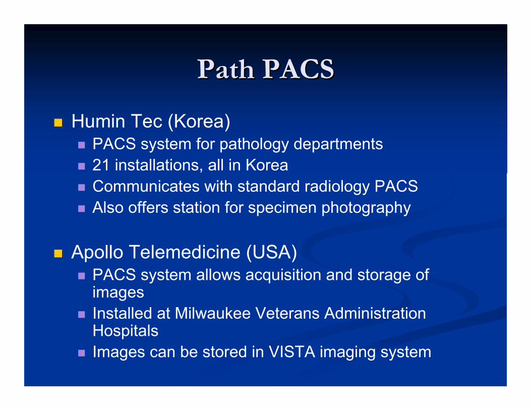
Path PACSPath PACS
� Humin Tec (Korea)� PACS system for pathology departments
� 21 installations, all in Korea
� Communicates with standard radiology PACS
� Also offers station for specimen photography
� Apollo Telemedicine (USA)� PACS system allows acquisition and storage of images
� Installed at Milwaukee Veterans Administration Hospitals
� Images can be stored in VISTA imaging system

Other Vendor ActivityOther Vendor Activity
� Visual-med� Working with VA in Georgia and Washington to allow acquisition and forwarding of pathology images to VISTA PACS
� Has stand alone product that can DICOMize digital images for forwarding to PACS
� Olympus� Working on a product to capture images and send to PACS
� Aurora Interactive� Grant from government of Canada
� Researching feasibility of using DICOM for whole slide imaging and JPEG 2000 formats
� Considering what forms of metadata are appropriate or feasible to include

Academic Center EffortsAcademic Center Efforts
� Univ. of Pittsburgh� AP LIS is image aware
� Gross specimen photos and single field microscopic
images saved
� Transmitted to Enterprise Image Archive
� Clinicians can see only selected images on
completed cases
� Main clinician interest is specimen photos
� Main pathologist use is conferences

European ExamplesEuropean Examples
� Otto von Guericke University in
Magdeburg, Germany
� Installed combined PACS and departmental
information system in pathology
� University of Trieste, Italy
� Has integrated pathology into their PACS
implementation

Anatomic Pathology WorkflowAnatomic Pathology Workflow
� Tissue sample examined grossly (+/- photos)
� Small portions are selected and chemically
processed (fixed) and embedded in paraffin
� Paraffin blocks are used to make microscope
slides
� Slides are stained using various chemicals
� Slides are examined microscopically by
pathologist (+/- photos)

Pathology Workflow

Current State Future State?
Diagnosis and ImagingDiagnosis and Imaging

Storage and RetrievalStorage and Retrieval
OR

Barriers to Adoption of Barriers to Adoption of
Current ProductsCurrent Products
� Turf� PACS systems have traditionally been the domain of Radiology
� Movement toward storing all medical images in a central locationwith a single viewing mechanism still in infancy
� Workflow� May need to manually annotate files with image description, accession number, etc.
� If sending to PACS, need to order study first
� Cost� Image acquisition and annotation takes time – no extra reimbursement currently
� Slide scanners and storage are costly

Imaging ComparisonImaging Comparison
� Radiology� digital acquisition
� automatic image capture
� clinician interpretable
� many patient requests
� large storage needs
� digital images save money
� large budgets
� strong standards for storage and transfer
� Pathology� analog primary data
� manual image capture
� hard to interpret for non-pathologists
� few patient requests
� extreme storage needs
� digital imaging costs more
� modest budgets
� limited pathology specificstandards
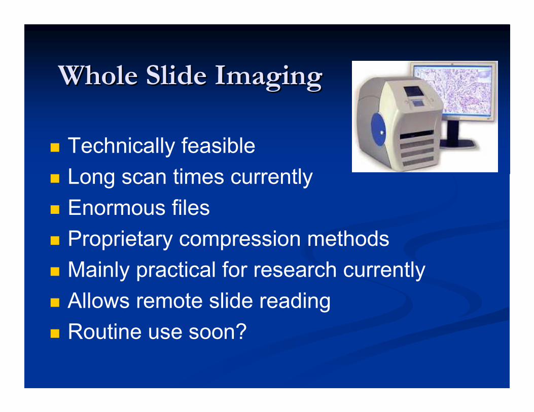
Whole Slide ImagingWhole Slide Imaging
� Technically feasible
� Long scan times currently
� Enormous files
� Proprietary compression methods
� Mainly practical for research currently
� Allows remote slide reading
� Routine use soon?

Whole Slide ImagingWhole Slide Imaging
� Multiple companies working on scanners
� Challenge is speed, file size, compression
� Current speed is around 5 minutes per
slide for conventional scanners
� Matrix scanners claim <1 minute per slide

Whole Slide ImagingWhole Slide Imaging
� Typical glass slide is 2.6 x 7.6 cm
� Tissue often occupies 1.9 x 2.75 cm
� Scanning at medium power 21,260 pixels/cm
40,394 x 58,465 = 2.4 billion pixels
x 24 bits color/pixel = 7 GB image file
� High power gives twice the resolution
7 GB x 2 x 2 = 28 GB
This is only in a single plane of focus!

Whole Slide ImagingWhole Slide Imaging
� Compression (lossy) may reduce file size
to 100 MB – 1 GB
� Assume cases have 5 slides on average
� Assume a volume of 30,000 cases per
year
0.5 GB x 5 x 30,000 =
75 Terabytes per year!
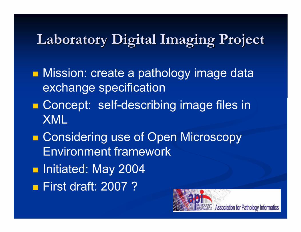
Laboratory Digital Imaging ProjectLaboratory Digital Imaging Project
� Mission: create a pathology image data
exchange specification
� Concept: self-describing image files in
XML
� Considering use of Open Microscopy
Environment framework
� Initiated: May 2004
� First draft: 2007 ?

LDIP GoalsLDIP Goals
� Allow anyone who uses pathology images to exchange images and accompanying annotations in a format that can be completely understood by anyone
� Allow vendors to write simple software that will port their proprietary images into or out of the data exchange standard
� Allow easy interchange to and from DICOM
� Allow the integration of metadata/data pairs with related data in other databases.

Metadata of ImagesMetadata of Images
� Specimen / patient demographics / prior history
� Accession / slide / block number
� Anatomic location
� Stain / antibody / procedure
� Magnification / capture equipment
� Pathology report / diagnosis
� Description of image or slide contents
� Research protocol information
� Other characterization of tissue (genotype etc.)

Commercial
Trestle
Apollo
dmetrix.com
Bioimagene
Aperio
Nikon
Olympus
Academic & Other
Ohio State Univ.
Harvard Univ.
Univ. of Michigan
Univ. of Florida
Henry Ford Hospital
Cleveland Clinic
Walter Reed Army Medical Center
National Institutes of Health
Armed Forces Institute of Path.
Assoc. Soc. Investigative Path.
LDIP ParticipantsLDIP Participants

RoadmapRoadmap
� DICOM
� WG-10 (strategic advisory) looking into pathology
� Submit updated work item proposal for pathology
� Build relationships with pathology vendors and LDIP
� LDIP
� Continue work on data specification
� Ensure DICOM compatibility
� Liaison with DICOM and others
� Other organizations?

Issues to Address in DICOMIssues to Address in DICOM
� Support for whole-slide microscopic images
� Support for navigating and selecting a region
of interest from within entire slide image
� Support for multi-resolution formats including
multiple pyramid voxel conventions
� Support for multispectral and hyperspectral
modality images
� Workflow model for pathology

Selected ReferencesSelected References
� Whole slide imaging
Aperio www.aperio.com
Bacus www.bacuslabs.com
Trestle www.trestlecorp.com
Dmetrix www.dmetrix.net
� Laboratory Digital Imaging Project
www.ldip.org
www.openmicroscopy.org

Selected ReferencesSelected References
� Pathology implementations
Magdeburg: Pathology Research and Practice,
Nov. 2002. 198:679-684
Trieste: Medicon 2001
http://www.tbs.ts.it/archives/medicon01-
belloni.pdf