AY 20 NEUROSCIENCE NEWSLETTER - uni-goettingen.de
32
MAY 2019 NEWSLETTER Georg-August-Universität Göttingen ∙ International Max Planck Research School NEUROSCIENCE Welcome to the 6 th Neuro-Newslet- ter published by the Göttingen Inter- national Master/PhD/MD-PhD Pro- gram and International Max Planck Research School (IMPRS) Neuro- sciences. While 2017 was the year to look ahead, to write reports and propos- als, and to prepare for the next period of a successful study program, 2018 simply threw us back and reminded us how close joy and sorrow can be to each other. While we still had the chance to raise our glasses together in a toast to the successful prolongation of the IMPRS Neurosciences, it was not even one year later that we had to say goodbye to our dear friend and colleague Prof. Dr. Michael Hörner. Michael had been the coordinator of the IMPRS Neurosciences since 2005 and program speaker of the GGNB PhD Program Molecular Physiology of the Brain since 2009. Additionally, he established the electrophysiology training lab at the ENI and was an ac- tive organizer of international cours- es, summer schools, and symposia in the field of neurosciences. We cher- ish the memories of his kind and gen- erous personality. One of Michael’s last activities in the program was the visit of the opening ceremony of NEURIZONS 2018 and The Neuroscience Program... Editorial ....................................................... 1 Towards novel drugs to treat anxiety ............ 3 Brain Illuminatio... ........................................ 5 Crack the Sell... ............................................ 8 Touching upon Piezo2 function... ................10 Master’s class 2017/18 .................................12 Master’s class 2018/19 .................................13 PhD projects started in 2017 and 2018 ........14 The Masters of 2017 and 2018.....................16 The Doctors of 2017 and 2018 ....................18 The circle of academic life ......................... 20 Studying Brain States in the States .............. 22 Beginning a new line of research................ 23 Creutzfeldt Award ...................................... 25 Joining the program since 2017 ...................26 Left the program since 2017 ........................26 Neurizons 2018 .......................................... 29 Surprise in Thessaloniki .............................. 30 Family reunion by the Kiez ..........................31 in Sorrow and Joy CONTENT NEURIZONS Symposium 2018 at the Max Planck Institute for Biophysical Chemistry
Transcript of AY 20 NEUROSCIENCE NEWSLETTER - uni-goettingen.de

MAY2019
NEWSLETTERGeorg-August-Universität Göttingen ∙ International Max Planck Research School
N E U R O S C I E N C E
Welcome to the 6th Neuro-Newslet-ter published by the Göttingen Inter-national Master/PhD/MD-PhD Pro-gram and International Max Planck Research School (IMPRS) Neuro-sciences.
While 2017 was the year to look ahead, to write reports and propos-als, and to prepare for the next period of a successful study program, 2018 simply threw us back and reminded us how close joy and sorrow can be to each other. While we still had the chance to raise our glasses together in a toast to the successful prolongation of the IMPRS Neurosciences, it was not even one year later that we had
to say goodbye to our dear friend and colleague Prof. Dr. Michael Hörner. Michael had been the coordinator of the IMPRS Neurosciences since 2005 and program speaker of the GGNB PhD Program Molecular Physiology of the Brain since 2009. Additionally, he established the electrophysiology training lab at the ENI and was an ac-tive organizer of international cours-es, summer schools, and symposia in the field of neurosciences. We cher-ish the memories of his kind and gen-erous personality.
One of Michael’s last activities in the program was the visit of the opening ceremony of NEURIZONS 2018 and
The Neuroscience Program...Editorial ....................................................... 1
Towards novel drugs to treat anxiety ............ 3
Brain Illuminatio... ........................................ 5
Crack the Sell... ............................................ 8
Touching upon Piezo2 function... ................10
Master’s class 2017/18 .................................12
Master’s class 2018/19 .................................13
PhD projects started in 2017 and 2018 ........14
The Masters of 2017 and 2018 .....................16
The Doctors of 2017 and 2018 ....................18
The circle of academic life ......................... 20
Studying Brain States in the States .............. 22
Beginning a new line of research ................ 23
Creutzfeldt Award ...................................... 25
Joining the program since 2017 ...................26
Left the program since 2017 ........................26
Neurizons 2018 .......................................... 29
Surprise in Thessaloniki .............................. 30
Family reunion by the Kiez ..........................31
in Sorrow and Joy
CONTENT
NEURIZONS Symposium 2018 at the Max Planck Institute for Biophysical Chemistry

N E U R O S C I E N C E2
Neurosciencei n G ö t t i n g e n …
the awarding of the Otto Creutzfeldt Ph.D. Prize 2018. The biennial NEU-RIZONS symposium organized by the PhD students of the Neuroscience Program again attracted national and international scientists and alumni to visit Göttingen. Likewise, the ELEC-TRAIN course in electrophysiology - organized and conducted by Michael and other enthusiastic “Electrainers” in the ENI teaching labs - was suc-cessful again, and it makes us happy that the course instructors decided to continue Michael’s legacy and offer the course also this year.
The renewal of the IMPRS is of utmost importance for the Neuroscience Program and its continuation. The IMPRS Neuroscience as well as our twin program of the IMPRS Molecu-lar Biology have continuously been successful in attracting high num-bers of applicants with an excellent academic quality from all around the world. Still: competition never rests and we will use our chances to fur-ther improve the program and select the best candidates to be embedded
in an excellent research environment and become a part of our Göttingen Neuroscience family.
The Neuroscience coordination team has been completed again after Dr. Jonas Barth joined us as new scien-tific coordinator in March this year. Being an alumnus of our program himself and coming from the posi-tion of scientific coordinator of the Georg-August University School of Science (GAUSS) which he helped to develop to its current state since late 2015, Jonas brings the best qualifica-tions for the job and is eager to fur-ther improve the program.
Work and fun should never be mu-tually exclusive, so we held our an-nual PhD retreat and enjoyed some wonderful and hot summer days in the air-conditioned seminar room of the Wälderhaus in Hamburg and en-joyed the city and harbor as balance for an intense scientific program (see article in this newsletter edition). We have started combining the retreats with career sessions at which alumni
of various professional backgrounds (academia and private sector) give short presentations about their indi-vidual career paths and their current positions. The talks are followed by round-table discussions in a speed-dating format. These events proved to be very successful to bring our PhD students in touch with former members of our program who have already made good progress in their profession.
This summer we will head north again and visit Schloss Etelsen and the city of Bremen.
Sandra DrubeAdministrative Coordinator
Jonas BarthScientific Coordinator
DISCLAIMER / IMPRINT
Publisher: Coordination Office of the international MSc/PhD/MD-PhD Neuroscience Program
Design and page layout: LifeTechMedia (Martin Nolte, Eva-Maria Twehues) and
Neuroscience Coordination Office (Jonas Barth, Sandra Drube)
The views expressed in this newsletter by the authors of articles are not necessarily those of the Neuroscience program. All reasonable measures
have been taken to ensure that the material contained in this newsletter is correct. However, the Neuroscience program gives no warranty and
accepts no responsibility for the accuracy or the completeness of the material.

3N E U R O S C I E N C E
Science Spotlight2 0 1 9
Most of us are familiar with anxiety that may strike before detrimental events. This feeling is normal as long as it is proportional to the event; anxiety experienced by a student the night before an important exam should be milder than this of a sol-dier before he enters into enemy’s territory. Activation of anxiety cir-cuitry is crucial to our survival be-cause it increases alertness during dangerous situations or makes us avoid them altogether. But while the behavior of the soldier who deserts the battlefield out of fear of death is normal, the student who avoids tak-ing exams because of fear of failure may suffer from pathological anxiety.
Luckily for the anxious student, there are available treatments. Ben-zodiazepines, the most widely used drugs for anxiety-related disorders, enhance inhibitory transmission in the brain network that produces avoidance behavior. In the heart of this network lies a group of intercon-nected nuclei known as the basal and centromedial amygdala. Amyg-dala constantly evaluates the envi-ronment via processing of sensory information. Inputs that are related to potentially dangerous situations, for example, a smell of a burning pan, will trigger the activation of the amygdala. Like a smoke detector, amygdala (particularly its centrome-dial part) then will alert the rest of the brain to activate defense mecha-nisms including freezing, escape or avoidance from entering the danger zone. Benzodiazepines silence the
by Olga Babaev
amygdala, which disproportional triggering by very mild or even non-existent danger-related inputs un-derlies anxiety disorders. The major downside of benzodiazepines is that they impede the neural activity not only of amygdala but of the whole brain – while the student will make it to the exam, he may find it difficult to concentrate on the questions.
Despite the side effects of benzodi-azepines, there are currently no bet-ter treatments available, mainly due to the poor understanding of the mo-lecular mechanisms underlying anxi-ety. The goal of my Ph.D. work with Dilja Krueger Burg and Nils Brose was to identify proteins that regulate anxiety-related neural activity, hop-ing to find pharmacological targets that will offer an alternative to ben-zodiazepines.
To be efficient, anxiety drugs should predominantly target proteins that acutely regulate both the neural transmission within the amygdala and the anxiety-related behavior. I focused on two synaptic proteins: Neuroligin 2 (Nlgn2) and IgSF9b. Unlike Nlgn2 that had an estab-lished role in anxiety in mice and humans, nothing was known about the function of IgSF9b. Nevertheless, we became interested in IgSF9b after an almost accidental discovery that deletion of IgSF9b has a dramatic ef-fect on anxiety.
Using an open field test that meas-ures the time mice spend in anxio-
genic center of the field, we showed that Nlgn2 KO mice are anxious because they avoid the center to a far greater extent than their WT lit-termates. However, this anxiety of Nlgn2 KO mice can be “fixed”, or rescued, once we additionally delete IgSF9b. These double KO mice dem-onstrated a completely normal level of anxiety, suggesting that blocking IgSF9b is a potential approach to treat anxiety.
Next, we asked whether IgSF9b regulates neural transmission in the amygdala. Both counting the number of activated neurons (using the expression of activation marker cFOS) in the amygdala and meas-uring amygdala activity in vivo in mice that explore anxiogenic envi-ronment demonstrates that deletion of IgSF9b suppresses the highly ac-tivated centromedial amygdala of Nlgn2 KO mice. This downregula-tion of neural activity happens due to enhanced inhibitory transmission and increased number of inhibitory synapses in IgSF9b KO mice. Most importantly, blocking IgSF9b expres-sion in the centromedial amygdala of adult Nlgn2 KO mice enhances their inhibitory synapses and relieves their anxiety almost as efficiently as con-genital KO of IgSF9b. Together, these findings demonstrate that deletion of IgSF9b, that has an acute anxiolytic effect in anxious mice because it puts breaks on the main output region of the amygdala, may be a potential therapeutic approach to anxiety.
Towards novel drugs to treat anxiety

N E U R O S C I E N C E4
Science Spotlight2 0 1 9
WT Nlgn2 KO IgSF9b KO Double KOWT
Nlgn2 KO
IgSF9b KO
Double KO
**
Tim
e in
cen
ter (
sec)
Nlgn2: p < 0.01IgSF9b: p < 0.001
050
100150200250300350
###
BACeM
LA
CeL
Amygdala anxietycircuitry
90 min
WT Nlgn2 KO IgSF9b KO Double KO
**
Num
ber o
f neu
rons
0
100
200
300
400Nlgn2 p < 0.01
#
***
*
Pow
er (µ
V2 /H
z x
104 )
2
6
0
##
40
30
20
10
0
40
30
20
10
0
40
30
20
10
0
40
30
20
10
00 2 4 6 8 0 2 4 6 8 0 2 6 8 0 2 4 6 8
WT Nlgn2 KO IgSF9b KO Double KO
Freq
uenc
y (H
z)
Time (sec) Time (sec) Time (sec) Time (sec)
11.5
9.5
7.5
5.5
Pow
er (µV
2/Hz x 10
4)
Freq
uenc
y (H
z)
Freq
uenc
y (H
z)
Freq
uenc
y (H
z)
4
Nlgn2: p < 0.01IgSF9b: p < 0.01
###
BACeM
1.0
*
20 msec
5 pA
WT
Nlgn2 KO
IgSF9b KO
Double KO20 pA
200 msec
0.8
0.6
0.4
0.2
0.020151050
Cum
ulat
ive
prob
abili
ty
Inter-event interval (sec)
Cum
ulat
ive
prob
abili
ty
Mean frequency (Hz)0 1 2
0.0
0.5
1.0
*** IgSF9b KO** Double KO
Freq
uenc
y (H
z)
IgSF9b: p < 0.05
0
1.25
2.50mIPSCs
mIPSC frequency mIPSC frequency
BA
CeM
Immunohisto-chemistry in CeM
in CeM
WT Nlgn2 KO IgSF9b KO Double KO
VIAAT
VIAAT
cFOS
**
Num
ber o
f pun
cta/
µm
0.0
0.5
1.0
1.5
2.0IgSF9b: p < 0.05
6 weeks24 h
Pre-injection OF Post-injection OFAAV injection Pre-injection Post-injection
**Ctrl IgSF Ctrl IgSF
WT Nlgn2 KO
0
150
300
450
IgSF9b p < 0.05
Tim
e (%
pre
)
Ctrl shRNA
IgSF9b shRNA
VIAAT in CeM
*
0
2
4
6
8
Ctrl IgSFCtrl IgSF
IgSF9b: p < 0.05
Nlgn2 KOWT
Num
ber o
f pun
cta/
µm
a
b
c
d
e
f g
5N E U R O S C I E N C E
Science Spotlight2 0 1 9
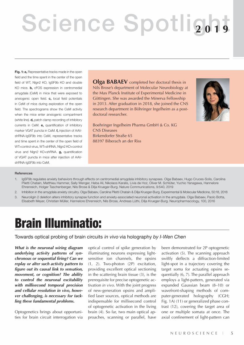
Fig. 1: a, Representative tracks made in the open
field and the time spent in the center of the open
field of WT, Nlgn2 KO, IgSF9b KO and double
KO mice. b, cFOS expression in centromedial
amygdala (CeM) in mice that were exposed to
anxiogenic open field. c, local field potentials
in CeM of mice during exploration of the open
field. The spectrograms show the CeM activity
when the mice enter anxiogenic compartment
(white line). d, patch clamp recording of inhibitory
currents in CeM. e, quantification of inhibitory
marker VGAT puncta in CeM. f, injection of AAV-
shRNA-IgSF9b into CeM, representative tracks
and time spent in the center of the open field of
WT+control virus, WT+shRNA, Nlgn2 KO+control
virus and Nlgn2 KO+shRNA. g, quantification
of VGAT puncta in mice after injection of AAV-
shRNA-IgSF9b into CeM.
What is the neuronal wiring diagram underlying activity patterns of syn-chronous or sequential firing? Can we replay or alter such activity pattern to figure out its causal link to sensation, movement, or cognition? The ability to control the neuronal excitability with millisecond temporal precision and cellular resolution in vivo, howe-ver challenging, is necessary for tack-ling these fundamental problems.
Optogenetics brings about opportuni-ties for brain circuit interrogation via
optical control of spike generation by illuminating neurons expressing light-sensitive ion channels, the opsins (1, 2). Two-photon (2P) excitation, providing excellent optical sectioning in the scattering brain tissue (3), is the prerequisite for precise optogenetic ac-tivation in vivo. With the joint progress of new-generation opsins and ampli-fied laser sources, optical methods are indispensable for millisecond control of optogenetic activation in the living brain (4). So far, two main optical ap-proaches, scanning or parallel, have
been demonstrated for 2P optogenetic activation (5). The scanning approach swiftly deflects a diffraction-limited light-spot in a trajectory covering the target soma for actuating opsins se-quentially (6, 7). The parallel approach employs a light-pattern, generated via expanded Gaussian beam (8–10) or wavefront-shaping methods of com-puter-generated holography (CGH; Fig. 1A) (11) or generalized phase con-trast (12), covering the target area of one or multiple somata at once. The axial confinement of light-pattern can
References
1. IgSF9b regulates anxiety behaviors through effects on centromedial amygdala inhibitory synapses. Olga Babaev, Hugo Cruces-Solis, Carolina Piletti Chatain, Matthieu Hammer, Sally Wenger, Heba Ali, Nikolaos Karalis, Livia de Hoz, Oliver M. Schlüter, Yuchio Yanagawa, Hannelore Ehrenreich, Holger Taschenberger, Nils Brose & Dilja Krueger-Burg. Nature Communications, 9:540, 2019
2. Inhibition in the amygdala anxiety circuitry. Olga Babaev, Carolina Piletti Chatain & Dilja Krueger-Burg. Experimental & Molecular Medicine, 50:18, 2018
3. Neuroligin 2 deletion alters inhibitory synapse function and anxiety-associated neuronal activation in the amygdala. Olga Babaev, Paolo Botta, Elisabeth Meyer, Christian Müller, Hannelore Ehrenreich, Nils Brose, Andreas Lüthi, Dilja Krueger-Burg. Neuropharmacology, 100, 2016
Olga BABAEV completed her doctoral thesis in Nils Brose’s department of Molecular Neurobiology at the Max Planck Institute of Experimental Medicine in Göttingen. She was awarded the Minerva Fellowship in 2013. After graduation in 2018, she joined the CNS research department in Böhringer Ingelheim as a post-doctoral researcher.
Boehringer Ingelheim Pharma GmbH & Co. KGCNS DiseasesBirkendorfer Straße 6588397 Biberach an der Riss
Brain Illuminatio: Towards optical probing of brain circuits in vivo via holography by I-Wen Chen

N E U R O S C I E N C E6
Science Spotlight2 0 1 9
be preserved into the brain by inte-grating temporal focusing (TF) (8–13). The advantage of parallel approach is simultaneous actuation of all opsins under illumination of the desired light-pattern (14), thus permitting fast and precise neuronal activation for opsins of a wider range of channel kinetics (15–17).
My post-doc project, being carried out in an interdisciplinary team led by Prof. Valentina Emiliani (Vision Institute,
Paris, France), aims at applying the in-novative wavefront-shaping methods for investigating the functional wiring in the mouse visual cortex in vivo. Till today, only few studies reported in vivo 2P optogenetic activation (7, 9, 10, 18, 19), without sufficient details of its temporal properties. We sought to ap-ply the parallel method of TF-CGH for in vivo spike generation for 3 opsins of different channel closing time constants: ReaChR of ~100 ms (20), CoChR of ~30 ms, and ChrimsonR of
~15 ms (21). Using 2P-guided patch recordings in the layer 2/3 neurons of anesthetized mice, we found the illu-mination conditions enabling action potential induction of <10 ms peak la-tency and <1 ms jitter for the 3 opsins (Fig. 1B). Spike trains can be precisely induced by repetitive illuminations at the target neuron (Fig. 1C). We dem-onstrated multi-cell activation in an all-optical manner by simultaneous holographic illumination onto multi-ple target cells while monitoring cal-
Figure 1: Holographic activation of one or multiple neurons in vivo. A) The wavefront-shaping method of CGH achieves intensity modulation
at the front focal plane (FFP) via phase modulation at the back focal plane (BFP) using spatial light modulator (SLM). B) Example traces of holography-
induced spike for ReaChR, CoChR, ChrimsonR and raster plot upon increasing illumination intensity. C) Holography-induced spike train at 20 Hz for the
3 opsins and raster plots upon different illumination frequencies. D) In the example field-of-field, 8 target cells are stimulated. Their calcium signal and
those from nearby non-target cells are presented at the right.

7N E U R O S C I E N C E
Science Spotlight2 0 1 9
References
1. Boyden ES, Zhang F, Bamberg E, Nagel G, Deisseroth K (2005) Millisecond-timescale, genetically targeted optical control of neural activity. Nat Neurosci 8(9):1263–1268.
2. Emiliani V, Cohen AE, Deisseroth K, Häusser M (2015) All-optical interrogation of neural circuits. J Neurosci 35(41):13917–13926.
3. Helmchen F, Denk W (2005) Deep tissue two-photon microscopy. Nat Methods 2(12):932–940.
4. Papagiakoumou E (2013) Optical developments for optogenetics. Biol Cell 105(10):443–464.
5. Oron D, Papagiakoumou E, Anselmi F, Emiliani V (2012) Two-photon optogenetics (Elsevier B.V.). 1st Ed. doi:10.1016/B978-0-444-59426-6.00007-0.
6. Rickgauer JP, Tank DW (2009) Two-photon excitation of channelrhodopsin-2 at saturation. Proc Natl Acad Sci U S A 106(35):15025–15030.
7. Packer AM, Russell LE, Dalgleish HWP, Häusser M (2015) Simultaneous all-optical manipulation and recording of neural circuit activity with cellular resolution in vivo. Nat Methods 12(2):140–146.
8. Andrasfalvy BK, Zemelman B V, Tang J, Vaziri A (2010) Two-photon single-cell optogenetic control of neuronal activity by sculpted light. Proc Natl Acad Sci U S A 107(26):11981–11986.
9. Rickgauer JP, Deisseroth K, Tank DW (2014) Simultaneous cellular-resolution optical perturbation and imaging of place cell firing fields. Nat Neurosci 17(12):1816–1824.
10. Pegard NC, et al. (2017) Three-dimensional scanless holographic optogenetics with temporal focusing (3D-SHOT). Nat Commun 8(1):1228.
11. Bègue A, et al. (2013) Two-photon excitation in scattering media by spatiotemporally shaped beams and their application in optogenetic stimulation. Biomed Opt Express 4(12):5391–5401.
12. Papagiakoumou E, et al. (2010) Scanless two-photon excitation of channelrhodopsin-2. Nat Methods 7(10):848–854.
13. Papagiakoumou E, et al. (2013) Functional patterned multiphoton excitation deep inside scattering tissue. Nat Photonics 7(4):274–278.
14. Ronzitti E, et al. (2017) Recent advances in patterned photostimulation for optogenetics. J Opt 19(11):113001.
15. Chaigneau E, et al. (2016) Two-photon holographic stimulation of ReaChR. Front Cell Neurosci 10:234.
16. Shemesh OA, et al. (2017) Temporally precise single-cell-resolution optogenetics. Nat Neurosci 20(12):1796–1806.
17. Ronzitti E, et al. (2017) Sub-millisecond optogenetic control of neuronal firing by two-photon holographic photoactivation of Chronos. J Neurosci 37(44):10679–10689.
18. Yang W, Carrillo-reid L, Bando Y, Peterka DS, Yuste R (2018) Simultaneous two-photon imaging and two-photon optogenetics of cortical circuits in three dimensions. Elife 7(e32671):1–21.
19. Mardinly AR, et al. (2018) Precise multimodal optical control of neural ensemble activity. Nat Neurosci 21(6):881–893.
20. Lin JY, Knutsen PM, Muller A, Kleinfeld D, Tsien RY (2013) ReaChR: a red-shifted variant of channelrhodopsin enables deep transcranial optogenetic excitation. Nat Neurosci 16(10):1499–1508.
21. Klapoetke NC, et al. (2014) Independent optical excitation of distinct neural populations. Nat Methods 11(3):338–346.
22. Chen TW, et al. (2013) Ultrasensitive fluorescent proteins for imaging neuronal activity. Nature 499(7458):295–300.
23. Chen I-W, et al. (2018) Parallel holographic illumination enables sub-millisecond two-photon optogenetic activation in mouse visual cortex in vivo. J Neurosci. 39 (18) 3484-3497.
24. Chen I-W, Papagiakoumou E, Emiliani V (2018) Towards circuit optogenetics. Curr Opin Neurobiol 50:179–189.
cium signal in a neuronal population expressing both opsins and GCaMP6 (22) in anesthetized and awake mice (Fig. 1D). These results are summarized in a research article in Journal of Neu-roscience (23). We are now working towards probing the relationship be-tween neuronal wiring and functional properties, e.g. visual selectivity. In sum, the light-shaping technology of holography provides unprecedent spa-tiotemporal resolution and precision for brain circuit manipulation (24).
I-Wen CHEN After finishing the Neuroscience Msc program in Goettingen in 2010, I-Wen CHEN did her doctoral thesis in Fritjof Helmchen’s laboratory at the Brain Research Institute, University of Zurich. After completing her dissertation, she then joined as a post-doc in Valentina Emiliani’s laboratory at Paris Descartes University/Vision Institute. She was awarded a Marie Sklodowska Curie Individual Fellowship in 2017.
Vision Institute (Prof. Valentina Emiliani)Department of PhotonicsWavefront-Engineering Microscopy Group17 Rue Moreau, 75012 Paris

N E U R O S C I E N C E8
Science Spotlight2 0 1 9
In order to maintain high-fidelity synaptic transmission, newly endo-cytosed synaptic vesicles (SVs) must accumulate large amounts of neu-rotransmitters quickly before subse-quent exocytosis. It is known that the major endocytic pathways at the pr-
esynaptic terminals involves forma-tion of a clathrin coat around the nas-cent vesicles. Whether newly formed clathrin-coated vesicles (CCVs) can be filled with neurotransmitters has remained unknown. The first and indispensable step in vesicle filling
is the formation of a proton electro-chemical gradient (∆µH+) across the vesicular membrane by the Vacuolar ATPases (V-ATPase). Thus, to answer the question whether CCVs can be filled with neurotransmitters, we iso-lated CCVs from mouse brain and
Crack the SellClathrin Chokes V-ATPase on Synaptic Vesicles by Zohreh Farsi

9N E U R O S C I E N C E
Science Spotlight2 0 1 9
measured the ∆µH+ across their mem-brane using a recently developed sin-gle-vesicle imaging approach (Farsi, et al. 2016).
Briefly, we measured ∆pH in CCVs isolated from brains of mice express-ing super-ecliptic pHluorin in the ve-sicular lumen (spH-CCVs), and per-formed ∆ψ measurements in CCVs, isolated from the wild-type mouse brains, after labeling with the poten-tiometric probe VF2.1.Cl. Upon ATP addition, we observed significant im-pairment of ∆µH+ formation in CCVs as compared to SVs. We detected key V-ATPase subunits in both CCVs and SVs by immunoblotting, indicating
Figure 1: Averaged fluorescence traces of
single spH-vesicles (A) and VF2.1.Cl-labeled
vesicles (B) over time in response to ATP.
C) Immunoblots of isolated SVs and CCVs
for different subunits of Vo and V1 of the
vATPase. D) ATPase activity measured in 1.3
µg of isolated SVs and CCVs. E) Electron
micrographs of negatively stained CCVs
before and after uncoating with Tris-buffer (pH
9.0). Luminal pH (F) and membrane potential
(G) of acidified SVs and CCVs before and
after treatment with Tris-buffer. H-I) Model
of V-ATPase block by clathrin ring: solved
structures of V-ATPase, clathrin coat and AP2
were used to show how V-ATPase fits within
the clathrin lattice. The plasma membrane
is depicted in light beige, clathrin triskelia
in brown, V-ATPase complex in gray (when
inactive) and light blue (when active). As
clathrin triskelia are recruited (possibly through
AP2), clathrin ring starts building around the
V-ATPase complex. Insertion of the last clathrin
triskelion would collide with the regulatory
H-subunit of V-ATPase (I), thus we hypothesize
that the displacement the regulatory H-subunit
blocks the V-ATPase activity.
that lack of acidification is not due to the absence of V-ATPase on the CCVs. Next, we measured the enzy-matic ATPase activity in both CCVs and SVs, and observed that CCVs show significantly less ATPase activ-ity compared to the same amount of SVs. One explanation for these results is that intact and functional V-ATPases are present on CCVs but are not able to pump protons most probably due to the clathrin coat. To test this hypothesis, we performed an in vitro uncoating assay by treating the CCVs with an alkaline Tris buffer (pH 9.0). Intriguingly, we observed that uncoated vesicles reached the same luminal pH and membrane potential
as SVs upon application of ATP. This demonstrates unequivocally that the V-ATPase activity is indeed inhibited in the presence of a fully assembled clathrin coat, and the V-ATPase re-gains its function once the coat is removed. Based on available struc-tural information, we believe that the steric hindrance provided by the formation of the clathrin coat around the vesicles results in dislocation of one of the key subunits of V-ATPase which in turn would block the activ-ity of the proton pump. In this mod-el, the clathrin coat may conserve ATP at the synapse and allows for V-ATPase function only when the vesicle is properly formed.
Zohreh FARSI Zohreh FARSI did her doctoral thesis in Reinhard Jahn`s department at the Max-Planck Institute for Biophysical Chemistry, Goettingen. After graduation in 2015, she then joined MDC-BIMSB as a postdoc in the group of Andrew Woehler in Berlin. She was awarded the Otto-Hahn Award from MPI and a postdoctoral fellowship from Peter und Traudl Engel-horn Foundation in 2017. She will join Morgan Sheng’s group in the Broad Institute, Cambridge, MA in September 2019.

N E U R O S C I E N C E10
Science Spotlight2 0 1 9
The sense of touch is often taken for granted, despite the fact that our our everyday life greatly depends on this sense. The ability to perceive our en-vironment to alert us of danger or to further social interactions, such as mother-child bonding are all es-sential to our survival. Our sense of touch relies on the conversion of me-chanical stimuli to electrical signals (this is known as mechanotransduc-tion), which then travel to the brain to be processed. This task is fulfilled by specific ion channels called Pie-zo2, which are activated when cells are exposed to pressure and other mechanical forces. These channels can be found in sensory nerves and specialized structures in the skin. These channels help detect physical
contact, roughness of surfaces and the position of our body parts.
From this screen we identified anoth-er protein namely, myotubularin re-lated protein-2, or Mtmr2 for short. In our work published in 2018 (Naray-anan et al., 2018), we have explored the function of mtmr2 in modulating piezo2.
To test if Mtmr2 played a role in mechanotransduction, we studied if changing the levels of mtmr2 in sensory neurons of mice grown in the laboratory, would affect Piezo2 mediated mechanotransduction. We found that when Mtmr2 levels were low, the activity of Piezo2 channels increased. However, when the pro-
tein levels were high, Piezo2 chan-nels were inhibited. These results suggest that Mtmr2 can control the activity of Piezo2. In order to further study the molecular basis of mtmr2 function, we explored the known roles of mtmr2. Mtmr2 is known to catalyse conversion of phosphatidyl inositols. It’s preferred substrate is phosphatidylinositol (3,5) diphos-phate [PI(3,5)P2].
An inhibition of Mtmr2 would be ex-pected to increase PI(3,5)P2. To test if this increase of PI(3,5)P2 lead to the potentiation of Piezo 2, as mentioned earlier, we used Apilimod to counter-act increase of PI(3,5)P2 upon Mtmr2 knockdown. Apilimod is a specific blocker of the PI(3,5)P2 synthesiz-ing enzyme PIKfyve (Vaccari et al., 2011). We found that it’s application indeed reverses the Piezo2 potentia-tion seen upon Mtmr2 knockdown. We also found additional evidence that PIP2 directly binds to piezo2 through biochemical bidding assays. Based on our study we proposed a potential mechanism of piezo2 mod-ulation by mtmr2 and pip2, as shown in figure 1.
Touching upon Piezo2 function Understanding the role of modulatory proteins and membrane lipids in the functioning of Piezo2 by Pratibha Narayanan
Figure 1: Working model: Local control of Piezo2 function by interdependent actions
of Mtmr2 and PI(3,5)P2. Mtmr2 controls the abundance of PI(3,5)P2 by dephosphorylation.
Mtmr2 and Piezo2 expression as well as PI(3,5)P2 might be compartmentalized in membrane
microdomains. Piezo2 localization in Mtmr2-negative microdomains would facilitate its access to
local PI(3,5)P2 and consequently potentiate Piezo2 RA-MA currents (left side). On the other hand,
high Mtmr2 levels and its localization in the proximity of Piezo2 would augment PI(3,5)P2 turnover,
thereby decreasing local PI(3,5)P2 availability and suppressing Piezo2 RA-MA currents (right side).
One could further speculate that Mtmr2, via binding Piezo2, might recruit Piezo2 to membrane
microdomains depleted of PI(3,5)P2. This would provide an active mechanism to inhibit Piezo2 RA-
MA currents in membrane compartments – may they be at the plasma membrane or intracellular
membranes.

11N E U R O S C I E N C E
Science Spotlight2 0 1 9
References
1. Coste, B., Mathur, J., Schmidt, M., Earley, T.J., Ranade, S., Petrus, M.J., Dubin, A.E., and Patapoutian, A. (2010). Piezo1 and Piezo2 are essential components of distinct mechanically-activated cation channels. Science 330, 55-60.
2. Coste, B., Xiao, B., Santos, J.S., Syeda, R., Grandl, J., Spencer, K.S., Kim, S.E., Schmidt, M., Mathur, J., Dubin, A.E., et al. (2012). Piezo proteins are pore-forming subunits of mechanically activated channels. Nature 483, 176-181.
3. Hu, J., Chiang, L.-Y., Koch, M., and Lewin, G.R. (2010). Evidence for a protein tether involved in somatic touch. The EMBO journal 29, 855-867.
4. Narayanan, P., Sondermann, J., Rouwette, T., Karaca, S., Urlaub, H., Mitkovski, M., GomezVarela, D., and Schmidt, M. (2016). Native Piezo2 interactomics identifies pericentrin as a novel regulator of Piezo2 in somatosensory neurons. Journal of Proteome Research 15, 2676-2687.
5. Narayanan P., Hütte M., Kudryasheva G., Taberner FJ., Lechner SG., Rehfeldt F., Gomez-Varela D., Schmidt M. (2018). Myotubularin related protein-2 and its phospholipid substrate PIP2 control Piezo2-mediated mechanotransduction in peripheral sensory neurons. Elife. 2018 Mar 9;7.
6. Poole, K., Herget, R., Lapatsina, L., Ngo, H.-D., and Lewin, G.R. (2014). Tuning Piezo ion channels to detect molecular-scale movements relevant for fine touch. Nature communications 5, 3520.
7. Qi, Y., Andolfi, L., Frattini, F., Mayer, F., Lazzarino, M., and Hu, J. (2015). Membrane stiffening by STOML3 facilitates mechanosensation in sensory neurons. Nature Communications 6, 8512.
8. Vaccari, I., Dina, G., Tronchere, H.l.n., Kaufman, E., Chicanne, G.t., Cerri, F., Wrabetz, L., Payrastre, B., Quattrini, A., Weisman, L.S., et al. (2011). Genetic interaction between MTMR2 and FIG4 phospholipid phosphatases involved in Charcot-Marie-Tooth neuropathies. PLoS Genetics 7.
9. Wetzel, C., Hu, J., Riethmacher, D., Benckendorff, A., Harder, L., Eilers, A., Moshourab, R., Kozlenkov, A., Labuz, D., Caspani, O., et al. (2007). A stomatin-domain protein essential for touch sensation in the mouse. Nature 445, 206-209.
This study is the first step towards identifying how the complex sense of touch is mediated by an orchestra of proteins and lipids. This information would be essential not only to under-stand the sense of touch but also the mechanism of Piezo2 when the skin has become injured or upon inflam-mation. Understanding the compo-nents of the machinery will then be imperative to finding potential targets and therapies for inflammation.
Pratibha NARAYANAN completed her PhD at the Max Plank Institute of Experimental Medicine, in the lab of Dr. Manuela Schmidt. During her PhD, she worked on the characterization of Piezo2, a protein crucial to the sense of touch in mammals. Currently she is in India, with a non-profit organization, Teach for India, which focuses on education for under-privileged children.
Fellow 2018-2020Teach For India,Mumbai

N E U R O S C I E N C E12
StudentsC u r r e n t
Master’s class 2017/18Irene Melati Aji Indonesia, BSc from Philipps University Marburg, Germany
Aishwarya Bhonsle India, MSc from University of Bristol, UK
Tony Carricarte Cuba, BSc from Uni-versity of Havana, Cuba
Daniela Doda Albania, BSc from Uni-versity of Genoa, Italy
Delane Espinueva Canada, BSc from University of British Columbia, Canada
Conor Heins USA, BSc from Swarthmore College, USA
Hendrik Heiser Germany, BSc from Georg August University Göttingen, Germany
Inés Hojas García-Plaza Spain, BSc from Universidad Autónoma de Ma-drid, SpainAnna Marie Müllen Germany, BSc from Georg August University Göttin-gen, Germany
Dmytro Nesterenko Ukraine, MSc from Taras Shevchenko University of Kyiv, Ukraine
Tarana Nigam India, BTech from VIT University Vellore, India
Melanie Nuesch Germano Uruguay, BSc from Universidad de la República, Uruguay
Adrián Palacios Muñoz Mexico, BSc from Universidad Autónoma de Nuevo León, Mexico
Sabine Rannio Germany, BSc from University of Bristol, UK
Marina Slashcheva Russian Federation, BSc from St. Petersburg State Univer-sity, Russian Federation
Jesse St. Amand USA, BSc from Arizo-na State University, USA
Yannan Su P.R. China, BSc from Nan-jing University, P.R. China
Mariia Zeziulia Ukraine, BSc from Taras Shevchenko University of Kyiv, Ukraine
Applications 2017
In the year 2017, the Neuroscience program received 420 applications from 72 countries.
Germany 23other Western Europe 32Eastern Europe 10North America 21Central/South America 25North Africa 29Central/South Africa 42Asia / Near East 77Central Asia / Far East 156Australia 1

13N E U R O S C I E N C E
StudentsC u r r e n t
Master’s class 2018/19Hebatallah Abdelrasol Egypt, BSc from Helwan University, Egpyt
Mohammed Abdelwahab Osman Mohammed Sudan, MBBS from Uni-versity of Khartoum, Sudan
Lukas Amann Germany, BSc from Georg August University Göttingen, Germany
Mathis Baßler Germany, BSc from University of Heidelberg, Germany
Jasmina Bier Germany, BSc from Jacobs University Bremen, Germany
Max Crayen Germany, BSc from Johannes Gutenberg University Mainz, Germany
Julia Dziubek Poland, BSc from Uni-versity of Wroclaw, Poland
Jonas Hemesath Germany, BSc from Georg August University Göttingen, Germany
Nare Karagulyan Armenia, BSc from Yerevan State University, Armenia
Priyanka Kislai India, MSc from St. Xa-vier’s College, Mumbai, India
Anna Liashenko Ukraine, BSc from Taras Shevchenko National University of Kyiv, Ukraine
Selene Lickfett Germany, BSc from Ulm University, Germany
Yifan Mayr P.R. China, BSc (double degree) from Johannes Kepler Univer-sity Linz, Austria / University of South Bohemia, Czech Republic
Tor Memhave Denmark, BSc from Uni-versity of Copenhagen, Denmark
Aditi Methi India, BS-MS (dual degree) from Indian Institute of Technology Madras, Chennai, India
Andrew Sasmita Indonesia, BSc from International Medical University, Malaysia
Patricia Schikorra Germany, BSc from University Duisburg-Essen, Germany
Ivan Skorodumov Russian Federation, Pharmacy Specialist from Lomonosov Moscow State University, Russian Federation
Applications 2018
In the year 2018, the Neuroscience program received 481 applications from 70 countries.
Germany 37other Western Europe 30Eastern Europe 9North America 21Central/South America 30North Africa 24Central/South Africa 64Asia / Near East 82Central Asia / Far East 183Australia 1

N E U R O S C I E N C E14
StudentsN e w
PhD projects started in 2017 and 2018
Heba Ali Role of novel Neuroli-gin2-interacting proteins in amygdala fear and anxiety circuits Nils Brose, Camin Dean, Hannelore Ehrenreich
Theocharis Alvanos Quantitative Molecu-lar Physiology of Active Zones at Calyceal Synaps-es of the Auditory Pathway Tobias Moser, Silvio Rizzoli, Erwin Neher
Tal Dankovich Imaging the brain in time: molecular mechanisms of long-lasting synaptic plasticity Silvio Rizzoli, Oliver Schlüter, André Fischer
Yunus Can Erol Encoding of Global Motion Signals in the Mammalian Retina Tim Gollisch, Stefan Treue, Jan Clemens
Burak Gür Synapse specific analysis of circuit functioning Marion Silies, Silvio Rizzoli, Jan Clemens
Dimokratis Karamanlis How nonlinear processing shapes natural stimulus encoding in the retina Tim Gollisch, Alexander Gail, Marion Silies
Madhura Ketkar Network dynamics of the Drosophila brain Marion Silies, Tim Gollisch, Viola Priesemann
Henry Klemp Establishing parameters for the characterization of neurometabolic and neu-rodegenerative dieseases using mass-spectroscopy based Metabolomics André Fischer, Klaus-Armin Nave, Jutta Gärtner
Ronja Markworth Role of protein-protein interactions and posttrans-lational modifications of presynaptic proteins in the regulation of calcium-triggered exocytosis Mathias Bähr, Silvio Rizzoli, Reinhard Jahn

15N E U R O S C I E N C E
StudentsN e w
Sebastian Molina Obando ON and OFF pathway interactions in the Dros-ophila visual system Marion Silies, André Fiala, Tim Gollisch
Helena Maria (Linda) Olsthoorn Ionic mechanisms of vesicular neurotransmitter transporters Reinhard Jahn, Tobias Moser, Ira Milosevic
Sonja Pribicevic Molecular mechanism of the kinetics of SNAREs Reinhard Jahn, Tobias Moser, Marina Rodnina
Jenifer Rachel Inhibitory feedback con-nectivity of supragranular VIP cells to layer IV target neurons: postsynaptic cell-type specificity and impact of presynaptic fir-ing patterns Jochen Staiger, Camin Dean, Oliver Schlüter
Alejandro Restrepo Arango Oligodendroglial commu-nication of axonal activity to the vasculature Klaus-Armin Nave, Susann Boretius, Nuno Raimundo
Nikoloz Sirmpilatze Influence of anesthesia on brain function, metabo-lism and blood perfusion – implications for preclini-cal neuroimaging Susann Boretius, Jochen Staiger, Hansjörg Scherberger
Elsa Steinfath Neural basis of acoustic communication in Drosophila Jan Clemens, Marion Silies, Viola Priesemann
Agnes Steixner Phenotype-based genetic association studies - from myelin to cognition Hannelore Ehrenreich, Susann Boretius, Walter Paulus
Chrystalleni Vassiliou The role of the TRPV1 channel and OLM in-terneurons in Sharp-wave ripples, place maps and spatial memory Camin Dean, Hansjörg Scherberger, Tobias Moser

N E U R O S C I E N C E16
StudentsG r a d u a t e d
The Masters of 2017 and 2018
Heba Ali (A. Fischer) Effect of Glia-mediated Immune Responses on the Neuronal Transcriptome
Theocharis Alvanos (T. Moser) The role of RIM-binding proteins at the Endbulb of Held syn-apse of the auditory pathway
Çağatay Aydın (M. Silies) Characterization of visual wide-field neurons in Drosophila
Allison Barry (M. Schmidt) Modulation of TRPV1 by Interaction Partners
Tizibt Bogale (K.-A. Nave) Oligodendrocytes as modifiers of disease progression in a mouse model of inherited Amyo-trophic Lateral Sclerosis (ALS)
Tal Dankovich (S. Rizzoli) Super Resolution Imaging of Synaptic Plasticity in Cultured Hippocampal Neurons
Yunus Can Erol (T. Gollisch) Effect of receptive field surround on luminance dependent ON-OFF response changes in mouse retinal ganglion cells
Elisabeth Fritsch (S. Rizzoli) Developing a Correlative Light and Electron Microscopy ap-proach for studying the role of neu-ronal membrane glycolipid protein nmgp-1 in synaptic pruning in C. elegans.
Burak Gür (M. Silies) Molecular mechanisms shaping functional properties of early visual processing
Alina Heukamp (T. Gollisch) Establishing a two-photon microscope setup to study the effects of acetylcholine on response proper-ties of distinct types of retinal ganglion cells
Nehal Johri (H. Scherberger) Effective connectiv-ity of the macaque grasping network: Granger causality analysis of resting-state fMRI
Dimokratis Karamanlis (T. Gollisch) Spatial integration in mouse retinal ganglion cells
Danai Katsere (M. Bähr) Chapter 1: TrkB-dependent re-routing of the CaSR into late endosomes - Chapter 2: Rab7 mutations potentially affect vesicle trafficking speeds and recruitment of EndophilinA2, a proposed component of the retromer complex
Henry Klemp (T. Friede) Metabolomics in rare neurological diseases
Ima Mansori (T. Bayer) Assessing the Preventive Potential of Cannabidiol on Alzheimer’s Disease Pathology
Ronja Markworth (C. Dean) The Effect of Co-activation on Endosomal Sorting of TrkB and CaSR
Sebastian Molina Obando (M. Silies) Molecular mechanisms that shape ON responses in the Drosophila visual system
Vasyl Mykytiuk (N. Brose) Regulation of mice reward-seeking behavior and locomotion by optogenetic stimulation of dopamin-ergic inputs from the ventral tegmental area to the lateral septum and the lateral hypothalamus
Helena Maria (Linda) Olsthoorn (R. Jahn) Reconstitution and Functional Characterization of the Vesicular GABA Transporter
Carolina Piletti Chatain (N. Brose) Region-specific Roles of Nlgn2 and lgSF9b in the Anxiety Circuitry

17N E U R O S C I E N C E
StudentsG r a d u a t e d
Sonja Pribicevic (H. Kawabe) The role of HECT-type E3 ligases in developing neurons
Juan Diego Prieto (T. Moser) Evaluation of the role of the CaV1.3 Ca2+-channel in the calcium current of rod bipolar cells of the mouse retina
Jenifer Rachel (J. Staiger) Optogenetic Characteriza-tion of Calretinin or CCK expressing VIP interneuron projections in primary somatosensory cortex of mouse
Alejandro Restrepo Arango (A. Flügel) Role of pericytes in regulating the blood-brain barrier in homeostasis in adult brain
Yasmine Shorafa (A. Antal) The Effects of Concurrent Finger Movements on Transcranial Alternating Current Stimulation (tACS)-Induced Aftereffects
Nikoloz Sirmpilatze (S. Boretius) The temporal stability of BOLD fMRI measurements in medetomidineanesthetized rats
Elsa Steinfath (M. Silies) Glial Ca2+ activity in the visual system of Drosophila melanogaster
Agnes Steixner (H. Ehrenreich) Phenotype-based genetic association studies on myelin-genes in neuropsychiatric diseases
Clara Tepohl (T. Gollisch) Chronic functional imaging of dendritic spines in the ferret primary visual cortex
Diana Toscano Tejeida (M. Wilke) Neural Variability Quenching and its Relation to Cognitive Performance in Young and Old Adults
Özge Uslu (A. Pooresmaeili) The Effect of Subliminal Reward Signals on Sensory-Motor Processing
Juan Felipe Vargas Fique (M. Silies) The role of GABAergic and glutamatergic fast inhibitory trans-mission in the Drosophila ON visual pathway
Chrystalleni Vassiliou (C. Dean) Investigating the Role of the TRPV1 channel and OLM interneurons in Sharp-Wave Ripples
Kwok Yui Reymond (Tony) Yip (N. Brose) Probing the Neural Circuits of Anxiety in Nlgn2 Knockout Mice using Whole-Brain Activity Mapping
Deniz Yüzak (M. Silies) Spatio-temporal receptive field properties of direction selective cells in Drosophila
Yu Zhao (A. Fischer) DNA hydroxymethylation changes accompany the formation and maintenance of memory
Lin Zhou (A. Gail) Training Recurrent Neural Networks for Reaching Tasks
N E U R O S C I E N C E18
StudentsG r a d u a t e d
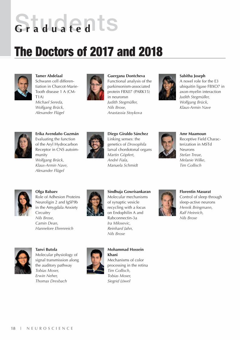
The Doctors of 2017 and 2018
Tamer Abdelaal Schwann cell differen-tiation in Charcot-Marie-Tooth disease 1 A (CM-T1A) Michael Sereda, Wolfgang Brück, Alexander Flügel
Erika Avendaño Guzmán Evaluating the function of the Aryl Hydrocarbon Receptor in CNS autoim-munity Wolfgang Brück, Klaus-Armin Nave, Alexander Flügel
Olga Babaev Role of Adhesion Proteins Neuroligin 2 and IgSF9b in the Amygdala Anxiety Circuitry Nils Brose, Camin Dean, Hannelore Ehrenreich
Tanvi Butola Molecular physiology of signal transmission along the auditory pathway Tobias Moser, Erwin Neher, Thomas Dresbach
Guergana Dontcheva Functional analysis of the parkinsonism-associated protein FBX07 (PARK15) in neuronsn Judith Stegmüller, Nils Brose, Anastassia Stoykova
Diego Giraldo Sánchez Linking senses: the genetics of Drosophila larval chordotonal organs Martin Göpfert, André Fiala, Manuela Schmidt
Sindhuja Gowrisankaran Molecular mechanisms of synaptic vesicle recycling with a focus on Endophilin A and Rabconnectin-3a Ira Milosevic, Reinhard Jahn, Nils Brose
Mohammad Hossein Khani Mechanisms of color processing in the retina Tim Gollisch,Tobias Moser, Siegrid Löwel
Sabitha Joseph A novel role for the E3 ubiquitin ligase FBXO7 in axon-myelin interaction Judith Stegmüller, Wolfgang Brück, Klaus-Armin Nave
Amr Maamoun Receptive Field Charac-terization in MSTd Neurons Stefan Treue, Melanie Wilke, Tim Gollisch
Florentin Masurat Control of sleep through sleep-active neurons Henrik Bringmann, Ralf Heinrich, Nils Brose

19N E U R O S C I E N C E
StudentsG r a d u a t e d
Sharlen Moore Corona The role of oligoden-drocytes in higher-order circuit functions Klaus-Armin Nave, Mikael Simons, Swen Hülsmann
Ramanathan Narayanan BAF155 regulates the genesis of basal progeni-tors through both Pax6-de-pendent and independent Jochen Staiger, André Fischer, Klaus-Armin Nave
Pratibha Narayanan Touching upon regulators of Piezo2 in mouse soma-tosensation Manuela Schmidt, Martin Göpfert, Luis Pardo
Ahmad Nazzal Visual and Auditory Perceptual Decision- Making in The Human Brain as Investigated by fMRI and Lesion Melanie Wilke, Mathias Bähr, Tobias Moser
Dennis Nestvogel Electrophysiological Analysis of the Synaptic Vesicle Priming Processt Nils Brose, Erwin Neher, Thomas Dresbach
Julio Santos Viotti The presynaptic protein Mover buffers synaptic plasticity at the hippo-campal mossy fiber synapse Thomas Dresbach, Tobias Moser, Michael Müller
Julia Sondermann Identification and char-acterization of protein complexes involved in different pain states in vertebrates Manuela Schmidt, Martin Göpfert, Henning Urlaub
Man Ho Wong Synapse refinement in mouse visual cortex during development Oliver Schlüter, Siegrid Löwel, Tobias Moser
King Faisal Yambire Lysosomal and mitochon-drial crosstalk: the role of lysosomal signaling on mitochondrial biogenesis and function Ira Milosevic, André Fischer, Klaus-Armin Nave

N E U R O S C I E N C E20
AlumniA c a d e m i c C a r e e r s
The circle of academic life
Göttingen was my home for seven ye-ars, 2011-2018. I joined the IMPRS-Neuroscience program as a Master’s student and stayed on to pursue doc-toral research in Prof. Tobias Moser’s lab. Six months ago, I started a new phase in my academic career as a postdoctoral research fellow in Dr. Jayeeta Basu’s lab at the New York University Medical Center (NYUMC) in New York City.
Dr. Basu’s lab focuses on synapses, circuits, and learning within the hippocampus-entorhinal cortex net-work. Much of the lab focuses on how the sensory input from entorhi-nal cortex influences memory and spatial navigation (place cell) func-tion in the hippocampus.
I have a slightly different approach. I look at how the hippocampus re-ciprocally feeds back into the ento-rhinal cortex to shape cortical sen-sory processing. My work asks how ‘real-time’ sensory information is processed in the context of long-term memories of past experiences. The aim is to decipher the circuit prin-ciples that allow for quick adaptation of behaviour in response to changing environmental demands.
Another project that I have taken up here is in collaboration with the Comprehensive Epilepsy Center at the NYUMC. I have access to re-sected human hippocampal tissue from patients with refractory medial temporal lobe epilepsy. I record from acute human hippocampal slices as a means to validate the applicability of the circuit dynamics derived from rodent experiments in humans. Here, my aim is to establish the connecti-
vity, synaptic strength, dendritic inte-gration, and plasticity rules governing the human hippocampus-entorhinal cortex connection.
My PhD in Göttingen was a perfect starting point to delve into the field of circuit function. During my PhD I worked on synaptic transmission. I used giant synapses of the auditory system, like the calyx and endbulb of Held, as model systems to study the molecular players involved in signal
transfer between neurons. I investi-gated how a single synapse functions and how the pre-synapse is coupled to the post-synapse to bring about time-locked reliable signal transmis-sion. My focus was a single synapse.
In my first year of PhD, I attended the Gordon Research Conference for Synaptic Transmission in the US. It is a close-knit conference attended by, at most 200 people dedicated to the research topic. You spend a week in
a far-off retreat spot, away from civi-lization for an absolute immersion in science. In the span of a week you meet all the giants in the field in a casual setting, free to approach anyo-ne with your questions. You can talk about the latest developments in ve-sicle retrieval or neurotransmitter re-lease while choosing the ingredients for your omelette.
This is where I met Dr. Jayeeta Basu in 2014. She was then a post-doc in
Dr. Steven Siegelbaum’s lab at Co-lumbia University. She presented her research on the newly discovered long-range inhibitory input from the entorhinal cortex into the hippocam-pus, and how it modulates coding and learning behaviour. Her talk and her research fascinated me. I spoke to her after her talk and at my poster. Her meticulous dissection of the ba-lance of individual synaptic excitati-on and inhibition dictating the output of a circuit gave me a sense of future
Lab brunch and Christmas gift exchange 2018
by Tanvi Butola

21N E U R O S C I E N C E
AlumniA c a d e m i c C a r e e r s
direction to build upon my current initiation into the field of synaptic transmission.
Two years later in 2016, I was wrap-ping up my doctoral thesis. The same year Dr. Basu published her work, some of which she had already pre-sent at the 2014 conference. Her study again ignited in me several questions and ideas. What does the micro-circuit look like that is mani-pulated by long-range inhibition? How does the timing of signal trans-mission between synapses influence the dynamics of circuit function? How would a synaptic discrepancy (impairment of neurotransmitter re-lease or Ca2+ channel or vesicle recy-cling) affect the local and hetero-sy-
naptic plasticity, circuit output, and animal’s learning behaviour? I was thinking of ways to extend my know-ledge of a single synapse function to a collection of synapses: a neuronal circuit.
With this in mind, I reached out to Dr. Basu with my questions and ideas. She had just started her group at the Neuroscience Institute in the NYUMC. Several email exchanges later we arranged to meet at SFN in November 2016. We had a very sti-mulating discussion about the open questions in her lab and about my ideas. This was followed up several Skype meetings, more emails, inter-views with other labs, intense discus-sions with my doctoral mentors, all culminating in a big dilemma about which direction to choose.
The questions researched by the Basu lab were close to my research in-terests. In the end, I decided to join
Dr. Basu’s lab to be a part of a dyna-mic new lab. I wanted to be a part of the initial phase of the development of the lab: establishing a lab, procu-ring equipment and start-up grants, attracting and getting good scientists to join a new lab, forging collabo-rations, balancing bench work, and administrative responsibilities. One day, I hope to have my own lab and apply the hands-on training I receive with Dr. Basu as my mentor.
Contrary to what it seems, my jour-ney from the quaint and peaceful city of science, Göttingen, to the bustling metropolis of the new world, New York, is the epitome of ‘life coming a full circle’. Dr. Basu is an alumnus of the program and she worked in the same department and even the same building (3rd floor, Tower 6 MPI-BPC, Faßberg) where I spent hours at my setup. From within this circle of aca-demic life, I wish to go ahead and have my own research identity.
My Christmas gift from Jayeeta. (A, in hand)
The Ultimate Experiment- a coupon for three
2-hour dendritic patch sessions. (B, on the
face) Happy me.
Tanvi BUTOLA did her doctoral work in Tobias Moser’s laboratory ‘Synaptic Nanophysiology’ at the Max Planck Institute for Biophysical Chemistry Göttingen. After graduating in 2017, she joined New York University School of Medicine as a postdoctoral researcher with Jayeeta Basu. She currently works with learning and memory in the context of sensory processing.

N E U R O S C I E N C E22
AlumniA c a d e m i c C a r e e r s
Studying Brain States in the States:
A fresh and delicious smell of fir trees, green grass and mossy shrub-bery; people are incredibly friendly, patient, tolerant and have an amaz-ing sense of humor; in the east, there are mountains covered in snow and in the west there is the beautiful Pa-cific Ocean – all of this is Oregon. Much of it is reminiscent of J.R.R. Tolkien’s description of the shire in the book The Lord of the Rings. My family and I have moved here for my Postdoc more than a year ago, exact-ly on my 30th birthday – and we are loving it!
My host lab moved to Oregon at the exact time as we did, and my col-
leagues and I have mainly focused on building a new lab during the first year of my Postdoc. Building a new lab can be challenging; particularly when setting up new and complex equipment. However, it also forges strong bonds between the members of a lab and it offers a unique oppor-tunity to know all of the equipment by heart.
While living in Goettingen as a PhD student, I carried out research in the field of Molecular Neurobiology. Back then, I was specifically interest-ed in understanding how presynaptic proteins work together in establishing the high speed and fidelity of n e u r o t r a n s -mission in the central nervous system. The knowledge and technical ex-pertise I gained during my PhD have laid a solid foundation, on which I am now building as a postdoctoral research scholar. For my postdoc in Oregon, I am now study-ing the brain on a systems level, rath-er than on a molecular level. Chang-ing fields never is easy and it requires much learning. However, I feel that it has already paid off in my case. Apart from the deep love and apprecia-tion that I have developed for study-ing questions directly related to the “neural” code and to neural circuits driving behavior, I have been able to greatly broaden my technical exper-tise. I really enjoy the spirit and the atmosphere in the fields of systems neuroscience. Similar to scientists in the fields of astro- and particle phys-ics, most labs in systems neurosci-ence tend to be extremely collabora-tive and do not shy away from sharing their data and their reagents with oth-ers before publication. This kind of behavior is particularly evident in the habit of most scientists in the field to publish their work on preprint servers and by sharing their mouse lines and
tools freely with others without much hesitation. The positive spirit is also particularly evident by the work of the Allen Institute for Brain Research in Seattle, with which my host lab frequently exchanges various kinds
of information and which is located within driving-distance.
In my current postdoc project, I study the impact of the behavioral state of animals on neural informa-tion processing in the brain. Process-ing of sensory information within neural networks has been shown to be highly dependent on behavioral state. This is particularly relevant for students in an early morning lecture who are striving to listen but find themselves drifting through periods of low- and high-arousal, taking in only a fraction of the words spoken by the lecturer. To study the influence of arousal and behavioral state on sensory processing, I employ in-vivo electrophysiology (intra- and extra-cellular recordings) in combination with behavioral analysis in awake mice. In correspondence with what has been described in humans, the relationship between the level of arousal and task performance follows
My Life as Postdoc in Oregon by Dennis Nestvogel

23N E U R O S C I E N C E
AlumniA c a d e m i c C a r e e r s
an inverted u-shape in mice, with the best performance in sensory tasks occurring during intermediate states of arousal. In my host lab, we are specifically interested in the neural mechanisms that enable “optimal” task performance and how these may be disturbed in patients suffering of psychiatric disorders.
This is a very exciting time to be con-ducting research in the neurosciences. The development of many new tools, such as optogenetics, improved calci-um sensors, CRISPR and high-density electrophysiology recordings are ena-bling us now to conduct experiments, which other scientists only dreamed of several years ago. There are sev-
eral long-standing questions, which can now be addressed with these new tools and I am very happy to be a sci-entist at this exciting time. I am look-
ing forward to spending more time in Oregon together with my family, be-fore we eventually move back to live closer to our parents and siblings.
Dennis NESTVOGEL carried out his PhD work under the supervision of JeongSeop Rhee and Nils Brose at the Max-Planck Institute of Experimental Medicine. After his graduation in 2017, he joined the research group of David McCormick at the University of Oregon. His postdoctoral project deals with state-dependent sensory processing in the visual system of mice.
Beginning a new line of research ...at one of World`s top University by Ramanathan Narayanan
Almost a decade ago, during my Bachelor studies when I was award-ed the Indian Academy of Sciences Summer Fellowship, little did I know that these two months of my life are going to shape the many years to fol-low. My first exposure to a research environment not only enhanced my passion for Science but also attract-ed me to the exciting field of Neu-roscience. After my Bachelor`s de-gree, I worked at the National Brain Research Centre, India as a project fellow during which I received the great opportunity to join the IMPRS Neuroscience program. Coming from
Chennai, one of the most populous and bustling cities of India, taking a stroll through the quiet and charming streets of Goettingen seemed like a different world in all sense. With nu-merous academic and research insti-tutions dotting its map and nurturing excellent scientists for centuries, the city rightfully earned its name `Stadt die Wissenschaft`.
After rigorous coursework, I started my doctoral studies at the UMG Cen-tre for Anatomy in the department of Prof. Jochen Staiger to work on tran-scriptional and chromatin remodel-
ling mechanisms underlying cortical development. As time progressed, on the one hand learning new tech-niques and getting positive results excited me, while on the other a glimpse of the pitfalls that surround ` Science & Research ` prompted me to re-analyse my passion for academ-ic research. Besides many factors, I believe that it is this threshold point that tests a PhD student either to continue in academic research or to choose alternative career paths. But I didn’t give up…the freedom and joy exploratory research offers made me choose an academic career once

N E U R O S C I E N C E24
AlumniA c a d e m i c C a r e e r s
again…but the next obvious ques-tion, where? Choosing a postdoc pro-ject is not that easier than I thought. Finding the necessary funds and a project that interests you the most is an obvious thing to go for but should not be the sole reason to choose a particular position. It requires a care-ful balance of three aspects: a project that needs some (if not all) of your prior expertise, an environment that enables you to learn new concepts/techniques and a mentor who is will-ing and committed to promoting you professionally. Through my experi-ence and that of my close contacts, it is unfortunate that these aspects are often poorly addressed.
Having worked in neurodevelop-ment, I wanted to expand my horizon
into neurodevelopmental disorders during my postdoctoral phase. For-tunately, I received an attractive of-fer from the Swiss Federal Institute of Technology, ETH Zurich matching my expectations and promising in terms of future opportunities. Before relo-cating to Zurich, I was at least certain that I will get more sunshine (enough of the dull weather!) and will be at ease getting used to the city since it’s in the German-speaking part of Swit-zerland (though I am still not perfect in German!). However, after a few weeks, it turned out that not only I love the city but my new workplace too. ETH Zurich is constantly ranked one of the best Universities world-wide and together with the Univer-sity of Zurich, has a strong Neuro-science community. I joined as a
postdoc in the group of Prof. Gerhard Schratt, one of the pioneers in the field of micro-RNA and synapse re-search. With very helpful team mem-bers and excellent infrastructure, I had a head start into my project that investigates the role of a mammalian micro-RNA cluster in social behav-iour and autism spectrum disorder. As a significant recognition, I was awarded the ETH Zurich Postdoc-toral Fellowship co-funded by Marie Sklodowska Curie Actions COFUND Program, including an additional budget for research and travel costs. Both ETH Zurich and the Swiss Na-tional Science Foundation offers sev-eral opportunities to obtain funding and to attain independence, there-by providing an ambient environ- ment for early-career researchers to develop professionally. I hope the warm welcome “Grüezi” in Zurich will continue, giving the right bal-ance and the impetus to continue in academic research.
Ramanathan NARAYANAN did his doctoral thesis in Jochen Staiger`s department at the Institute of Neuroanatomy, University Medical Centre Goettingen. After graduation in 2017, he then joined ETH Zurich as a postdoc in the group of Gerhard Schratt. He was awarded a fellowship under ETH Zurich – Marie Sklo-dowska Curie Actions COFUND Program in 2018.
Institute for Neuroscience (Prof. Gerhard Schratt)Department of Health Science & Technology (D-HEST)Swiss Federal Institute of Technology, ETH ZurichWinterthurerstrasse 1908050 Zurich

25N E U R O S C I E N C E
Honors &A w a r d s
Creutzfeldt Award
Creutzfeldt PhD Prize The Creutzfeldt PhD Prize is awarded for the best PhD thesis in memoriam of Prof. Dr. Otto Detlev Creutzfeldt, founding director of the department of Neurobiology at the Max Planck Institute for Biophysical Chemistry in Göttingen. The prize is awarded since 2007 to PhD graduates of the Neuro-science program based on excellent achievements during the PhD and the grading of the written dissertation and the oral defense. In 2011 for the first time 2 winners have been selected for the Creutzfeldt Prize.
Traditionally, the award ceremony is part of the official opening of the NEU-RIZONS Symposium and takes place in the presence of the spokespersons of the MSc/PhD/MD-PhD Program & International Max Planck Research School for Neurosciences, a represent-ative of Sartorius stedim AG and Mary Creutzfeldt.
The award includes the book pre-sent ‘Cortex Cerebri’ written by Otto Creutzfeldt and a gift of 500,-€ spon-sored by the Göttingen company Sar-torius stedim biotech AG, which has generously supported the Neurosci-ence program since its foundation
2007 Prize winner: Dr. Irina DudanovaMax Planck Institute of Neurobiology Martinsried
2009 Prize winner: Dr. Henry Lütcke Brain Research Institute Zurich, Switzerland
2011 Prize winners: Dr. Ioanna BethaniGoethe-Universität Frankfurt
Dr. Stephan JunekMax Planck Institute for Brain Research Frankfurt
2013 Prize winners: Sadim Jawhar, Ph.D. Biomedical Research Institute, Doha, Qatar
Dr. David OwaldOxford University, United Kingdom
2015 Prize winners: Dr. Natalia Revelo Nuncira Radboud umc, Institute for Molecular Life Sciences Nijmegen, Netherlands
Nicolas Snaidero, Ph.D. Technical University / Ludwig-Maximilians University München
2018 Prize winners: Dr. Pratibha Narayanan Teach for India, New Delhi, India
Dr. Dennis NestvogelUniversity of Oregon, Eugene, USA

N E U R O S C I E N C E26
FacultyN e w / L e a v i n g
Joining the program since 2017
Jan Clemens How acoustic commu-nication signals are pro-cessed to inform behavior is the major interest of Dr. Clemens’ group ‘Neu-
ral Computation and Behavior’ at the European Neuroscience Institute. He teaches the development of the insect nervous system and neural modelling to our students and currently supervis-es his first Neuroscience Master’s and PhD students.Further information: http://www.uni-goettingen.de/en/601849.html
Igor Kagan Well acquainted with the Göttingen research envi-ronment, Dr. Kagan now also joined our Neuro-science program. He has
been a group leader at the German Pri-mate Center since 2011 and has been active as a lab rotation supervisor and tutor for quite a while. His major re-search interests encompass neurophys-iology, functional imaging of decision-making, and cognitive and visuomotor functions in primates. Further information: http://www.uni-goettingen.de/en/365803.html
Caspar Schwiedrzik graduated in 2011 and worked as a postdoc at the Rockefeller University in New York. Since 2017, he has headed the Neu-
ral Circuits and Cognition Lab at the European Neuroscience Institute. His work involves invasive and noninvasive approaches to study different forms of learning in the visual system.Further information: http://www.uni-goettingen.de/en/589682.html
Theo Geisel was director and scientific member of the Max Planck Institute for Dynamics and Self-Organization and Pro-fessor at the University of
Göttingen from 1996 until 2016. A world leader in theoretical neurosci-ence and one of the “pioneers” of the chaos theory, he joined the Neurosci-ence Program in 2004 and remained a member until his retirement. Theo Geisel was awarded numerous prizes, including the Gottfried-Wilhelm-Leib-niz-Prize and the Gentner-Kastler-Prize of the Deutsche Physikalische Gesells-chaft and the Société Francaise de Phy-sique, and was our program’s key link to the physics faculty
Hiroshi Kawabe taught our students in bio-chemical techniques and supervised them during lab rotation projects from 2014 until 2017. Shortly
after becoming a member of our pro-gram, Dr. Kawabe took the opportunity to further pursue his research at the University of Kobe in Japan, where he continues his research program on the role of protein ubiquitination in nerve cell development.
Till Marquardt was a group leader at the European Neuroscience Institute (ENI-G) from 2007 until 2016 where he stud-ied key aspects of nerv-
ous system development and function. Dr. Marquardt was an Emmy Noether Young Investigator (DFG) and a Europe-an Research Council (ERC) grant holder. In 2016, he was appointed as Professor for Neurobiological Research at the RWTH Aachen.
Left the program since 2017

27N E U R O S C I E N C E
FacultyL e a v i n g
Judith Stegmüller graduated from the Univer-sity of Heidelberg and did a postdoc at Harvard Med-ical School, Boston, USA. Dr. Stegmüller became an
independent group leader at the Max Planck Institute of Experimental Medi-cine in Göttingen in 2008 where she pursued a research program on the role of the ubiquitin proteasome system (UPS) in axon growth and regeneration. In 2016, she accepted a position at the Neurological Clinic, University Clinics Aachen, where she continues her work on the UPS and on genes involved in degenerative diseases.Further information: http://www.neu-roscience-aachen.de/arbeitsgruppe-stegmueller.html
Mikael Simons came to Göttingen as a jun-ior group leader at the Cen-tre for Biochemistry and Molecular Cell Biology in 2004. In 2008, he became
a group leader (Cellular Neuroscience Lab) at the Max Planck Institute of Ex-perimental Medicine, with which he is still associated. In the same year, Prof. Simons became a faculty member of our program, where he supervised sev-eral MSc and PhD theses and took care of a number of lab rotation students. His current research at the Technical Uni-versity Munich – where he joined the German Center for Neurodegenerative Diseases (DZNE) in 2017 – focuses on myelin biogenesis, regeneration in the CNS, and neurodegeneration.Further information: http://www.neuro-science.med.tum.de/index.php?id=25
Current Faculty Members
Andrea Antal
Matthias Bähr
Thomas Bayer
Susann Boretius
Henrik Bringmann
Nils Brose
Wolfgang Brück
Jan Clemens
Camin Dean
Peter Dechent
Thomas Dresbach
Hannelore Ehrenreich
Gregor Eichele
André Fiala
André Fischer
Alexander Flügel
Jens Frahm
Tim Friede
Alexander Gail
Tim Gollisch
Martin Göpfert
Robert Gütig
Ralf Heinrich
Stefan Hell
Swen Hülsmann
Reinhard Jahn
Igor Kagan
Siegrid Löwel
Ivan Manzini
Ira Milosevic
Tobias Moser
Klaus-Armin Nave
Tiago Outeiro
Luis Pardo
Walter Paulus
Arezoo Pooresmaeili
Jeong Seop Rhee
Michael Rickmann
Silvio Rizzoli
Annekathrin Schacht
Hansjörg Scherberger
Oliver Schlüter
Manuela Schmidt
Caspar Schwiedrzik
Michael Sereda
Marion Silies
Jochen Staiger
Anastassia Stoykova
Stefan Treue
Melanie Wilke
Sonja Wojcik
Fred Wolf
Fred Wouters
For details regarding the research of all faculty members, please see www.gpneuro.uni-goettingen.de/content/c_faculty.php

N E U R O S C I E N C E28
FacultyL e a v i n g
Michael Hörner ObituaryProfessor Dr. Michael Hörner, our program co-ordinator and member of our faculty, lost his fight against a malicious dis-
ease after long suffering in October 2018. This is painful for all of us, but of course in the first place for his fam-ily, especially his beloved son. Our thoughts are with them.
Since 2005, Michael had coordinated the IMPRS Neurosciences programme with great enthusiasm and success. In 2009, Michael also became the speak-er of the PhD programme Molecular Physiology of the Brain. At the Europe-an Neuroscience Institute Göttingen, Michael ran a teaching lab where he trained students in electrophysiology, including the famous intracellular re-cordings from leech Retzius cells. Mi-chael was highly engaged in teaching and lecturing neuroscience, covering a wide spectrum of neuroscientific top-ics, such as signalling in electric fish, acoustic communication, or immuno-histochemistry.
Michael completed his dissertation in 1989 in the Department of Cellular Bi-ology at Georg August University un-der the supervision of Prof. Friedrich-Wilhelm Schürmann. In those early
days, Michael closely worked together with Klaus Schildberger (now professor at Leipzig University), and the two pro-duced a remarkable paper document-ing the effects of intracellular neuronal manipulations on cricket phonotactic behaviour. The respective setup had been developed in cooperation with their colleague Heribert Gras, with whom Michael had remained in close contact ever since. This was Michael as we all know him – highly cooperative, innovative, staying in contact, and hav-ing a true interest in the people around him and in what (and how) they are doing. These networking skills, to-gether with his joy for teaching and re-search, also brought him a position as guest professor at the Hong Kong Uni-versity of Science and Technology and manager of the DAAD German Centre in Hong Kong from 2002 to 2004. Be-fore, between his dissertation (in 1989) and habilitation (in 1997), Michael had worked as an assistant professor at the Institute of Zoology at the Georg August University and, as a guest re-searcher, in Tucson, Woods Hole, and Boston, where he stayed from 1994 to 1995 as a Feodor-Lynen/Humboldt Fel-low. Afterwards, he became an Associ-ate Professor, again in Göttingen, be-fore he moved to Hong Kong in 2002.Through his diverse activities, Michael was well known in the neuroscience
community, and we are very lucky to have had him here in Göttingen, run-ning the IMPRs Neuroscience program and turning it into a great success. As we all know, Michael was remarkably talented in student mentoring, and together with his team, he organized memorable student retreats. He also had a particular love for long hikes and bird watching, walking with his binoc-ulars, across Spiekeroog for instance, when time allowed during retreats.
Nearly everybody in the neurosci-ence community in Göttingen knows Michael Hörner, and he was one of the prime experts on MSc and PhD programs and research school organi-zation. Many of our students profited tremendously from his compassionate individual guidance and his enthusi-asm for teaching and research. This deep involvement of Michael would not have been possible without the strong support from his family, and we are very grateful for this. Honestly, it will be virtually impossible to fill the gap that Michael’s death has torn into our community. Still, and not unex-pectedly, he prepared things very well, so that the quality of our program can and will be maintained. But for our program, Michael was so influential that any future development will carry his stamp.
Andreas Stumpner, Martin Göpfert, Nils Brose

29N E U R O S C I E N C E
CampusE v e n t s
Neurizons 2018
In May 2018 scientists from all over the world came together in the Max-Planck institute for Biophysical Che-mistry full of anticipation. They were there for Neurizons, the biennial con-ference organized by the students of the MSc/PhD/MD-PhD Program and International Max Planck Research School (IMPRS) for Neurosciences. But actually, it all started many months before. While the then Master studen-ts were busy finishing their theses, the
first gathering had commenced to start the organization. Helpful advice and experiences from previous organizers was passed on and then the new orga-nizers were ready for the immense task ahead of them: organizing a four-day neuroscience conference.
Fast forward twelve months and you would have found yourself in the middle of a buzzing conference where Nicholas Humphrey provided a more philosophical keynote talk with a whole new perspective. A range of talks were available from molecular neuroscience to higher brain func-tions. You could meet Amit Agarwal, an alumnus from our program in the middle of the process of starting his own group in Heidelberg, or have an inspiring conversation with Christo-pher Colwell about learning and me-
mory. You might have been surprised by the fascinating behavior of Daniel Kronauer ants. Many inspiring talks formed the basis of a successful Neu-rizons.
A conference is so much more than just its talks. At Neurizons students participated in ‘speeddating’ sessions with CoachMe to learn more about their favorite speakers, further deve-loped their soft skills by joining the
soft skill work-shop about ca-reer planning by Schiller & Mer-tens, challenged themselves by participating in the Young Investi-gator Contest and presented their own research in the posters sessi-
ons with some wine or beer in their hands. Neurizons provided ample opportunity to have fun and socialize with your peers and the speakers in the amazing neuroscience themed pub quiz on the first night, the con-ference party in EinsB, the wine-and-cheese/beer-and-pretzel poster sessi-ons and the final BBQ.
From the outside it seemed as if everything was running smoothly
which is not surprising as behind the scenes more than 20 students dressed up in turquoise were addressing pro-blems and emergencies as they came up. While one is calling with a spea-ker to frantically organize a new flight as the original flight was cancelled, another gets a call with the very legit question: ‘Do we actually have wine openers for the wine-and-cheese sessi-on which is in an hour?’. Problems big and small were solved at rapid speed so that participants would not notice a thing. It was challenging to orga-nize an event this size and work with so many different people at once, but in the end the memory that remains is one of a very successful conference full of great scientists and events. We
hope we fired, wired and inspi-red your interest in neuroscience again. The prepa-ration for the next conference will start again soon. We hope to see you in 2020.
Fire, wire, inspire – by Linda Olsthoorn

N E U R O S C I E N C E30
CampusE v e n t s
Surprise in Thessaloniki A neuro-inspired workshop at the 15th Annual Conference on Complex Systems by Conor Heins
In late September of 2018, my colleague Brennan Klein and I hosted a one-day satellite event called ‘Complexity from Cells to Consciousness,’ one of several events taking place at the annual Confer-ence on Complex Systems (CCS 2018). Featuring keynote speakers such as Karl Friston from University College London and Jessica Flack from the Santa Fe In-stitute, the workshop revolved around the application of normative theories (most notably, the Free Energy Principle or FEP) to the study of complex systems. Described by a recent Wired magazine article as “the most all-encompassing idea since Darwin’s theory of natural selection,” the Free Energy Principle is a framework articulated by theoretical neurobiologist Karl Friston to describe the essential goal of nervous systems as predicting and modeling their sensory environments.
Brennan and I learned about FEP through an article written by philosopher Andy Clark that we discovered in 2013. Titled Whatever Next? Predictive Brains, Situ-ated Agents, and the Future of Cogni-tive Science, Clark’s review addresses an influential idea that’s now known as the ‘Predictive Processing Framework.’ According to the most distilled form of Predictive Processing, the fundamen-tal imperative of a nervous system is to minimize the surprise it encounters over time. At first glance, the notion of ‘mini-mizing surprise’ might sound vague or underspecified, but surprise has a for-mal definition in information theory and statistics: surprise scores the probabil-ity assigned to an event, given a model specifying how that event occurs. The mathematical consequences of surprise fuel the ability of theories like FEP to ex-plain the structure and function of nerv-ous systems. According to Clark and
other proponents of this framework, by minimizing surprise the brain acts like an approximate ‘prediction engine,’ en-gaging in inference and model-building about the world it’s embedded in.
The wide-reaching ideas introduced to us in Clark’s review not only piqued our interest in predictive processing and FEP, but it also paved the way for extension of the principle to other fields of science. Brennan and I were immensely curious to learn whether and how FEP could be extended to describe complex phenom-ena encountered in other, non-neuro-scientific domains. FEP provides a ge-neric explanation for why any com-plex system, sim-ply by existing, will appear to adapt to and anticipate its external world. We were therefore invigorated upon discovering that practitioners of vari-ous disciplines, from plant biologists to financial economists, were successfully applying FEP to investigate fundamental questions in their respective disciplines. Motivated by what we sensed was an emerging, interdisciplinary shift in the approach to understand complex sys-tems, Brennan and I decided to organ-ize the satellite workshop at CCS, invit-ing participants from across the world to discuss the ability of broad frameworks such as FEP to unify the study of com-plexity across disciplines.
CCS 2018 was hosted by Aristotle Uni-versity, bringing the event to the city of Thessaloniki, Greece. The day was filled with discussions about the benefits
and pitfalls of using theoretical guid-ing principles like FEP to explain phe-nomena across domains, conversations that were simultaneously validating and challenging. Invited talks addressed the application of free energy to a plethora of subjects, from quantum mechanical phenomena to the use of ‘artificial cu-riosity’ in reinforcement learning. We heard Jessica Flack stress the need to un-derstand the application of these princi-ples across spatial and temporal scales, especially the extent to which predictive processing occurs in biological collec-
tives. Karl Friston examined the simple, dynamical conditions under which an-ticipation and surprise-minimization must naturally emerge. The workshop also featured a poster session, providing the opportunity for young researchers to exhibit their creative applications of FEP to problems in their own fields. It was humbling and encouraging to see researchers from seemingly-unrelated fields come together to contextualize their work in terms of a unifying princi-ple: the drive to minimize surprise.
More information about the Com-plexity from Cells to Consciousness workshopcan be found at the follow-ing url: http://ccs2018.web.auth.gr/ fromCellsToConsciousness.

31N E U R O S C I E N C E
AlumniFamily reunion by the KiezThe annual Neuroscience retreat was held in the vibrant city of Hamburg by Georg Hafner
During the master studies we see our classmates every day. We grow to-gether like a family. There is the su-per smart sister, the funny brother, the cousin who always shows up late and the aunt who likes to have one too many in the evenings. Of course, there is the serious uncle who always shakes the head in disbelief when the unre-flected niece asks a silly question.
After this phase everybody joins a different lab and pursues a different path of science. The family members start losing sight of each other. Except for the closest friends, former class-mates become a face you greet in the Mensa or at Rewe. Therefore, a few years ago student representatives and the coordination office came up with the idea of an annual family reunion. Since then the scattered neuroscience students assemble in a place in Ger-many and catch up with their former classmates and the people from other batches. In 2018, the retreat was held in Hamburg from 2nd-4th of August. The
initial excitement of seeing each other at the train station already imposed a serious threat to the main mission: to reach Hamburg united. Except for the cousin who is always late, we made it as one group to the target destination, the Wälderhaus in the district of Ham-burg-Wilhelmsburg. The presence of
so much condensed intelligence in one spot might have overwhelmed the reception there and they took some extra time to as-sign the rooms. Without hesita-tion we jumped into the first scientific ses-sion, dominat-ed by research on vision in fly and mouse followed by a quick poster session. The guided city tour in the evening introduced us to the spirit of a pulsating harbor city, revealed to us the story, value and cost of iconic buildings like the Elbphilharmony and encouraged us to explore the city our-selves at night.
The second day was opened with a session about the auditory system. It contained talks about how calcium regulates the release of neurotransmit-
ter and the exciting topic of hearing restoration with optogenetic cochlear implants – perfect to finally exile the noise induced trauma caused by the loud night life at Reeperbahn.
Already empowered by the scientific discussions during the second poster
session, we felt like giants in the Min-iatur Wunderland. It displayed incredi-bly detailed models of whole countries
and cities, compressing the world into a building. The visit there was framed by a boat ride through the biggest har-bor of Germany in sunny weather and a dinner in the city center. On the next morning, to blow away the brain fog caused by yesterday`s extended city tour, the students ex-hausted the coffee supply and listened to interesting talks about the molecu-lar profile of a synapse and the brain-wide effects of anesthesia. The retreat was concluded by the discussion with three alumni on their career after the PhD. Amit Agarwal illuminated the career path of the academic. He em-phasized the advantage to extend the PhD period as much as possible so that publications can be attributed to the PhD and not the Post-Doc period. Benjamin Wilhelm described his two very busy years at McKinsey before he settled into a more enjoyable position at Stonehaven consulting. Florentin Masarut, who had graduated this year, told about his quest to find a position in industry in Hamburg.
All in all, the retreat was a great mix of scientific and personal exchange and the family reunion we wished for.
Our alumni activities of the year 2019 will focus on our new Alumni Men-toring Program. This specific one-to-one mentoring offers a targeted and intensified relationship over a pe-riod of 6 months. The mission is to facilitate meaningful connections between experienced alumni and current PhD students and junior post-docs across a wide variety of careers and research fields in- and outside of academia. Focusing on career ad-vancement, professional development and networking, this program aims to be a rewarding and inspiring experi-ence for all participants. In addition to the regular individual contact of
the mentor-mentee pairs, the mentees can apply for travel support to meet their mentors at their workplace. Our IMPRS Offices and Career Service will offer guidance throughout the mentor-ing process and organize joint events and career-related workshops.
The pilot phase starting in 2019 with the first intake of 6-10 mentees will be run within the IMPRS Molbio/Neuro community. Subject to successful evaluation it will be expanded to the GGNB and GAUSS community in sub-sequent years. Both mentees as well as mentors need to apply for the pro-gram. The matching of each pair will
be based on the mentee’s successful application and interview as well as relevant expertise of the mentor. Fol-lowing our basic rules set out in the mentorship agreement, they are flex-ible to decide together on the timing, format and content of the meetings.
We hope that this exciting opportunity will be well-received by our alumni and PhD students. We are very much looking forward to the applications for the first call in early summer.
Stefanie Klug
ht tp: //w w w.gpneuro.uni -goet t ingen .de/
DISCLAIMER / IMPRINT
Publisher: Coordination Office of the international MSc/PhD/MD-PhD Neuroscience Program
Design and page layout: LifeTechMedia (Martin Nolte, Eva-Maria Twehues) and
Neuroscience Coordination Office (Jonas Barth, Sandra Drube)
The views expressed in this newsletter by the authors of articles are not necessarily those of the Neuroscience program. All reasonable measures
have been taken to ensure that the material contained in this newsletter is correct. However, the Neuroscience program gives no warranty and
accepts no responsibility for the accuracy or the completeness of the material.
Our new Alumni Mentoring Program