
Award Number: W81XWH-12-1-0516 TITLE: Cadherin-11 ... INVESTIGATOR: Sandeep K. Agarwal, M.D., Ph.D....
15
Award Number: W81XWH-12-1-0516 TITLE: Cadherin-11 Regulation of Fibrosis through Modulation of Epithelial-to- Mesenchymal Transition: Implications for Pulmonary Fibrosis in Scleroderma PRINCIPAL INVESTIGATOR: Sandeep K. Agarwal, M.D., Ph.D. CONTRACTING ORGANIZATION: Baylor College of Medicine Houston, TX 77030-3411 REPORT DATE: May 2016 TYPE OF REPORT: Final PREPARED FOR: U.S. Army Medical Research and Materiel Command Fort Detrick, Maryland 21702-5012 DISTRIBUTION STATEMENT: Approved for Public Release; Distribution Unlimited The views, opinions and/or findings contained in this report are those of the author(s) and should not be construed as an official Department of the Army position, policy or decision unless so designated by other documentation.
Transcript of Award Number: W81XWH-12-1-0516 TITLE: Cadherin-11 ... INVESTIGATOR: Sandeep K. Agarwal, M.D., Ph.D....

Award Number: W81XWH-12-1-0516
TITLE: Cadherin-11 Regulation of Fibrosis through Modulation of Epithelial-to-Mesenchymal Transition: Implications for Pulmonary Fibrosis in Scleroderma
PRINCIPAL INVESTIGATOR: Sandeep K. Agarwal, M.D., Ph.D.
CONTRACTING ORGANIZATION: Baylor College of Medicine Houston, TX 77030-3411
REPORT DATE: May 2016
TYPE OF REPORT: Final
PREPARED FOR: U.S. Army Medical Research and Materiel Command Fort Detrick, Maryland 21702-5012
DISTRIBUTION STATEMENT: Approved for Public Release; Distribution Unlimited
The views, opinions and/or findings contained in this report are those of the author(s) and should not be construed as an official Department of the Army position, policy or decision unless so designated by other documentation.

REPORT DOCUMENTATION PAGE Form Approved
OMB No. 0704-0188 Public reporting burden for this collection of information is estimated to average 1 hour per response, including the time for reviewing instructions, searching existing data sources, gathering and maintaining the data needed, and completing and reviewing this collection of information. Send comments regarding this burden estimate or any other aspect of this collection of information, including suggestions for reducing this burden to Department of Defense, Washington Headquarters Services, Directorate for Information Operations and Reports (0704-0188), 1215 Jefferson Davis Highway, Suite 1204, Arlington, VA 22202-4302. Respondents should be aware that notwithstanding any other provision of law, no person shall be subject to any penalty for failing to comply with a collection of information if it does not display a currently valid OMB control number. PLEASE DO NOT RETURN YOUR FORM TO THE ABOVE ADDRESS. 1. REPORT DATEMay 2016
2. REPORT TYPEFinal
3. DATES COVERED30Sep2012 - 29Feb2016
4. TITLE AND SUBTITLE 5a. CONTRACT NUMBER
5b. GRANT NUMBER W81XWH-12-1-0516 5c. PROGRAM ELEMENT NUMBER
6. AUTHOR(S)Sandeep K. Agarwal, MD, PhD
5d. PROJECT NUMBER
5e. TASK NUMBER
E-Mail: [email protected]
5f. WORK UNIT NUMBER
7. PERFORMING ORGANIZATION NAME(S) AND ADDRESS(ES)Baylor College of Medicine
8. PERFORMING ORGANIZATION REPORTNUMBER
One Baylor Plaza, BCM 310 Houston, TX 77030-3411
9. SPONSORING / MONITORING AGENCY NAME(S) AND ADDRESS(ES) 10. SPONSOR/MONITOR’S ACRONYM(S)U.S. Army Medical Research and Materiel Command Fort Detrick, Maryland 21702-5012
11. SPONSOR/MONITOR’S REPORTNUMBER(S)
12. DISTRIBUTION / AVAILABILITY STATEMENTApproved for Public Release; Distribution Unlimited
13. SUPPLEMENTARY NOTES
14. ABSTRACTMy laboratory focuses on the potential role of cadherin-11 (Cad11) in fibrosis. We have previously reported that Cad11 expression is increased in fibrotic tissues from lungs of patients with idiopathic pulmonary fibrosis and skin of patients with systemic sclerosis. Subsequent studies have demonstrated that Cad11 is a critical mediator of lung and skin fibrosis using the intratracheal (IT) and subcutaneous bleomycin models. Preliminary studies suggest that Cad11 may regulate type 2 alveolar epithelial cell epithelial-to-mesenchymal transition (EMT), a process that contributes to the development of lung fibrosis. As opposed to the IT bleomycin lung fibrosis model, repeated administration of bleomycin via the intraperitoneal (IP) route is considered to better mimic human lung fibrosis and the process of EMT. This proposal builds on these recent observations and utilizes the IP bleomycin pulmonary fibrosis model. We hypothesize that Cad11 regulates the EMT in AEC during the development of pulmonary fibrosis and that cadherin-11 is therapeutic target in the intraperitoneal bleomycin model of pulmonary fibrosis. This proposal will be the first identify novel mechanisms by which Cad11 regulates EMT and build the foundation for additional translational studies seeking to develop Cad11 as a therapeutic target for SSc-ILD.
15. SUBJECT TERMS Nothing listed
16. SECURITY CLASSIFICATION OF: 17. LIMITATIONOF ABSTRACT
18. NUMBEROF PAGES
19a. NAME OF RESPONSIBLE PERSONUSAMRMC
a. REPORTU
b. ABSTRACTU
c. THIS PAGEU UU
15 19b. TELEPHONE NUMBER (include area code)
Cadherin-11 Regulation of Fibrosis through Modulation of Epithelial-to-Mesenchymal Transition: Implications for Pulmonary Fibrosis in Scleroderma

Principal Investigator/Program Director (Last, First, Middle): Agarwal, Sandeep, Krishna
OMB No. 0925-0001/0002 (Rev. 08/12 Approved Through 8/31/2015) Page 1
Table of Contents
Page
Introduction…………………………………………………………….………..….. 2
Body…………………………………………………………………………………………………………… 3-11
Key Research Accomplishments………………………………………….…….. 11
Reportable Outcomes……………………………………………………………… 12
Conclusion…………………………………………………………………………… 13
References……………………………………………………………………………. 13
Appendices…………………………………………………………………………… 13
Supporting Data...…………………………………………………………………… 13

Principal Investigator/Program Director (Last, First, Middle): Agarwal, Sandeep, Krishna
OMB No. 0925-0001/0002 (Rev. 08/12 Approved Through 8/31/2015) Page 2
Figure 1. Pulmonary phenotype following intraperitoneal bleomycin exposure in Cad11 deficient mice. This figure displays representative histology of lungs from wild type (WT) mice treated with PBS (left) and bleomycin (BLM, middle). Cad11 deficient mice (KO) exposed to BLM are shown in the right panels. Examination of lung histology through H&E staining (top) and Mason’s Trichome (MT, bottom) revealed that KO
mice
exposed to bleomycin displayed a reduction in lung inflammation and pulmonary fibrosis. Scale bars: 200 mm (H&E), 100 mm (MT); n= 8 mice per group
Final Report Summary. Grant DoD Award W81XWH-12-1-0516, Cadherin-11 Regulation of Fibrosis through Modulation of Epithelial-to-Mesenchymal Transition: Implications for Pulmonary Fibrosis in Scleroderma
INTRODUCTION. My laboratory focuses on the potential role of cadherin-11 (Cad11) in fibrosis. We have previously reported that Cad11 expression is increased in fibrotic tissues from lungs of patients with idiopathic pulmonary fibrosis and skin of patients with systemic sclerosis. Subsequent studies have demonstrated that Cad11 is a critical mediator of lung and skin fibrosis using the intratracheal (IT) and subcutaneous bleomycin models. Preliminary studies suggest that Cad11 may regulate type 2 alveolar epithelial cell epithelial-to-mesenchymal transition (EMT), a process that contributes to the development of lung fibrosis. As opposed to the IT bleomycin lung fibrosis model, repeated administration of bleomycin via the intraperitoneal (IP) route is considered to better mimic human lung fibrosis and the process of EMT. This proposal builds on these recent observations and utilizes the IP bleomycin pulmonary fibrosis model. We hypothesize that Cad11 regulates the EMT in AEC during the development of pulmonary fibrosis and that cadherin-11 is therapeutic target in the intraperitoneal bleomycin model of pulmonary fibrosis. This proposal will be the first identify novel mechanisms by which Cad11 regulates EMT and build the foundation for additional translational studies seeking to develop Cad11 as a therapeutic target for SSc-ILD.
BODY RESEARCH RESULTS Specific Aim 1. Determine the role of cadherin-11 in the intraperitoneal bleomycin model of pulmonary fibrosis and the extent to which cadherin-11 modulates epithelial-to-mesenchymal transition in vivo.
We have performed the intraperitoneal pulmonary fibrosis model in wild type and cadherin-11 deficient mice. As seen in figure 1, wild type (WT) mice develop subpleural fibrosis when administered bleomycin (BLM). This is evidence on both H&E staining and Masson’s trichrome staining which stains the extracellular matrix and fibrosis blue. In contrast, the cadherin-11 deficient mice (KO) have an attenuated fibrotic response when repeatedly given BLM via the IP route.
The extent of fibrosis was further quantified as seen in figure 2. WT mice had lower oxygen levels when administrated BLM but the KO mice had normal levels of oxygen, indicating less pulmonary fibrosis (fig 2a). KO mice given BLM also had lower cell counts in the bronchoalveolar lavage fluid (BAL), less BAL collagen

Principal Investigator/Program Director (Last, First, Middle): Agarwal, Sandeep, Krishna
OMB No. 0925-0001/0002 (Rev. 08/12 Approved Through 8/31/2015) Page 3
Fig 2. Quantitative analyses of fibrosis in WT and KO mice. (A) Measurement of arterial oxygen saturation. Data presented as percentage of oxygen saturation. (B) Total cells numbers obtained from bronchoalveolar lavage (BAL). (C) Soluble collagen protein levels were measured using Sircol Assay. Data presented as mean mg collagen/ml BAL fluid. (D) Ashcroft scores were used to determine the degree of fibrosis. Data are presented as mean ± SEM. (*, p ≤ 0.05 WT PBS vs. WT BLM and #, p ≤ 0.05 WT BLM vs. KO BLM; n=8 mice per group).
Figure 3. Pulmonary phenotype following intraperitoneal bleomycin exposure in Cad11 deficient mice. This figure displays representative immunohistochemistry of lungs from wild type (WT) mice treated with PBS (left) and bleomycin (BLM, middle). Cad11 deficient mice (KO) exposed to BLM are shown in the right panels. Examination of lung histology through alpha smooth muscle actin IHC (top) and beta-catenin IHC (bottom) revealed that KO mice exposed to bleomycin displayed a reduction in myofibroblasts and beta catenin expression. n= 8 mice per group
and lower histologic scores of fibrosis (Ashcroft Scores) compared to WT mice given BLM (figure 2). Together these data demonstrate that Cad11 deficient mice have decrease fibrosis in the IP BLM pulmonary fibrosis model and that Cad11 is an important mediator of pulmonary fibrosis.
Additional experiments shown in figure 3, demonstrate that the levels of alpha smooth muscle actin was also decreased. Alpha smooth muscle actin is a marker of the myofibroblast, the major producer of extracellular matrix in the development of pulmonary fibrosis. These data indicate that in the absence of cadherin-11 the number of myofibroblasts that are present in the lung is decreased in Cad11 deficient mice. In addition previous data have supported an important role for beta catenin in the development of pulmonary fibrosis and skin fibrosis. Beta catenin IHC was performed and demonstrated that beta-catenin expression was lower in cadherin-11 deficient mice.
Principal Investigator/Program Director (Last, First, Middle): Agarwal, Sandeep, Krishna
OMB No. 0925-0001/0002 (Rev. 08/12 Approved Through 8/31/2015) Page 4
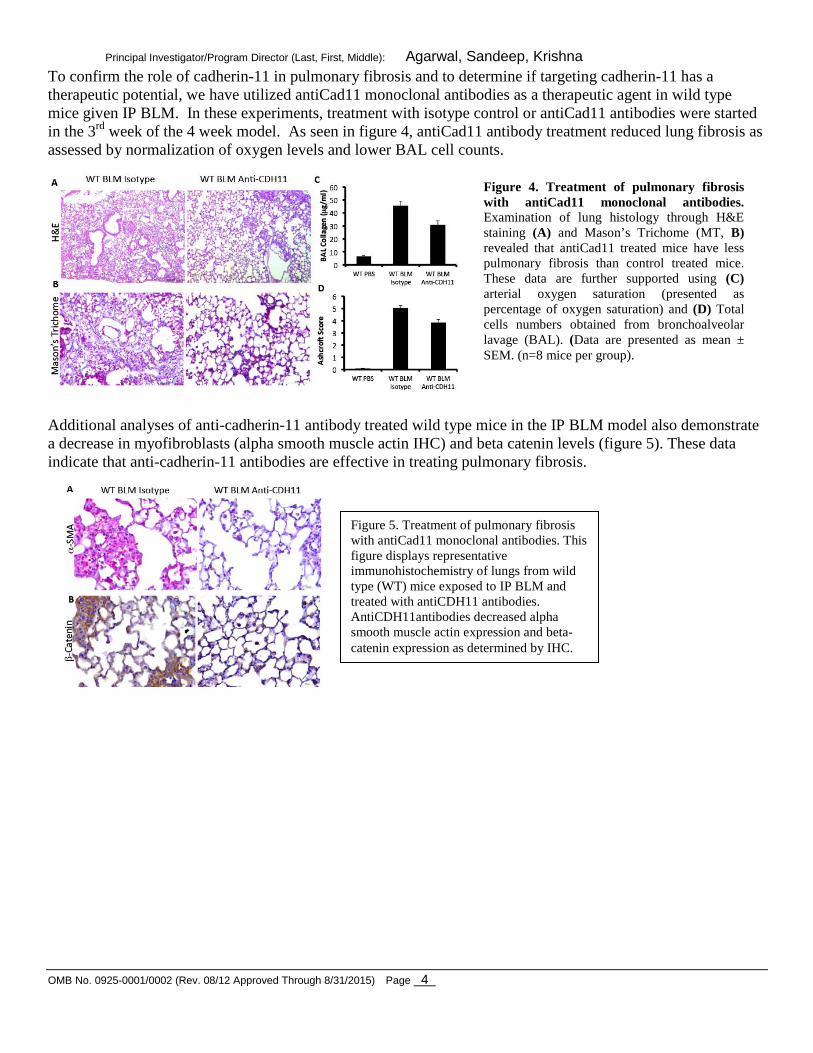
Figure 4. Treatment of pulmonary fibrosis with antiCad11 monoclonal antibodies. Examination of lung histology through H&E staining (A) and Mason’s Trichome (MT, B) revealed that antiCad11 treated mice have less pulmonary fibrosis than control treated mice. These data are further supported using (C) arterial oxygen saturation (presented as percentage of oxygen saturation) and (D) Total cells numbers obtained from bronchoalveolar lavage (BAL). (Data are presented as mean ± SEM. (n=8 mice per group).
To confirm the role of cadherin-11 in pulmonary fibrosis and to determine if targeting cadherin-11 has a therapeutic potential, we have utilized antiCad11 monoclonal antibodies as a therapeutic agent in wild type mice given IP BLM. In these experiments, treatment with isotype control or antiCad11 antibodies were started in the 3rd week of the 4 week model. As seen in figure 4, antiCad11 antibody treatment reduced lung fibrosis as assessed by normalization of oxygen levels and lower BAL cell counts.
Additional analyses of anti-cadherin-11 antibody treated wild type mice in the IP BLM model also demonstrate a decrease in myofibroblasts (alpha smooth muscle actin IHC) and beta catenin levels (figure 5). These data indicate that anti-cadherin-11 antibodies are effective in treating pulmonary fibrosis.
Figure 5. Treatment of pulmonary fibrosis with antiCad11 monoclonal antibodies. This figure displays representative immunohistochemistry of lungs from wild type (WT) mice exposed to IP BLM and treated with antiCDH11 antibodies. AntiCDH11antibodies decreased alpha smooth muscle actin expression and beta-catenin expression as determined by IHC.

Principal Investigator/Program Director (Last, First, Middle): Agarwal, Sandeep, Krishna
OMB No. 0925-0001/0002 (Rev. 08/12 Approved Through 8/31/2015) Page 5
To determine if cadherin-11 regulates the process of epithelial to mesenchymal transition (EMT) during the development of lung fibrosis in vivo, total RNA was obtained from lungs. As see in figure 6, both Cad11 KO mice and antiCad11 treated mice had decreased mRNA of Twist and Snail, two transcription factors that regulate EMT. These data suggest that Cad11 may regulate EMT in vivo.
To determine if cadherin-11 regulates the process of epithelial injury during the development of lung fibrosis in vivo, total RNA was obtained from lungs. As see in figure 7, both Cad11 KO mice and antiCad11 treated mice had decreased mRNA of Hif1alpha, HSP70 and PAI1, which are genes upregulated during epithelial injury. These data suggest that Cad11 may regulate epithelial injury.
Together these data from Aim 1, demonstrate that cadherin-11 is an important regulator of pulmonary fibrosis in mice. Finally, targeting cadherin-11 may be an effective strategy to treatment pulmonary fibrosis.
Figure 6. Cadherin-11 regulates Twist and Snail expression in vivo during the development of pulmonary fibrosis induced by IP bleomycin. These data demonstrate an important role for Cadherin-11 in regulating EMT.
Figure 7. Cadherin-11 regulates HSP70, Hif-1-alpha, and PAI1 expression in vivo during the development of pulmonary fibrosis induced by IP bleomycin. These data demonstrate an important role for Cadherin-11 in regulating epithelial injury in vivo.

Principal Investigator/Program Director (Last, First, Middle): Agarwal, Sandeep, Krishna
OMB No. 0925-0001/0002 (Rev. 08/12 Approved Through 8/31/2015) Page 6
Finally, we have acquired the Tomato Red SP-C-Cre, Rosa26 lacZ reporter mice and the breeding colony. These mice express Tomato Red under the direction of the SP-C promoter, and are supposed to be specific for type II alveolar epithelial cells. To induce lung fibrosis, these mice have been challenged with intraperitoneal bleomycin twice a week for four weeks. Lungs were harvested and frozen sections were obtained for dual color IF analyses (figure 8). AntiCadherin-11 antibody was used to stain the lungs (green) and type II alveolar epithelial cells appear red. The images in figure 1 demonstrate expression of cadherin-11 and SPC but the background levels are high. There are some cells that co-express these markers, suggesting that a subset of cadherin-11 expressing cells, likely fibroblast, are derived from SPC expressing type II alveolar epithelial cells. We are currently optimizing staining and fluorescent conditions as well as looking at expression of these markers during different time points in the model. The goal of these experiments is to track epithelial to mesenchymal transition during lung fibrosis, track the expression of cadherin-11 during fibrosis and determine if cadherin-11 plays a role in EMT during fibrosis.
In summary, in aim 1 we can conclude that cadherin-11 is a key regulator of lung fibrosis in mice.
Figure 8. Tomato Red SP-C-Cre, Rosa26 lacZ reporter mice were given bleomycin to induce lung fibrosis. Lungs were also stained with anticadherin-11 antibodies (green) for IF analyses.

Principal Investigator/Program Director (Last, First, Middle): Agarwal, Sandeep, Krishna
OMB No. 0925-0001/0002 (Rev. 08/12 Approved Through 8/31/2015) Page 7
Figure 9. Cadherin-11 regulates EMT in vitro in MLE12 cells.
Specific Aim 2. Determine the contribution of cadherin-11 to process of epithelial-to-mesenchymal transition (EMT) and modulation of Rho-GTPases in airway epithelial cells (AECs) in vitro.
Type II alveolar epithelial cell lines (AEC) were used to determine the role of Cad11 in AEC EMT. MLE-12 cells, an AEC, were cultured and TGFbeta was used to drive the process of EMT. During EMT, E-cadherin decreases and collagen increases, therefore these mRNA transcripts were used for quantifying EMT. The results of these experiments are provided in figure 9. To block Cad11 function, soluble Cad11 Fc fusion protein was added to cultures. As seen in figure 9A, TGFbeta decreased E-cadherin expression and increase Col1a1 expression in MLE12 cells. Soluble Cad11 Fc fusion protein inhibited EMT induced by TGFbeta as noted my higher E-cadherin levels and a significant reduction in Col1a1 mRNA. In contrast, when Cad11 Fc fusion protein was immobilized onto the tissue culture plate, providing an activating signal through Cad11 (figure 9B), a different result was observed. First, immobilized Cad11 Fc fusion protein alone was able to induce Col1a1 expression at the 50 ug/ml concentration, although E-cadherin expression was also increased. In the presence of TGFbeta, immobilized Cad11 synergistically increased Col1a1 expression, indicating that Cad11 engagement can increase collagen expression. Finally, Cad11 siRNA was utilized to block Cad11 expression in MLE12 cells (figure 9C). Cad11 siRNA alone resulted in a slight increase in E-cadherin expression and decrease in collagen expression. In the presence of TGFbeta this response was magnified where MLE12 cells transfected with Cad11 siRNA but not Ncad siRNA were unable to undergo EMT as indicated by high levels of E-cadherin and lower levels of Col1a1. These data together indicate that Cadherin-11 is a regulator of MLE12 EMT. Similar data were observed with the A549 cell line (data not shown).

Principal Investigator/Program Director (Last, First, Middle): Agarwal, Sandeep, Krishna
OMB No. 0925-0001/0002 (Rev. 08/12 Approved Through 8/31/2015) Page 8
We extended these observations utilizing primary AECs isolated from wild type and cadherin-11 deficient mice. Primary AECs from wild type and cadherin-11 deficient mice were isolated and cultured with TGF-beta to drive the process of EMT. During EMT, E-cadherin decreases and collagen increases, therefore these mRNA transcripts were used for quantifying EMT. The results of these experiments are provided in figure 10. TGF-beta decreased E-cadherin expression and increased collagen expression in wild type AECs, which was attenuated by soluble cadherin-11 fusion protein (which inhibits cadherin-11 function). In addition, in comparison to wild type AECs, primary AECs isolated from cadherin-11 deficient mice had a reduction in the changes in E-cadherin and collagen expression induced by TGF-beta. Together with the data from yea 1 in MLE-12 cells, these new data indicate that cadherin-11 is a regulator of epithelial-mesenchymal transformation in alveolar epithelial cells. These data together indicate that Cadherin-11 is a regulator of epithelial to mesenchymal transition in type II alveolar epithelial cell lines. Given the expression of cadherin-11 on lung fibroblasts, we wanted to determine if cadherin-11 also regulated the production of collagen and other ECM proteins. As seen in figure 11, murine lung fibroblasts increased expression of Col1a1 when stimulated with TGFbeta alone. Interestingly, culturing lung fibroblasts on immobilized Cadherin-11 but not Ecadherin also increased Col1a1 production.
In addition, using soluble Cadherin-11 FC fusion protein to block cadherin-11 function the production of collagen induced by TGFbeta is decreased (figure 12). Similar trends were seen with other ECM and matrix related genes including SMA, CTGF and TGFbeta (data not shown).
Figure 10. Cadherin-11 regulates EMT in vitro in primary alveolar epithelial cells.
Figure 11. Cadherin-11 regulates collagen production by murine lung fibroblasts.
Figure 12. Cadherin-11 regulates collagen production by murine lung fibroblasts.

Principal Investigator/Program Director (Last, First, Middle): Agarwal, Sandeep, Krishna
OMB No. 0925-0001/0002 (Rev. 08/12 Approved Through 8/31/2015) Page 9
Figure 13. Soluble Cad-11 ELISA detects human (blue) and mouse (red) Cad-11
Figure 14. Serum cadherin-11 levels are increased in certain autoantibody subsets of systemic sclerosis using a commercial Soluble Cad-11 ELISA.
Therefore, in aim 2, we can conclude that cadherin-11 is an important regulator of EMT and the mesenchymal gene expression in lung fibroblasts. Specific Aim 3. Determine the circulating levels of cadherin-11 in scleroderma patients with interstitial lung disease. In the past year a lot of effort has been dedicated to the studies in Specific Aim 3. This aim seeks to determine the circulating levels of cadherin-11 in scleroderma patients with interstitial lung disease. At the outset of these experiments, there was not a commercial Cad-11 ELISA. Therefore, we developed an ELISA and optimize our conditions using 2 anti-Cad-11 antibodies (clones 3H10 and 23C6). As seen in figure 13, our ELISA can detect both human and mouse soluble Cad-11.
More recently, a commercial ELISA has become available (R&D Systems). We have obtained this ELISA and conducted experiments with it. This ELISA was first tested on a sera from healthy patients (n=20), systemic lupus erythematosus patients (n=29) and systemic sclerosis patients (n=20). Patients with lupus and systemic sclerosis both had an increase in circulating soluble cadherin-11 levels that was statistically significant over levels seen in healthy controls (p<0.01). As seen in figure 14, levels of cadherin-11 were more elevated in systemic sclerosis patients with the anticentromere antibody (ACA)
and anti RNA polymerase III antibody (RNA POL). Furthermore, patients with other autoantibodies also had a remarkably elevated level. These data suggest that cadherin-11 levels are increased in
patients with systemic sclerosis and lupus. These data use a small set of samples therefore, the goal in Aim 3 is to test a larger cohort of patients for serum cadherin-11 levels.

Principal Investigator/Program Director (Last, First, Middle): Agarwal, Sandeep, Krishna
OMB No. 0925-0001/0002 (Rev. 08/12 Approved Through 8/31/2015) Page 10
To further characterize the levels of soluble cadherin-11 in systemic sclerosis patients, we have obtained baseline serum samples from patients and healthy controls enrolled in the GENISOS study (UTHSC). Table 1 presents the basic demographics of the healthy controls and patients. TABLE 1.
Control SSc N 153 300 Age 48.8 +/- 14.2 50.1 +/- 13.1
Male (%) 26 (17%) 50 (17%) Age (49.2 +/- 14.4) Age (49.7 +/- 15.2)
Female (%) 127 (83%) 250 (83%) Age (48.7 +/- 14.2) Age (50.1 +/- 12.6)
Race (%) Caucasian (%) 71 (46%) 141 (47%)
Hispanic (%) 43 (28%) 85 (28%) Black (%) 33 (22%) 64 (21%) Asian (%) 0 (0%) 9 (3%) Other (%) 6 (4%) 1 (<1%)
Table 2 presents the clinical characteristics of the systemic sclerosis patients at the time of enrollment in GENISOS.
TABLE 2 Number of patients (total number 300)
Disease duration 3.95 +/- 2.93 years, range 0.17-17 yrs
Limited 125 Diffuse 171 Subset unknown 4
ANA positive 275 ANA negative 14 ANA not done 11
Anti-centromere positive 40 Anti-centromere negative 249 Anti-centromere not done/unknown 11
Anti-topoisomerase positive 46 Anti-topoisomerase negative 243 Anti-topoisomerase not done/unknown 11
Anti polymerase III positive 64

Principal Investigator/Program Director (Last, First, Middle): Agarwal, Sandeep, Krishna
OMB No. 0925-0001/0002 (Rev. 08/12 Approved Through 8/31/2015) Page 11
Figure 15. Serum cadherin-11 levels are increased in a large cohort of systemic sclerosis. P=0.0001
Figure 16. Serum cadherin-11 levels are increased in patients classified as either limited or diffuse scleroderma. P=0.0001 P<0.0001
Anti polymerase III negative 225 Anti polymerase III not done/unknown 11 As seen in figure 15, patients with systemic sclerosis have a significantly increased level of soluble cadherin-11 relative to age/matched healthy controls.
Controls
All SSc
0
200
400
600
800
As seen in figure 16, patients with limited and diffuse forms of systemic sclerosis had a significantly increased level of soluble cadherin-11 relative to age/matched healthy controls.
Controls
Limite
d
DIffuse
0
200
400
600
800
1000
We are currently performing additional analyses to determine if cadherin-11 levels are a biomarker for certain clinical subsets of systemic sclerosis and are predictive of the development of lung fibrosis in systemic sclerosis. Preliminary studies do not clearly show a an association but more complex statistical modeling is being considered. KEY RESEARCH ACCOMPLISHMENTS 1. Cadherin-11 deficient mice have decrease pulmonary fibrosis in the intraperitoneal model of pulmonary fibrosis 2. AntiCad11 antibodies are effective in treating lung fibrosis in the intraperitoneal model of pulmonary fibrosis this model 3. Cad11 regulates the in vitro TGF-beta induced epithelial-to-mesenchymal-transition (EMT) in MLE-12 cells, a mouse alveolar epithelial cell line 4. Cad11 regulates extracellular matrix production by lung fibroblasts. 5. Cadherin-11 levels are increased in some subsets of scleroderma patients suggesting a potential role for cadherin-11 as a scleroderma biomarker.

Principal Investigator/Program Director (Last, First, Middle): Agarwal, Sandeep, Krishna
OMB No. 0925-0001/0002 (Rev. 08/12 Approved Through 8/31/2015) Page 12
REPORTABLE OUTCOMES. Seminars Presented
1. “Cadherin-11 in Scleroderma and Fibrosis”. Baylor College of Medicine. Section of Immunology, Allergy and Rheumatology Research Seminar. Houston, TX. January, 2011.
2. “Cadherin-11 in pulmonary fibrosis”. Baylor College of Medicine, Section of Immunology, Allergy and Rheumatology Research Seminar. Houston, TX. May, 2011.
3. “Role for Cadherin-11 in Tissue Fibrosis and Autoimmune Diseases”. Baylor College of Medicine, Graduate Immunology Research Seminar. Houston, Texas, August 2012.
4. “Cadherin-11 in pulmonary fibrosis”. Baylor College of Medicine, Section of Pulmonology Research Seminar. Houston, TX. December 2012.
5. “Cadherin-11 in pulmonary fibrosis”. National Jewish Health, Autoimmune Interstitial Lung Disease Program Research Conference. Denver, CO, December 2012.
6. “Cadherin-11 in fibrosis”. Baylor College of Medicine, Section of Nephrology Research Seminar. Houston, TX. December 2013.
7. “Role of cadherin-11 in fibrosis and lung diseases”. Michael E. DeBakey Veterans Administration Medical Center Center for Translational Research on Inflammatory Diseases Research Seminar. August 2015.
8. “Role of cadherin-11 in lung fibrosis”. Baylor College of Medicine Biology of Inflammation Center Research Seminar. October 2015.
Publications
1. Wu M, Pedroza M, George WT, Schneider DJ, Lafyatis R, Mayes MD, Tan FK, Brenner MB, Blackburn MR, Agarwal, SK. Identification of cadherin-11 as a mediator of dermal fibrosis and possible role in systemic sclerosis. Arthritis and Rheumatism. 66 (4): 1010-21, 2014.
2. Alimperti S, You H, George AT, Agarwal SK, Andreadis ST. Cadherin-11 Regulates stem cell stem cell differentiation into smooth muscle cells and development of contractile function in vivo. Journal of Cell Science. 127:2627-38, 2014.
3. Le TT, Karmouty-Quintana H, Melicoff E, Le TT, Chen NY, Weng, T, Pedroza M, George AT, Garcia-Morales LJ, Bunge RR, Bruckner BA, Loebe M, Seethamraju H, Agarwal SK, Blackburn MR. Blockade of interleukin-6 trans signaling attenuates pulmonary fibrosis. J. Immunology. 193(7): 3755-68, 2014.
4. Pedroza M, Le TT, Lewis K, Karmouty-Quintana H, To S, George AT, Blackburn MR, Tweardy DJ, Agarwal SK. STAT3 contributes to pulmonary fibrosis through epithelial injury and fibroblast-myofibroblast differentiation. FASEB J. 30(1): 129-40, 2016.
5. Row S, Liu Y, Alimperti S, Agarwal SK, Andreadis ST. Cadherin-11 is a novel regulator of extracellular matrix synthesis and tissue mechanics. Journal of Cell Science. In revision.
Manuscripts in progress
1. Pedroza M, To BT, Blackburn MR, Agarwal SK. Cadherin-11 is a regulator of lung fibrosis and epithelial to mesenchymal transition. Likely to be submitted to Journal of Cell Biology.
2. Welschhans L, Mayes M, Assassi S, Agarwal SK. Soluble Cadherin-11 levels of scleroderma patients. Likely to be submitted to Arthritis and Research Therapy.

Principal Investigator/Program Director (Last, First, Middle): Agarwal, Sandeep, Krishna
OMB No. 0925-0001/0002 (Rev. 08/12 Approved Through 8/31/2015) Page 13
CONCLUSIONS Cadherin-11 is a mediator of lung fibrosis and can regulate epithelial-to-mesenchymal-transition (EMT) in MLE-12 cells, a mouse alveolar epithelial cell line.
REFERENCES
None for current report
APPENDICES None for current report
SUPPORTING DATA No additional data for current report, see “BODY” section above for data.


![CD146 mediates an E-cadherin-to-N-cadherin switch during TGF-β … · 2018. 7. 10. · expression [16–19]. N-cadherin is reported to be upregulated by TGF-β signaling [20,21].](https://static.fdocuments.in/doc/165x107/6126bb2c5b910b6f974c32bd/cd146-mediates-an-e-cadherin-to-n-cadherin-switch-during-tgf-2018-7-10-expression.jpg)


![Research Paper Desacetylvinblastine Monohydrazide Disrupts ... · promote VE-cadherin internalization, which increases endothelial cell permeability -30]. Whether [27 VE-cadherin](https://static.fdocuments.in/doc/165x107/60b3e368cf71b2652b121d17/research-paper-desacetylvinblastine-monohydrazide-disrupts-promote-ve-cadherin.jpg)


